1. Introduction
Obesity is a global public health priority. With the improvement of people’s standard of living, the number of obese Chinese has grown to an annual rate of 38.1% over the past several years [
1]. Obesity is currently one of the major health concerns in developed countries. With more than 1.9 billion people now obese or overweight, the eventual impact of expected adverse events such as diabetes, hypertension, sleep apnea, cardiac failure, reflux, and major orthopedic problems, will take a major toll on public health [
2,
3]. Obesity, which is defined as the “New World Syndrome”, is one of the most important problems of the modern age. The prevalence of obesity in all age groups is observed throughout the World [
4].
For this purpose, the importance of substances to be used in the diet is increasing. During this project, we will study the usability of bee bread, which is gaining importance each day with its rich nutrient content, as a dietary supplement, and this study is the first to be carried out in this field. Following the detailed literature reviews (e.g., Pubmed, Web of Science, Sciencedirect), the scarcity of studies on bee bread draws attention and this study is expected to contribute greatly to the literature [
5,
6]. IL-6 is a pleotropic cytokine with a broad spectrum of biological activity involved in regulation of inflammation, cell differentiation, cell proliferation, regulation of the immune system, hemopoiesis and tumor formation [
7].
This project will contribute to the determination of natural and healthy alternative diet factors such as bee bread for the fight against obesity, which is seen as a serious health problem all over the world and in our country, while it will be possible to bring in new data to be obtained from bee bread to world literature.
2. Experimental Procedure
In this study, 40 Sprague Dawley species adult female rats, weighing 200–250 g will be used. Rats will be randomly divided into 4 groups (n = 8). For this study, the first step was to separate rats as the control group (n = 8) and the obesity group (n = 32). The rats, on which obesity will be formed, (n = 32) will be fed with high fat content diet. The rats, on which obesity will be formed, will be fed with this supplement for 4 weeks. Rats in the control group will be fed with standard rat supplement during this period (n = 8). The rats, on which obesity will be formed, will be separated into experiment groups with 8 members in each group (group 2, 3, 4, 5). The weight of rats will be recorded at the beginning and end of the study.
1st Group: Control: Group fed with standard rat supplement (n = 8)
2nd Group: Group fed with high fat diet (n = 8)
3nd Group: Group fed with 100 mg/kg/day bee bread (n = 8)
4rd Group: Group fed with 200 mg/kg/day bee bread (n = 8)
5th Group: Group administered with Metformin 300 mg/kg/day as positive control (n = 8). Metformin is an agent that improves insulin sensitivity, also it is well known to reduce body weight. For this reason, it will be used as positive control.
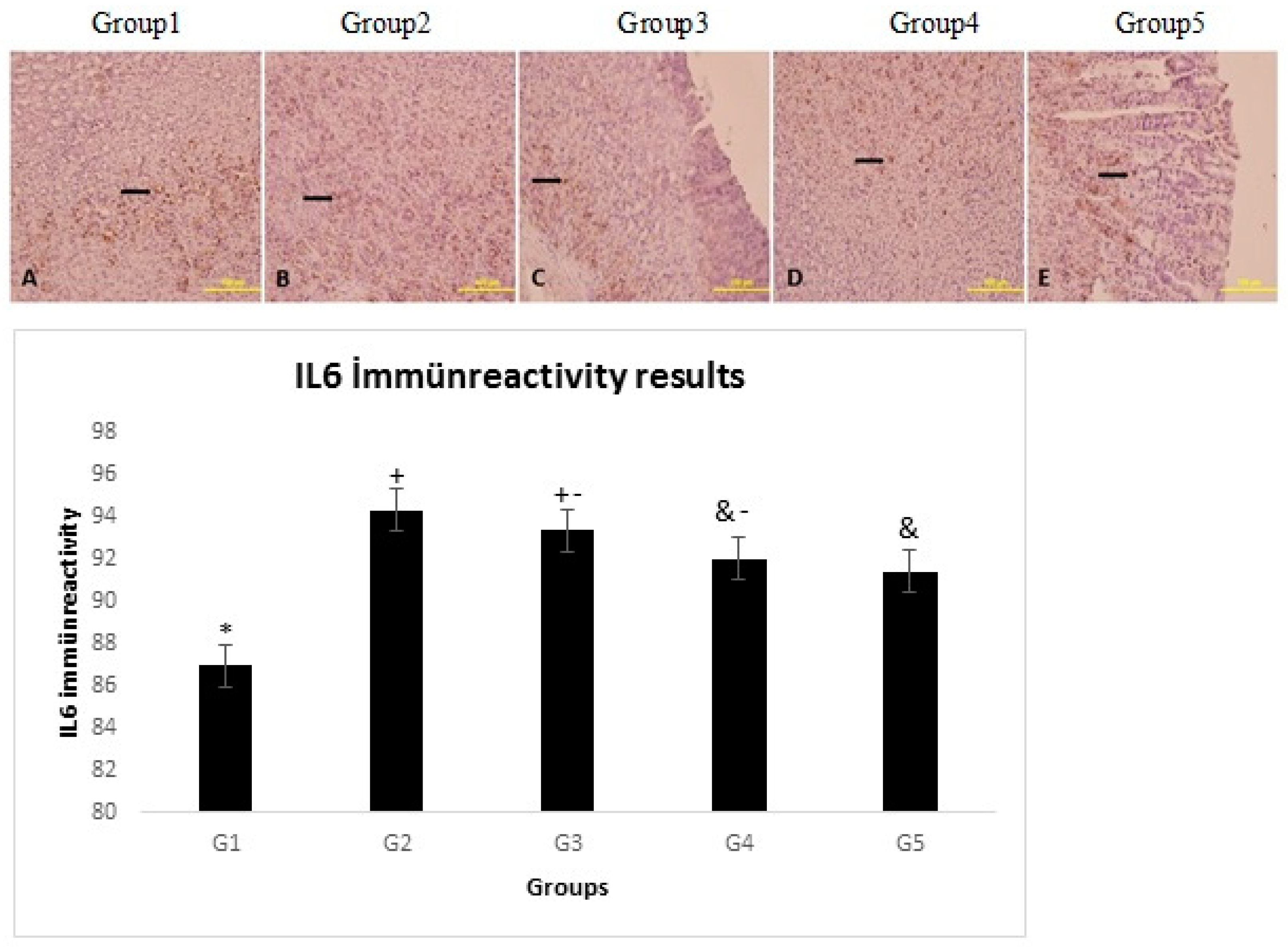
When the experiment protocol is complete at the end of the four week feeding period, the stomach tissues from the rats will be collected, and histological and immunohistochemical evaluations will be made in some of the tissues. In the remaining tissue, IL6 level will be measured by immunohistochemistry.
2.1. Immunohistochemistry
IL6 was detected immunohistochemically using a polyclonal antibody and the streptavidin–biotin–peroxidase technique. Some of the sections were deparaffinized in xylene, rehydrated and rinsed in deionized water. Antigen retrieval was performed by microwave treatment in 0.01 M sodium citrate buffer, pH 6.0, at 95_C for 5 min. The slides were cooled rapidly and held at room temperature for 20 min. Sections were washed with phosphate-buffered saline (PBS) and endogenous peroxidase activity was inhibited by immersion in 3% (w/v) H2O2 in methanol for 10 min. For the next stages, a Lab VisionTM UltraVisionTM Large Volume Detection System (TA-125-HDX, Thermo Fisher Scientific, Wal-tham, MA, USA) was used. All cross sections were washed with PBS and then Ultra V block was applied for 10 min at room temperature to block outside the antigenic fields. The sections were incubated with an IL6 specific polyclonal antibody (sc.1265-R; Santa Cruz Biotechnology, Santa Cruz, CA, USA) diluted to 2.5 g/mL in antibody diluents buffer (TA-125-ADQ, Thermo Fisher Scientific, Waltham, MA, USA) overnight at 4_C. As a negative control, PBS was used instead of primary antibody. After washing with PBS, the sections were incubated with biotinylated secondary antibodies (TA-125-HDX, Thermo Fisher Scientific, Waltham, MA, USA). The immunoreaction was amplified using the streptavidin–avidin–peroxidase complex and the sections were visualized using 3,30-p-diaminobenzidine tetrahydrochloride (TA-060-HDX, Thermo Fisher Scientific, Waltham, MA, USA) and lightly counter-stained with Gill hematoxylin. For the final step, increasing alcohol serial concentrations were used to remove water, the sections were then passed through xylene, and finally, they were covered with an entellan. Under the light microscope (olympus BX51), and with a digital camera (DP71), images were obtained. From each of the subjects, five different areas were evaluated in terms of the expression differences using the image J program.
2.2. Statistical Analysis
All statistical analyses were carried out using SPSS statistical software (SPSS for windows, SPSS Inc, Chicago, IL, USA, version 22.0). The Kolmogorov-Smirnov test was used to identify normal distribution of data. In case of normal distribution, quantitative variables were compared using one-way analysis of variance (ANOVA) and posthoc Tukey test. Results are presented as mean ± SEM. A p value of <0.05 was considered statistically significant.

