Proceeding Paper

Identification of the FGFR3G380R Mutant As a Likely Cause of Psychomotor Delay in an Achondroplastic Child: A Combined Clinical Exome Sequencing and Biomolecular Modeling Approach †

1
Department of Medical Biochemistry, Faculty of Medicine, Near East University, 99138 Nicosia, Mersin 10, Turkey
2
Department of Medical Genetics, Faculty of Medicine, Uludağ University,16059 Bursa, Turkey
3
Department of Pediatric Endocrinology, Faculty of Medicine, Uludağ University, 16059 Bursa, Turkey
*
Author to whom correspondence should be addressed.
Presented at the 2nd International Cell Death Research Congress, Izmir, Turkey, 1–4 November 2018.
Proceedings 2018, 2(25), 1551; https://doi.org/10.3390/proceedings2251551
Published: 5 December 2018
(This article belongs to the Proceedings of The 2nd International Cell Death Research Congress)

Abstract

:
Mutations in the gene for fibroblast growth factor receptor 3 (FGFR3) are implicated in achondroplasia, an autosomal-dominant form of short-limbed dwarfism. The present study involves a combination of clinical exome sequencing, targeted resequencing and protein modeling methods to decipher the pathobiology of achondroplasia with psychomotor delay in a two-year-old child. Accordingly, the resulting genetic information establishes the frequent FGFR3 c.1138G>A (p.G380R) mutation as the single hit causing pediatric achondroplasia with psychomotor delay, while the predicted model stresses the importance of a phenylalanyl residue (F384) in enhancing the dimerization potential of the receptor’s transmembrane domain via a cation‒π interaction with the newly introduced arginyl residue. Overall, the likely involvement of FGFR3G380R in psychomotor delay calls for comprehensive clinical assessment in achondroplastic children, although the precise mechanism by which the mutant receptor results in the development of neurological manifestations awaits further investigation.

1. Introduction

Achondroplasia is the most common form of disproportionate dwarfism in humans. It is characterized by short proximal long bones (rhizomelia) and macrocephaly, yet affected individuals are of normal intelligence. The inheritance pattern in achondroplasia is autosomal dominant; over 97% of all cases are caused by the completely-penetrant c.1138G>A (p.G380R) mutation in the FGFR3 gene encoding fibroblast growth factor receptor 3 [1]. FGFR signaling plays pivotal roles in the proliferation, differentiation and apoptotic death of chondrocytes [2]. Experimental evidence argues that pathogenic mutations, such as p.G380R, in the transmembrane domain of FGFR3 confer gain-of-function properties to the receptor, leading to its constitutive activation [3]. The consequent inhibition of chondrocyte proliferation results in abnormal endochondral bone formation. Here, I aim at exploring the genetic and mechanistic basis of psychomotor delay in a two-year-old male showing typical clinical and radiological manifestations of achondroplasia.

2. Materials and Methods

2.1. Clinical Exome Sequencing

Genetic testing, for which written informed consent has been obtained from the pediatric patient’s parents, was performed on genomic DNA extracted from whole blood. The coding regions of a total of 4,813 genes associated with known clinical phenotypes were sequenced on a MiSeq platform (Illumina) using the TruSight One Sequencing Panel (Illumina). The presence of the c.1138G>A transition was confirmed by polymerase chain reaction coupled with direct sequencing of the FGFR3 gene on a CEQ 8800 system (Beckman Coulter).

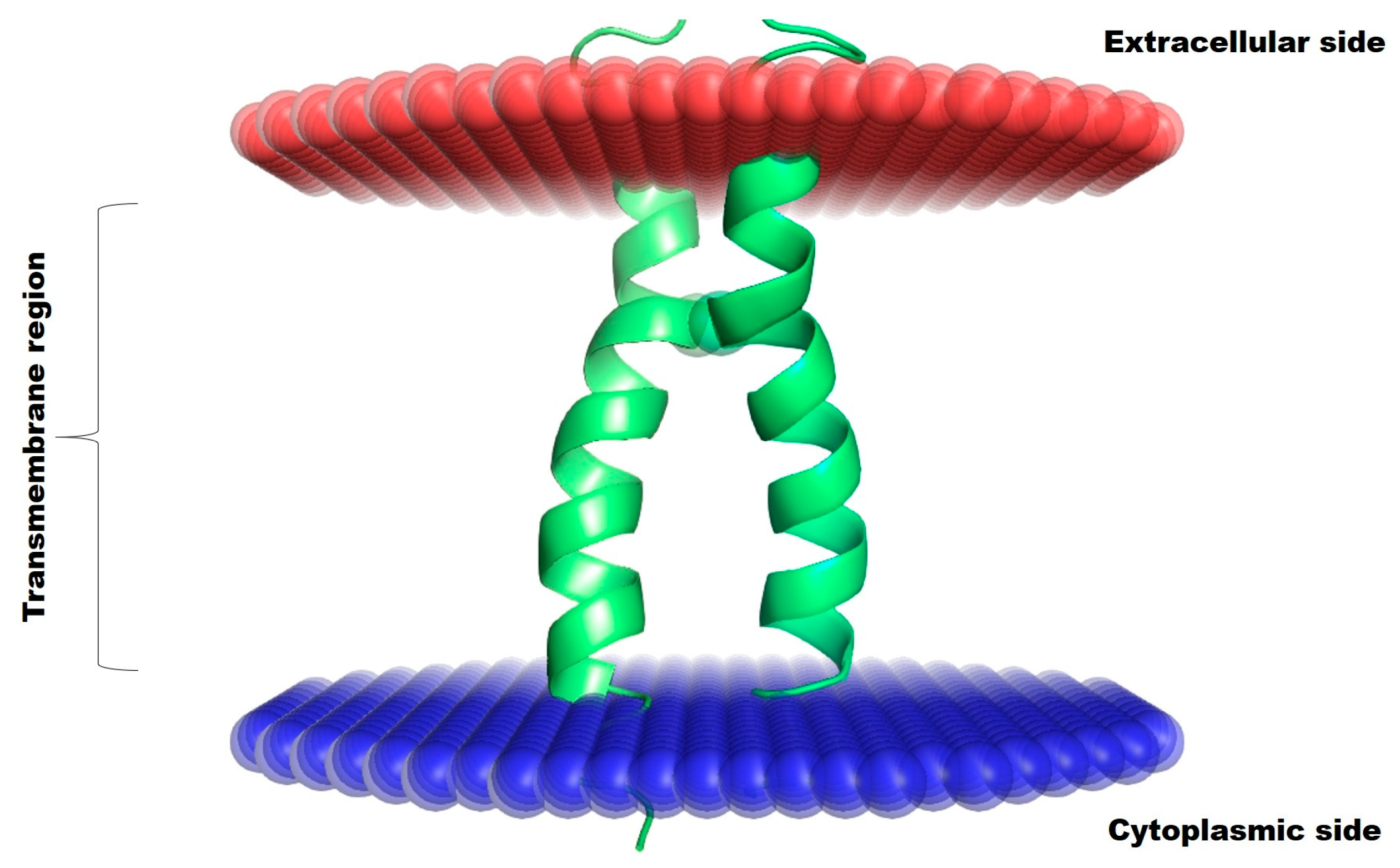
2.2. Biomolecular Modeling

The spatial structure of the human FGFR3 transmembrane domain dimer in a membrane-mimicking environment (PDB ID: 2LZL) was downloaded from the Orientations of Proteins in Membranes (OMP) database [4]. The p.G380R substitution was introduced in silico after applying two iterative cycles of the MutaBind method [5]. Intermonomeric noncovalent interactions occurring between the constituent amino acid residues were computed using the Protein Interactions Calculator (PIC) Web server [6]. Manual inspection and subsequent processing of the built model was achieved using the PyMOL Molecular Graphics System, Version 1.8 (Schrödinger).

3. Results

Post-sequencing data analysis revealed that the FGFR3 c.1138G>A (p.G380R) mutation, which has arisen de novo in the pediatric patient, was a likely cause of achondroplasia with psychomotor delay (Figure 1).
The two glycyl residues, which are subject to substitution in the disease state, in the native FGFR3 transmembrane domain dimer were unable to engage in any protein‒protein interactions described by the PIC Web server. They, however, lie at the juxtaposition of the two monomers, allowing only very limited mobility for the side chains of bulkier residues that are to replace them (Figure 2).
The in silico mutagenized and energy-minimized model of the receptor showed that the side chains of the two arginyl residues could be accommodated at the juxtaposition of the transmembrane helices in a symmetric configuration without any steric clashes (Figure 3, left panel). Furthermore, there was a sufficiently long distance between the positive charge centers of the arginyl residues to prevent electrostatic repulsions. The PIC analysis of the predicted model revealed intermonomeric cation‒π interactions between the aromatic rings of the F384 residues and the positively charged guanidinium groups of the opposite R380 residues, suggesting a plausible means by which the receptor is forced into the constitutively active state (Figure 3, right panel).

4. Discussion

The clinical exome represents a greatly enriched subset of the human genome, facilitating the search for both exonic and exon-flanking (e.g. regulatory and splice-site) regions harboring known variants associated with Mendelian diseases. In the present study, the FGFR3 c.1138G>A (p.G380R) mutation emerges as the only biologically relevant variant in an achondroplastic child with symptoms suggestive of neurological impairment. Biomolecular modeling is useful for gaining insights into the behavior of mutant proteins in an in silico setting. Here, cation‒π interactions between the arginyl residues and the vicinal phenylalanyl residues are predicted to have a stabilizing effect on the FGFR3G380R dimer. Cation‒π interactions are relatively strong (~3 kcal mol-1), reflecting the preferential (non-random) localization of positively charged side chains in the vicinity of aromatic side chains at protein‒protein interfaces [7]. Although the proposed cation‒π interactions may contribute to the stabilization and thus the constitutive activation of the dimeric FGFR3 complex, the characterization of the complex pleiotropic outcomes of this activation (i.e. downstream signaling events and consequent neuronal responses) remains a major challenge. It should also be noted here that the lipid environment may produce unusual and unpredicted effects in the dimerization of FGFR3 transmembrane helices. To conclude, given the fact that the FGFR3 c.1138G>A (p.G380R) mutation is the most common etiologic factor in achondroplasia, the neurological manifestations of pediatric achondroplasia can be frequent and significant, demanding in-depth clinical assessment even in asymptomatic children.

Acknowledgements

The author acknowledges the following for their complete help in this study: Dr. Mahmut Çerkez Ergören, Associate Professor of Medical Genetics at Near East University; and the MAGI Group and its founder/president Matteo Bertelli.

Conflicts of Interest

The author declares no conflict of interest.

References

  1. Ornitz, D.M.; Legeai-Mallet, L. Achondroplasia: development, pathogenesis, and therapy. Dev. Dyn. 2017, 246, 291–309. [Google Scholar] [CrossRef] [PubMed]
  2. Su, N.; Jin, M.; Chen, L. Role of FGF/FGFR signaling in skeletal development and homeostasis: learning from mouse models. Bone Res. 2014, 2, 14003. [Google Scholar] [CrossRef] [PubMed]
  3. Webster, M.K.; Donoghue, D.J. Constitutive activation of fibroblast growth factor receptor 3 by the transmembrane domain point mutation found in achondroplasia. EMBO J. 1996, 15, 520–527. [Google Scholar] [CrossRef] [PubMed]
  4. Lomize, M.A.; Pogozheva, I.D.; Joo, H.; Mosberg, H.I.; Lomize, A.L. OPM database and PPM web server: resources for positioning of proteins in membranes. Nucleic Acids Res. 2012, 40, D370–D376. [Google Scholar] [CrossRef] [PubMed]
  5. Li, M.; Simonetti, F.L.; Goncearenco, A.; Panchenko, A.R. MutaBind estimates and interprets the effects of sequence variants on protein‒protein interactions. Nucleic Acids Res. 2016, 44, W494–W501. [Google Scholar] [CrossRef] [PubMed]
  6. Tina, K.G.; Bhadra, R.; Srinivasan, N. PIC: Protein Interactions Calculator. Nucleic Acids Res. 2007, 35, W473–W476. [Google Scholar] [CrossRef] [PubMed]
  7. Crowley, P.B.; Golovin, A. Cation‒π interactions in protein‒protein interfaces. Proteins 2005, 59, 231–239. [Google Scholar] [CrossRef] [PubMed]
Figure 1. Sequence chromatogram showing the heterozygous FGFR3 c.1138G>A (p.G380R) mutation in the pediatric patient.
Proceedings 02 01551 g001
Figure 2. Spatial position of the nuclear magnetic resonance (NMR) structure of the human FGFR3 transmembrane domain dimer (PDB ID: 2LZL) with respect to the lipid bilayer. The two glycyl residues, which have been substituted with the arginyl residues in the achondroplastic child, are shown as spheres. The transmembrane helices of the FGFR3 monomers are colored in pale green and bright green, respectively.
Proceedings 02 01551 g002
Figure 3. Left panel. Mutant FGFR3 superposed on native FGFR3. The transmembrane helices of the FGFR3G380R monomers are colored in pale orange and bright orange, respectively. Right panel. Favorable noncovalent interactions between the arginyl and phenylalanyl residues. Distance is the length of space between the positive charge center and the centroid of the aromatic ring; angle is the amount of rotation between the line joining the cation to the centroid and the normal to the aromatic plane at the centroid.
Proceedings 02 01551 g003
Publisher’s Note: MDPI stays neutral with regard to jurisdictional claims in published maps and institutional affiliations.

