Fluid Region Analysis and Identification via Optical Coherence Tomography Image Samples †

 ,
,  ,
,  , and
, and 
Abstract
:1. Introduction
2. Methodology
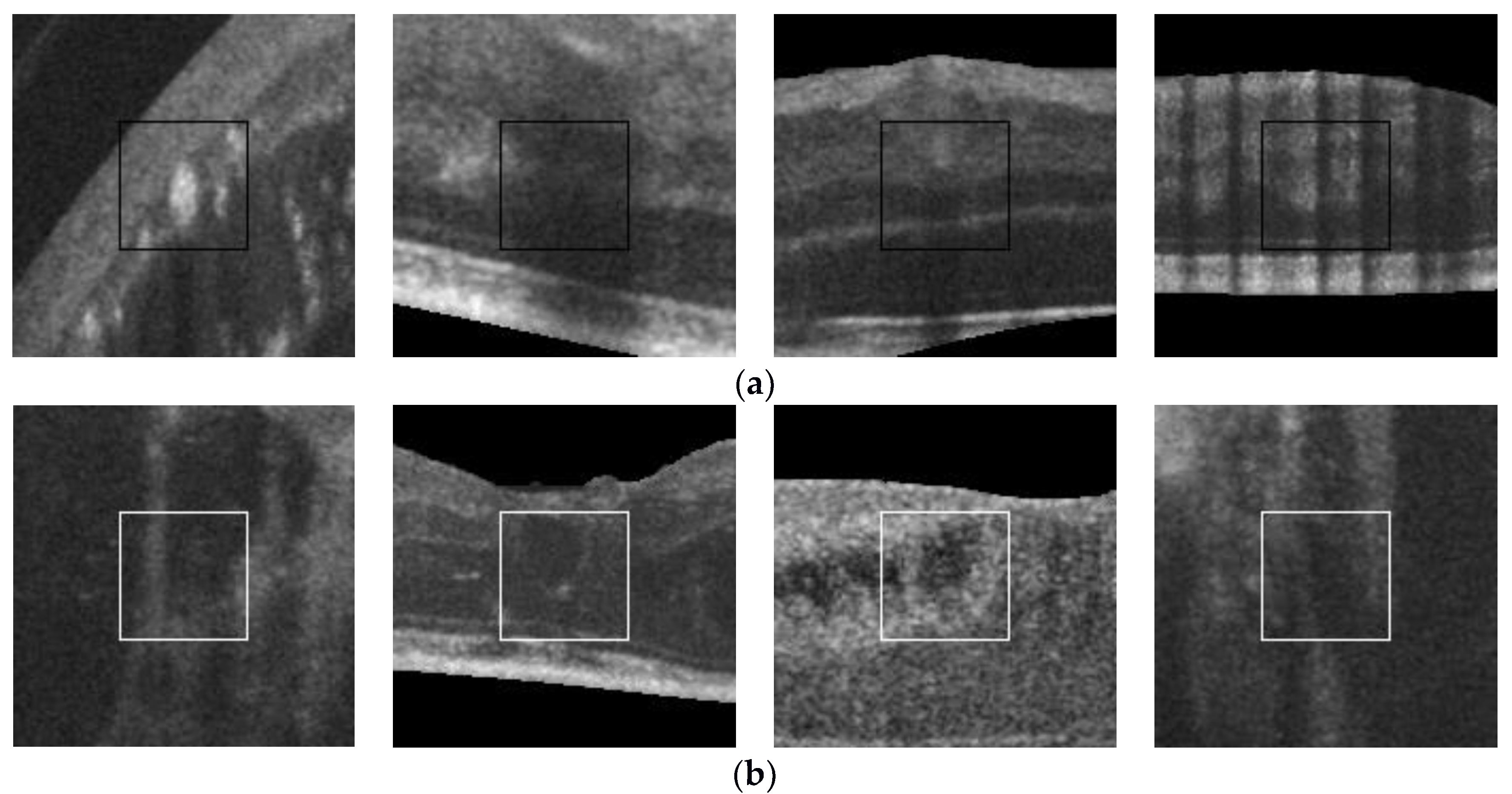
3. Results
Author Contributions
Acknowledgments
Conflicts of Interest
References
- De Moura, J.; Vidal, P.L.; Novo, J.; Rouco, J.; Ortega, M. Feature definition, analysis and selection for cystoid region characterization in optical coherence tomography. Procedia Comput. Sci. 2017, 112, 1369–1377. [Google Scholar] [CrossRef]
Publisher’s Note: MDPI stays neutral with regard to jurisdictional claims in published maps and institutional affiliations. |
© 2022 by the authors. Licensee MDPI, Basel, Switzerland. This article is an open access article distributed under the terms and conditions of the Creative Commons Attribution (CC BY) license (https://creativecommons.org/licenses/by/4.0/).
Share and Cite
Vidal, P.L.; Moura, J.d.; Novo, J.; Rouco, J.; Ortega, M. Fluid Region Analysis and Identification via Optical Coherence Tomography Image Samples. Proceedings 2018, 2, 1180. https://doi.org/10.3390/proceedings2181180
Vidal PL, Moura Jd, Novo J, Rouco J, Ortega M. Fluid Region Analysis and Identification via Optical Coherence Tomography Image Samples. Proceedings. 2018; 2(18):1180. https://doi.org/10.3390/proceedings2181180
Chicago/Turabian StyleVidal, Plácido L., Joaquim de Moura, Jorge Novo, José Rouco, and Marcos Ortega. 2018. "Fluid Region Analysis and Identification via Optical Coherence Tomography Image Samples" Proceedings 2, no. 18: 1180. https://doi.org/10.3390/proceedings2181180
APA StyleVidal, P. L., Moura, J. d., Novo, J., Rouco, J., & Ortega, M. (2018). Fluid Region Analysis and Identification via Optical Coherence Tomography Image Samples. Proceedings, 2(18), 1180. https://doi.org/10.3390/proceedings2181180