Proceeding Paper

Regenerable Bead-Based Microfluidic Device with integrated THIN-Film Photodiodes for Real Time Monitoring of DNA Detection †

1
Instituto de Engenharia de Sistemas e Computadores—Microsistemas e Nanotecnologias (INESC MN) and Institute of Nanoscience and Nanotechnology (IN), 1000-029 Lisbon, Portugal
2
IBB—Institute for Bioengineering and Biosciences, Instituto Superior Técnico, 1049-001 Lisbon, Portugal
3
Department of Bioengineering, Instituto Superior Técnico, 1049-001 Lisbon, Portugal
*
Author to whom correspondence should be addressed.
Presented at the Eurosensors 2018 Conference, Graz, Austria, 9–12 September 2018.
Proceedings 2018, 2(13), 953; https://doi.org/10.3390/proceedings2130953
Published: 10 December 2018
(This article belongs to the Proceedings of EUROSENSORS 2018)

Abstract

:
Nanoporous microbead-based microfluidic systems for biosensing applications allow enhanced sensitivities, while being low cost and amenable for miniaturization. The regeneration of the microfluidic biosensing system results in a further decrease in costs while the integration of on-chip signal transduction enhances portability. Here, we present a regenerable bead-based microfluidic device, with integrated thin-film photodiodes, for real-time monitoring of molecular recognition between a target DNA and complementary DNA (cDNA). High-sensitivity assay cycles could be performed without significant loss of probe DNA density and activity, demonstrating the potential for reusability, portability and reproducibility of the system.

1. Introduction

Biosensing devices for medical and environmental applications that are portable, low cost, accurate, and sensitive are in high demand, making microfluidics an attractive solution for point-of-care applications. In particular, DNA hybridization-based biosensors allow for decentralized DNA testing with high specificity and sensitivity while being faster, simpler and cheaper than traditional testing. There have been many recent efforts to develop DNA hybridization-based microfluidic biosensors. Many use electrochemical detection techniques that are prone to variability due to change in the surrounding environment and have complex and time consuming protocols [1], or magnetic-bead based detections that usually requires bulky machinery increasing the cost of the device [2], and few have focused on the regeneration of the microfluidic device [1]. Regeneration not only allows performing more assays on a single chip, but also enables the measurement of a control sample under the same experimental conditions. Our group has previously demonstrated that using agarose nanoporous microbeads inside microchannels enhances the sensitivity of detection of different biomarkers, mostly by increasing the available active surface area and decreasing diffusion times [3,4]. In this work, we report a regenerable microfluidic microbead-based DNA detection system with integrated fluorescence detection.

2. Materials and Methods

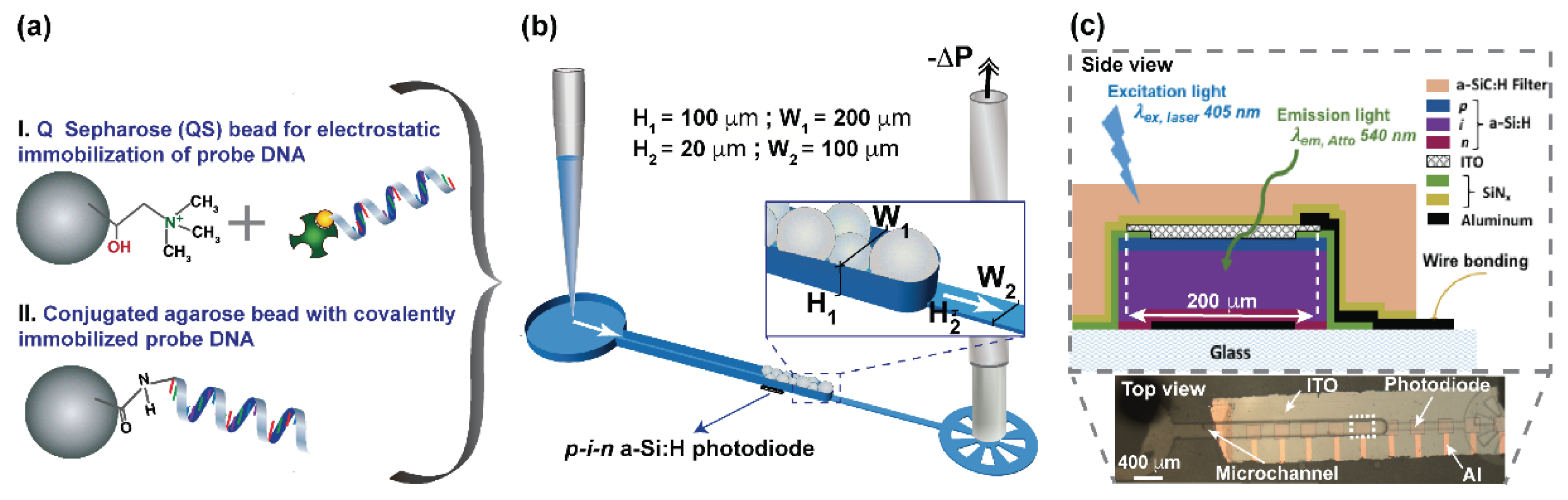
2.1. DNA Immobilization Strategies

Electrostatic and covalent immobilization strategies were used to immobilized a 23 bp single-stranded DNA analogous to micro RNA (MIR145) (StabVida, Lisbon, Portugal) to nanoporous agarose beads (Figure 1a). In the first strategy, Q Sepharose (QS) Fast Flow beads (GE Healthcare, Uppsala, Sweden) (~90 μm average size) were suspended in a solution of biotinylated DNA:streptavidin in a 1:4 M ratio previously incubated for 10 min, to allow adsorption of the DNA:streptavidin complex to the beads via electrostatic interactions. In the second strategy, NHS-Activated Sepharose 4 Fast Flow beads were used to covalently immobilize aminolink C6-modified probe DNA.

2.2. Fabrication of Microchannel and Bead Trapping

The microfluidic channels were fabricated using standard polydimethylsiloxane (PDMS) soft-lithography techniques, described in detail elsewhere [3]. Briefly, aluminum hard masks were fabricated by direct write optical lithography (Heidelberg DWLII, Heidelberg, Germany). The master mold fabricated in SU-8 (Microchem Corp, Newton, MA, USA) comprised a 20 µm high segment (SU-8 2015), and a 100 µm high segment (SU-8 50). The PDMS structures were fabricated using Sylgard 184 silicone elastomer kit (Dow Corning, Midland, MI, USA). The PDMS structures were sealed against a 250 µm PDMS slab using an oxygen plasma treatment (Harrick Plasma, Ithaca, NY, USA). The beads were packed inside the taller microfluidic channel by adapting a pipette tip at the inlet and applying a negative pressure at the outlet, using a syringe pre-filled with water connected to the microfluidic structure via capillary tubing and metal coupler (Figure 1b).

2.3. Fabrication of a-Si:H Photodiodes

The hydrogenated amorphous silicon (a-Si:H) p-i-n photodiodes (Figure 1c) were fabricated in house as described in detail elsewhere [5]. Briefly, the structure has a bottom contact of 200 nm of aluminum, a mesa structure of 10 nm n+-a-Si:H/500 nm intrinsic a-Si:H/10 nm of p+-a-Si:H, a passivation layer of 100 nm SiNx through which a via was etch, and a top transparent electrode of 50 nm indium tin oxide (ITO). Finally, a second passivation layer of 200 nm SiNx was deposited over the full structure with vias opened at the contact pads for electrical contact. A high-pass 1.8 µm thick a-SiC:H absorption filter was deposited by rf-PECVD to cut the fluorescence excitation light.

3. Results and Discussion

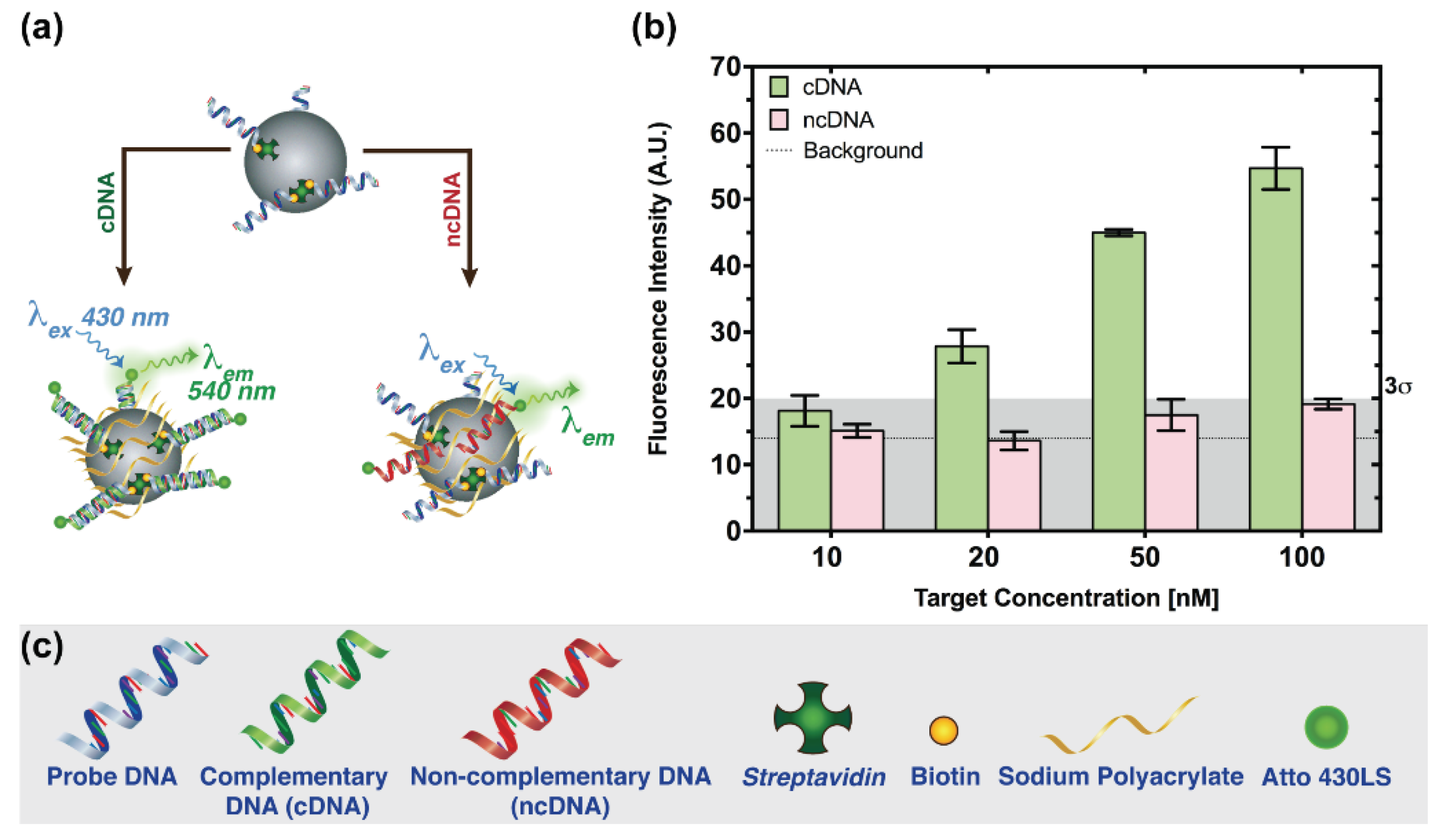
3.1. Probe Immobilization Strategies

DNA probe immobilization to agarose nanoporous beads was achieved through either electrostatic interactions or a covalent bond. In the former, QS beads with a positively charged surface were used to interact with the DNA:streptavidin complex, that immobilizes electrostatically to the beads mainly due to the negative charge of the DNA (Figure 2a). Using this immobilization strategy, calibration curves for increasing concentrations of target DNA (cDNA and ncDNA) labelled with an organic fluorophore (Atto 430LS) were measured. The limit of detection (LoD) obtained was 20 nM based on the 3σ criterion, with σ being the standard deviation of a blank assay (Figure 2b). The ncDNA signal is below the 3σ threshold for the range of concentrations tested. For the concentrations of cDNA above the threshold, specificity ratios between (2.05 ± 0.28) and (2.86 ± 0.20) were obtained. Nevertheless, the achievement of lower LoDs is limited by the background variability, which is intrinsic to assay design and adsorption of non-specific molecules to the charged beads. Noticeably, some of the inter-assay variability can be attributed to different microchannel conditions, such as packing of the beads, in between different assays, which may influence the flow rates and the density of probe immobilized on the beads’ surface. In order to reduce variability, a covalent immobilization strategy was developed, allowing the regeneration of the system.

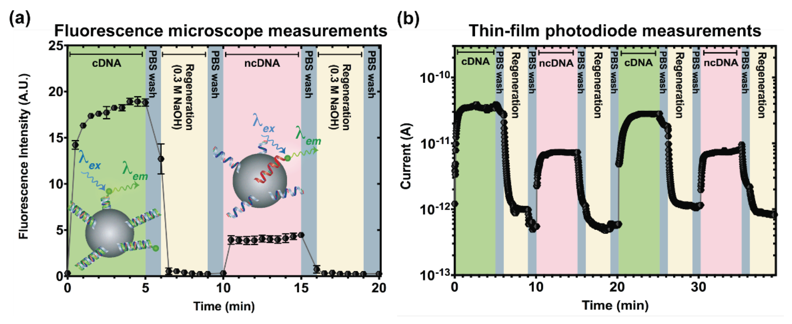
3.2. Microcolumn Regeneration

For the regeneration strategy, covalent immobilization of the DNA probe was used to avoid significant loss of probe density. A regeneration cycle was successfully obtained by first flowing cDNA, followed by 0.3 M NaOH for column regeneration, and finally ncDNA using first a fluorescence microscope for signal acquisition (Figure 3a). Inter-solutions washing steps were performed using PBS. A 17-fold difference between the measured fluorescence of cDNA and ncDNA (after PBS washing) was observed. The full regeneration of the column was achieved with the fluorescence values, after regeneration, being comparable to the background. In order to make this system compatible with a portable setup, the fluorescence acquisition of two regeneration cycles was performed using a 200 × 200 μm thin-film p-i-n a-Si:H photodiode (Figure 3b) aligned beneath the area in the microchannel where the beads were packed. The result shows the variation in photocurrent resulting from the measurement of fluorescence by the photodiode over two successive regeneration cycles. We can see that the trend is maintained and the results are consistent with those from the microscope.

4. Conclusions

In this work, we showed the proof-of-concept of a regenerable microfluidic bead-based DNA detection system with integrated fluorescence detection. The electrostatic probe immobilization strategy proved to be fast and simple, achieving a LoD between 10–20 nM. Furthermore, the detection of the target DNA is performed in 5 min. In systems where regeneration is used, the immobilization of probe DNA by covalent bonding to the beads was required, allowing the regeneration of the system without significant loss of probe DNA. The integration of thin-film photodiodes demonstrates the potential of the system as a point-of-use screening tool for biomedical, food and environmental analysis or even biodefense.

Author Contributions

C.R.F.C. conceived, designed and performed the experiments. D.R.S. fabricated and characterized the thin film p-i-n photodiodes. V.C. and J.P.C. supervised the project. All the authors contributed to the writing of the manuscript.

Acknowledgments

C.R.F.C. and D.R.S. acknowledge Fundação para a Ciência e Tecnologia (FCT) for their doctoral grants PD/BD/135274/2017 and SFRH/BD/95079/2013, respectively. INESC-MN acknowledges FCT for funding through the Associated Lab IN (UID/NAN/50024/2013) and the project OptLoc (PTDC/BBB-NAN/5927/2014).

Conflicts of Interest

The authors declare no conflict of interest.

References

  1. Ferguson, B.S.; Buchsbaum, S.F.; Swensen, J.S.; Hsieh, K.; Lou, X.; Soh, H.T. Integrated microfluidic electrochemical DNA sensor. Anal. Chem. 2009, 81, 6503–6508. [Google Scholar] [CrossRef] [PubMed]
  2. Wang, S.; Sun, Y.; Gan, W.; Liu, Y.; Xiang, G.; Wang, D.; Wang, L.; Cheng, J.; Liu, P. An automated microfluidic system for single-stranded DNA preparation and magnetic bead-based microarray analysis. Biomicrofluidics 2015, 9, 024102. [Google Scholar] [CrossRef] [PubMed]
  3. Pinto, I.F.; Caneira, C.R.; Soares, R.R.; Madaboosi, N.; Aires-Barros, M.R.; Conde, J.P.; Azevedo, A.M.; Chu, V. The application of microbeads to microfluidic systems for enhanced detection and purification of biomolecules. Methods 2017, 116, 112–124. [Google Scholar] [CrossRef] [PubMed]
  4. Pinto, I.F.; Santos, D.R.; Caneira, C.R.F.; Soares, R.R.G.; Azevedo, A.; Chu, V.; Conde, J.P. Optical biosensing in microfluidics using nanoporous microbeads and amorphous silicon thin-film photodiodes: Quantitative analysis of molecular recognition and signal transduction. J. Micromech. Microeng. 2018, 28, 094004. [Google Scholar] [CrossRef]
  5. Santos, D.R.; Soares, R.R.G.; Chu, V.; Conde, J.P. Performance of hydrogenated amorphous silicon thin film photosensors at ultra-low light levels: Towards attomole sensitivities in lab-on-chip biosensing applications. IEEE Sens. J. 2017, 17, 6895–6903. [Google Scholar] [CrossRef]
Figure 1. (a) Schematics of the beads used. The DNA probe was immobilized through (I) electrostatic interaction to positively charged agarose beads or (II) via covalent bonding between amino-modified probe DNA and NHS activated agarose beads. (b) Schematics of the microfluidic device used. (c) Micrograph of the microcolumn aligned with a photodiode array and structural schematics of the p-i-n a-Si:H photodiode with an integrated a-SiC:H filter, suitable for fluorescence measurements.
Proceedings 02 00953 g001
Figure 2. (a) Schematics of the assay used to detect DNA using an electrostatic immobilization of probe DNA to agarose beads. (b) Fluorescence detection of target DNA (10–100 nM) using the electrostatic immobilization of probe DNA, measured with a fluorescence microscope. It should be noticed that in this case two different columns were used to detect complementary and non-complementary DNA. The error bars correspond to the standard deviation of three separated assays performed in different microcolumns. (c) Schematics of the molecules used in the assays.
Proceedings 02 00953 g002
Figure 3. (a) Measurement of one hybridization assay cycle including microcolumn regeneration using a fluorescence microscope. The error bars correspond to the standard deviation of two individual assays performed in two different microcolumns. DNA hybridization steps were performed using 1 µM of target DNA, flowed through the microcolumn during five minutes. All the steps were performed at 7 µL/min. (b) Sequential measurement of two hybridization and regeneration assay cycles performed using the thin-film p-i-n a-Si:H photodiode at 0 V bias.
Proceedings 02 00953 g003
Publisher’s Note: MDPI stays neutral with regard to jurisdictional claims in published maps and institutional affiliations.

