Abstract
A gold-silver alloy film based spectral surface plasmon resonance imaging (SPRi) sensor has been prepared for in-situ quantitative detection of biochemical analytes at the sensor surface. This novel sensor has lower detection cost yet higher sensitivity relative to the conventional counterpart with a gold film. Using the laboratory-made multifunctional SPR sensing platform, both the resonant color images and the resonant spectra for the Au-Ag alloy film were measured at different incident angles. The quantitative relationship between the resonant wavelength and the average hue of corresponding resonant color image was established. With this relationship the most hue-sensitive spectral range was determined. After setting the initial resonant wavelength in the hue-sensitive spectral range, the refractive-index sensitivity of the Au-Ag alloy film based SPRi sensor was measured as Δhue/Δnc = 29,879/RIU, being 8 times higher than that obtained with the gold-film SPRi sensor. The immunodetection of benzo(a)pyrene (BaP) in water was fulfilled using the Au-Ag alloy film based SPRi sensor. The average hue of the SPR color image linearly increases with increasing the BaP concentration up to C = 0.5 μg/L and the slope is Δhue/ΔC = 132.2/(μg/L). The sensor is responsive to a change of BaP concentration as low as ΔC = 0.01 μg/L.
1. Introduction
SPRi is a well-established powerful technique for in situ, real-time, label-free and high-throughput analysis of biomolecular interaction on the surface of SPR chip. The commercial SPRi sensors are easily available nowadays, and they generally operate with a light source of a laser, providing users with time-resolved intensity images that allow for rapid analysis of kinetics of interfacial biomolecular interaction within a narrow linear intensity-response range. In most cases, SPR intensity images merely indicate the on-resonance and off-resonance areas of the sensor chip, enabling users to intuitively distinguish the biomolecular interaction areas from the no biomolecular interaction areas. It is difficult to quantify the surface bound biomolecules with SPR intensity images because the measured intensity is a relative parameter that cannot be accurately fitted through the Fresnel formula. The advent of charge-coupled device (CCD) based full-color RGB imaging sensor makes colored SPR images realized. The colored SPRi sensor is informative and has a very large dynamic response range and can utilize different colors to reflect the degree of biomolecular interaction on different micrometer-scale areas of the SPR chip. From these points of view, the colored SPRi sensor is superior to the intensity-interrogated SPRi sensor. The different colors on different areas of the colored SPR image are dominated by the resonant wavelengths for the surface plasmon modes on the corresponding areas. Once the information on the resonant wavelength involved in the colored SPR image is attained, quantitative detection by means of colored SPRi sensor can be easily realized. As a matter of fact, the use of colored SPRi sensor for quantitative detection has been recently fulfilled based on calculation of the hue component of the SPR color image [1,2]. By processing a SPR color image in the HSV color space using the hue-extracting algorithm, the color image with the RGB data at each pixel can be transformed into the 2D profile of hue component. The previous work demonstrated that the hue component at a pixel of the SPR color image has a quantitative correlation with the biomolecular interaction at the corresponding surface area of the SPR chip [1]. This work reports on the application of the hue-based SPRi sensor for in situ, quantitative trace detection of benzo(a)pyrene (BaP) in water. BaP is a small molecule with high carcinogenicity. The environmental protection agency (EPA) of America regulated that the Maximum Contaminant Level (MCL) of BaP in drinking water is 0.0002 mg/L (i.e., 0.2 ppb) [3]. The Chinese government has set national standards for drinking water quality in 1985 that show the MCL of 0.01 μg/L (i.e., 10 ppt) for BaP in drinking water [3]. Low-concentration BaP in air and water is colorless and odorless and thus cannot be easily detected. For in situ trace BaP detection, the SPR chips used in this work were prepared by sputtering of AuAg alloy films on glass substrates. AuAg alloy films are more stable than pure silver films and have higher SPR sensitivity than pure gold layers. Furthermore, AuAg alloy film allows for readily functionalization with the established gold-thiol chemistry. These outstanding characteristics of AuAg alloy films are the basis of the remarkable sensing properties of the SPRi sensor prepared herein. In this work, the relationship between the resonance wavelength and the average hue of the SPR color image was obtained, and the hue-sensitive wavelength range was determined, and the AuAg alloy film was successfully modified with the antibody molecules to result in a SPRi immunosensor for BaP detection. The sensor’s performance was investigated.
2. Experimental Conditions
Chemical reagents including 1-(3-Dimethylaminopropyl)-3-ethylcarbodiimide hydrochloride (EDC), n-hydroxysuccinimide (NHS) and 3-mercaptopropionic acid (3-MPA) were purchased from Aladdin Reagent Co. (Shanghai, China); Benzo[a]pyrene antibody (BaP-13, sc-51508, 1 μg/mL) was obtained from Santa Cruz Biotechnology Company, USA; Benzo[a]pyrene standard solution (4 μg/mL in methanol) was purchased from National Institute of Metrology, China; Milli-Q water was used for aqueous solution preparation. Sputtering target of Au(50 wt%)Ag(50 wt%) alloy was obtained from Triiion Metals Co., LTD., Beijing, China; Slide glass substrates (n = 1.522 @633 nm) were purchased from Matsunami glass Co., Osaka, Japan.
A number of SPR chips were prepared by successive sputtering of a 3-nm Cr layer and a 50-nm AuAg alloy film on the cleaned glass substrates. To enhance both the sensitivity and selectivity to BaP in water, the as-prepared SPR chips were modified with BaP antibody molecules according to the following steps: first to soak the SPR chip in the ethanolic MPA solution (30 mg/mL) at room temperature for 12 h for formation of the self-assembled MPA monolayer on the AuAg alloy film; secondly to immerse the MPA-modified SPR chip in an aqueous mixed solution containing NHS (0.05 mol/L) and EDC (0.1 mol/L) for 40 min for activating the MPA monolayer; Finally to dip the activated SPR chip into the antibody solution (0.02 μg/mL BAP-13 in PBS) for 30 min for binding antibody molecules to the surface. The modified SPR chips were kept in refrigerator before use.
The colored SPRi platform was constructed using a tungsten-halogen lamp (LS-1, Ocean Optics, Portland, MI, USA), a glass prism (n = 1.799 @633 nm, Beidong Photoelectric Automation, Wenzhou, China), a fibre collimator and a linear polarizer (Daheng Optics, Beijing, China), an optical spectrum analyzer (USB2000+, Ocean Optics, USA), a CCD digital camera (Xiansheng Instrument, Shenzhen, China), a microscope lens tube, and a fluidic chamber [2]. The prism is mounted on a goniometer and the SPR chip is closely sandwiched between the glass prism and the fluidic chamber. The broadband light from the lamp passes through the fiber collimator and the linear polarizer to become a p-polarized collimated beam (~5 mm diameter, divergence angle < 0.2°). The beam is incident upon the prism and undergoes total internal reflection at the glass-metal interface of the SPR chip. The reflected beam is received with the microscope lens tube in which the beam is split into two beams with the semitransparent mirror mounted inside. The two beams reached the CCD digital camera and the CCD spectrometer, respectively, for simultaneous measurements of SPR image and SPR spectrum. It is worth noting that the CCD spectrometer used here is for determining the resonance wavelength and finally establishing the relationship between the hue component of the SPR color image and the resonance wavelength. With this relationship the hue-sensitive spectral range can be determined.
3. Results and Discussions
The surface modification process was monitored in real time by SPR spectroscopy. The MPA self-assembly causes the resonance wavelength to shift from 551.28 nm to 579.27 nm (ΔλR = 28 nm), and the antibody immobilization leads to a redshift of ΔλR = 22.4 nm. The SPR spectroscopic results demonstrated that the surface modification of SPR chips is successful.
To establish the relationship between the hue component of the SPR color image and the
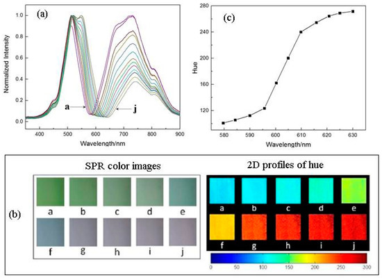
resonance wavelength, both the SPR spectra and SPR images were measured at different incident angles. Figure 1a,b show the ten SPR spectra and ten SPR color images obtained at ten angles of incidence using the water-covered Au-Ag alloy film. The ten 2D profiles of hue derived from the ten SPR color images were also shown in Figure 1b. Figure 1c displays a plot of the average hue against the resonance wavelength. A linear relationship between the average hue and the resonance wavelength exists only in a small range of wavelength from 595.6 nm to 609.8 nm and the slope is Δhue/ΔλR = 8.2/nm. In this narrow spectral region the refractive-index (RI) sensitivity in terms of ΔλR/Δnc was measured to be ca. 2950 nm/RIU for the Au-Ag alloy film based SPR sensor. According to Δhue/ΔλR= 8.2/nm, the RI sensitivity in terms of Δhue/Δnc should be 24,202/RIU. The measured value of Δhue/Δnc is equal to 29,879/RIU, being very close to the expected value.

Figure 1.
(a) ten SPR spectra and (b) ten SPR colour images and the corresponding 2D hue distributions (both spectra and images were measured at ten angles of incidence using the water-covered AuAg alloy film), (c) the average hue versus the resonance wavelength determined with the SPR spectra.
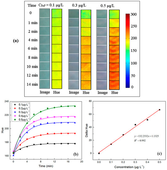
The immunodetection of BaP in water was performed using the colored SPRi sensor. Figure 2a shows the SPR color images successively recorded during the course of the surface immunoreaction with a given concentration of BaP solution. Figure 2b displays the corresponding 2D profiles of hue, with which the average hue was determined. Plotting the average hue against the detection time leads to the five curves corresponding to five concentrations of BaP solutions. Each curve represents a kinetic process of surface immunoreaction with a given concentration of BaP solution. Figure 2c displays the relationship between the change of average hue and the BaP concentration. The change of average hue linearly increases with increasing the BaP concentration up to 0.5 μg/L and the slope is Δhue/ΔC = 132.2 (μg/L) − 1. With the hue resolution of Δhue = 5 for the CCD camera used, the present SPRi sensor enables to distinguish a change of lower than 0.01 μg/L in BaP concentration.

Figure 2.
(a) SPR color images and corresponding 2D hue profiles recorded at different immunoreaction times with different concentrations of aqueous BaP solutions, (b) average hue of SPR images versus the immunoreaction time of BaP at different concentrations, (c) Average hue variation as a function of BaP concentration.
Acknowledgments
This work was supported by the National Key Basic Research Program of China (973) (2015CB352100) and National Natural Science Foundation of China (61377064, 61675203).
References
- Wong, C.L.; Olivo, M. Surface Plasmon Resonance Imaging Sensors: A Review. Plasmonics 2014, 9, 809–824. [Google Scholar] [CrossRef]
- Fan, Z.; Gong, X.; Lu, D.; Gao, R.; Qi, Z. Benzo[a]pyrene Sensing Properties of Surface Plasmon Resonance Imaging Sensor Based on the Hue Algorithm. Acta Phys.-Chim. Sin. 2017, 33, 1001–1009. [Google Scholar] [CrossRef]
- Wang, L.; Wan, X.; Gao, R.; Lu, D.; Qi, Z. Nanoporous Gold Films Prepared by a Combination of Sputtering and Dealloying for Trace Detection of Benzo[a]pyrene Based on Surface Plasmon Resonance Spectroscopy. Sensors 2017, 17, 1255. [Google Scholar] [CrossRef] [PubMed]
Publisher’s Note: MDPI stays neutral with regard to jurisdictional claims in published maps and institutional affiliations. |
© 2018 by the authors. Licensee MDPI, Basel, Switzerland. This article is an open access article distributed under the terms and conditions of the Creative Commons Attribution (CC BY) license (https://creativecommons.org/licenses/by/4.0/).