Proceeding Paper

Fast Formation of Lipid Bilayer Membranes for Simultaneous Analysis of Molecular Transport Using Parylene Coated Chips †

by
Tanzir Ahmed
1,*,
Sander van den Driesche
1,
Martin Oellers
1,
Roland Hemmler
2,
Karsten Gall
2,
Satya Prathyusha Bhamidimarri
3,
Mathias Winterhalter
3,
Richard Wagner
3 and
Michael J. Vellekoop
1
1
Institute for Microsensors, Microactuators and Microsystems (IMSAS), University of Bremen, Microsystems Center Bremen (MCB), D-28359 Bremen, Germany
2
Ionovation GmbH, D-49143 Bissendorf, Germany
3
Department of Life Sciences & Chemistry, Jacobs University, D-28759 Bremen, Germany
*
Author to whom correspondence should be addressed.
Presented at the Eurosensors 2018 Conference, Graz, Austria, 9–12 September 2018.
Proceedings 2018, 2(13), 920; https://doi.org/10.3390/proceedings2130920
Published: 23 November 2018
(This article belongs to the Proceedings of EUROSENSORS 2018)

Abstract

:
Artificial lipid bilayers are an essential tool to investigate channel forming proteins. A particular challenge is to study antibiotic uptake through bacterial porins requiring low volume and parallelization. Here, we present a lipid bilayer silicon chip having a Parylene-C coated silicon nitride membrane with different aperture sizes. The Parylene-C allows very fast lipid bilayer membrane fabrication, 30 to 130 s. The realization-success is very high and an average lifetime of at least 9 h was established. Furthermore, a 3D-printed holder is realized where parallel assembly of the chips, including fluid inlets for the pipetting robot, is demonstrated.

1. Introduction

A standard method to achieve a hydrophobic aperture in a Teflon foil for artificial bilayer measurements is by performing mechanical machining (such as punching mechanically) or by applying electrical sparks on the foil [1]. The result in a coarse aperture with a non-predictable diameter is not optimal for the lipid bilayer formation. For a quick and automated bilayer measurement, apertures with smooth hydrophobic surface and precise sizes are needed [2]. In this contribution, micro-apertures with different diameters were fabricated in a silicon nitride membrane and later coated with Parylene-C, a hydrophobic and biocompatible material to support artificial lipid bilayers.
The presented micro-aperture chip together with a 3D printed holder enables easy parallel chip mounting including fluidic accessibility to industrial grade pipetting robots for simultaneous bilayer analysis.

2. Materials and Methods

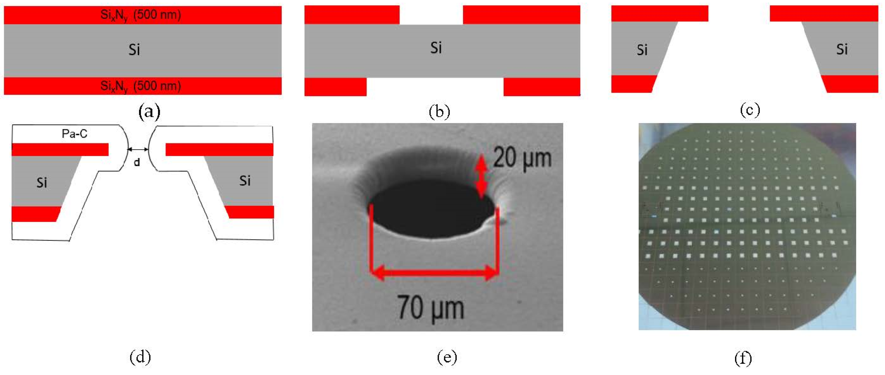
2.1. Lipid Bilayer Chip Fabrication

The bilayer chip was realized by standard microfabrication techniques. A double polished silicon wafer (380 µm thick) was utilized as base substrate. Photolithography and reactive ion etching steps were conducted to realize the membrane and aperture. KOH anisotropic etching was performed from the backside of the wafer to open the silicon nitride membrane (Figure 1a–c). The adhesion promoter 3-trimethoxysilylpropyl methacrylate was used to improve the adhesion between the silicon nitride membrane and parylene-C coating. In Figure 1e, a membrane with a 70 µm aperture, coated with 10 µm of parylene-C, is depicted. After parylene deposition, the wafer was diced into 5 × 5 mm2 chips (Figure 1f).

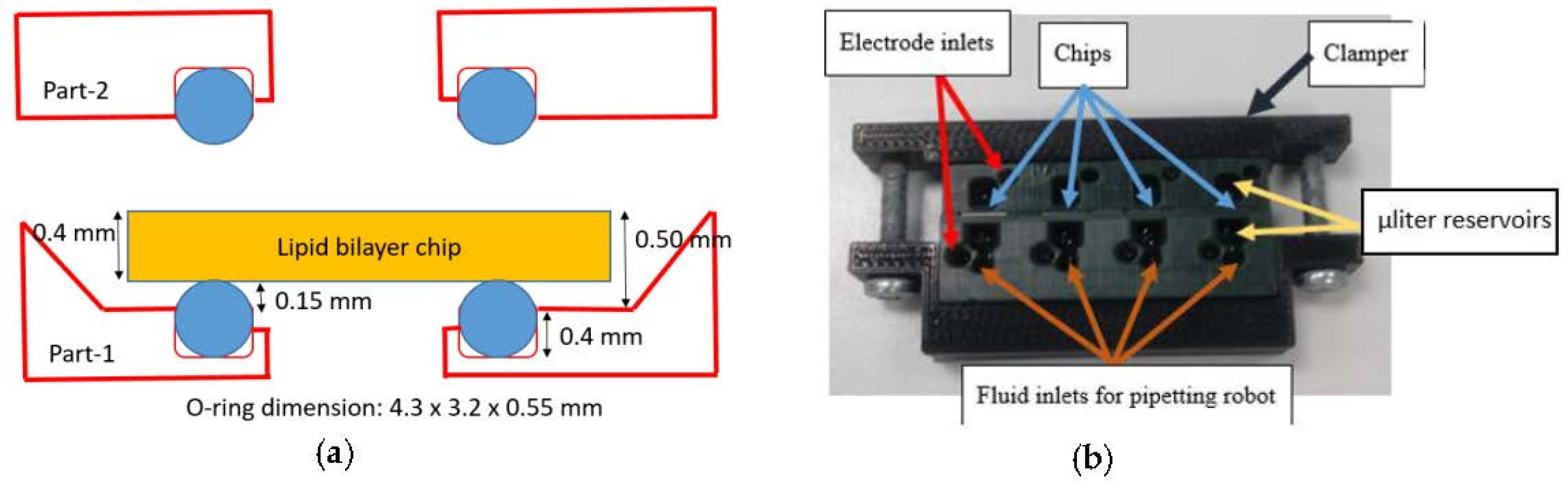
2.2. Designing and Printing the Parallel Bilayer Analysis Platform
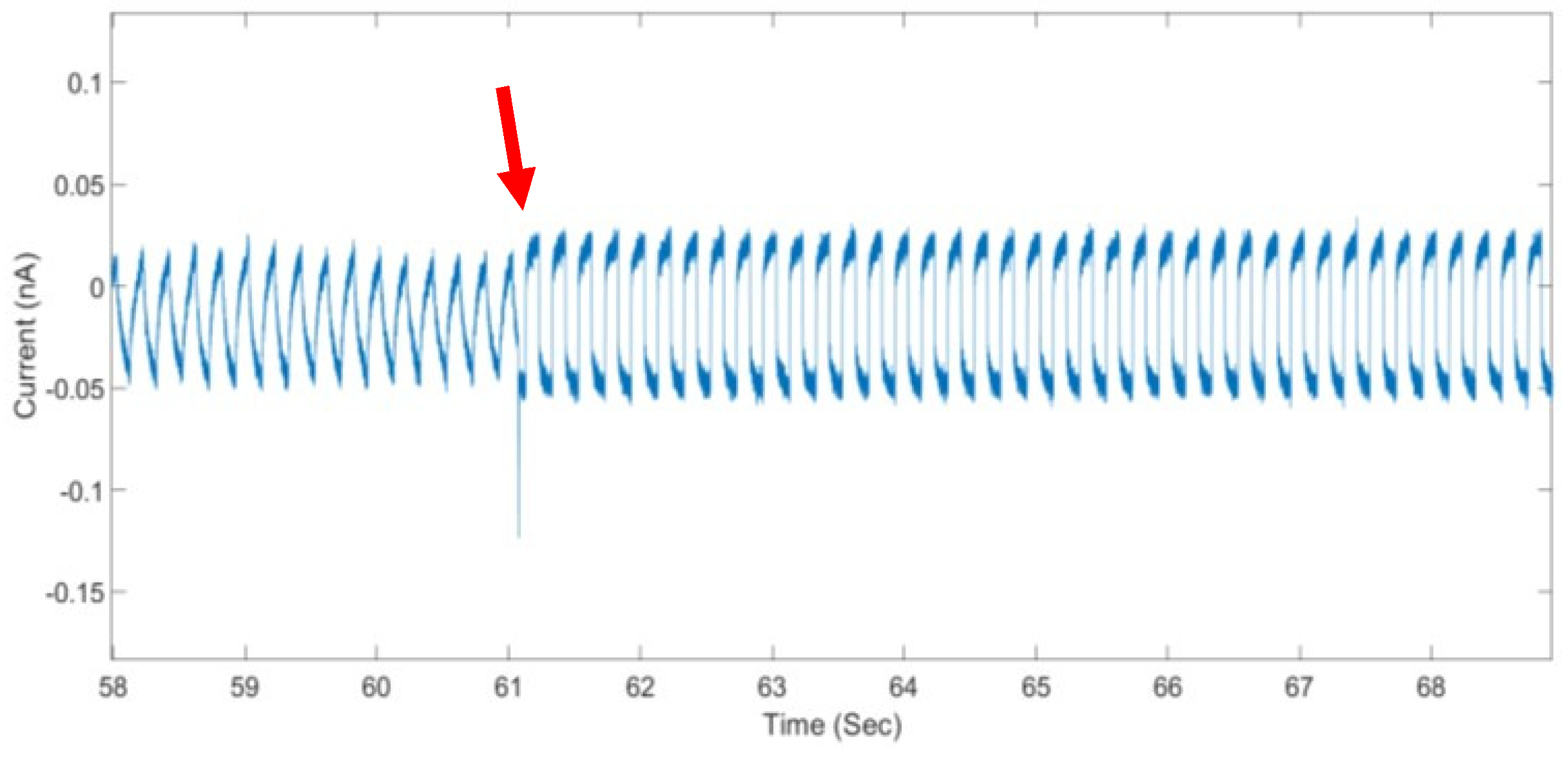
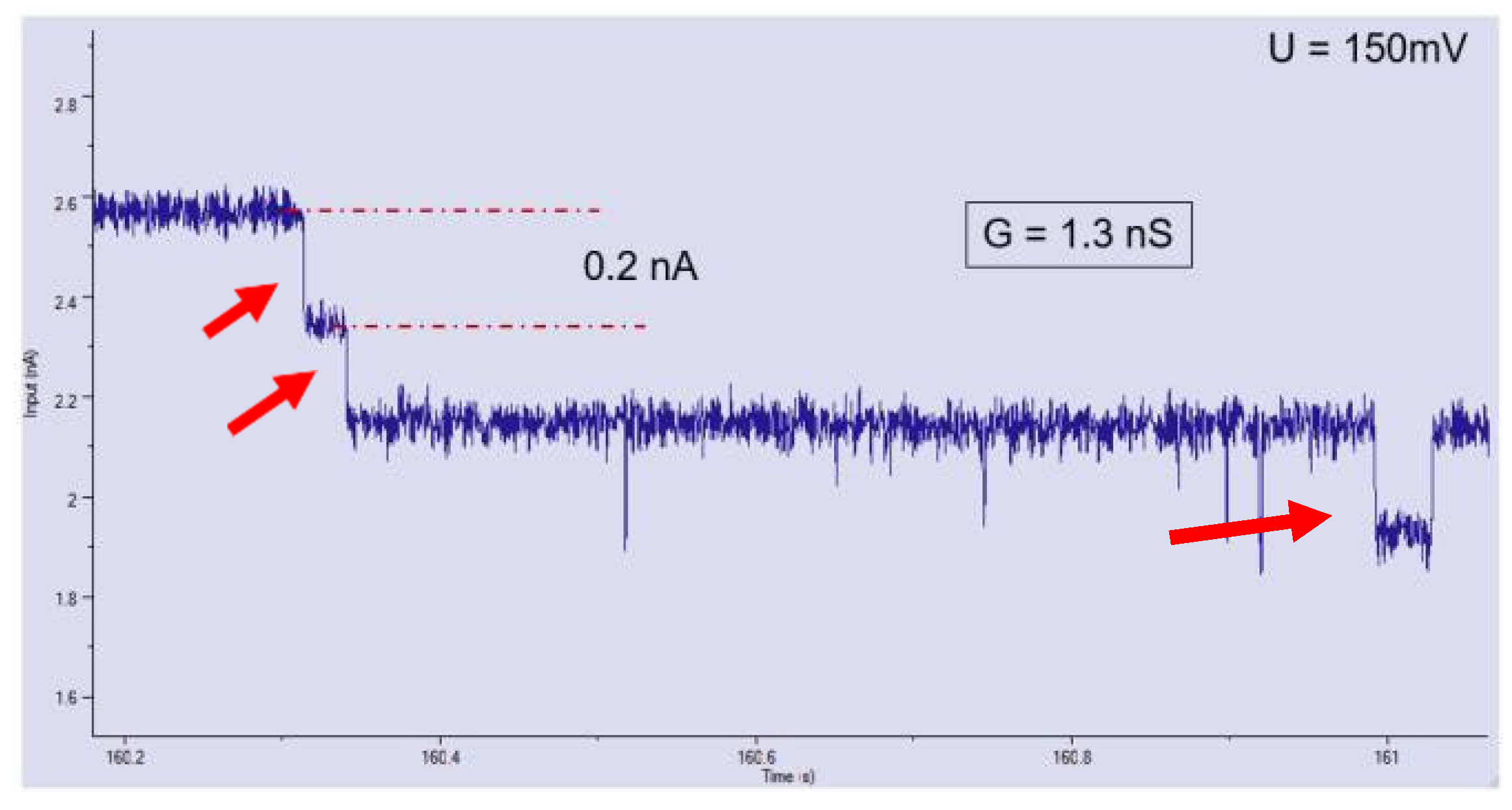
A lipid bilayer analysis platform with four aperture chips was designed and realized for simultaneous investigations of ion transport through proteins located in synthetic lipid membranes. The 3D printed chip holder contains two separate parts with each four reservoirs (c.a. 40 µL), compatible with standard industrial pipetting robots. The mounting method for the bilayer chips is depicted in Figure 2a [3]. The chip is mounted in a chip slot located in a 3D printed holder (Part-1). In the holder, O-rings (4.30 × 3.20 × 0.55 mm) were used to ensure the sealing between the chip and chip-holder. This platform allows easy replacement of the chips. The holder design was drawn in Autodesk Inventor 2018 software. After designing, the 3D holder platform was printed. In this work, a Micro HiRes (EnvisionTEC Inc., Dearborn, MI, USA) printer was used to fabricate the platform. The printing material was HTM140M, a high-resolution, high-temperature, low viscosity acrylic polymer resin. After printing, the constructed parts were cleaned with isopropanol and dried with nitrogen gas. Finally, the printed parts were placed in a UV flood chamber for five minutes to crosslink the remaining non-cured resin. A completed parallel platform with four bilayer chips is shown in Figure 2b.

3. Results

The fabricated chips allowed very fast bilayer realization. After applying the lipid (DPhPC/N-decane) on the aperture, successful formation of the lipid bilayer was always achieved within 130 s and sometimes already after 30 s. Figure 3 shows the formation of a stable bilayer in a chip with an 80 µm aperture, after 61 s. Here, a 1 molar NaCl buffer solution was pipetted at both sides of the membrane to conduct ion transport measurements. The measured bilayer capacitance was 21 pF.
The average lifetime of a stable synthetic membrane in an 80 µm aperture chip was at least 9 h. The setup was placed in a petri dish covered with Parafilm to reduce buffer evaporation. Alternatively, the buffer evaporation can be further reduced by utilizing a syringe pump. In this contribution, chips with an aperture of 80 µm and 90 µm were used. Out of total nine trials, the bilayer was formed eight times, yielding 90% realization success. Here, for the chip with 80 µm aperture, the bilayer capacitance varied between 28 pF to 30 pF respectively whereas the chip with 90 µm aperture showed slightly higher bilayer capacitance between 32 pF to 36 pF.
In addition, to prove the functionality of the presented synthetic membrane setup, ion-channel activity through an outer membrane porin was successfully conducted. For this experiment, a chip with a 200 µm aperture was used. At a 150 mV holding potential, the resultant current change is 0.2 nA, resembling a single port opening enabling ion transport through the pore, see Figure 4. The measured conductance of 1.3 nS was in accordance with published values [4].

4. Conclusions

We have demonstrated a 3D printed lipid bilayer platform with parylene-C coated silicon nitride apertures to perform ion transport investigations through porins fused into a synthetic membrane. The 3D printed platform allows easy replacement of the aperture chips. The synthetic membrane can be created very fast, between 30 and 130 s. The average lifetime of a stable bilayer is at least 9 h. Careful removing the excess protein using an automatic syringe pump improves the quality of the electrical recording. Moreover, to prove the ion channel activities in a 200 µm Parylene coated aperture, the outer membrane porin (OmpF) could be successfully integrated in the bilayer and displayed its characteristic electrical fingerprint.

Acknowledgments

This work is a part of the research project “Boomer”—Bacterial periplasmic organelles and outer membrane vesicles, project number: ZF4176703AJ6, which is supported by the Federal Ministry for Economic Affairs and Energy (BMWi) under the framework of the Central Innovation Programme for SMWs (ZIM). SPB was financed through IMI-Translocation (115525).

Conflicts of Interest

The authors declare no conflict of interest.

References

  1. Mayer, M.; Kriebel, J.K.; Tosteson, M.T.; Whitesides, G.M. Microfabricated teflon membranes for low-noise recordings of ion channels in planar lipid bilayers. Biophys. J. 2003, 85, 2684–2695. [Google Scholar] [CrossRef]
  2. O’Shaughnessy, T.J.; Hu, J.E.; Kulp, J.L.; Daly, S.M.; Ligler, F.S. Laser ablation of micropores for formation of artificial planar lipid bilayers. Biomed. Microdevices 2007, 9, 863–868. [Google Scholar] [CrossRef] [PubMed]
  3. van den Driesche, S.; Lucklum, F.; Bunge, F.; Vellekoop, M.J. 3D printing solutions for microfluidic chip-to-world connections. Micromachines 2018, 9, 71. [Google Scholar] [CrossRef] [PubMed]
  4. Danelon, C.; Suenaga, A.; Winterhalter, M.; Yamato, I. Molecular Origin of the Cation Selectivity in OmpF Porin. Single Channel Conductances versus Free Energy Calculation. Biophys. Chem. 2003, 104, 591–603. [Google Scholar] [CrossRef]
Figure 1. (ad) Fabrication processes of micro apertures on silicon nitride membranes where a 380 µm double sided polished silicon wafer was used as base substrate; (e) Parylene-C coated aperture (70 µm diameter); (f) finalized wafer after dicing (chip dimension 5 × 5 mm2).
Proceedings 02 00920 g001
Figure 2. Parallel bilayer analysis platform: (a) Chip slot design with O-ring incorporation; (b) Complete measuring setup fixed by a clamper.
Proceedings 02 00920 g002
Figure 3. Formation of a lipid bilayer pointed by the arrow, here the bilayer formed 61 s after applying the lipid.
Proceedings 02 00920 g003
Figure 4. Current response of porin OmpF after successful integration in a synthetic bilayer. The lipid bilayer is supported in a 200 µm Parylene coated aperture. The measured current increment is 0.2 nA per pore opening (pointed by arrows) resembling ion transport through the protein pores fused in the bilayer at a 150 mV holding potential.
Proceedings 02 00920 g004
Publisher’s Note: MDPI stays neutral with regard to jurisdictional claims in published maps and institutional affiliations.

