Proceeding Paper

Application of a Novel Low-Cost Hyperspectral Imaging Setup Operating in the Mid-Infrared Region †

Research Center for Non Destructive Testing GmbH (RECENDT), 4040 Linz, Austria
*
Author to whom correspondence should be addressed.
Presented at the Eurosensors 2018 Conference, Graz, Austria, 9–12 September 2018.
Proceedings 2018, 2(13), 800; https://doi.org/10.3390/proceedings2130800
Published: 30 November 2018
(This article belongs to the Proceedings of EUROSENSORS 2018)

Abstract

:
In this contribution, we demonstrate the realization and application of a low-cost, flexible, small and fast hyperspectral imaging approach operating in the mid-infrared fingerprint region where most molecules exhibit their fundamental vibrations. Following this approach, the recording of chemical images of macroscopic-sized samples at standoff distances in reflection geometry is possible. The optical setup is based on spectral identification by means of a MEMS-based Fabry-Pérot interferometer combined with 2D-snapshot spatial resolution using a bolometer camera. Results show the successful spatially resolved (resolution below 500 µm) chemical identification of different samples deposited on a metal surface (FOV = 6 × 5 cm) at a working distance of 35 cm.

1. Introduction

Spatially resolved chemical identification of macroscopic-sized samples is of importance in many fields including industrial process analytics, e.g., in manufacturing processes or food industry, as well as in biomedical and forensic applications, cultural properties science or for atmospheric gas sensing. The mid-infrared (MIR, 2.5–25 µm) wavelength region is especially interesting for spectroscopic evaluation because it covers the narrowband fundamental molecular vibrations. Thereby higher sensitivities—ca. 1000-fold compared to the near-infrared (NIR, 0.8–2.5 µm)—can be achieved and proper multi-component analysis becomes possible as multiple narrow absorption bands are covered. Nevertheless, most of the available infrared (IR) hyperspectral imaging (HSI) systems operate in the NIR region where weak and spectrally broad overtone vibrations are observed. This has mainly two reasons, firstly NIR light sources (e.g., halogen lamps) provide enough optical power for the illumination of large sample areas simultaneously enabling high SNR, and secondly short-wave IR (SWIR, 0.9–1.7 µm) cameras are already available at affordable prices (~1 cent per pixel) compared to MIR cameras (~10 cent per pixel). Furthermore, MIR HSI systems need to be cooled and in many cases possess unfavorable characteristics such as being not portable, not capable of real-time acquisition and bound to fixed sample sizes.
However, most recently two technological developments allow for low-cost MIR imaging which has been mutually exclusive up to now. Firstly, new low-cost and robust tunable MEMS Fabry-Pérot interferometers (FPI) operating in the MIR range offer broad spectral coverage with real-time capability and are especially suitable for applications demanding miniaturization and field applicability. Secondly, low-cost (~1 cent per pixel), small-sized and sensitive uncooled bolometer cameras operating in the wavelength range of 8 to 14 µm recently became available. By combining a bolometer camera with a spectrometer, a MIR HSI system can be realized as already shown in [1]. So far MIR HSI systems have been mostly based on grating, prism or Fourier-transform spectrometers and on HgCdTe focal plane arrays (FPA) leading to expensive and bulky systems mostly restricted to laboratory applications [2]. A tunable FPI has already been successfully implemented for HSI in the MIR range coupled to an HgCdTe FPA enabling passive, chemical standoff detection of gas plumes [3].
In this work we demonstrate a novel MIR HSI system consisting of a MEMS FPI, a bolometer camera and an objective system. All employed components can be fully integrated, thus the setup has the potential to be implemented in a mobile device. We show its successful application by recording highly resolved chemical images of different kinds of samples, such as polymers, oils and paints at standoff distances of 35 cm in reflection geometry. The different materials in the spectral images can be analyzed by a machine-learning based classification algorithm allowing also real-time analyzation. The presented approach makes the technique of MIR hyperspectral imaging affordable for the first time (hardware costs below 500 €) using small-sized and field-applicable hardware. Thereby, MIR HSI becomes more accessible for many applications where spatially resolved chemical identification of large-sized samples in real-time is of interest.

2. Materials and Methods

The small and portable HSI setup—see photo in Figure 1a—consists of a custom-made MEMS FPI, a bolometer camera and an objective system. The FPI can be tuned from 8 to 10.5 µm with a filter time constant of few milliseconds. The bolometer camera consists of 160 × 120 pixels with a pixel pitch of 12 µm and a frame rate of 9 Hz. For the setup an objective system was designed in order to be able to change the field of view (FOV) and the spatial resolution depending on the specific application. The presented results are conducted with an angular FOV of 10° leading to a spatial resolution of below 500 µm and a measuring area of approximately 6 × 5 cm, see recorded image of a sliding caliper in Figure 1b. For sample illumination a common low-cost thermal emitter was used. The HSI system and the light source were mounted in specular reflection geometry with an angle of incidence of ~10°.
The oil samples measured in this work were a motor oil (labeled as oil1) and an olive oil (labeled as oil2). The two measured paints were a printed circuit lacquer (labeled as paint1) and a protective lacquer (labeled as paint2). The polymer films included polycarbonate (PC), polyethylene terephthalate (PET) and polypropylene (PP). Samples were deposited on a reflecting metal plate. The analysis of the spectral images was conducted using a machine-learning based hyperspectral image classification software (perClass Mira) which allows user-defined multi-class classification, automatically selects machine learning models and enables real-time implementation [4].

3. Results and Discussion

3.1. Oils and Paints

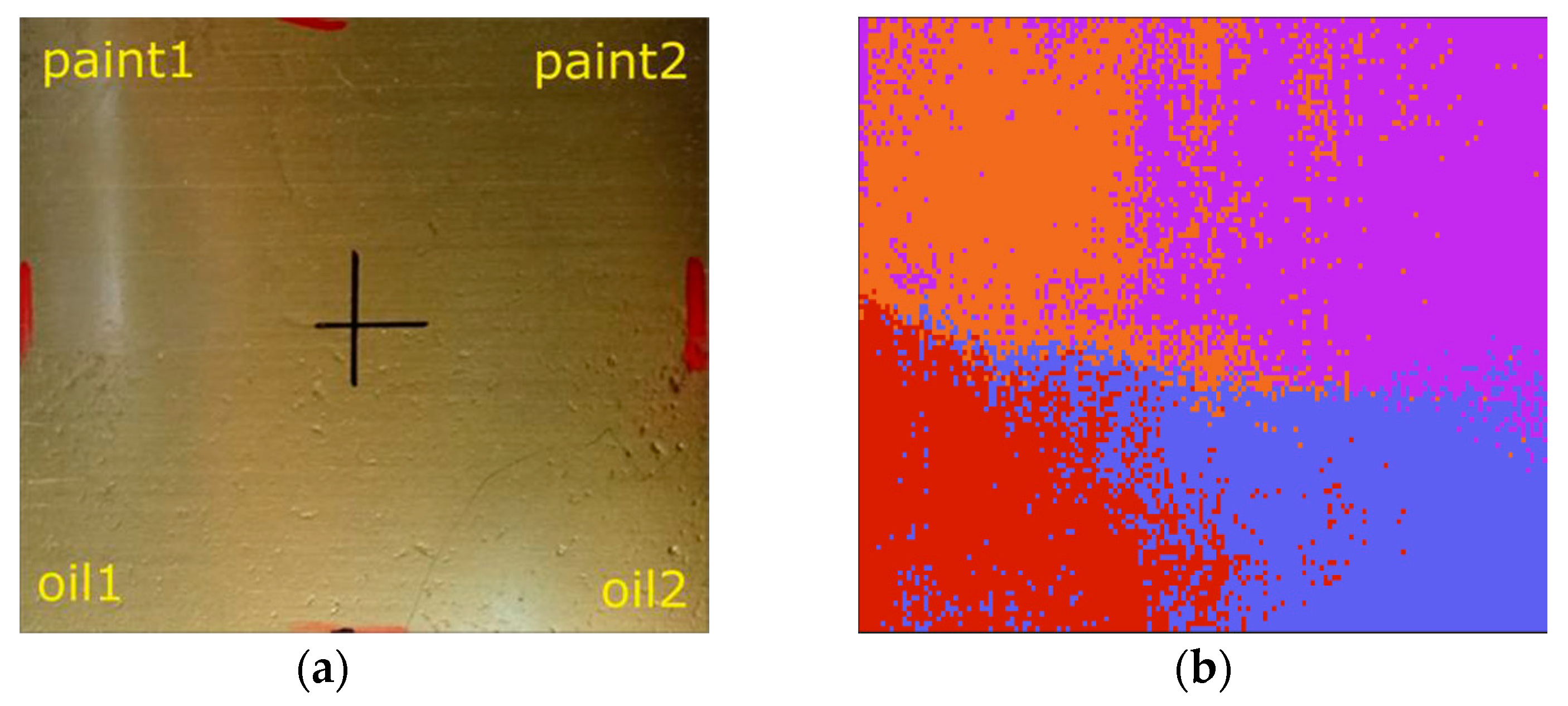
A metal plate with the two different oils and paints, see Figure 2a, was illuminated by the thermal light source and the reflected light was collected by the HSI system. The narrowband wavelength transmission of the filter was tuned over the whole spectral range and an image was recorded for every filter position. A single class illustrated by a different color was assigned to each material using the acquired spectral information in the respective area of the hyperspectral image. By means of machine-learning based classification, the false-color image in Figure 2b was generated. It can be seen that the different chemical components can be clearly distinguished. Furthermore there are some overlaps of the colors which demonstrates that the spectra of oils just as the spectra of the paints are very similar, but it could also indicate that the samples partly mixed during the deposition.

3.2. Polymer Films

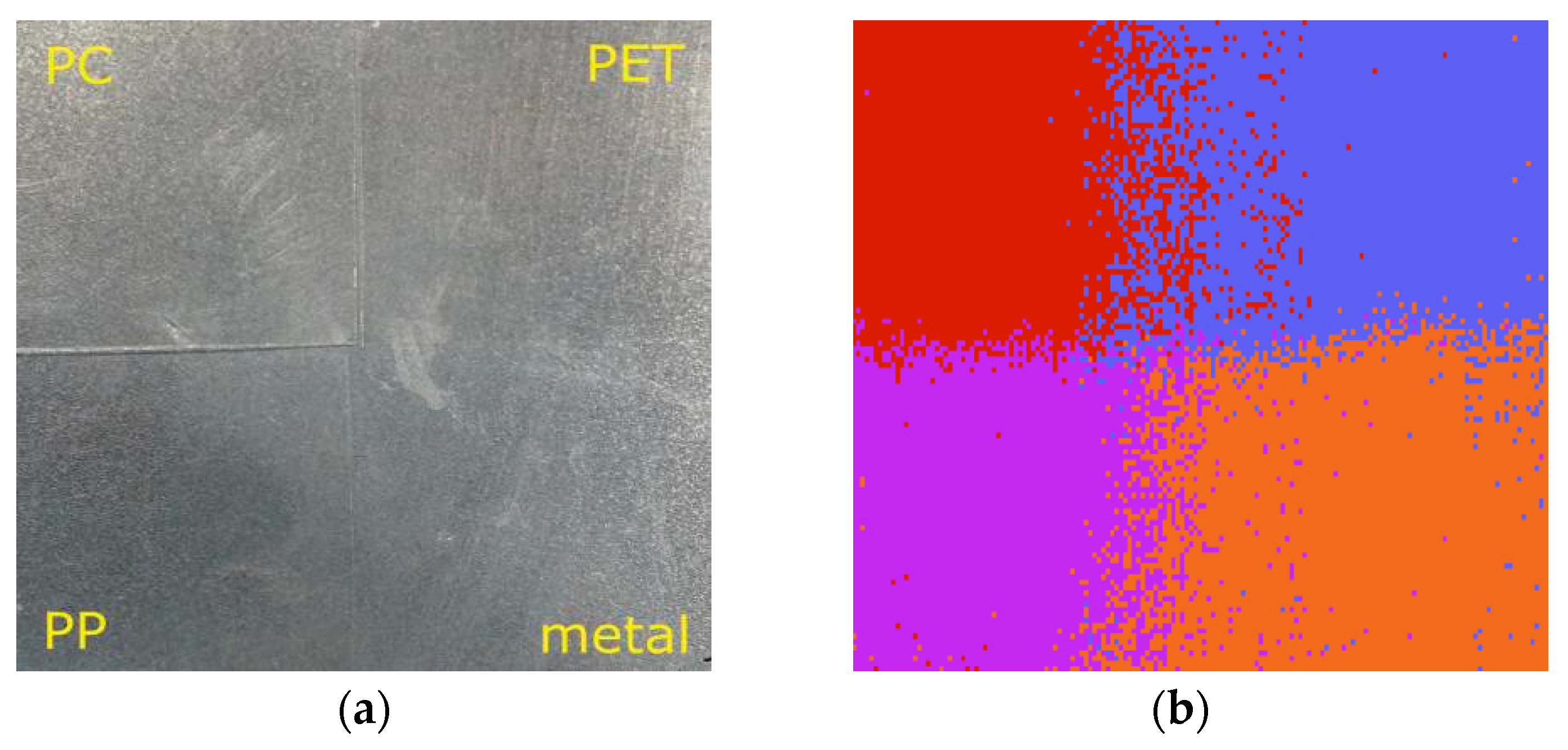
The same measurement configuration as described above was used for imaging the three different polymer films which were put onto a metal plate, as shown in Figure 3a. The differentiation of the samples was executed again by classification analysis. As the polymers show different absorption bands at different wavelengths, the films can be well distinguished showing only small overlaps in the false-color image, as can be seen in Figure 3b.

4. Conclusions and Outlook

We showed the successful application of a novel low-cost hyperspectral imaging system by spatial and chemical resolution of different materials by means of hyperspectral image analysis. Hyperspectral imaging is basically SNR-limited for standoff measurements at a macroscopic scale especially in the MIR region due to the low signal intensity. This indicates the importance of the optical design and the sample illumination. Depending on the specific demands of the application, the illumination can also be executed by newly available MIR laser-based light sources such as quantum cascade lasers [5] or supercontinuum lasers [6]. For the first time hyperspectral imaging in the decisive fingerprint region was realized to be low-cost and small-sized. By operating in this spectral region the amplitude of the spectral respectively molecular response—illustrating the key parameter of hyperspectral imaging—can be dramatically improved. The presented system opens up new possibilities in the field of hyperspectral imaging especially for process analytics by being miniaturized, portable and real-time capable. And finally the barrier of high costs—so far connected to MIR hyperspectral imaging systems—can be crossed making this promising technology more widely accessible in the future.

Author Contributions

M.B., J.K. and G.L. conceived and designed the experiments; J.K. and K.D. performed the experiments; J.K. and R.Z. analyzed the data; F.H. designed and built the electronic drivers; J.K., R.Z. and M.B. wrote the paper. All authors have read and agreed to the published version of the manuscript.

Funding

This work has been supported by the project “multimodal and in-situ characterization of inhomogeneous materials” (MiCi) by the federal government of Upper Austria and the European Regional Development Fund (EFRE) in the framework of the EU-program IWB2020. This work was also supported by the strategic economic- research program “Innovative Upper Austria 2020” of the province of Upper Austria.

Conflicts of Interest

The authors declare no conflict of interest. The founding sponsors had no role in the design of the study; in the collection, analyses, or interpretation of data; in the writing of the manuscript, and in the decision to publish the results.

References

  1. Sugawara, S.; Nakayama, Y.; Taniguchi, H.; Ishimaru, I. Wide-field mid-infrared hyperspectral imaging of adhesives using a bolometer camera. Sci. Rep. 2017, 7, 12395. [Google Scholar] [CrossRef] [PubMed]
  2. Türker-Kaya, S.; Huck, C. A Review of Mid-Infrared and Near-Infrared Imaging: Principles, Concepts and Applications in Plant Tissue Analysis. Molecules 2017, 22, 168. [Google Scholar] [CrossRef] [PubMed]
  3. Marinelli, W.J.; Gittins, C.M.; Gelb, A.H.; Green, B.D. Tunable Fabry-Perot etalon-based long-wavelength infrared imaging spectroradiometer. Appl. Opt. 1999, 38, 2594–2604. [Google Scholar] [CrossRef] [PubMed]
  4. Paclik, P.; Lai, C. perClass Mira User Guide. Available online: http://doc.perclass.com/perClass_Mira_1.0/Introduction.html (accessed on 18 July 2018).
  5. Schwaighofer, A.; Brandstetter, M.; Lendl, B. Quantum cascade lasers (QCLs) in biomedical spectroscopy. Chem. Soc. Rev. 2017, 46, 5903–5924. [Google Scholar] [CrossRef] [PubMed]
  6. Kilgus, J.; Duswald, K.; Langer, G.; Brandstetter, M. Mid-Infrared Standoff Spectroscopy Using a Supercontinuum Laser with Compact Fabry–Pérot Filter Spectrometers. Appl. Spectrosc. 2018, 72, 634–642. [Google Scholar] [CrossRef] [PubMed]
Figure 1. (a) Photograph of the built HSI system. The size of the camera and the FPI is compared to a 1 cent coin, which is fittingly the price of a single pixel of the bolometer camera; (b) Image of a sliding caliper on a breadboard recorded with the bolometer camera at a random filter position confirming the basic functionality of the optical system and the FOV of 6 × 5 cm.
Proceedings 02 00800 g001
Figure 2. (a) Visible image of the oil and paint samples deposited in 4 areas on a metal plate. The cross indicates the center where all samples are tangent; (b) Generated false-color image using a classification algorithm.
Proceedings 02 00800 g002
Figure 3. (a) Visible image of the three different polymer films on a metal plate; (b) False-color image generated with a classification algorithm.
Proceedings 02 00800 g003
Publisher’s Note: MDPI stays neutral with regard to jurisdictional claims in published maps and institutional affiliations.