Proceeding Paper

A New Type of LSPR Sensor Featuring Immobilized Liposome or Phospholipid Single Layer †

1
Electronics, Kyoto Institute of Technology, Kyoto 606-8585, Japan
2
Graduate School of Engineering, Osaka University, Suita 565-0871, Japan
*
Author to whom correspondence should be addressed.
Presented at the Eurosensors 2018 Conference, Graz, Austria, 9–12 September 2018.
Proceedings 2018, 2(13), 791; https://doi.org/10.3390/proceedings2130791
Published: 4 December 2018
(This article belongs to the Proceedings of EUROSENSORS 2018)

Abstract

:
We have fabricated a new type of LSPR sensor featuring immobilized liposome or phospholipid single layer. LSPR principally shows an ultrahigh sensitivity on surface dielectric environmental change due to interaction with target, but little has been reported so far on applying phospholipid membranes and/or liposomes as model cell membrane. We newly tried to investigate biosensing capabilities using the membranes of the both structures on Au nanostructures of LSPR sensor chip. As a result, it was confirmed that the phospholipid single layer is more effective to improve the sensitivity than the liposome. Finally, we have clearly detected 100 nM target protein of CAB and estimated a possible detection of 10 nM range from wavelength resolution by interaction with the phospholipid single layer.

1. Introduction

As for one of the reasons why the application of phospholipid and its effective performance on Localized Surface Plasmon Resonance (LSPR) sensing have been little shown so far, we consider that the phenomena of near-field light would not be so strong in the interaction on the membrane immobilized on Au nanostructure, compared to the case in antigen-antibody reaction occurring directly on Au surface.
We have fabricated a new type of LSPR sensor featuring immobilized liposome or phospholipid single layer. LSPR principally shows an ultrahigh sensitivity on surface dielectric environmental change due to interaction with target, but little has been reported so far on applying phospholipid membranes [1] and/or liposomes as model cell membrane, although the phospholipid membrane has a variety of attractive capabilities to detect target molecular dynamics. We newly tried to investigate biosensing capabilities using the membranes of the both structures on Au nanostructures of LSPR sensor chip. As a result, it was confirmed that the phospholipid single layer is more effective to improve the sensitivity than the liposome. Finally, we have clearly detected 100 nM target protein of CAB and estimated a possible detection of 10 nM range from wavelength resolution by interaction with the phospholipid single layer.

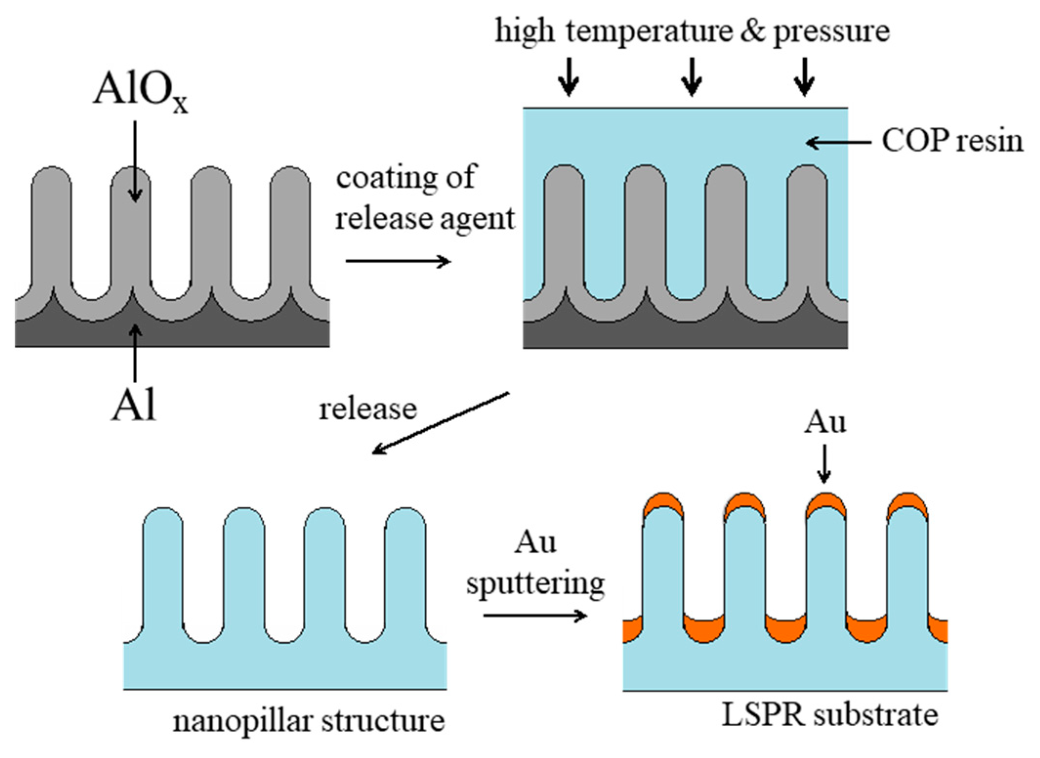
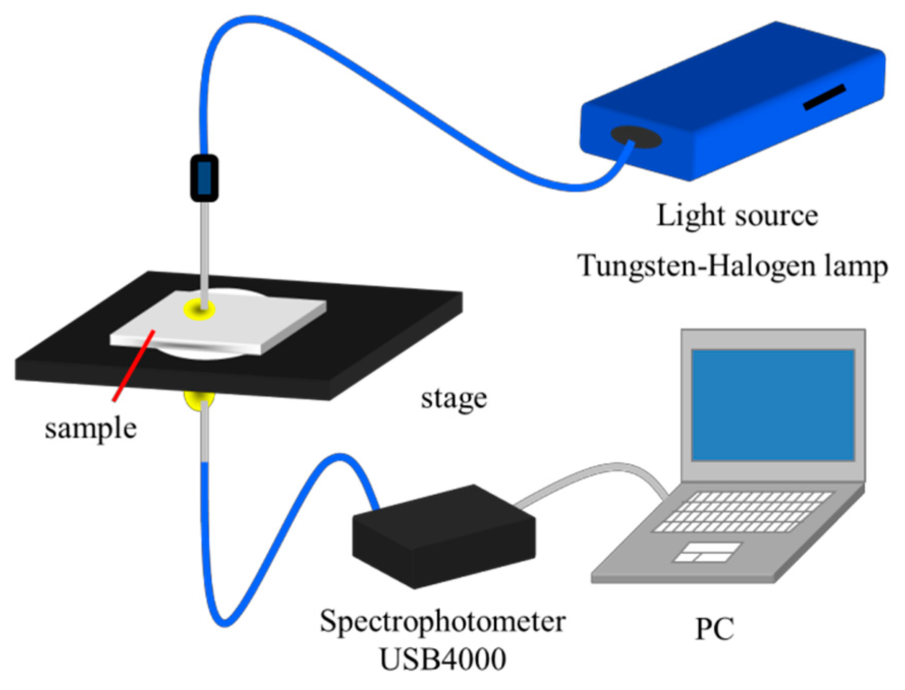
2. Materials and Methods

For the Au nanostructures, we used gold-capped nanopillars imprinted thermally on a polymer film, which was developed previously by one of our authors [2]. The polymer is Cyclo-olefin polymer (COP). The fabrication steps of the structure is illustrated in Figure 1; (a) fabrication of porous alumina mold, (b) thermal nanoimprinting with high temperature and pressure to the COP resin. (c) formation of the nanopillar structure by releasing from the alumina porous mold, and (d) final Au-sputtered and capped nanopillar structures for the LSPR substrate. Figure 2 shows an SEM view of fabricated Au nanopillars. It is observed that designed structures are successfully formed.
Figure 3 show cross-sectional illustrations of LSPR sensor immobilized with (a) liposomes and (b) phospholipid single layer. Au island was formed on the tip of nanopillar. DMPC liposomes or its single-layered phospholipids were immobilized on the nanostructures. Carbonic anhydrase from bovine (CAB) was used as a target protein because its interaction was confirmed with some different phospholipids.
Figure 4 shows our LSPR measurement system. Absorption spectra of nanopillar films were measured with a spectroscopy system consisting of a spectrophotometer (USB-4000-UV–vis), a tungsten halogen light source (LS-1), optical fibers (R-400-7 UV/vis, fiber core diameter: 200 μm), and collimating lenses (74-UV), all purchased from Ocean Optics. Nanopillar films immobilized on a glass microscope slide with carbon tapes were put on the sample stage of the spectroscopy system.

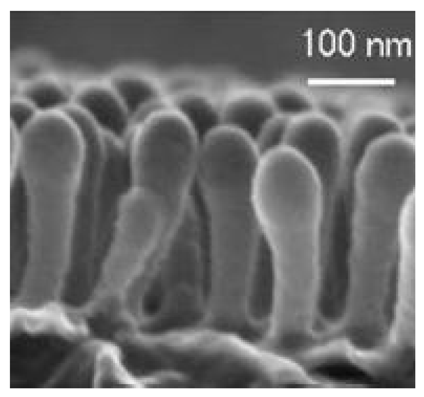
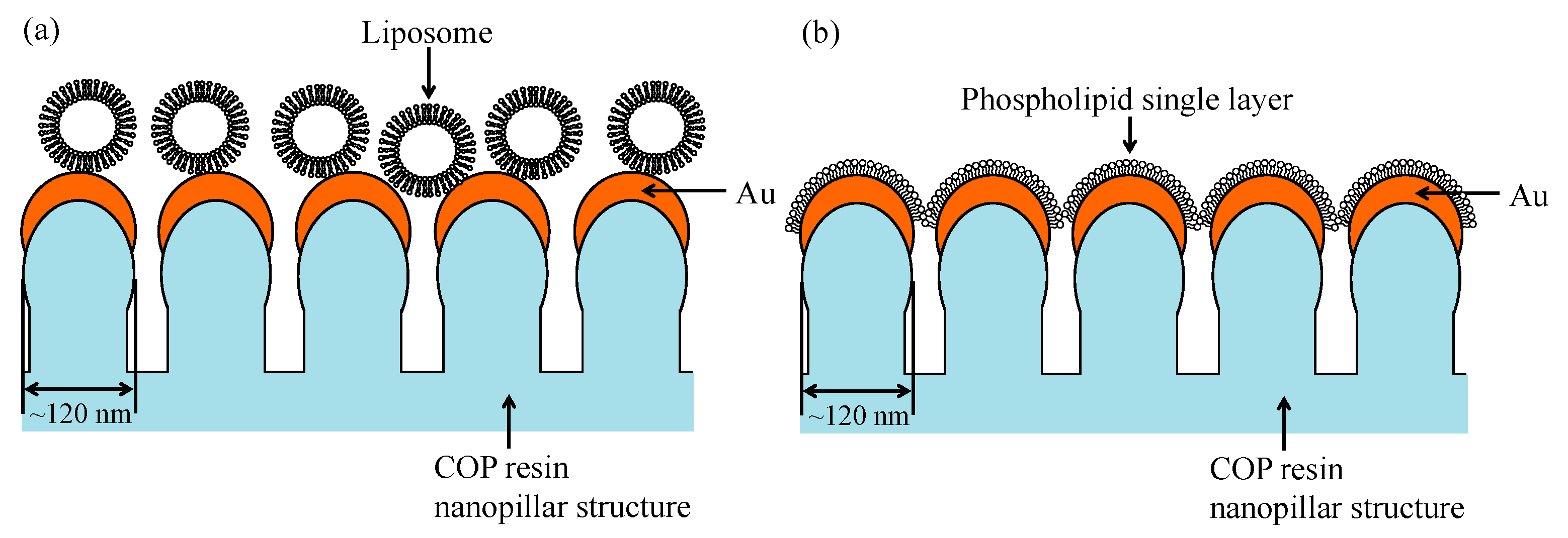
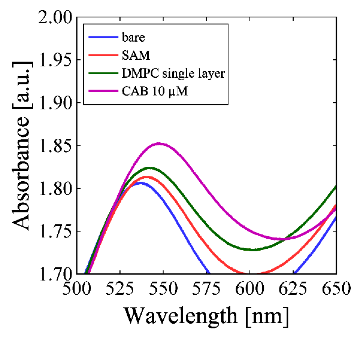
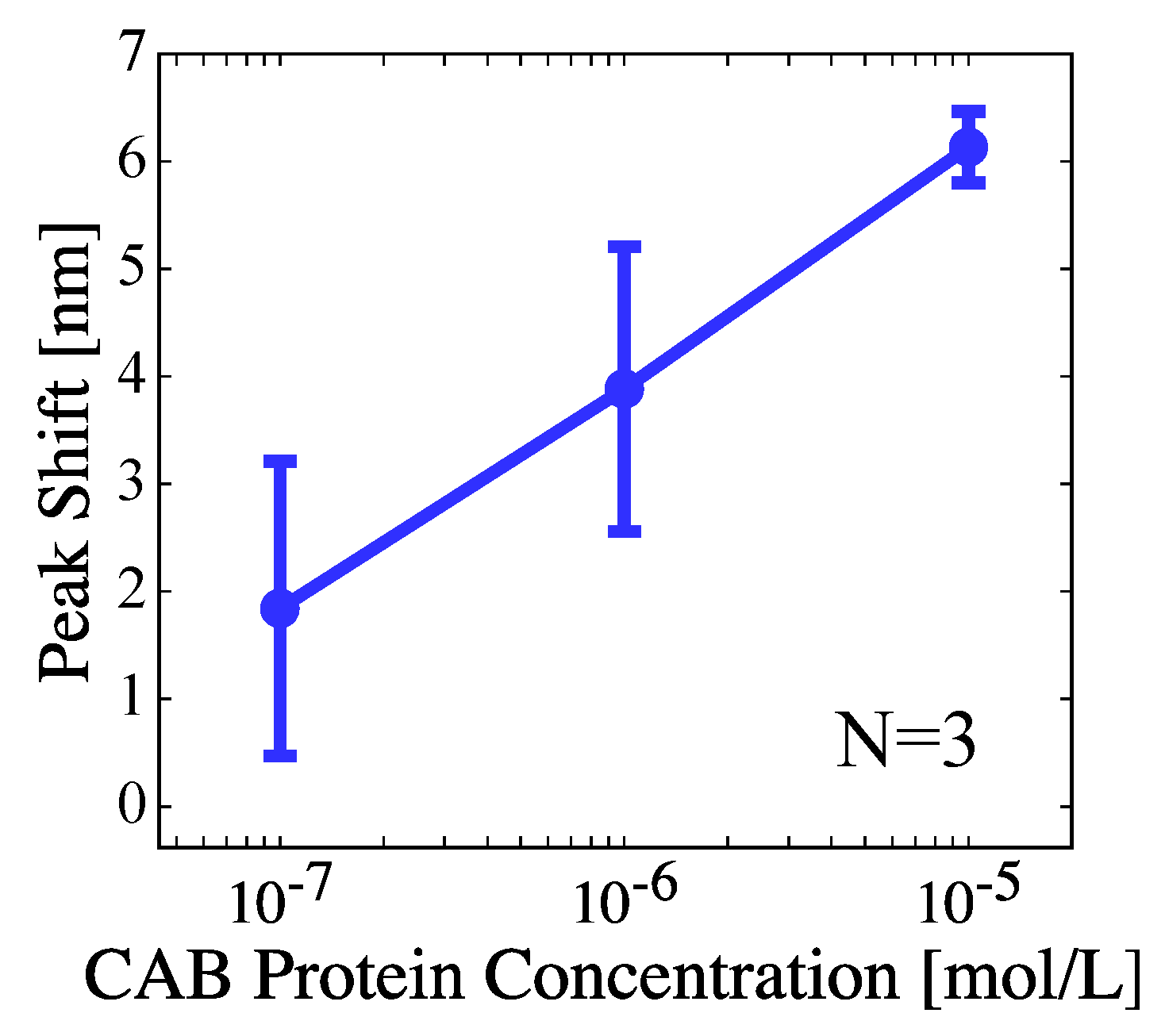
3. Results and Discussion

As shown in Figure 5, absorption spectrum of a bare Au/nanopillar shows maximum peak corresponding to LSPR of Au nanostructures at ~540 nm. With immobilized liposome, LSPR peak wavelength shifts showed continuously positive increase from bare Au surface, after SAM deposition, immobilization of liposome, and finally after addition of 10 μM CAB. But the sensitivity would degrade as the size of liposome (about 100 nm) was larger than the range of near-field effect (a few to tens of nm) from the Au nanostructure. From Figure 5, the characteristic peak wavelength also shows clearly a monotonous shift from bare Au surface, after SAM deposition, immobilization of DMPC single layer, and finally after addition of 10 μM CAB. The sensitivity is expected to improve more for the membrane, because its size (thickness) is around 3 nm and within the range of the near-field effect. After decreasing the target CAB concentration, finally, Figure 6 suggests that 100 nM CAB was clearly detected and the sensitivity improved by about 2 orders of magnitude from the case with the liposome.

4. Conclusions

We have fabricated a new type of LSPR sensor featuring immobilized liposome or phospholipid single layer. By the sensor, we tried to investigate biosensing capabilities using the membranes of the both structures on Au nanostructures of LSPR sensor chip. As a result, a monotonous increase in the shift of LSPR peak wavelength was successfully observed depending on successive formation on the Au nanopillars of SAM layer, immobilized liposome or phospholipid single layer and interacted target protein. Thereafter, it was confirmed that the phospholipid single layer is more effective to improve the sensitivity than the liposome. Finally, we have clearly detected 100 nM target protein of CAB and estimated a possible detection of 10 nM range from wavelength resolution by interaction with the phospholipid single layer.

References

  1. Hiep, H.M.; Endo, T.; Saito, M.; Chikae, M.; Kim, D.K.; Yamamura, S.; Takamura, Y.; Tamiya, E. Label-Free Detection of Melittin Binding to a Membrane Using Electrochemical-Localized Surface Plasmon Resonance. Anal. Chem. 2008, 80, 1859–1864. [Google Scholar] [CrossRef] [PubMed]
  2. Saito, M.; Kitamura, A.; Murahashi, M.; Yamanaka, K.; Hoa le, Q.; Yamaguchi, Y.; Tamiya, E. Novel Gold-Capped Nanopillars Imprinted on a Polymer Film for Highly Sensitive Plasmonic Biosensing. Anal. Chem. 2012, 84, 5494–5500. [Google Scholar] [CrossRef] [PubMed]
Figure 1. The used fabrication steps of the Au nanopillar structure [2].
Proceedings 02 00791 g001
Figure 2. An SEM view of fabricated Au nanopillars [2].
Proceedings 02 00791 g002
Figure 3. Cross-sectional illustrations of LSPR sensor immobilized with (a) liposomes and (b) phospholipid single layer.
Proceedings 02 00791 g003
Figure 4. LSPR measurement system.
Proceedings 02 00791 g004
Figure 5. Absorbance for immobilized with DMPC single layer vs LSPR wavelength.
Proceedings 02 00791 g005
Figure 6. LSPR peak wavelength shift vs target CAB protein concentration.
Proceedings 02 00791 g006
Publisher’s Note: MDPI stays neutral with regard to jurisdictional claims in published maps and institutional affiliations.

