Proceeding Paper

Photoelectrochemical Imaging Using Carbon Dots (CDs) Derived from Chitosan †

by
Dewen Zhang
1,
Nikolaos Papaioannou
3,
Maria-Magdalena Titirici
2 and
Steffi Krause
2,*
1
Institute of Medical Engineering, School of Basic Medical Sciences, Xi’an Jiaotong University Health Science Center, Xi’an 710061, China
2
School of Engineering and Materials Science, Queen Mary University of London, London E1 4NS, UK
3
School of Physics and Astronomy, Queen Mary University of London, London E1 4NS, UK
*
Author to whom correspondence should be addressed.
Presented at the Eurosensors 2018 Conference, Graz, Austria, 9–12 September 2018.
Proceedings 2018, 2(13), 778; https://doi.org/10.3390/proceedings2130778
Published: 10 December 2018
(This article belongs to the Proceedings of EUROSENSORS 2018)

Abstract

:
Carbon dots (CDs) derived from chitosan via a solvothermal method were covalently linked to an indium tin oxide (ITO) surface and showed a direct photoelectrochemical response. We attribute the photocurrent of the ITO-silane-CD surface to a photogenerated electron-transfer process by CDs under illumination. The ITO-silane-CD surface was successfully used for ac-photocurrent imaging. This opens up new applications for CDs as biocompatible and light-addressable electrochemical sensors in bioimaging applications.

1. Introduction

Carbon dots (CDs) with nanostructures have generated excitement as a semiconductor-like and metal-free photocatalysts [1]. CDs have been widely applied to enhance photocatalytic activities of heterostructured photocatalysts in photoelectric conversion [2], pollutant photodegradation [3], water splitting [4], CO2 conversion [5] and organic synthesis [6]. CDs played important roles in photoreactions by light absorption, electron-hole pair generation, photoexcited charge separation and migration, which showed both electron donor and acceptor abilities under illumination. However, the photoinduced electron transfer process of CDs is still unclear. Self-assembled monolayers of conventional quantum dots have been used in light-addressable electrodes for electrochemical sensing [7]. Compared to conventional quantum dots, CDs are cheap, non-poisonous and biocompatible, which are ideal for bioimaging. Herein, we report the photoelectrochemical behaviour of CDs prepared by simple bottom-up solvothermal approach from chitosan and their use in electrochemical imaging sensors.

2. Materials and Methods

2.1. The Preparation of CDs

CDs were prepared by a one-step solvothermal carbonization of chitosan dispersed in ethanol (4% w/v) at 200 °C for 12 h. The dark brown liquid phase obtained was centrifuged at 20,000 rpm for 10 min to separate the liquid containing fluorescent CDs from the solid black carbonaceous precipitate. The liquid phase containing CDs was then filtered using standard syringe filters.

2.2. The Preparation of ITO-Silane-CD Surfaces

The indium tin oxide (ITO) coated glass (50 Ω/sq, Diamond Coatings Limited, UK) was cut into 1 cm × 1 cm pieces. They were cleaned by ultrasonication for 15 min with acetone, isopropanol and ultrapure water. After drying with nitrogen, the ITO substrates were immersed in a solution of 1:1:5 (v/v) H2O2 (30%)/NH4OH (30%)/H2O for 1 h, washed with ultrapure water and dried with nitrogen. Then, ITO substrates were immersed in a 1% 3-glycidoxypropyldimethoxymethylsilane ethanol solution for 20 min. After drying, they were placed in an oven at 110 °C for 1 h, washed with ethanol thoroughly and dried with nitrogen. The ITO-silane surfaces were incubated with the CD ethanol solution overnight at room temperature (Scheme 1a). Finally, the ITO-silane-CD surfaces were washed with ultrapure water. After drying, the ITO-silane-CD surface was kept at room temperature before use [8].

2.3. Photocurrent Measurements

Linear sweep voltammetry (LSV) was carried out in 10 mM pH 7.4 phosphate buffer with 0.1 M KCl using an Autolab PGSTAT30/FRA2 (Windsor Scientific Ltd., UK) in a custom made electrochemical cell consisting of a platinum electrode and an Ag/AgCl electrode as the counter and reference electrodes, respectively. The scan rate was 5 mV/s. An adjustable diode laser (wavelength 400–450 nm, max 500 mW) with a UV filter that blocked light of wavelengths lower than 420 nm was used as the chopped back side illumination source. The irradiance was measured as 10 mW/cm2 with a calibrated power meter.

2.4. AC Photocurrent Imaging

As shown in Scheme 1b, a focused diode laser (400–450 nm) with a UV filter that blocked the light of wavelengths lower than 420 nm was used for charge carrier generation. The modulation frequency was 10 Hz. The position of the sample holder was adjusted by an M-VP-25XL XYZ positioning system with a 50 nm motion sensitivity on all axes (Newport, UK). AC photocurrents were measured in pH 7.4 PBS buffer solution by an EG&G 7260 lock-in amplifier using a platinum electrode and an Ag/AgCl electrode as the counter and reference electrodes, respectively.

3. Results and Discussion

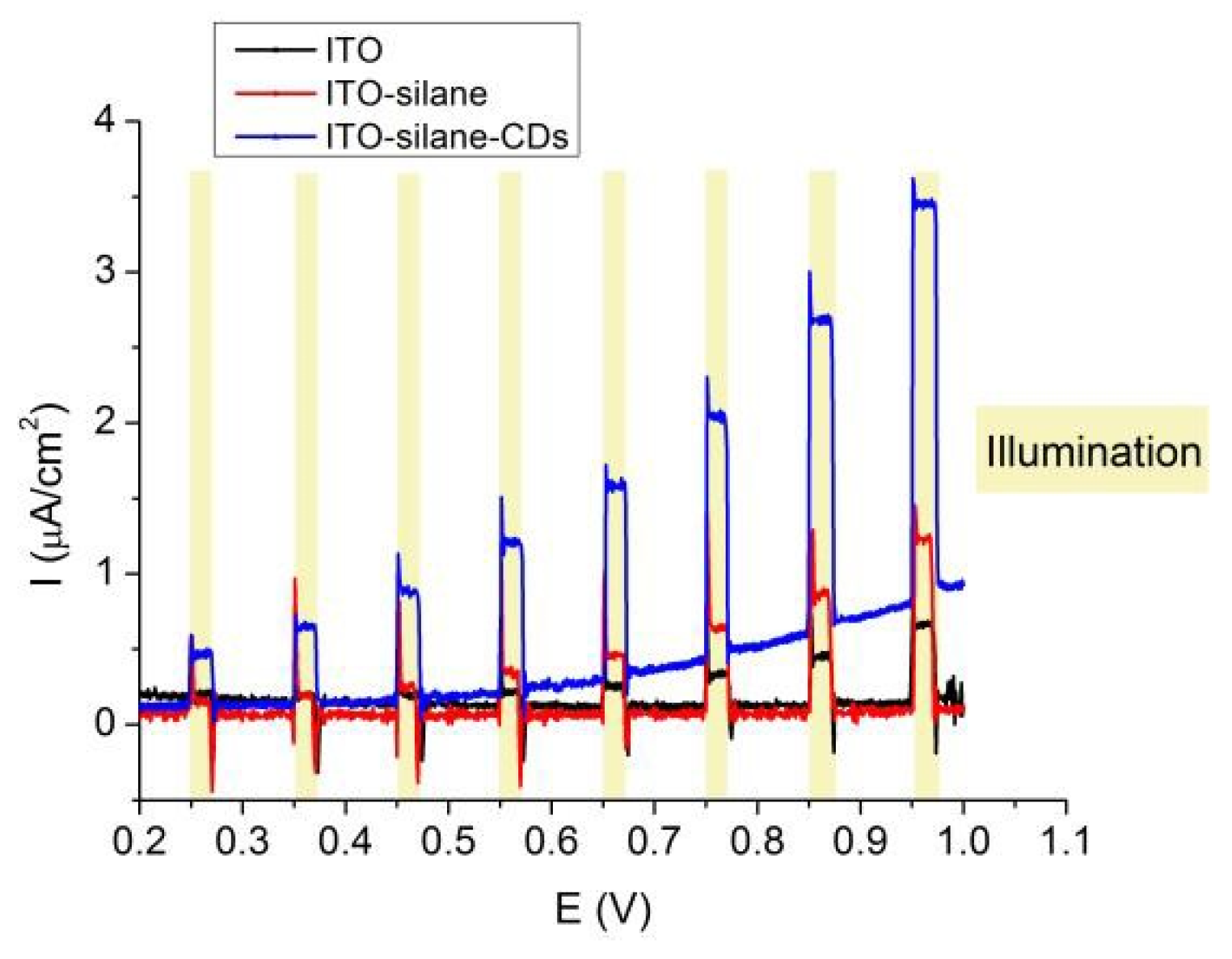
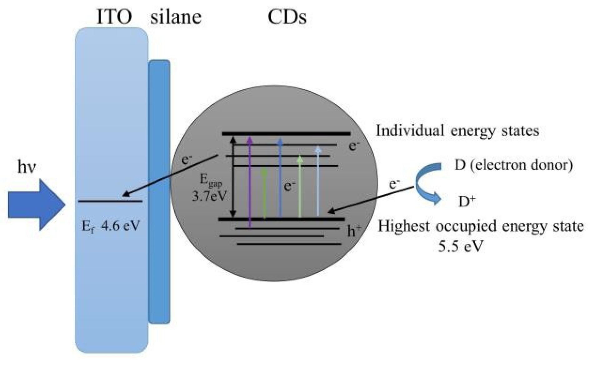
CDs were prepared by a one-step solvothermal carbonization of chitosan dispersed in ethanol (4% w/v), which showed spherical morphologies and an average diameter of around 4 nm in a transmission electron microscopy (TEM) image. To investigate the photoelectric conversion behaviour, CDs were covalently linked to an indium tin oxide (ITO) surface through a 3-glycidoxypropyldimethoxymethylsilane monolayer. X-ray photoelectron spectroscopy (XPS) confirms the linking of silane and CDs by the appearance of a nitrogen peak assigned to the amine in the CDs from chitosan. Photoelectrochemical response measurements of ITO-silane-CDs were carried out by linear sweep voltammetry (LSV) in 10 mM pH 7.4 phosphate buffer with 0.1 M KCl. The results (Figure 1) confirmed the direct photoelectrochemical response of CDs under 420–450 nm laser illumination. The photocurrent generated by CDs and the mechanism have been studied (Figure 2). The photocurrent was attributed to the enhanced electron-transfer process catalyzed by CDs under illumination.
The ITO-silane-CD surface was applied to ac-photocurrent imaging, which measures the local photocurrent by illuminating the substrate with a focused, modulated laser beam. Figure 3a shows circular islands of PMMA with a diameter of 40 µm and 30 µm gaps on an ITO-silane-CD surface obtained by microcontact printing. Figure 3b shows the corresponding ac-photocurrent image measured at a bias voltage of 1.8 V and a modulation frequency of 10 Hz with micron scale lateral resolution. This discovery opens up new applications for CDs as light-addressable electrochemical sensors in bioimaging applications.

Author Contributions

D.Z., M.-M.T., and S.K. designed the experiments. N.P. produced the CD samples and performed the characterization of CDs. D.Z. performed the binding of CDs onto the ITO surface, photocurrent measurements and ac-photocurrent imaging. All authors contributed to the interpretation of the results. D.Z. and S.K. wrote the paper.

Funding

The authors are grateful to the Biotechnology and Biological Sciences Research Council (BBSRC) of UK for providing research funding (BB/P026788/1), and the EU for providing a Marie Skłodowska-Curie Individual Fellowship (H2020-MSCA-IF-2014-660489).

Conflicts of Interest

The authors declare no conflict of interest.

References

  1. Yu, H.; Shi, R.; Zhao, Y.; Waterhouse, G.I.N.; Wu, L.-Z.; Tung, C.-H.; Zhang, T. Smart utilization of carbon dots in semiconductor photocatalysis. Adv. Mater. 2016, 28, 9454–9477. [Google Scholar] [CrossRef] [PubMed]
  2. Bian, J.; Huang, C.; Wang, L.; Hung, T.; Daoud, W.A.; Zhang, R. Carbon dot loading and TiO2 nanorod length dependence of photoelectrochemical properties in carbon dot/TiO2 nanorod array nanocomposites. ACS Appl. Mater. Interfaces 2014, 6, 4883–4890. [Google Scholar] [CrossRef] [PubMed]
  3. Yu, H.; Zhang, H.; Huang, H.; Liu, Y.; Li, H.; Ming, H.; Kang, Z. ZnO/carbon quantum dots nanocomposites: One-step fabrication and superior photocatalytic ability for toxic gas degradation under visible light at room temperature. New J. Chem. 2012, 36, 1031–1035. [Google Scholar] [CrossRef]
  4. Zhang, X.; Wang, F.; Huang, H.; Li, H.; Han, X.; Liu, Y.; Kang, Z. Carbon quantum dot sensitized TiO2 nanotube arrays for photoelectrochemical hydrogen generation under visible light. Nanoscale 2013, 5, 2274–2278. [Google Scholar] [CrossRef] [PubMed]
  5. Yang, K.D.; Ha, Y.; Sim, U.; An, J.; Lee, C.W.; Jin, K.; Kim, Y.; Park, J.; Hong, J.S.; Lee, J.H.; et al. Graphene quantum sheet catalyzed silicon photocathode for selective CO2 conversion to CO. Adv. Funct. Mater. 2016, 26, 233–242. [Google Scholar] [CrossRef]
  6. Liu, R.; Huang, H.; Li, H.; Liu, Y.; Zhong, J.; Li, Y.; Zhang, S.; Kang, Z. Metal nanoparticle/carbon quantum dot composite as a photocatalyst for high-efficiency cyclohexane oxidation. ACS Catal. 2014, 4, 328–336. [Google Scholar] [CrossRef]
  7. Khalid, W.; el Helou, M.; Murböck, T.; Yue, Z.; Montenegro, J.-M.; Schubert, K.; Göbel, G.; Lisdat, F.; Witte, G.; Parak, W.J. Immobilization of quantum dots via conjugated self-assembled monolayers and their application as a light-controlled sensor for the detection of hydrogen peroxide. ACS Nano 2011, 5, 9870–9876. [Google Scholar] [CrossRef] [PubMed]
  8. Zhang, D.-W.; Papaioannou, N.; David, N.M.; Luo, H.; Gao, H.; Tanase, L.C.; Degousée, T.; Samorì, P.; Sapelkin, A.; Fenwick, O.; et al. Photoelectrochemical response of carbon dots (CDs) derived from chitosan and their use in electrochemical imaging. Mater. Horiz. 2018, 5, 423–428. [Google Scholar] [CrossRef]
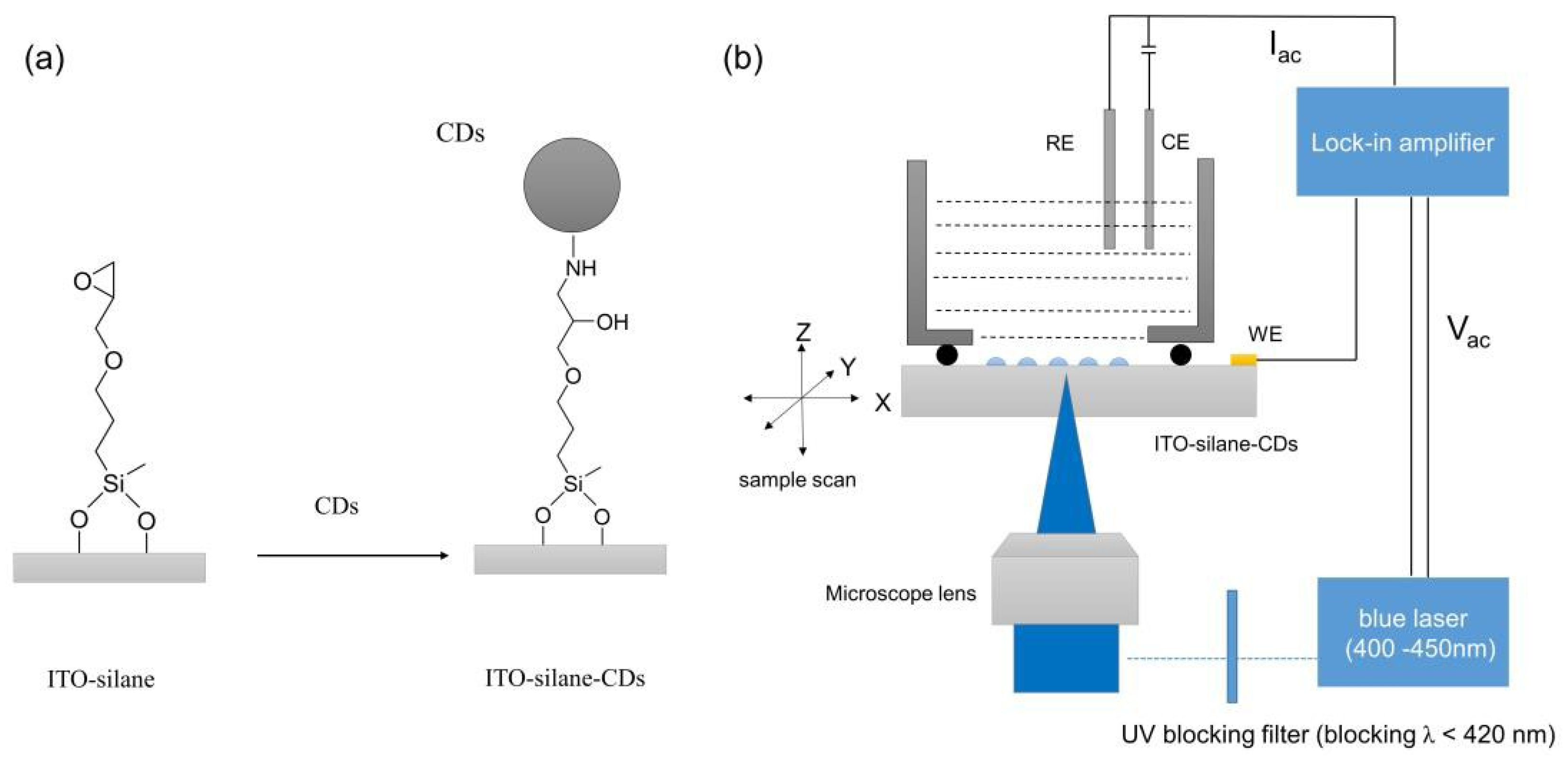
Scheme 1. (a) Scheme of CDs binding onto ITO via silane reaction; (b) Scheme of the ac-photocurrent image setup by scanning a focused, modulated laser beam across the ITO-silane-CD surface.
Proceedings 02 00778 sch001
Figure 1. LSV curves of ITO, ITO-silane, and ITO-silane-CDs with chopped illumination (420–450 nm, 10 mW/cm2).
Proceedings 02 00778 g001
Figure 2. The mechanism of photoelectric conversion of CDs.
Proceedings 02 00778 g002
Figure 3. (a) Optical micrograph of a PMMA dot array on ITO-silane-CD surface by microcontact printing; (b) Corresponding ac-photocurrent image of (a) measured at 1.8 V.
Proceedings 02 00778 g003
Publisher’s Note: MDPI stays neutral with regard to jurisdictional claims in published maps and institutional affiliations.