Proceeding Paper

Imaging Fluorophore-Labelled Intestinal Tissue via Fluorescence Endoscope Capsule  †

1
School of Engineering, University of Glasgow, Glasgow, Scotland G12 8LT, UK
2
School of Science and Engineering, University of Dundee, Scotland DD1 4HN, UK
*
Author to whom correspondence should be addressed.
Presented at the Eurosensors 2018 Conference, Graz, Austria, 9–12 September 2018.
Proceedings 2018, 2(13), 766; https://doi.org/10.3390/proceedings2130766
Published: 27 November 2018
(This article belongs to the Proceedings of EUROSENSORS 2018)

Abstract

:
The authors have developed a wireless fluorescence imaging capsule endoscope, potentially capable of detecting early signs of disease in the human intestine which can be missed by white-light imaging (WLI) capsule endoscopy (Figure 1). Intestinal fluorescence imaging exploits variations in tissue autofluorescence between healthy and diseased areas in response to illumination, or application of fluorescent labels which preferentially bind to diseased sites. To validate the capsule’s capability to image fluorescently-labelled tissue, a small area of a sample of ex vivo porcine small intestine was sonicated with 6 nm CdZnMg fluorescent quantum dots, and the labelled area clearly differentiated from surrounding tissue by the fluorescence imaging capsule.

1. Introduction

Wireless capsule endoscopy using white-light imaging (WLI) permits examination of the entire intestine for disease, and has been found to be effective for occult bleeding but can miss early signs of other diseases including cancer. Fluorescence endoscopy can improve detection of these early signs [1], assisting early diagnosis and consequently improving treatment outcomes. Given that that bowel cancer accounts for 16,000 deaths per year in the UK alone [2], with an additional 500 annual deaths from small intestine cancer, there is clear value in improved diagnostic techniques. However, current endoscope-based fluorescence imaging systems are incapable of examining most of the small intestine. Autofluorescence imaging exploits the fact that endogenous fluorophores in human intestinal tissue, which fluoresce green in response to blue illumination, exhibit a fluorescent response up to three times lower in cancerous areas than in healthy tissue. Previous work [3] has validated our capsule’s capacity to induce and image autofluorescence in intestinal tissue but this is still a relatively weak effect. In contrast, labels which preferentially attach to diseased areas, and exhibit increased fluorescent response in comparison to surrounding unlabeled tissue can be used to enhance detection [4] and can generate a relatively strong response. The work presented here demonstrates our capsule’s ability to image fluorescently-labelled intestinal tissue.

2. Materials and Methods

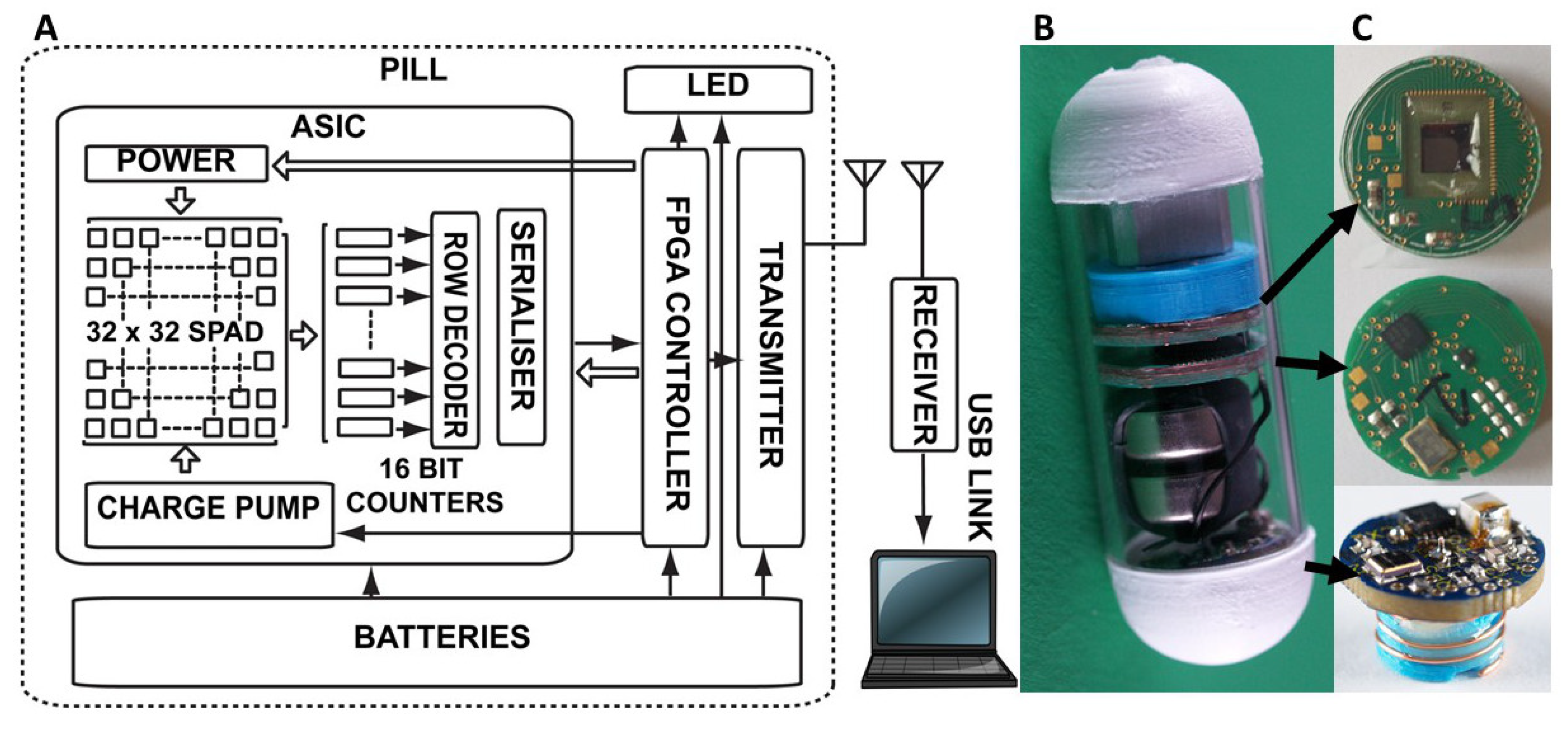
The core of the capsule (Figure 1 and Figure 2) is an application-specific integrated circuit (ASIC) implemented in a 0.18µ high-voltage CMOS process, featuring a 32 × 32 single-photon avalanche diode (SPAD) imaging array, each pixel of which generates a pulse in response to photon incidence, pulse counters, array addressing and power management. A machined aluminum optical block implements a blue illumination LED along with a monochromator, filters and lenses focusing the fluorescent image onto the ASIC while attenuating illumination light. A field-programmable gate array controls counter pulse readout, packetisation and transmission via an 868 MHz wireless transmitter. Images are transmitted at 2 frames/s. An external wireless receiver, embedded processor and PC with MATLAB application receive and display acquired fluorescence imaging video.
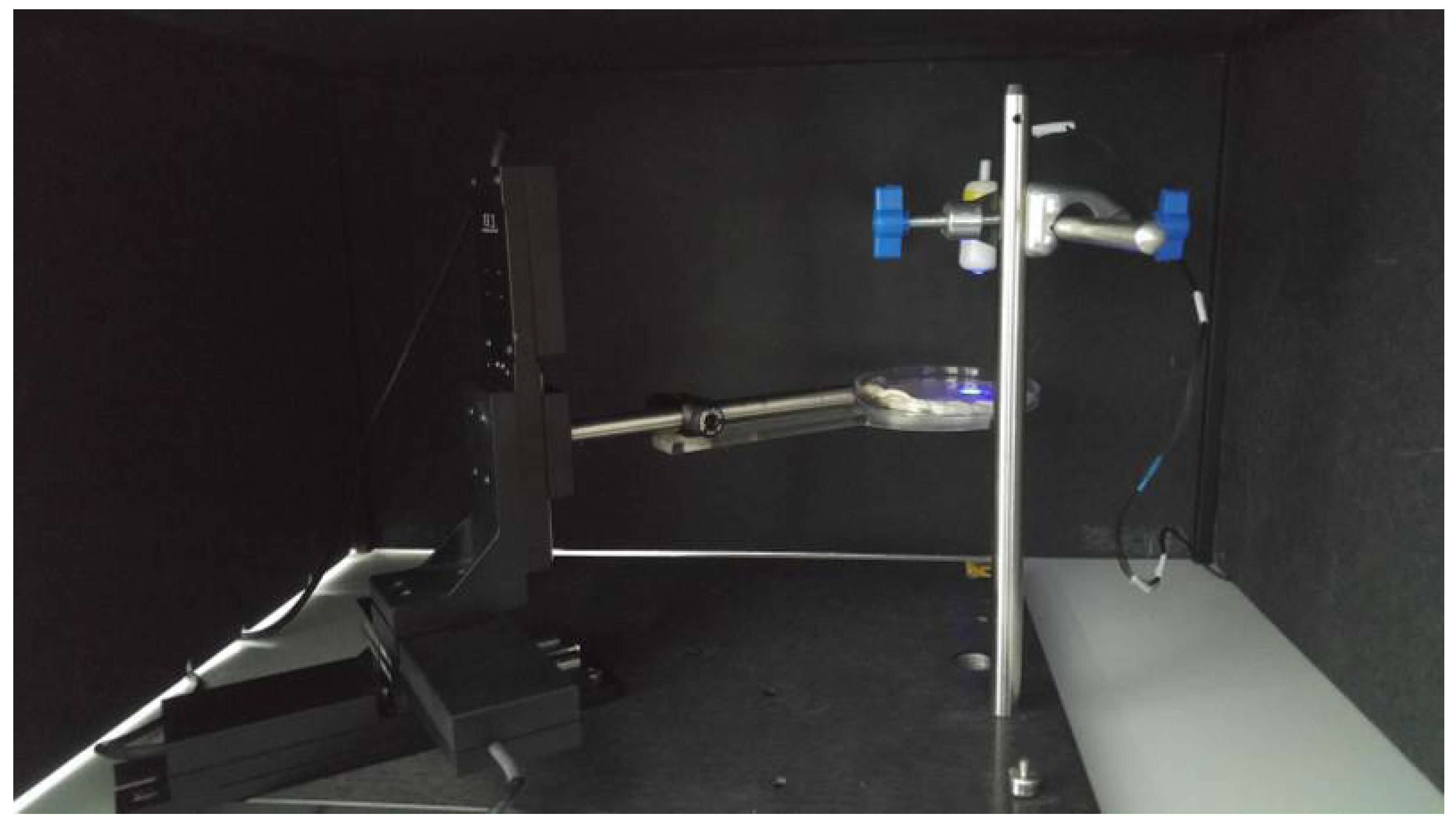
In order to demonstrate the capsule’s ability to image fluorescently labelled intestinal tissue, an area approx. 8 mm diameter on a sample of ex vivo porcine intestinal tissue was sonicated simultaneously with delivering a suspension of 0.2 mg/mL CdSeS/ZnS fluorescent quantum dots (Sigma, Gillingham, UK) and 15% microbubbles (2 to 5 × 10⁸ microbubbles/mL in saline, 2–8 µm average diameter, SonoVue, Bracco, Italy) for 2 min at a rate of 1 mL/min (Figure 3a). Ultrasound was applied through a drug delivery capsule with a bespoke focused ultrasound transducer (4 MHz, 1016 kPa, 15 mm focal distance, cross-sectional area at focus 1.8 mm2) [4]. A wired version of the capsule enclosed in a 3D-printed casing was positioned facing vertically downwards 15 mm above the tissue sample (Figure 4). The sample was positioned in petri dish and moved horizontally at 0.1 mm/s by a motorized translation stage. The whole system was housed in an optically absorbent enclosure with taped gaps to exclude light.

3. Results

Figure 3b shows an image of the sonication-labelled tissue area obtained via the fluorescence imager capsule. The labelled area is clearly visible. A mean 48,473 counts/s were registered from the labelled area and 32,828 counts/s from the surrounding unlabeled region.

4. Discussion and Conclusions

The image shown in Figure 3b clearly demonstrates the fluorescence imaging capsule’s ability to differentiate a fluorescently-labelled area from surrounding unlabeled intestinal tissue, with a fluorescence response c. 47% higher than that of the surrounding tissue. Given the success of this preliminary investigation the logical next steps are investigation of the ability to image to diseased areas of intestinal tissue with attached labels, and in-vivo testing of the capsule with labelled tissue.

Author Contributions

J.B. and M.A.-R. developed the fluorescence capsule ASIC, electronics, PCBs, optics and software. G.M. developed packaging. F.S. developed the sonication capsule system. Experimental work was designed, planned, implemented and results analysed by J.B., M.A.-R., G.M. and M.T. S.C. and D.C. managed and supervised. J.B. wrote this paper.

Acknowledgments

The project was funded by EPSRC grant EP/K034537/1.

Conflicts of Interest

The authors declare no conflict of interest. The founding sponsors had no role in the design of the study; in the collection, analyses, or interpretation of data; in the writing of the manuscript, and in the decision to publish the results.

References

  1. Song, L.W.M.K.; Wilson, B.C. Endoscopic detection of early upper GI cancers. Best Pract. Res. Clin. Gastroenterol. 2005, 19, 833–856. [Google Scholar] [CrossRef] [PubMed]
  2. Cancer Research UK. Available online: http://www.cancerresearchuk.org/health-professional/cancer-statistics/statistics-by-cancer-type/bowel-cancer/mortality#heading-Zero (accessed on 18 July 2018).
  3. Al-Rawhani, M.A.; Beeley, J.; Cumming, D.R.S. Wireless fluorescence capsule for endoscopy using single photon-based detection. Nat. Sci. Rep. 2015, 5, 18591. [Google Scholar] [CrossRef] [PubMed]
  4. Kobayashi, H.; Ogawa, M.; Alford, R.; Choyke, P.L.; Urano, Y. New strategies for fluorescent probe design in medical diagnostic imaging. Chem. Rev. 2010, 110, 2620–2640. [Google Scholar] [CrossRef]
  5. Stewart, F.R.; Qiu, Y.; Lay, H.S.; Newton, I.P.; Cox, B.F.; Al-Rawhani, M.A.; Beeley, J.; Liu, Y.; Huang, Z.; Cumming, D.R.; et al. Acoustic sensing and ultrasonic drug delivery in multimodal theranostic capsule endoscopy. Sensors 2017, 1553. [Google Scholar] [CrossRef]
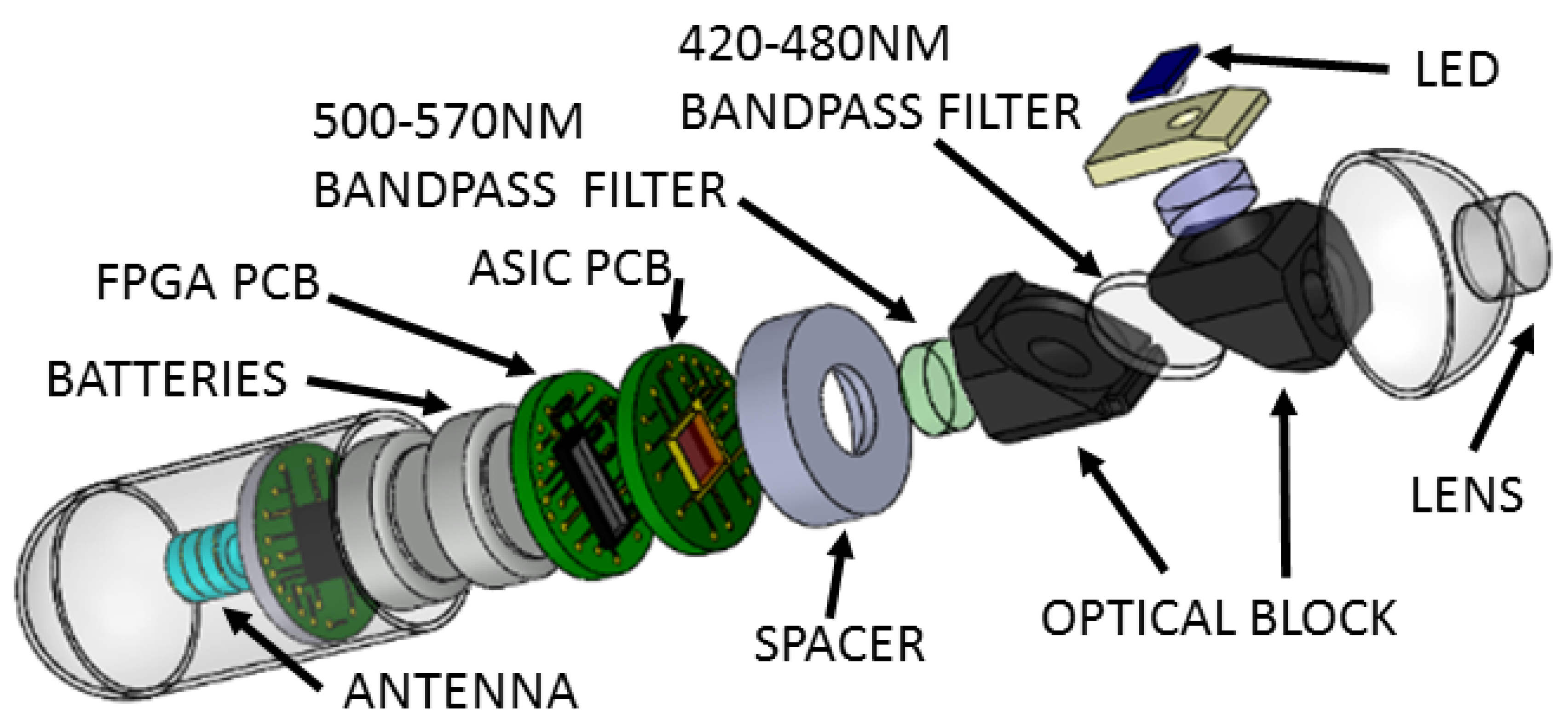
Figure 1. Exploded 3D view of the wireless fluorescence capsule endoscope.
Proceedings 02 00766 g001
Figure 2. (a) Wireless fluorescence imaging capsule schematic; (b,c) Capsule with filter/LED/monochromator optical block, SPAD array imager ASIC, control/communication FPGA and UHF wireless transmitter PCBs.
Proceedings 02 00766 g002
Figure 3. (a) Ex vivo porcine intestinal tissue insonated simultaneously with the application of CdSeS/ZnS fluorescent quantum dots and microbubbles. The 8 mm wide insonated area fluoresces under UV light. (b) Fluorescence image of the 8mm wide insonated area obtained from the imaging capsule.
Proceedings 02 00766 g003
Figure 4. The imager capsule, mounted on a stand (top right), faces downwards onto the fluorescently labelled intestinal tissue sample, which is moved by a motorized optical stage. An optically absorbent box with taped gaps surrounding the apparatus excludes external light.
Proceedings 02 00766 g004
Publisher’s Note: MDPI stays neutral with regard to jurisdictional claims in published maps and institutional affiliations.