Article

Massive Expansion and Diversified Expression Pattern of the Ammonium Transporters in the Living Fossil Lingula anatina


1
National Engineering Research Center for Marine Aquaculture, Zhejiang Ocean University, Zhoushan 316022, China
2
Zhejiang Marine Ecology and Environment Monitoring Center, Zhoushan 316021, China
3
Zhoushan Municipal Ecology and Environment Bureau, Zhoushan 316021, China
*
Author to whom correspondence should be addressed.
Nitrogen 2026, 7(2), 43; https://doi.org/10.3390/nitrogen7020043
Submission received: 18 March 2026 / Revised: 10 April 2026 / Accepted: 13 April 2026 / Published: 14 April 2026
(This article belongs to the Special Issue Nitrogen Metabolism and Degradation)

Abstract

Nitrogen metabolism is fundamental to all organisms, with ammonium transporters (Amt) playing a pivotal role in transmembrane ammonium transport. Brachiopods, as “living fossils”, offer unique insights into the evolutionary adaptation of marine invertebrates. This study systematically identified and characterized the Amt gene family in the brachiopod Lingula anatina. Five canonical Amt genes were identified, with nonrandom chromosomal distribution and evidence of lineage-specific duplication events. Phylogenetic analysis revealed that these Amt proteins cluster into three well-supported clades, showing closer affinity to Caenorhabditis elegans, reflecting conserved ancestral features predating protostome radiation. Structural predictions showed that LanAmtA and LanAmtB retain the canonical 11-transmembrane helix (TMH) topology with an extracellular N-terminus, while LanAmtC features a unique 12-TMH architecture with an intracellular N-terminus, resembling certain vertebrate Amt-related proteins. Critical functional residues involved in ammonium selectivity and transport were preserved across all paralogs. Expression profiling revealed non-redundant spatiotemporal patterns: LanAmtA1 and LanAmtB2 dominate early embryogenesis, with LanAmtB2 becoming the major isoform in late developmental stages; LanAmtC exhibits constitutive high expression across adult tissues. Collectively, our findings demonstrate that the L. anatina Amt family expanded via local duplications, evolving structural stability, regulatory diversity, and functional specificity. This study provides a comprehensive molecular framework for understanding the evolutionary adaptation of nitrogen-handling mechanisms in basal lophotrochozoans and sheds light on how intertidal organisms cope with dynamic environmental conditions.

1. Introduction

Nitrogen (N) is an indispensable cornerstone of biological systems, serving as a fundamental building block for nitrogen-containing biomolecules that underpin core metabolic processes across all domains of life [1]. Efficient transmembrane transport of nitrogenous compounds is critical for maintaining physiological homeostasis and adaptive fitness in organisms [2,3]. Among these compounds, ammonium represents the second most abundant nitrogen form on Earth and serves as a preferred nutrient source for many bacteria, fungi, and plants [4,5]. Conversely, in animals, ammonium primarily functions as a toxic metabolic byproduct requiring rapid excretion to avoid cellular damage [5,6].
Ammonium transmembrane transport is a ubiquitous biological process across all domains of life, underscoring its pivotal role in nitrogen metabolism, environmental adaptation, and evolutionary fitness [5,7]. Whether for nutrient acquisition in microbes and plants or waste elimination in animals, this transport is facilitated by a conserved group of membrane proteins collectively referred to as the ammonium transporter (Amt) superfamily [7]. While the physiological roles and molecular mechanisms of Amt proteins have been extensively studied in mammals and model organisms, significant gaps persist regarding the evolutionary history, gene family size, and functional diversification of Amts in invertebrates [8,9,10,11]. This is particularly evident in marine invertebrates, where the mechanisms of ammonium homeostasis remain poorly characterized despite the critical importance of nitrogen balance in aquatic environments [11]. Moreover, there is a notable paucity of data regarding the Amt family in lophotrochozoans, representing a major omission in our understanding of metazoan nitrogen-handling strategies. Addressing these gaps is essential for elucidating the broader evolutionary trajectory of nitrogen-handling strategies across metazoans and understanding how invertebrate species adapt to diverse ecological niches under varying pressures of nitrogen availability and toxicity.
Brachiopods (phylum Brachiopoda), as marine invertebrates, often referred to as “living fossils,” have maintained a highly conserved body plan over more than 500 million years of evolutionary history, enduring numerous mass extinction events from the early Cambrian to the present day [12]. Their extensive fossil record provides unparalleled insights into the long-term evolution of marine ecosystems and the adaptive strategies of sessile, filter-feeding organisms across a spectrum of environments, ranging from tropical reefs to deep-sea and polar habitats. Given their ancient lineage and remarkable ecological resilience, brachiopods represent an ideal model system for investigating the evolution of fundamental physiological processes in marine invertebrates [13]. Specifically, exploring the composition, structural diversity, and phylogenetic relationships of the Amt gene families in brachiopods holds significant promise for uncovering how nitrogen-handling mechanisms have evolved in response to environmental challenges. Such investigations not only deepen our understanding of the molecular basis of environmental adaptation but also shed light on the functional radiation of transmembrane transport systems in early-diverging lophotrochozoans.
The subphylum Linguliformea, represented by the genus Lingula, is widely recognized as the most basal lineage within Brachiopoda [14,15]. Its fossil record dates back to the early Cambrian, coinciding with the emergence of animal biomineralization. Among extant species, Lingula anatina (commonly known as the lamp shell) serves as a key model organism in brachiopod biology [16,17]. As a “living fossil”, L. anatina exhibits a suite of conserved anatomical traits, including an inarticulate (non-hinged) bivalved shell and a lophophore specialized for filter feeding, features that have enabled its persistence across diverse marine habitats over geological time. L. anatina plays a significant ecological role in intertidal and shallow subtidal ecosystems worldwide. As a suspension feeder, it contributes to nutrient cycling and helps maintain benthic–pelagic coupling in coastal environments. The intertidal zone, a dynamic interface between terrestrial and marine realms, is characterized by extreme fluctuations in salinity, temperature, oxygen availability, and ammonia concentrations [18]. These conditions render it both a sensitive indicator of environmental change and a valuable setting for studying physiological and molecular adaptations in marine invertebrates.
Recent advancements in molecular research, particularly the completion of the L. anatina genome project, have opened new avenues for investigating the evolutionary history of transmembrane transport systems. This genomic resource now enables systematic identification, characterization, and comparative analysis of Amt family proteins in a representative of one of the earliest-branching bilaterian lineages. Specifically, this study aims to achieve the following core objectives: (1) Perform genome-wide identification of Amt gene family members in L. anatina; (2) Elucidate the evolutionary relationships of Amts and characterize the patterns of gene duplication and synteny conservation; (3) Conduct structural bioinformatics analysis to resolve the conserved domains, transmembrane topology, and functional motifs of L. anatina Amts; and (4) Profile the spatial–temporal expression patterns of Amts to generate testable hypotheses regarding their functional roles in nitrogen homeostasis and environmental adaptation.

2. Materials and Methods

2.1. Materials

To comprehensively identify and characterize all members of the Amt gene family in L. anatina, we retrieved the chromosome-level genome assembly from the NCBI database (Accession number: GCA_051362555.1). Concurrently, we obtained RNA-seq transcriptome datasets spanning multiple tissues and key stages of embryonic developmental stages (see Table 1) to investigate the spatiotemporal expression profiles of Amt genes.
To mitigate potential artifacts arising from genome assembly, such as exon loss, fragmentation, or erroneous duplications, we performed a parameter-free de novo assembly of the mantle transcriptome (SRR2131237) using high-quality RNA-seq reads using Trinity v2.15.1 [19]. The resulting transcripts were then used to refine the genomic annotations of identified Amt genes and detect reproducible alternative splicing isoforms that may have been missed in the reference annotation. This integrative approach enhanced the accuracy of both gene structure prediction and isoform diversity assessment within the Amt family.

2.2. Identification of Amt Gene Family in L. anatina

To identify the complete set of Amt gene families in L. anatina, Amt protein sequences from Escherichia coli, Saccharomyces cerevisiae, Arabidopsis thaliana, Caenorhabditis elegans, Drosophila melanogaster and Homo sapiens were used as query sequences in local BLAST searches (BLAST+ v2.15.0) [20]. TBLASTN was employed against the L. anatina genome assembly with an E-value cutoff of 1 × 10⁻⁵, sequence coverage ≥ 50%, and amino acid identity ≥ 30% as primary filtering thresholds. Full-length coding sequences of the identified Amt genes were translated into amino acid sequences using the standard genetic code, and the resulting proteins were subjected to subsequent analyses.

2.3. Phylogenetic Analysis of Amt Gene Family in L. anatina

Two distinct multiple sequence alignments were performed for different analytical purposes. For sequence identity and conserved motif analysis of L. anatina Amt proteins and selected query sequences, ClustalX (v2.1) was used with default parameters [21]. The resulting alignments were visualized using DNAMAN X (v10.3.516) software. A pairwise identity matrix was calculated for each orthologous group, and heatmaps were generated using the built-in heatmap plugin in TBtools (v2.080) to graphically represent sequence conservation [22]. For phylogenetic reconstruction, only non-redundant full-length Amt proteins from L. anatina and representative query sequences were included; redundant isoforms and partial sequences were removed prior to alignment. Briefly, protein sequences were aligned using MAFFT (v7.520) under the L-INS-i algorithm [23]. Phylogenetic trees were inferred using the maximum likelihood (ML) method in IQ-TREE (v2.1.2) [24]. The best-fit amino acid substitution model was selected by ModelFinder (integrated in IQ-TREE). Branch support was evaluated with 5000 ultrafast bootstrap replicates (-bb 5000) and 1000 SH-aLRT tests (-alrt 1000). The final tree was visualized and annotated in FigTree (v1.4.5) (https://github.com/rambaut/figtree/releases/tag/v1.4.5pre, accessed on 15 October 2025).

2.4. Gene Structure and Alternative Splicing Analysis of Amt Genes in L. anatina

After determining the ORF sequences of each Amt gene in L. anatina, splice junctions were precisely identified using the “GXF Re-build from Sequence” program in TBtools, a sensitive alignment tool designed for spliced transcript mapping. Based on the obtained splicing junction information, exon–intron structures of each sequence were systematically visualized using the Gene Structure Display Server (GSDS) 2.0, a web-based platform for gene structure annotation [25].

2.5. Physicochemical analysis of the Amt Gene Family in L. anatina

To characterize the structural and functional features of the Amt genes in the brachiopod L. anatina, we performed comprehensive in silico analyses of the physicochemical properties and potential post-translational modification sites of all predicted Amt protein sequences. Key parameters, including protein length (number of amino acid residues), theoretical molecular weight (kDa), and isoelectric point (pI), were calculated using the ExPASy ProtParam tool (https://web.expasy.org/protparam/, accessed on 15 October 2025), which computes these values based on the primary amino acid sequence under standard physiological conditions [26]. To assess potential regulatory mechanisms mediated by phosphorylation, we predicted potential phosphorylation sites across all identified proteins using the NetPhos 3.1 server (https://services.healthtech.dtu.dk/service.php?NetPhos-3.1, accessed on 15 October 2025) [27]. NetPhos employs an artificial neural network trained on experimentally verified phosphorylation sites and assigns a prediction score between 0 and 1 for each residue; sites with scores ≥ 0.8 were considered high-confidence phosphorylation candidates in this study. All analyses were conducted using default parameters unless otherwise specified.

2.6. Structural Prediction of the Amt Gene Family in L. anatina

To gain insights into the structural architecture of Amt proteins in L. anatina, we performed integrated analyses of secondary structure topology and three-dimensional conformation. Transmembrane topology and secondary structural features were predicted and visualized using Protter v1.0 (https://wlab.ethz.ch/protter/, accessed on 15 October 2025) [28]. For high-resolution protein structural modeling, we submitted the full-length amino acid sequences of each identified Amt to the AlphaFold protein structure database via the web server (https://alphafoldserver.com/, accessed on 15 October 2025), which implements the deep learning-based AlphaFold3 system developed by DeepMind [29]. Predicted structures were visualized and analyzed in PyMOL (v2.5) to assess conserved structural motifs [30].

2.7. Expression Analysis of the Amt Genes in L. anatina

To assess the expression levels of the identified Amt genes, transcriptomic sequencing reads were aligned to the respective genome assembly using the Hisat2 aligner software (v2.2.1) with default parameters [31]. Transcript models were strictly aligned to the annotated gene loci, and only uniquely mapped reads were retained to avoid false-positive quantification; multi-mapped reads were discarded to ensure the accuracy of expression estimates. The number of reads mapping to each Amt gene region was quantified, and the matched reads were further processed to calculate FPKM (fragments per kilobase of transcript per million mapped reads) values using StringTie software (v2.2.0) [32]. This normalization procedure accounts for both gene length (to correct for bias associated with longer transcripts) and sequencing depth (to standardize across different sequencing libraries), providing a reliable and comparable measure of relative gene expression levels.

3. Results

3.1. Identification and Phylogenetic Analyses of the Amt Gene Family in L. anatina

Leveraging the chromosome-level genomic assembly of L. anatina, we conducted a comprehensive survey to identify the complete set of the Amt gene family. Through de novo BLAST screening, five canonical Amt genes were identified. Chromosomal mapping revealed their nonrandom distribution across the genome: four genes reside on chromosome 1 (Chr1: CM120640.1), while the fifth is located on chromosome 7 (Chr7: CM120646.1) (Figure 1A).
Closer inspection of their genomic arrangement provided evidence of lineage-specific duplication events. The first two Amt genes on Chr1 are arranged in a tandem configuration, strongly suggesting a recent tandem duplication. This hypothesis is corroborated by phylogenetic analysis and pairwise sequence comparisons, which show these paralogs share 56.37% amino acid identity, significantly higher than with other family members, and form a well-supported monophyletic clade (Figure 1B,C). Surprisingly, the other two Amt genes on Chr1, though not adjacent, exhibit even greater sequence similarity (64.08% identity) and also cluster together in phylogenetic trees (Figure 1B,C). Their physical separation implies an initial tandem duplication followed by local inter-chromosomal rearrangement, such as inversion or transposition, a phenomenon frequently observed in rapidly evolving genomes like that of L. anatina. In contrast, the singleton Amt gene on Chr7 shows no close paralog within the genome and forms a distinct lineage. To facilitate functional and evolutionary analyses, we assigned systematic nomenclature based on phylogeny: the tandem pair on Chr1 was designated LanAmtA1 and LanAmtA2, the second highly similar pair as LanAmtB1 and LanAmtB2, and the isolated gene on Chr7 as LanAmtC.
Given that lingulid brachiopods exhibit exceptionally high rates of gene family turnover among bilaterians, we compared L. anatina Amt gene sequences with orthologs from representative metazoans. Despite generally low overall sequence conservation across phyla, phylogenetic reconstruction resolved all Amt proteins into five well-supported clades (Figure 1B,C). Notably, three of these clades include both L. anatina and C. elegans Amt genes, which cluster more closely with each other than with homologs from deuterostomes or protostomes. While this pattern may reflect shared ancestral features retained in these lineages or convergent loss in others, it does not imply direct collinearity at the genomic level, as synteny is not conserved over such deep evolutionary distances. Rather, it suggests conservation of specific Amt subtypes predating the protostome radiation.

3.2. Gene Structure Analysis of the Amt Genes in L. anatina

To investigate potential post-transcriptional regulation in L. anatina Amt gene family, we performed a de novo transcriptome-free assembly followed by comprehensive alignment to identify alternative splicing isoforms. This approach revealed both conserved architectures and lineage-specific structural innovations within the Amt gene family.
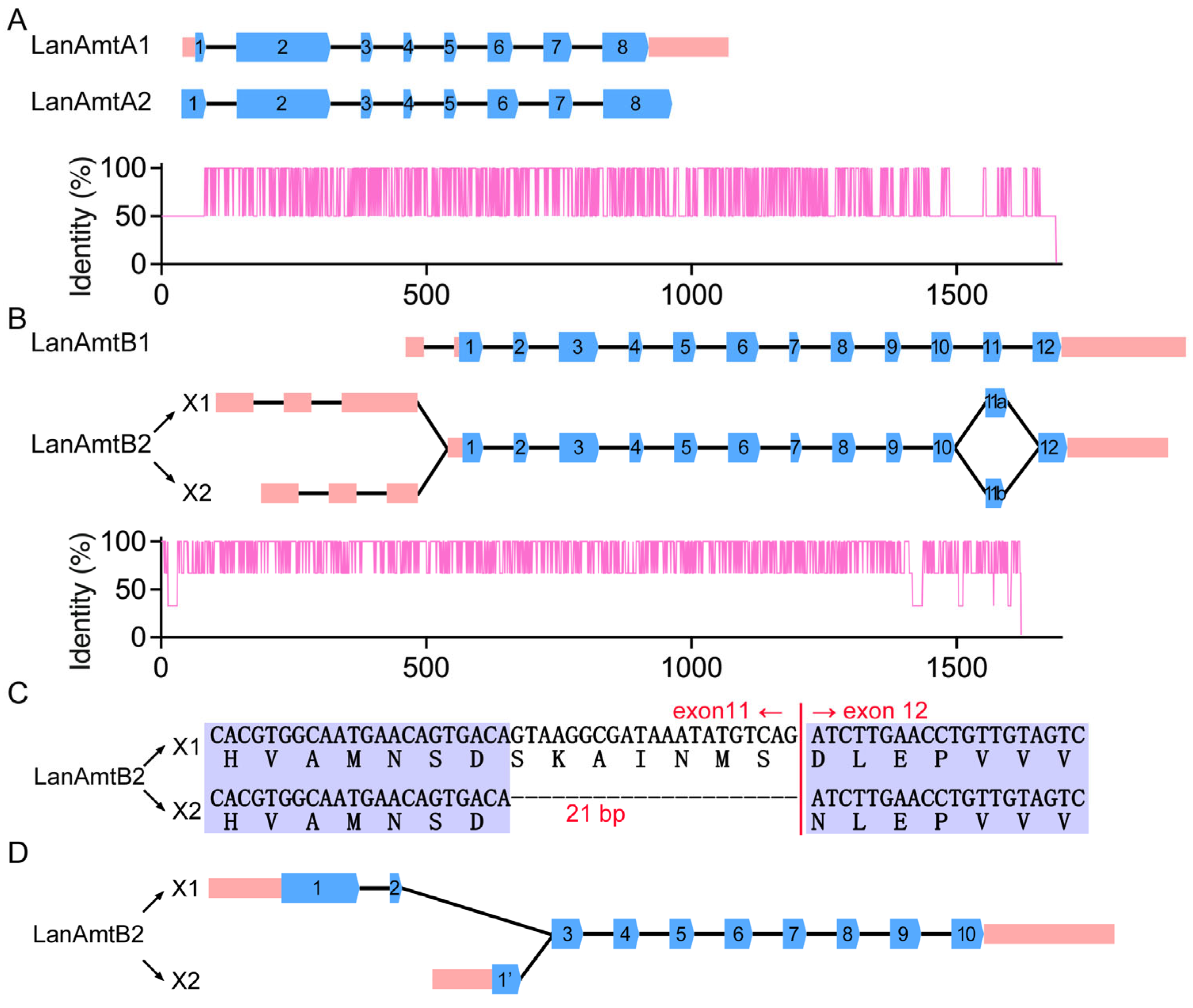
The AmtA subfamily (LanAmtA1 and LanAmtA2) displays nearly identical exon–intron structures, each comprising eight exons (Figure 2A). However, the terminal exons differ markedly in length, particularly at the 5′ and 3′ ends, likely reflecting divergence in regulatory regions. In contrast, AmtB subfamily members (LanAmtB1 and LanAmtB2) each contain 12 exons, a configuration distinct from the AmtA genes (Figure 2B). This shared architecture supports their derivation from an independent duplication event. Intriguingly, while LanAmtB1 produces a single transcript isoform, LanAmtB2 undergoes alternative splicing, yielding two variants. The primary difference lies in the 5′ untranslated region (UTR) and a 21-bp in-frame deletion at the 3′ end of exon 11 (Figure 2B,C). Because this deletion preserves the reading frame, both isoforms encode full-length, structurally intact proteins.
Similarly, LanAmtC gives rise to two alternative splice isoforms, differing in the composition of the first two exons (Figure 2D). These exons encompass both promoter-proximal regulatory sequences and coding segments, resulting in N-terminal protein variants with potentially distinct subcellular localizations or interaction partners. Such isoform diversification likely expands the functional repertoire of LanAmtC, possibly enabling context-dependent modulation of ammonia transport in response to developmental or environmental cues.

3.3. Physicochemical Properties of the Amt Gene Family in L. anatina

To better understand the structural and functional constraints shaping the Amt family in L. anatina, we systematically analyzed key physicochemical parameters of all five identified proteins. The length of predicted proteins varied considerably, ranging from 469 to 541 amino acid residues (Table 2). LanAmtA1 was the shortest (469 aa; ~50.3 kDa), while LanAmtA2 was the longest (541 aa; ~59.1 kDa) (Table 2). This ~72-residue difference, despite their origin from a recent tandem duplication, suggests differential domain retention post-duplication. The disparity in molecular mass likely stems not only from length variation but also from differences in amino acid composition, particularly in aromatic residues (e.g., Trp, Tyr, Phe), which contribute disproportionately to molecular weight. Such size divergence may influence transmembrane topology, pore architecture, or regulatory domain inclusion, potentially modulating transport kinetics or substrate specificity.
The theoretical isoelectric points (pI) of the Amt proteins spanned a moderately acidic range (4.94–6.18) (Table 2). LanAmtC exhibited the lowest pI (4.94), whereas LanAmtA1 had the highest (6.18). Given that the cytosolic pH in most eukaryotic cells is ~7.2, all LanAmt proteins are predicted to carry a net negative surface charge under physiological conditions. This electrostatic property could influence interactions with membrane lipids, cytosolic scaffolding proteins, or signaling molecules, and may also affect subcellular trafficking or stability in response to local pH fluctuations, particularly relevant in marine invertebrates exposed to variable environmental pH.
All five Amt proteins displayed low instability indices (ranging from 22.67 to 36.31), well below the threshold of 40 that typically distinguishes stable from unstable proteins (Table 2). This indicates high intrinsic structural stability, a feature likely essential for maintaining conformational integrity during repeated cycles of ammonia conduction and for resisting denaturation under osmotic or thermal stress in intertidal habitats. Furthermore, aliphatic indices, a measure of relative volume occupied by aliphatic side chains (Ala, Val, Ile, Leu), were uniformly high (90.17–101.04), with LanAmtB2 showing the highest value and LanAmtC the lowest (Table 2). Elevated aliphatic indices are characteristic of integral membrane proteins, reflecting enrichment in hydrophobic residues that facilitate stable integration into the lipid bilayer. The consistently high values across the LanAmt family strongly support their classification as multi-pass transmembrane transporters, consistent with the canonical Amt fold.

3.4. Structural Conservation of the Amt Genes in L. anatina

Across the tree of life, canonical Amt proteins typically adopt a structural architecture composed of 11 transmembrane helices, with the N-terminus oriented extracellularly and the C-terminus facing the cytoplasm [7]. This topology is remarkably conserved from prokaryotes to eukaryotes, including metazoan organisms such as plants, fungi, and invertebrates, and represents a unique fold with no homology to any other known membrane protein family. In contrast, vertebrate Amt-related proteins often exhibit an additional N-terminal TMH, resulting in a 12-TMH configuration [9].
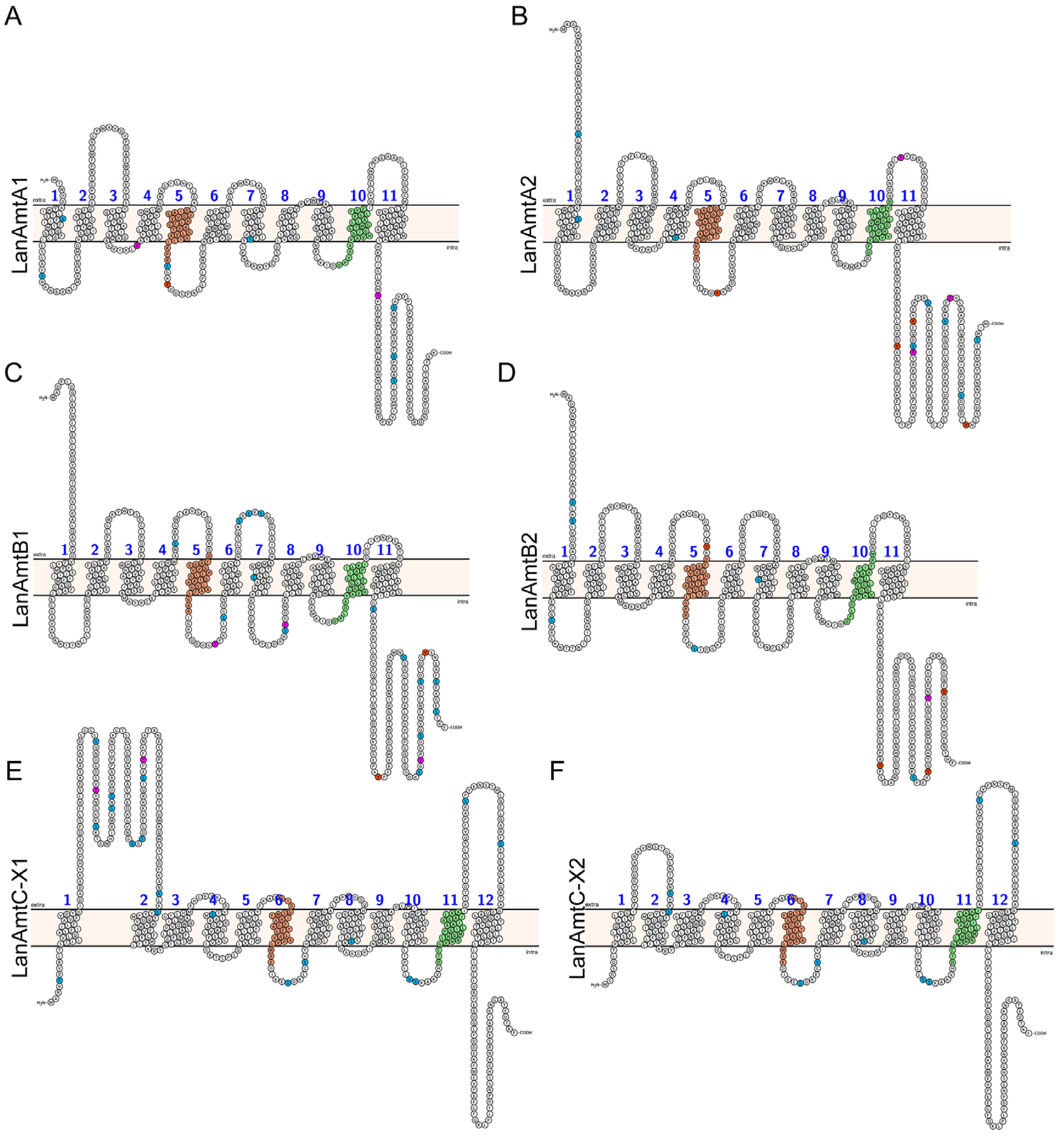
Among the five Amt genes identified in L. anatina, LanAmtC stands out due to its predicted 12-TMH architecture, which diverges from the canonical 11-TMH configuration (Figure 3A–F). Notably, LanAmtC displays an intracellular N-terminus, whereas all others conform to the ancestral extracellular N-terminus orientation. Phylogenetic analysis resolves LanAmtC into a distinct evolutionary clade, separate from the AmtA/B subfamilies, and highlights its closer affinity to certain vertebrate Amt proteins that also possess structural elaborations (Figure 1B). The retention of the ancestral 11-TMH topology in LanAmtA and LanAmtB aligns them with typical invertebrate and microbial Amt, suggesting conservation of core mechanisms governing ammonium conduction and membrane integration (Figure 3A–D).
In eukaryotes, post-translational modifications, particularly phosphorylation and dephosphorylation, play pivotal roles in regulating ammonia transporter activity [33]. To investigate the potential for such regulatory mechanisms in L. anatina, we systematically predicted phosphorylation sites across all members in the LanAmt gene family using the kinase-specific prediction algorithms. Interestingly, despite their recent origin from tandem duplications, LanAmtA1 and LanAmtA2 exhibit substantial sequence divergence, which extends to their post-translational modification landscapes. Prediction of phosphorylation sites revealed marked differences in the number, position, and kinase specificity of putative Ser/Thr/Tyr phosphorylation sites between these paralogs (Figure 3A,B). A similar pattern was observed within the LanAmtB subfamily (LanAmtB1 vs. LanAmtB2), suggesting that the sequence divergence and functional diversification following gene duplication may be accompanied by the evolution of distinct regulatory modules at the level of phosphorylation—though this hypothesis is based solely on computational predictions and requires experimental validation (Figure 3C,D).
Despite this topological divergence, LanAmtC retains substantial sequence conservation within its core transmembrane domains. Pairwise alignment across shared TM regions reveals 44.43% amino acid identity, markedly higher than the 24.92% identity observed across full-length sequences (Figure 4). This elevated conservation within membrane-embedded segments underscores their essential role in preserving fundamental transport functions, such as substrate selectivity, pore architecture, and conformational dynamics. In contrast, lower conservation observed in cytoplasmic and extracellular loops likely reflects greater evolutionary flexibility in regions involved in regulation or subcellular targeting. These findings reinforce the principle that transmembrane domains constitute the functional core of Amt proteins, under strong purifying selection even amid lineage-specific structural innovations.
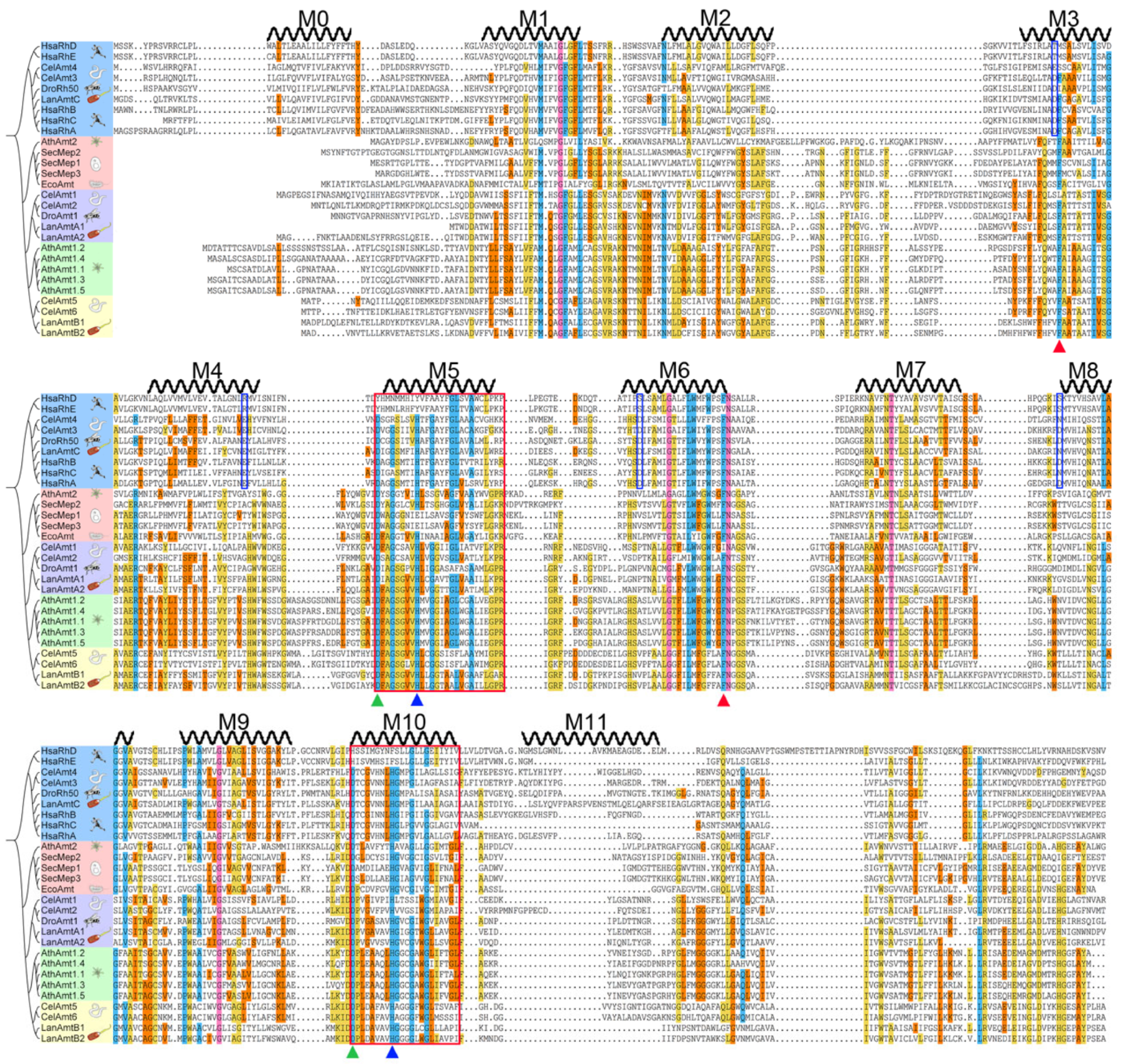
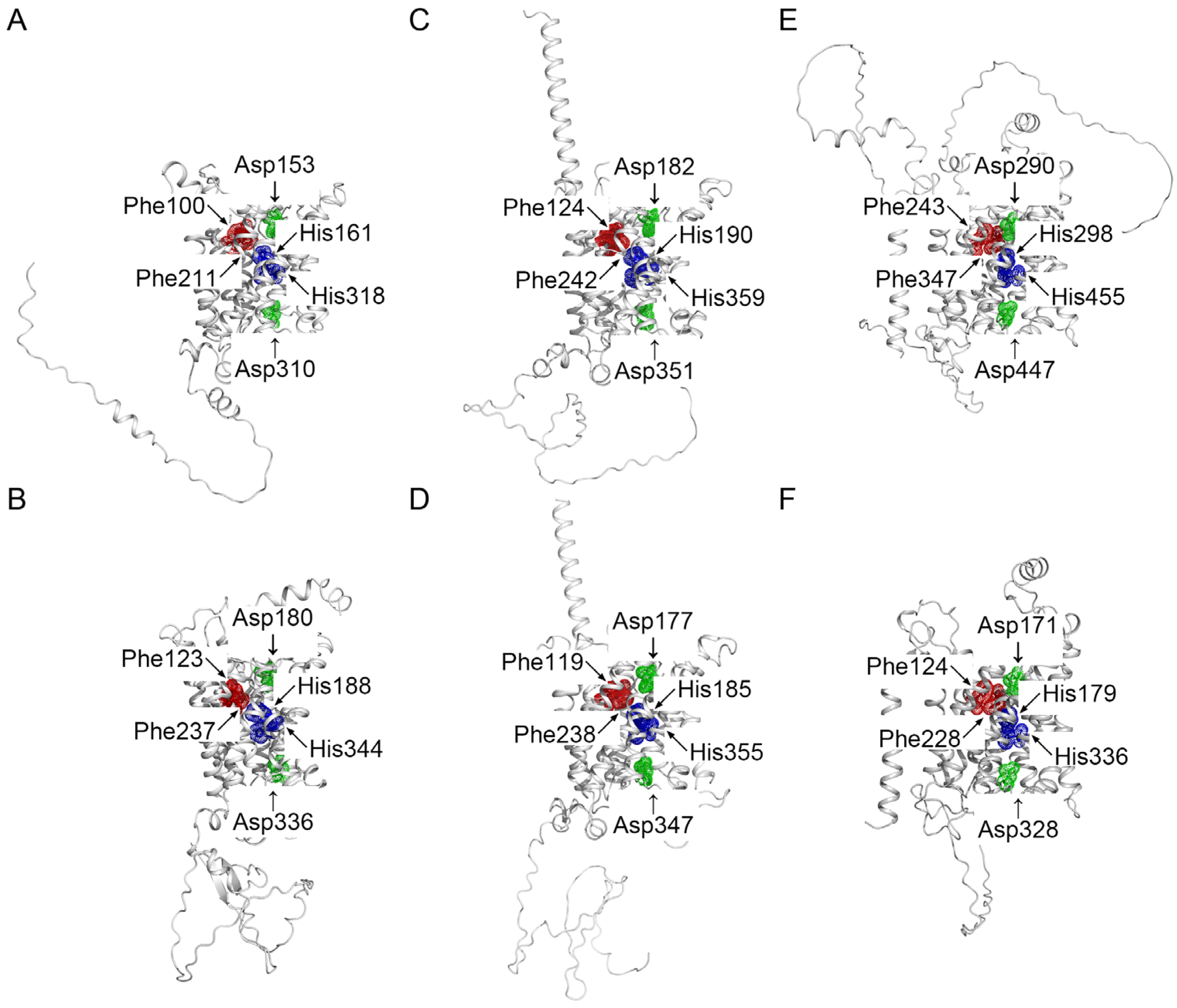
Critically, despite the dynamic evolutionary trajectory of the L. anatina genome, all five LanAmt genes retain key functional residues essential for ammonium transport (Figure 4). Structural and sequence analyses confirm the presence of highly conserved motifs in transmembrane helices 5 and 10 (Figure 3 and Figure 4). Notably, a pair of conserved His residues near the center of the pore forms part of the narrowest hydrophobic constriction, acting as a selectivity filter that permits passage of uncharged NH3 while excluding protons (Figure 4 and Figure 5). Additionally, all LanAmt proteins feature a characteristic extracellular aromatic gate composed of two Phe residues that regulate substrate access from the external environment (Figure 4 and Figure 5). At the extracellular vestibule, a strictly conserved Asp plays a critical structural role in stabilizing the entry pore, although it does not directly bind the substrate (Figure 4 and Figure 5). Similarly, a quasi-symmetric Asp residue is maintained near the cytoplasmic end of TM10, where it functions as an N-terminal helix capping residue, helping to orient the intracellular vestibule and preserve conformational integrity during the transport cycle (Figure 4 and Figure 5). The strict conservation of these residues across all five paralogs highlights their indispensable role in transport function. Collectively, these results demonstrate that while the Amt family in L. anatina has diversified in gene structure, membrane topology, and regulatory potential, its core transport mechanism remains deeply conserved, reflecting the fundamental importance of ammonia homeostasis in this ancient lophotrochozoan.
Of particular interest is LanAmtC, which exists in two alternative splice variants (LanAmtC-X1 and LanAmtC-X2). Both isoforms retain the canonical 12-transmembrane topology characteristic of this clade, but differ primarily in the length and composition of the first extracellular loop (Figure 3E,F). Intriguingly, LanAmtC-X1 harbors multiple high-confidence Ser and Thr phosphorylation sites within this variable loop region (Figure 3E,F). Given that extracellular loops can influence protein–protein interactions, membrane trafficking, or conformational dynamics, this differential phosphorylation potential suggests a mechanism for fine-tuning LanAmtC activity in response to environmental or developmental cues. Together, these findings indicate that the expansion of the LanAmt family in L. anatina has not only generated paralogs with divergent expression profiles and subtle structural variations but has also established layered regulatory complexity through differential post-translational modification. Such regulatory mechanisms likely enable precise spatiotemporal control of ammonia transport in this basal lophotrochozoan, reflecting an integrative strategy that combines transcriptional, structural, and post-translational regulation to achieve functional adaptability.

3.5. Expression Pattern of the Amt Genes in L. anatina

Tissue- and stage-specific gene expression is a fundamental mechanism for fine-tuning physiological functions in multicellular organisms, particularly among members of expanded gene families. To investigate the regulatory diversification of Amt genes in L. anatina, we analyzed their expression profiles across key embryonic stages and adult tissues using available transcriptomic datasets.
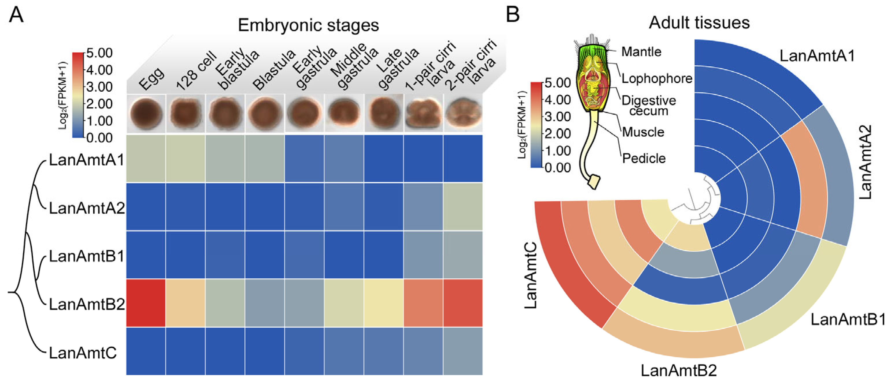
Remarkably, the five Amt genes exhibit distinct and non-redundant expression dynamics during development (Figure 6A). During early embryogenesis, LanAmtA1 and LanAmtB2 are the predominantly expressed family members, although their expression levels show a clear trend of gradual decrease (Figure 6A). As development proceeds, their expression trajectories diverge markedly: LanAmtA1 expression declines sharply following the transition from the blastula stage to gastrulation and becomes nearly undetectable after mid-gastrulation. In contrast, LanAmtB2 shows a progressive increase in transcript abundance, ultimately emerging as the dominant Amt gene from late gastrulation through larval stages. The remaining three paralogs are expressed at very low levels throughout embryogenesis, with only modest upregulation observed during the cirri larval stage, coinciding with the onset of active feeding and increased nitrogen metabolism (Figure 6A). Notably, none of these three genes show significant expression in early embryos, which may suggest potential functional specialization restricted to later developmental or adult contexts, though this inference requires further functional validation.
To further elucidate the potential functional diversification of the LanAmt gene family, we examined their expression profiles across adult tissues of L. anatina. It should be noted that all the expression patterns presented in this section are based on public RNA-seq datasets, and thus should be interpreted primarily as descriptive evidence rather than definitive functional conclusions. Strikingly, LanAmtC exhibits constitutively high expression in nearly all examined tissues, which may suggest a potential housekeeping or systemic role in ammonia homeostasis throughout the organism (Figure 6B); however, this proposed role requires additional functional data to be confirmed. This broad expression pattern is consistent with its potential specialization as a core component of basal nitrogen metabolism.
In contrast, LanAmtA1 is virtually undetectable in all adult tissues, consistent with its transcriptional silencing after early embryogenesis and reinforcing the possibility that it may play a role restricted to maternal or zygotic processes during initial cleavage stages (Figure 6A,B), though this functional inference remains to be supported by functional experiments. Its tandem duplicate, LanAmtA2, displays a complementary expression profile: while absent in early embryos, it becomes activated during late larval development and is predominantly expressed in the lophophore (the ciliated feeding and respiratory organ) in adults, with only trace levels detected in the mantle. This pattern implies a potential specialized function in ammonia handling at the interface between the organism and the external environment, which needs further functional verification.
Similarly, LanAmtB1, which initiates expression only at the end of embryogenesis, is primarily localized to the mantle, a tissue critical for ion exchange and waste excretion in brachiopods, and shows minimal expression in the lophophore (Figure 6A,B). Its paralog, LanAmtB2, exhibits a markedly broader tissue distribution in adults, being detectable in all sampled tissues except the digestive gland, where expression is either absent or below detection limits. Given its dominant expression during mid-to-late embryogenesis and sustained presence in multiple adult organs, LanAmtB2 may potentially serve as a versatile, developmentally persistent ammonia transporter, though this role requires confirmation by functional studies.

4. Discussion

The Amt/Mep/Rh superfamily comprises a group of transmembrane transport proteins that are ubiquitously conserved across prokaryotes and eukaryotes, playing pivotal roles in nitrogen metabolism, acid–base homeostasis, and osmoregulation [34]. In this study, we systematically identified and characterized the Amt gene family in the brachiopod L. anatina using whole-genome data. Our analyses encompassed chromosomal distribution, phylogenetic relationships, protein structural features, and signatures of functional conservation, thereby providing a molecular framework for understanding how L. anatina adapts to the physiologically challenging intertidal environment.
A total of five Amt genes that diversely originated were identified in the L. anatina genome. This genomic distribution architecture strongly indicates that local duplication events have driven the expansion of the Amt family in L. anatina. Such duplications likely provided the raw genetic material necessary for functional diversification, enabling fine-tuned regulation of ammonium uptake, excretion, and recycling in response to fluctuating environmental conditions characteristic of intertidal habitats. Phylogenetic analyses reveal that Amt proteins from diverse taxa generally cluster into well-supported, lineage-specific clades, reflecting independent evolutionary trajectories shaped by distinct modes of gene family expansion (Figure 1). Notably, the Amt repertoire in vertebrates, which served as the representative vertebrate in our analysis, also comprises five members. However, their expansion is primarily attributed to two rounds of whole-genome duplication in the early vertebrate ancestor, a major driver of genomic innovation in this lineage [35]. Consistent with this mechanism, vertebrate Amt genes form a monophyletic clade distinct from those of invertebrates, underscoring their divergence following large-scale genomic events. In contrast, the LanAmt genes appear to have expanded through small-scale, local duplications, and exhibit higher conservation than those in C. elegans, highlighting their fundamentally different evolutionary strategy for achieving functional complexity in an early-branching lophotrochozoan.
Expanded gene family members typically maintain highly conserved gene architectures, yet their coding sequences often display significant divergence. This pattern of structural conservation coupled with sequence differentiation is evident in the independent expansion of the AmtA and AmtB subfamilies within the L. anatina genome. Among the AmtA homologs, despite retaining conserved structural features consistent with their tandem duplication origin, the two paralogs exhibit remarkably low coding-sequence identity. This suggests a trajectory of rapid sequence evolution and potential neofunctionalization or subfunctionalization following the duplication event. In contrast, the AmtB homologs display a different evolutionary pattern: while their coding region architectures remain highly conserved, substantial divergence is observed in the non-coding regions. Notably, the 5′ region of LanAmtB2 exhibits alternative splicing, generating transcript variants that may differ in their regulatory potential. Such variation in the 5′ UTR could profoundly influence mRNA stability, translational efficiency, or responsiveness to upstream regulatory signals, thereby adding a layer of post-transcriptional complexity to the regulation of ammonium transport in this lineage.
It is noteworthy that certain invertebrate Amt family members, including those in C. elegans and L. anatina, cluster closely with vertebrate Amt genes, indicating a shared evolutionary origin (Figure 1B). This hypothesis is corroborated by a distinct structural signature: unlike the characteristic 11-transmembrane (11-TM) helix topology of canonical Amt transporters, both LanAmtC and the vertebrate Rh family possess a unique 12-TM topology [9]. The acquisition of this structural element may confer novel biophysical properties, such as altered membrane insertion kinetics, enhanced conformational stability, or modified gating dynamics during ammonium transport [9]. Moreover, the repositioning of the N-terminus to the cytosolic side, as predicted by topological modeling, could create a docking platform for intracellular regulatory factors [36]. Potential interactors might include protein kinases, pH-sensitive signaling molecules, or scaffolding proteins that modulate transporter activity in response to environmental fluctuations, such as changes in external ammonia concentration, salinity, or oxygen availability. Such regulatory capacity would be particularly advantageous for intertidal organisms like L. anatina, which must rapidly adjust nitrogen handling in response to dynamic habitat conditions. Thus, while invertebrate and vertebrate Amt proteins share deep homology and core functional mechanisms, lineage-specific structural innovations, such as the N-terminal extension in LanAmtC, likely underpin functional diversification and ecological adaptation.
Nevertheless, several important limitations inherent to bioinformatic predictions must be acknowledged when interpreting these results. All structural topology models, transmembrane helix predictions, and functional domain annotations were generated in silico using homology-based algorithms and secondary structure predictors, which rely on template structures and consensus sequence features rather than direct empirical data. Furthermore, transcriptomic inferences regarding expression patterns and alternative splicing rely on short-read assembly and abundance estimation, which can suffer from assembly artifacts, incomplete transcript recovery, or ambiguous isoform quantification, particularly in non-model organisms with limited transcriptome resources. Accordingly, experimental validation is essential to confirm the functional relevance of our bioinformatic predictions. Heterologous expression systems (e.g., Xenopus oocytes or yeast mutants) coupled with radiotracer uptake or electrophysiological assays are required to directly measure ammonium transport kinetics, substrate specificity, pH dependence, and regulatory responses of each LanAmt paralog.

5. Conclusions

In summary, through gene duplication and functional divergence during evolution, the Amt gene family in L. anatina has formed a protein system characterized by structural stability, differentiated expression patterns, and conserved core features, providing a molecular foundation for nitrogen utilization and ecological adaptation in the intertidal environment. Functional specialization, as suggested by the structural, phylogenetic, and expression-based indications in this study, remains a hypothesis to be validated in future research. Future studies involving expression profiling and functional experiments will help further elucidate the specific mechanisms of each member in physiological processes.

Author Contributions

Conceptualization, X.Y. and Y.J.; software, X.Y. and Y.J.; validation, X.Y., X.X. and J.P.; formal analysis, X.Y. and L.Y.; investigation, Y.J.; resources, X.L.; data curation, X.Y.; writing—original draft preparation, X.Y. and Y.J.; writing—review and editing, Y.J. and X.L.; visualization, Y.J. and X.L.; supervision, Y.J.; project administration, Y.J.; funding acquisition, Y.J. All authors have read and agreed to the published version of the manuscript.

Funding

This research was financially supported in part by grants from the Natural Science Foundation of China (32301408), the China Postdoctoral Science Foundation (2023M741837), the Special Grant of Zhoushan for Breeding Aquatic Animals (2024Y001-2), and the Key Laboratory of Mariculture of Ministry of Education, Ocean University of China (KLM202203).

Institutional Review Board Statement

Not applicable.

Data Availability Statement

All data analyzed during this study are included in this article.

Conflicts of Interest

The authors declare no conflicts of interest.

Abbreviations

The following abbreviations are used in this manuscript:
Amtammonium transporters
LanLingula anatina

References

  1. Cabello, P.; Roldán, M.D.; Castillo, F.; Moreno-Vivián, C. Nitrogen Cycle in Encyclopedia of Microbiology, 3rd ed.; Schaechter, M., Ed.; Academic Press: Oxford, UK, 2009; pp. 299–321. [Google Scholar]
  2. Miller, A.J.; Cramer, M.D. Root Nitrogen Acquisition and Assimilation. Plant Soil 2005, 274, 1–36. [Google Scholar] [CrossRef]
  3. Loqué, D.; von Wirén, N. Regulatory levels for the transport of ammonium in plant roots. J. Exp. Bot. 2004, 55, 1293–1305. [Google Scholar] [CrossRef] [PubMed]
  4. Wirén, N.V.; Merrick, M. Regulation and Function of Ammonium Carriers in Bacteria, Fungi, and Plants in Molecular Mechanisms Controlling Transmembrane Transport; Springer: Berlin/Heidelberg, Germany, 2004; pp. 95–120. [Google Scholar]
  5. Williamson, G.; Harris, T.; Bizior, A.; Hoskisson, P.A.; Pritchard, L.; Javelle, A. Biological ammonium transporters: Evolution and diversification. FEBS J. 2024, 291, 3786–3810. [Google Scholar] [CrossRef] [PubMed]
  6. Xu, Y.; Wang, J.; Wu, A.; Wang, M. Ammonia metabolism and ammonia-induced cell death: Role in cancer therapy. Cell Commun. Signal. 2025, 23, 468. [Google Scholar] [CrossRef] [PubMed]
  7. Khademi, S.; Stroud, R.M. The Amt/MEP/Rh family: Structure of AmtB and the mechanism of ammonia gas conduction. Physiology 2006, 21, 419–429. [Google Scholar] [CrossRef]
  8. Khademi, S.; O’Connell, J.; Remis, J.; Robles-Colmenares, Y.; Miercke, L.J.W.; Stroud, R.M. Mechanism of Ammonia Transport by Amt/MEP/Rh: Structure of AmtB at 1.35 Å. Science 2004, 305, 1587–1594. [Google Scholar] [CrossRef]
  9. Gruswitz, F.; Chaudhary, S.; Ho, J.D.; Schlessinger, A.; Pezeshki, B.; Ho, C.-M.; Sali, A.; Westhoff, C.M.; Stroud, R.M. Function of human Rh based on structure of RhCG at 2.1 A. Proc. Natl. Acad. Sci. USA 2010, 107, 9638–9643. [Google Scholar] [CrossRef]
  10. Williamson, G.; Tamburrino, G.; Bizior, A.; Boeckstaens, M.; Mirandela, G.D.; Bage, M.G.; Pisliakov, A.; Ives, C.M.; Terras, E.; A Hoskisson, P.; et al. A two-lane mechanism for selective biological ammonium transport. Elife 2020, 9, e57183. [Google Scholar] [CrossRef]
  11. Weihrauch, D.; Joseph, G.; Allen, P. Ammonia excretion in aquatic invertebrates: New insights and questions. J. Exp. Biol. 2018, 221, jeb169219. [Google Scholar] [CrossRef]
  12. Harper, D.A.T.; Popov, L.E.; Holmer, L.E. Brachiopods: Origin and early history. Palaeontology 2017, 60, 609–631. [Google Scholar] [CrossRef]
  13. Ruban, D.A. Brachiopod Diversity and Paleoenvironmental Changes in the Paleogene: Comparing the Available Long-Term Patterns. Diversity 2025, 17, 505. [Google Scholar] [CrossRef]
  14. Luo, Y.-J.; Takeuchi, T.; Koyanagi, R.; Yamada, L.; Kanda, M.; Khalturina, M.; Fujie, M.; Yamasaki, S.-I.; Endo, K.; Satoh, N. The Lingula genome provides insights into brachiopod evolution and the origin of phosphate biomineralization. Nat. Commun. 2015, 6, 8301. [Google Scholar] [CrossRef] [PubMed]
  15. Santagata, S. Brachiopoda in Evolutionary Developmental Biology of Invertebrates 2: Lophotrochozoa (Spiralia); Wanninger, A., Ed.; Springer: Vienna, Austria, 2015; pp. 263–277. [Google Scholar]
  16. Goto, R.; Takano, T.; Seike, K.; Yamashita, M.; Paulay, G.; Rodgers, K.S.; Hunter, C.L.; Tongkerd, P.; Sato, S.; Hong, J.-S.; et al. Stasis and diversity in living fossils: Species delimitation and evolution of lingulid brachiopods. Mol. Phylogenet. Evol. 2022, 175, 107460. [Google Scholar] [CrossRef] [PubMed]
  17. Gerdol, M.; Luo, Y.-J.; Satoh, N.; Pallavicini, A. Genetic and molecular basis of the immune system in the brachiopod Lingula anatina. Dev. Comp. Immunol. 2018, 82, 7–30. [Google Scholar] [CrossRef]
  18. Kon, K.; Shimanaga, M.; Horinouchi, M. Marine Ecology: Intertidal/Littoral Zone. In Japanese Marine Life; Inaba, K., Hall-Spencer, J., Eds.; Springer: Singapore, 2020. [Google Scholar] [CrossRef]
  19. Grabherr, M.G.; Haas, B.J.; Yassour, M.; Levin, J.Z.; Thompson, D.A.; Amit, I.; Adiconis, X.; Fan, L.; Raychowdhury, R.; Zeng, Q.D.; et al. Full-length transcriptome assembly from RNA-Seq data without a reference genome. Nat. Biotechnol. 2011, 29, 644–652. [Google Scholar] [CrossRef]
  20. Camacho, C.; Coulouris, G.; Avagyan, V.; Ma, N.; Papadopoulos, J.; Bealer, K.; Madden, T.L. BLAST+: Architecture and applications. BMC Bioinform. 2009, 10, 421. [Google Scholar] [CrossRef]
  21. Larkin, M.A.; Blackshields, G.; Brown, N.P.; Chenna, R.; McGettigan, P.A.; McWilliam, H.; Valentin, F.; Wallace, I.M.; Wilm, A.; Lopez, R.; et al. Clustal W and Clustal X version 2.0. Bioinformatics 2007, 23, 2947–2948. [Google Scholar] [CrossRef]
  22. Chen, C.; Wu, Y.; Li, J.; Wang, X.; Zeng, Z.; Xu, J.; Liu, Y.; Feng, J.; Chen, H.; He, Y.; et al. TBtools-II: A “one for all, all for one” bioinformatics platform for biological big-data mining. Mol. Plant 2023, 16, 1733–1742. [Google Scholar] [CrossRef]
  23. Katoh, K.; Toh, H. Recent developments in the MAFFT multiple sequence alignment program. Brief. Bioinform. 2008, 9, 286–298. [Google Scholar] [CrossRef]
  24. Nguyen, L.-T.; Schmidt, H.A.; Von Haeseler, A.; Minh, B.Q. IQ-TREE: A fast and effective stochastic algorithm for estimating Maximum-Likelihood phylogenies. Mol. Biol. Evol. 2015, 32, 268–274. [Google Scholar] [CrossRef]
  25. Hu, B.; Jin, J.; Guo, A.-Y.; Zhang, H.; Luo, J.; Gao, G. GSDS 2.0: An upgraded gene feature visualization server. Bioinformatics 2015, 31, 1296–1297. [Google Scholar] [CrossRef] [PubMed]
  26. Gasteiger, E.; Hoogland, C.; Gattiker, A.; Duvaud, S.E.; Wilkins, M.R.; Appel, R.D.; Bairoch, A. Protein Identification and Analysis Tools on the ExPASy Server in the Proteomics Protocols Handbook; Walker, J.M., Ed.; Humana Press: Totowa, NJ, USA, 2005; pp. 571–607. [Google Scholar]
  27. Blom, N.; Sicheritz-Pontén, T.; Gupta, R.; Gammeltoft, S.; Brunak, S. Prediction of post-translational glycosylation and phosphorylation of proteins from the amino acid sequence. Proteomics 2004, 4, 1633–1649. [Google Scholar] [CrossRef] [PubMed]
  28. Omasits, U.; Ahrens, C.H.; Müller, S.; Wollscheid, B. Protter: Interactive protein feature visualization and integration with experimental proteomic data. Bioinformatics 2014, 30, 884–886. [Google Scholar] [CrossRef]
  29. Abramson, J.; Adler, J.; Dunger, J.; Evans, R.; Green, T.; Pritzel, A.; Ronneberger, O.; Willmore, L.; Ballard, A.J.; Bambrick, J.; et al. Accurate structure prediction of biomolecular interactions with AlphaFold 3. Nature 2024, 630, 493–500. [Google Scholar] [CrossRef] [PubMed]
  30. Delano, W.L. The PyMol molecular graphics system. Proteins Struct. Funct. Bioinform. 2002, 30, 442–454. [Google Scholar]
  31. Kim, D.; Paggi, J.M.; Park, C.; Bennett, C.; Salzberg, S.L. Graph-based genome alignment and genotyping with HISAT2 and HISAT-genotype. Nat. Biotechnol. 2019, 37, 907–915. [Google Scholar] [CrossRef]
  32. Kovaka, S.; Zimin, A.V.; Pertea, G.M.; Razaghi, R.; Salzberg, S.L.; Pertea, M. Transcriptome assembly from long-read RNA-seq alignments with StringTie2. Genome Biol. 2019, 20, 278. [Google Scholar] [CrossRef]
  33. Berg, B.v.D.; Chembath, A.; Jefferies, D.; Basle, A.; Khalid, S.; Rutherford, J.C. Structural basis for Mep2 ammonium transceptor activation by phosphorylation. Nat. Commun. 2016, 7, 11337. [Google Scholar] [CrossRef]
  34. Sayer, M.H., Jr.; Reddy, V.S.; Tamang, D.G.; Västermark, Å. The Transporter Classification Database. Nucleic Acids Res. 2013, 42, D251–D258. [Google Scholar] [CrossRef]
  35. Holland, L.Z.; Daza, D.O. A new look at an old question: When did the second whole genome duplication occur in vertebrate evolution? Genome Biol. 2018, 19, 209. [Google Scholar] [CrossRef]
  36. Øye, H.; Lundekvam, M.; Caiella, A.; Hellesvik, M.; Arnesen, T. Protein N-terminal modifications: Molecular machineries and biological implications. Trends Biochem. Sci. 2025, 50, 290–310. [Google Scholar] [CrossRef]
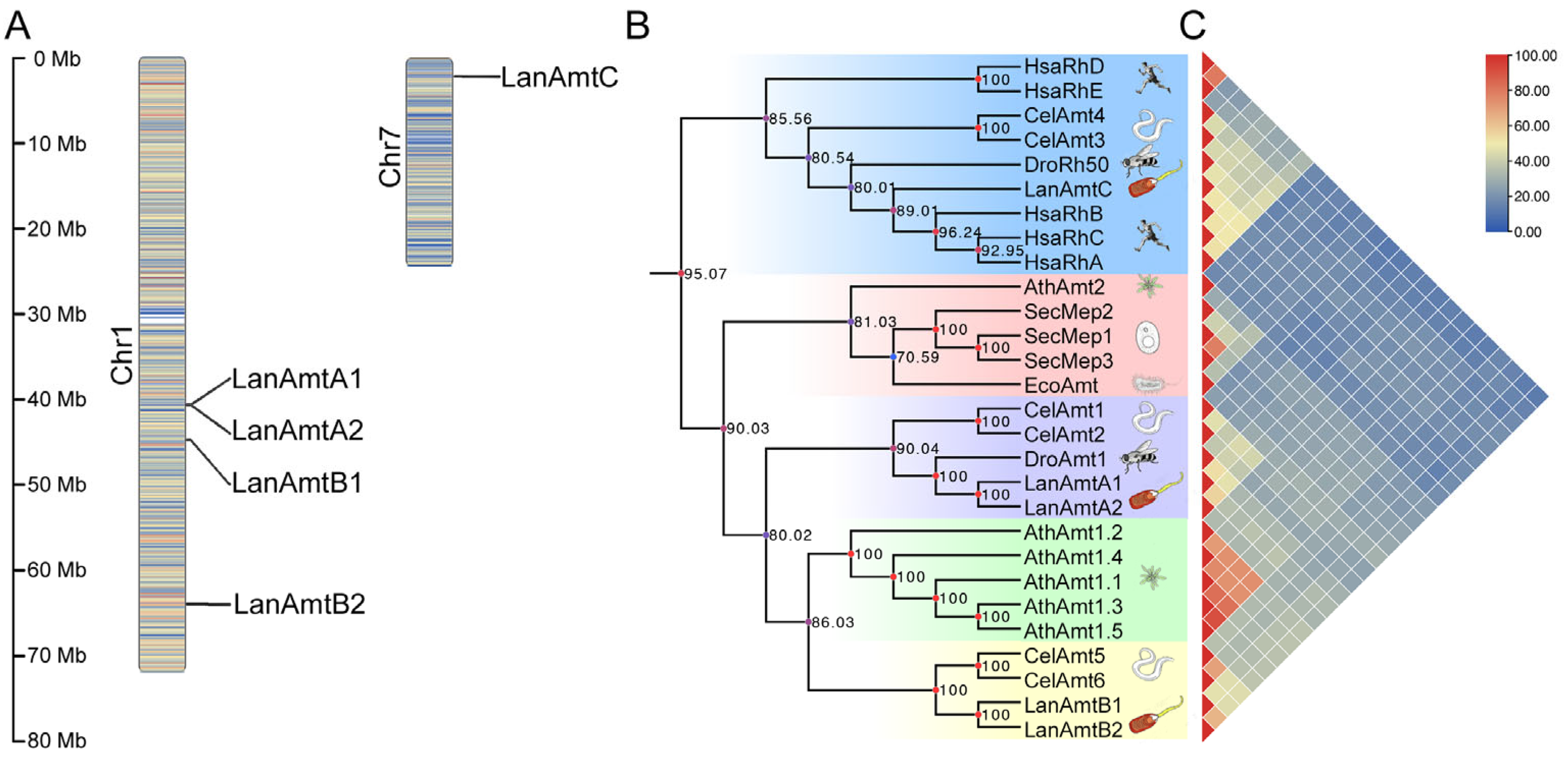
Figure 1. Genomic distribution and phylogenetic relationships of Amt family members in L. anatina. (A) Chromosomal localization of the five identified LanAmt genes. (B) Phylogenetic tree of Amt proteins from representative species across major metazoan phyla. (C) Pairwise protein sequence similarity matrix of Amt homologs from diverse taxa, calculated based on full-length alignments.
Nitrogen 07 00043 g001
Figure 2. Gene structure organization and alternative splicing patterns of Amt genes in L. anatina. (A) Exon–intron architecture and sequence divergence within the AmtA subfamily (LanAmtA1 and LanAmtA2). (B) Exon–intron architecture and sequence divergence within the AmtB subfamily (LanAmtB1 and LanAmtB2). (C) Local alignment of the genomic region spanning the junction between exon 11 and exon 12, comparing the two alternative splicing isoforms of LanAmtB2. (D) Gene models of the two alternative splicing isoforms of LanAmtC (designated LanAmtC-X1 and LanAmtC-X2).
Nitrogen 07 00043 g002
Figure 3. Predicted secondary structure topology of Amt proteins in L. anatina. (AF) Transmembrane topology diagrams for LanAmtA1 (A), LanAmtA2 (B), LanAmtB1 (C), LanAmtB2 (D), LanAmtC-X1 (E), and LanAmtC-X2 (F), generated using Protter. Conserved functional motifs within transmembrane helices 5 and 10 are highlighted in orange and green, respectively. Predicted phosphorylation sites on serine (Ser), threonine (Thr), and tyrosine (Tyr) residues are marked in light blue, purple, and red, respectively.
Nitrogen 07 00043 g003
Figure 4. Multiple sequence alignment of Amt proteins from L. anatina and representative homologs across diverse taxa. Conservation levels at each aligned position are color-coded as follows: pink (100% identity), light blue (≥75%), yellow (≥50%), and orange (≥33%). Transmembrane helices are labeled M0–M11 based on structural annotation and marked with wavy lines. Two highly conserved functional motifs located in transmembrane helices 5 and 10 are highlighted by red boxes. Key conserved residues critical for ammonium transport are annotated with colored triangles: Red triangles denote Phe residues that constitute the extracellular aromatic gate; Blue triangles indicate His residues in TM5 and TM10 positioned near the pore center, which form the narrowest hydrophobic constriction site; Green triangles mark two Asp residues located at the extracellular and intracellular vestibules, respectively. Acidic residues include an Glu and three Asp in the orthologs that related to vertebrate Rh family and serve to attract NH4+ are highlighted by blue boxes.
Nitrogen 07 00043 g004
Figure 5. Predicted three-dimensional structures of Amt proteins in the L. anatina. (AF) AlphaFold-predicted models for LanAmtA1 (A), LanAmtA2 (B), LanAmtB1 (C), LanAmtB2 (D), LanAmtC-X1 (E), and LanAmtC-X2 (F). Transmembrane helices are rendered as cartoons and colored in gray. Key conserved residues essential for ammonium transport are shown as stick-and-sphere representations and color-coded as follows: red for Phe residues forming the extracellular aromatic gate; blue for His residues in transmembrane helices 5 and 10, positioned near the pore center and constituting the narrowest hydrophobic constriction site; green for Asp residues located at the extracellular and intracellular vestibules, respectively.
Nitrogen 07 00043 g005
Figure 6. Spatiotemporal expression profiles of Amt genes in Lingula anatina. (A) Expression dynamics of LanAmt genes across key embryonic developmental stages. (B) Tissue-specific expression patterns of LanAmt genes in adult L. anatina. Gene expression levels are presented as FPKM (fragments per kilobase of transcript per million mapped reads). Illustrations of embryonic stages and schematic diagrams depicting internal anatomy were adapted from previously published sources [14,17].
Nitrogen 07 00043 g006
Table 1. Selected transcriptomic data for different developmental stages and tissues in this study.
Run No.Stages or TissuesBytes (bp)Application
SRR2131217Egg885601259Expression analysis
SRR2131219128 cells937404130Expression analysis
SRR2131221Early blastula858308241Expression analysis
SRR2131224Blastula896048608Expression analysis
SRR2131226Early gastrula1813873230Expression analysis
SRR2131227Middle gastrula1123581001Expression analysis
SRR2131229Late gastrula1713689257Expression analysis
SRR21312301-pair cirri larva3606932894Expression analysis
SRR21312322-pair cirri larva1122546685Expression analysis
SRR2131233Lophophore2861789013Expression analysis
SRR2131238Pedicle3887211456Expression analysis
SRR2131237Ventral mantle3848716217Expression analysis and Trinity assemble
SRR2131235Digestive cecum5176516384Expression analysis
SRR29286084Adductor muscle3297325545Expression analysis
Table 2. Physicochemical properties of Amt gene family in L. anatina.
GeneAmino Acid Sequence LengthMolecular Weight (Da)Isoelectric Point (pI)Instability IndexAliphatic Index
LanAmtA146950,210.246.1827.85101.04
LanAmtA254159,001.945.9636.3196.38
LanAmtB152356,429.635.0622.6790.17
LanAmtB252856,797.815.8125.0994.05
LanAmtC48452,328.604.9428.20100.79
Disclaimer/Publisher’s Note: The statements, opinions and data contained in all publications are solely those of the individual author(s) and contributor(s) and not of MDPI and/or the editor(s). MDPI and/or the editor(s) disclaim responsibility for any injury to people or property resulting from any ideas, methods, instructions or products referred to in the content.