Article

Relating the Morphology of Bipolar Neurons to Fractal Dimension

by
Bret Brouse, Jr.
1,2,
Conor Rowland
1,2 and
Richard P. Taylor
1,2,*
1
Department of Physics, University of Oregon, Eugene, OR 97403, USA
2
Materials Science Institute, University of Oregon, Eugene, OR 97403, USA
*
Author to whom correspondence should be addressed.
Fractal Fract. 2025, 9(1), 9; https://doi.org/10.3390/fractalfract9010009
Submission received: 1 October 2024 / Revised: 20 December 2024 / Accepted: 26 December 2024 / Published: 28 December 2024
(This article belongs to the Special Issue Fractal Analysis in Biology and Medicine)

Abstract

:
By analyzing reconstructed three-dimensional images of retinal bipolar neurons, we show that their dendritic arbors weave through space in a manner that generates fractal-like behavior quantified by an ‘effective’ fractal dimension. Examining this fractal weave along with traditional morphological parameters reveals a dependence of arbor fractal dimension on the summation of the lengths of the arbor’s dendrites. We discuss the implications of this behavior for healthy neurons and also for the morphological deterioration of unhealthy neurons in response to diseases.

1. Introduction

Extensive research has been devoted to examining the physical structure of neurons with the goal of investigating how their form influences their functionality [1,2,3,4]. In addition to healthy neurons, their unhealthy counterparts have also been investigated to understand how they deteriorate with disease [5,6,7,8]. It is well-established that dendrites branch out from the neuron’s cell body (its soma) to form an arbor that establishes electrical connectivity to neighboring neurons [9,10,11,12,13,14,15]. Here, we have investigated the relationship between the size and morphological structure of neuron arbors. For the latter, we focused on the repetition of dendrite patterns formed at different scales and quantified their fractal scaling properties.
Our research considered retinal bipolar cells due to their critical role in passing electrical signals through the visual system. This role makes the visual system vulnerable to diseases that impact the bipolar neuron’s ability to connect to their neighbors. Figure 1A,B show the connections that bipolar neurons establish to bridge between the retina’s ganglion cells and photoreceptors. Ganglion cells on the left-hand side (i.e., the inner surface) of the retina connect to bipolar neurons on the right, forming the signal communication route from the photoreceptors to the optic nerve and on to the brain. Zooming in on the retina’s right-hand side, the dendrites of the bipolar cells form a complex network of connections with horizontal cells and the photoreceptors. Known as the outer plexiform layer [16,17], the physical complexity of this network originates from the repeating fractal patterns of their dendrites. Figure 1C,D show an individual bipolar dendrite taken from a mouse retina and are shown from two orientations to help display their fractal complexity.
Although retinal diseases such as macular degeneration and retinitis pigmentosa predominantly impact the photoreceptors [18,19,20], the bipolar neurons are also affected [21]. Approaches to restoring sight include regrowing the damaged photoreceptors [18,22,23] or replacing them with retinal implants featuring photodiodes that act as artificial photoreceptors (Figure 1A) [24,25,26,27]. Both strategies will require re-establishing the bipolar neurons to interact with either the restored natural photoreceptors or their implanted artificial versions. For chemical strategies aimed at regrowing the bipolar neurons, fractal morphological studies will be necessary to determine when the neurons have re-established their healthy state [28]. The surfaces of future retinal implants will feature fractal patterns [29] that act as physical scaffolds for the bipolar neurons to adhere to and regrow their healthy form [30]. Fractal morphology studies will then inform the implant’s surface patterns.
Before examining variations in diseased neurons, it is necessary to establish the degree to which neurons vary within their healthy condition. Accordingly, healthy neurons were the focus of our study. By analyzing reconstructed three-dimensional images of mouse bipolar neurons, we first probed neuron size by employing two parameters—the arbor radius and the volume of space confined to a convex hull [31] that the neuron occupies. In terms of quantifying their fractal morphology, scaling analysis of neurons is not a new endeavor [32,33,34,35,36,37,38,39,40,41,42,43,44,45,46]. However, previous analyses were performed predominantly as a means to classify neurons based on morphology rather than to study the geometry of the dendritic arbors themselves. In contrast, our group recently examined the geometric properties of neurons from the hippocampal region of the brain in order to understand the morphological origin of neuron fractality and the functional purpose of assuming this form [47]. The study considered here extended this approach to the retina’s bipolar neurons.
What reason do neurons have to favor fractal geometry (in particular, multi-scaled branch patterns) over, say, the Euclidean designs that we find in the typical electrical circuitry of computers and cell phones (in particular, wires following one-dimensional lines)? Further, are there optimal values of fractal parameters that cause neurons to thrive? Our previous research demonstrated that neuron arbors enhance their ability to connect to their neighbors through their fractal characteristics [47,48]. Here, we built on this result and showed that, as with hippocampal neurons, the manner in which the bipolar dendrites weave through space is important for establishing their fractal-like behavior. Quantified by an ‘effective’ fractal dimension, this scaling parameter quantifies the relative amounts of coarse and fine structure contributing to the fractal mix that generates the physical complexity of the arbor’s dendritic patterns.
We examined variations in the arbor sizes, variations in their fractal dimensions, and the relationship between these two variations. In particular, we showed that large neurons are not simply bigger replicas of their smaller counterparts by identifying subtle changes in arbor pattern complexity with neuron size. We found that the arbor fractal dimension was dependent on the summation of the fork lengths of all of the dendrites in the arbor (see Section 2). We discuss the implications of this behavior for healthy neurons and also for the morphological deterioration of unhealthy neurons in response to diseases.

2. Materials and Methods
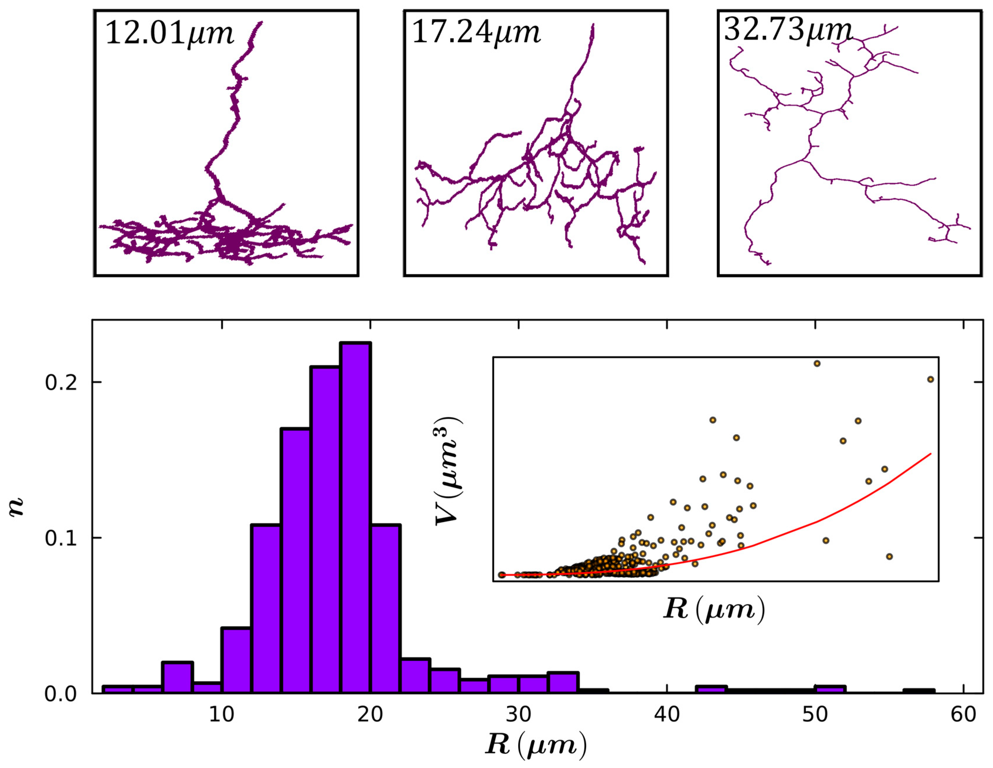
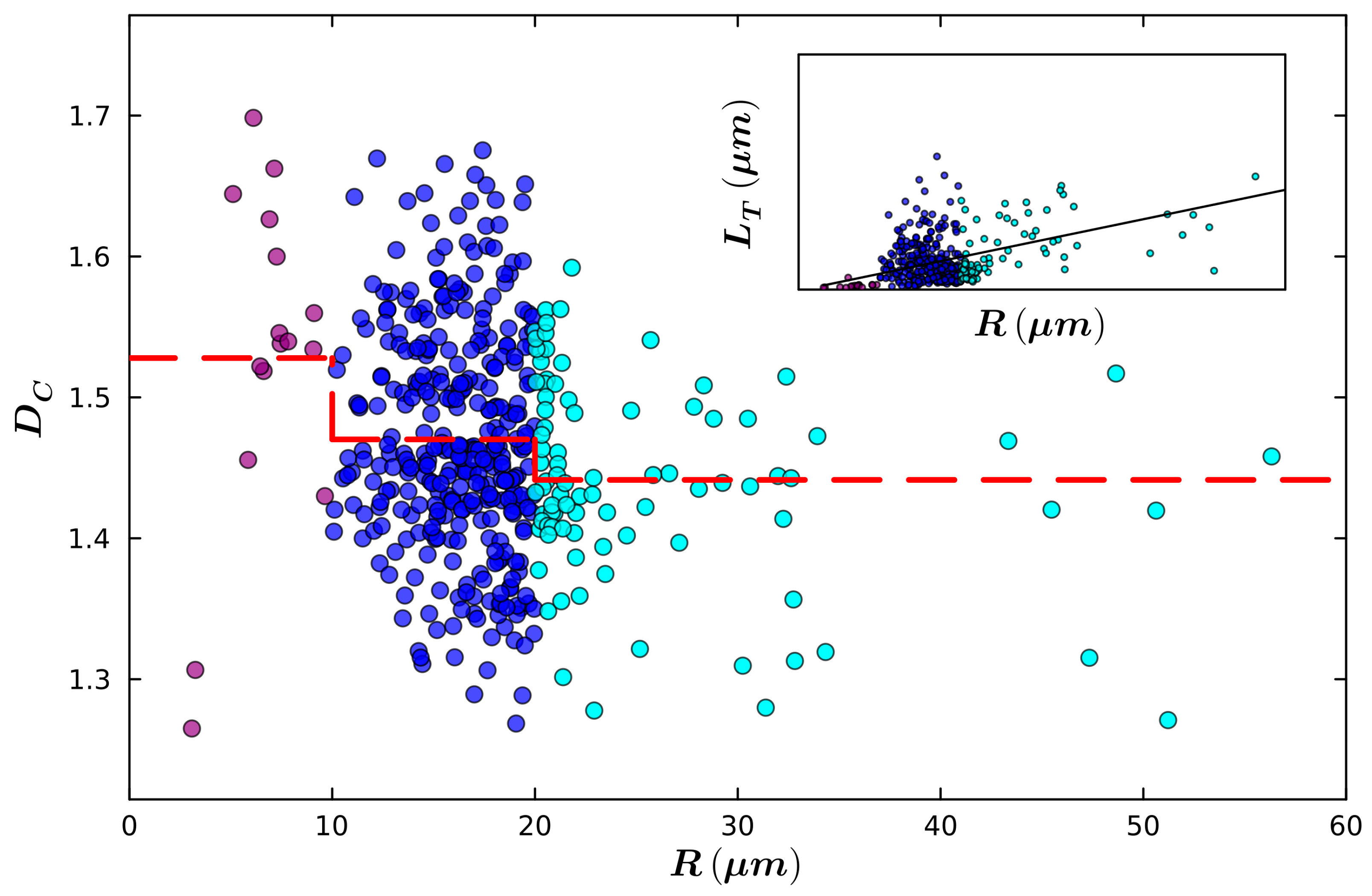
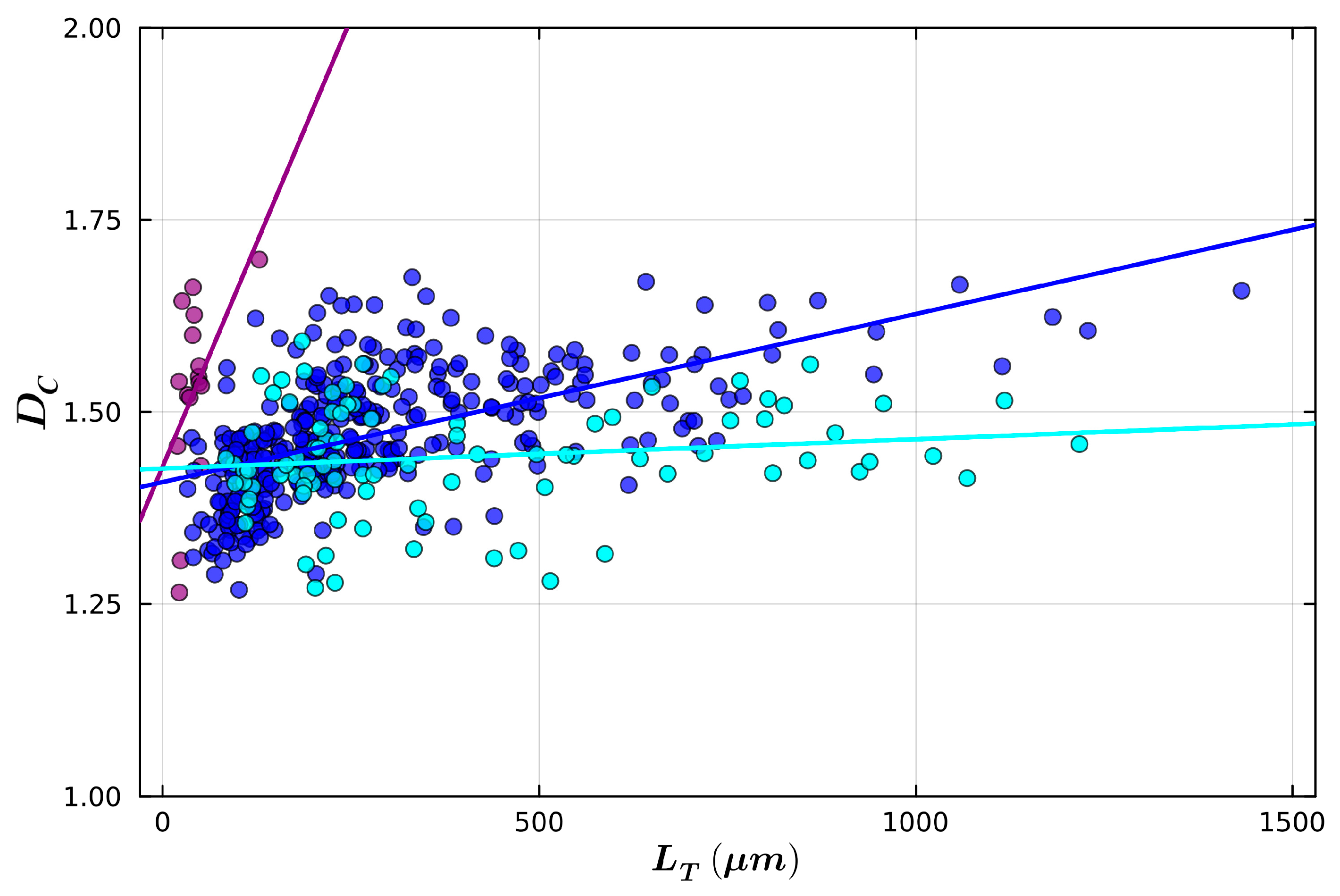
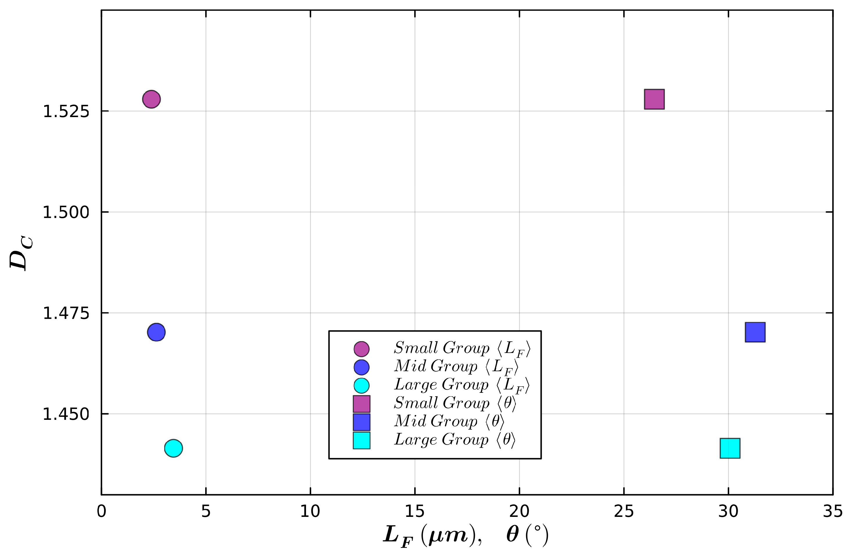
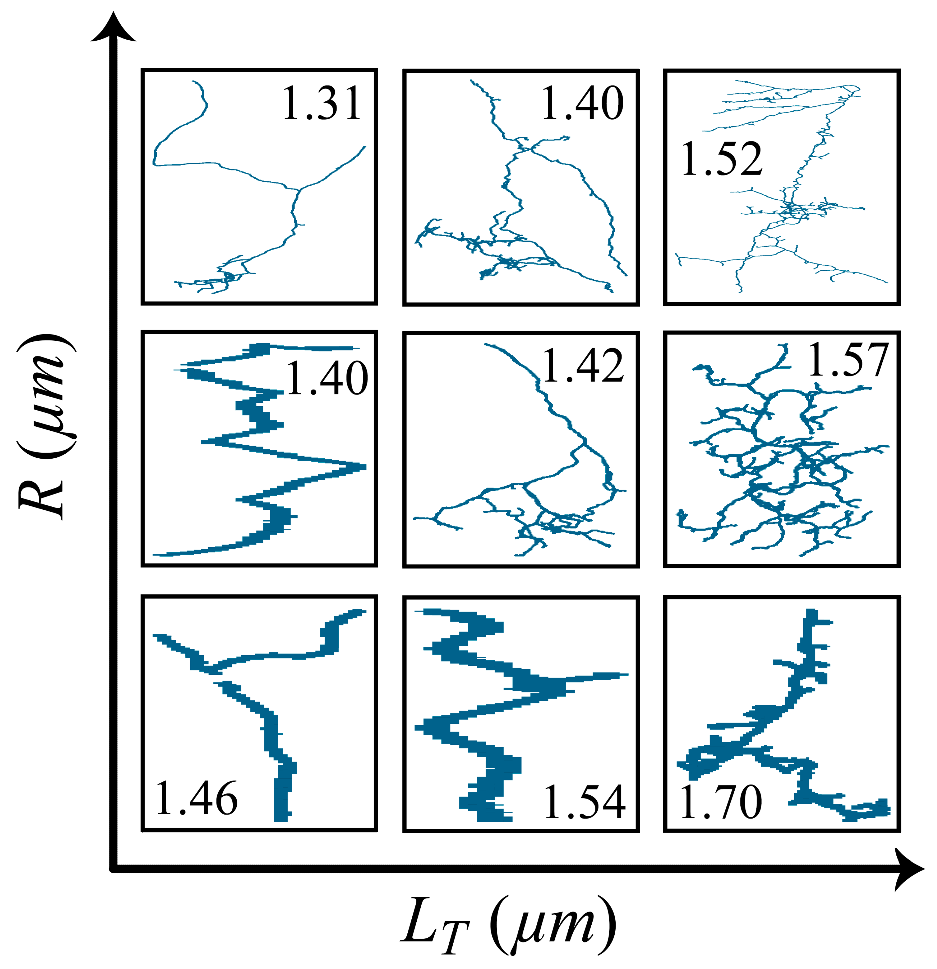
Our study focused on the data of 453 retinal bipolar neurons available on https://neuromorpho.org/ [49]. These in vitro images of healthy, 30-day-old C57BL/6 mouse neurons were obtained by other researchers using serial block-face electron microscopy (SBEM) after being prepared with selective staining techniques [50,51] and then reconstructed using a technique known as SegEM. This is achieved through a combination of trained human annotators manually tracing skeletons of neurites using 3D reconstruction software (KNOSSOS) and machine learning techniques [52]. In our reconstructions (Figure 1C,D), each dendrite is represented by a series of connected cylindrical segments of width W = 0.25 µm. These cylinders change direction, generating the ‘weaving’ behavior of the dendrite. The weave angle, θ, for each cylinder will be discussed in more detail below. In addition to the weaving behavior, the dendrites also fork. The dendrite length between forking events is referred to as the forking length, L F , and varies in magnitude. The total dendrite length, L T , is calculated by summing the L F values across all dendrites within a neuron’s arbor.
Two methods were used to quantify the size of the neuron’s arbor. The more traditional size parameter is called the arbor radius R [35,53]. When reconstructing arbors, the segments are not distributed uniformly, so to calculate the arbor radius, defined as its radius of gyration [35], the root mean square distance between any two pairs of points on the arbor can be calculated as
R 2 = 1 L T 2 i = 1 K j = 1 K δ l i δ l j r i r j 2
where δ l i is the length of a segment i , r i is the position vector corresponding to the segment i , and K is the total number of segments [12]. The second method for quantifying size considers the volume occupied by the arbor. Volume V measures the region constrained to a convex hull that contains the neuron.
The fractal analysis techniques are demonstrated in Figure 2 for two fractal structures. The left column considers fractal analysis performed on a traditional mathematical fractal known as the H-Tree [13,48]. The H-Tree shown in Figure 2A has six ‘branch levels’ (we assign the branch levels such that i = 1 corresponds to branch sections emerging from the center, i = 2 to branch sections emerging from the first forks, etc.). The right column considers the same analysis performed on an example bipolar neuron. This neuron is chosen because it is representative of the other neurons. For example, it has 12 ‘branch levels’, which is very near the median value of 13 across all 453 neurons. A scaling analysis of the forking lengths is shown in Figure 2C,D. In each case, the forking length, L F , is plotted along the x axis and is normalized to the maximum fork length, L m a x . The y axis plots the number of branches, N F , with a given L F / L m a x value. Figure 2E,F shows the equivalent plots in log–log space. The uncertainty bars shown in F represent the standard deviation of the mean fork length per branch level in log-space. Because the scale invariance of fractals is generated by the power law N F L F D F , fractal behavior is manifested by data lying on a slope with gradient D F (where the subscript of D F signifies its dependence on the fork length) [47,54]. Figure 2C,E include a power law line (red) with the D F value matched to that used when generating the H-Tree ( D F = 1.4) and an intercept based on the line passing through N F = 1 when L F / L m a x = 1 (this corresponds to one branch having the longest length). Figure 2D,F includes a similar power law line with D F = 1.47 (see Section 3).
The distributions of weave angles within the two structures shown in Figure 2A,B were examined using the histograms shown in Figure 2G,H, where the inset to panel G shows a schematic definition of the weave angle, θ, and fork length, L F . Note that our previous studies considered the angle between forks as an additional parameter [47,48,55,56]. For the purposes of the current study, we have considered the forking angle to belong to the distribution of weave angles. Finally, a traditional box counting analysis was performed and is shown in Figure 2I,J. The box counting technique determines the amount of space occupied by the structure by inserting it into a three-dimensional grid of the boxes and counting the number of boxes, N b o x , occupied by the branches [37]. This count was repeated across a range of box sizes, L B . The following relationship can be used to determine the structure’s ‘covering’ fractal dimension, D C :
N b o x ~ L B D C
The lower (i.e., fine scale) limit of box sizes is restricted by the branch width, W. The upper limit, L m a x , is restricted to one fifth of the largest extent of the arbor in x, y, or z-directions. This ensures sufficient boxes to achieve reliable counting statistics. Note that the x-axis is normalized using L m a x . The scaling plots shown in panels I and J clarify the role of D C in determining the physical complexity of the arbor. A larger D C value corresponds to a steeper slope and therefore to more fine-scale boxes occupied than an equivalent low D C arbor. This indicates that the arbor’s dendrites are generating a relatively large amount of fine-scale structure within their fractal patterns.

3. Results

To determine the origin of the neuron’s fractality, we compare its scaling behavior to that of the H-Tree (Figure 2). Panels C and E show that the forking lengths of the H-Tree follow the expected power law dependence (red lines) with a gradient corresponding to D F = 1.4—the input value used to generate the pattern. In contrast, this behavior is absent for the neuron: in panel D, L F / L m a x does not exhibit a systemic reduction in N F and consequently the data in panel F does not follow the well-defined slope of the red power law line. Hence, the scaling of the forking lengths cannot be quantified by a D F value. This result demonstrates that the L F distribution alone is insufficient for generating the fractal character of the neuron branches. In addition to the distribution of branch lengths, the weave angles must also play a role in generating the scale-invariance of the neuron.
As expected, Figure 2G shows that only weave angles of 0° are present in the H-Tree (being made entirely of straight lines between the forks). In contrast, the broad distribution of weave angles for the neuron arbor is shown in Figure 2H (which focuses on angles less than or equal to 90°). We found the mean weave angle to be 32 ± 1.0° and the median to be 28 ± 1.2°.
We return to the box-counting plots to examine how these distinctly different angle distributions of the H-Tree and neuron affect their fractality. Whereas the scaling plots of panels E and F are sensitive only to the size distributions of the forking lengths, the box counting panels, G and H, are sensitive to the combined effect of the fork lengths and weave angles. In panel G, we still observe a clear linear relationship when plotted in log–log space for the H-Tree. This is as expected because the weave angles lack any distribution in sizes and so D C = D F . In panel J, where both weave angle and length distributions are accounted for by using the box-counting technique, a clear linear relationship in log–log space is observed. The fractal dimension is measured as D C = 1.47 ± 0.01 for this particular neuron. This value is used for D F when generating the power law lines in panels D and F to emphasize the difference between the scaling characteristics of the branch lengths and the combined length-weave behavior: A fractal dimension of 1.47 captures the overall fractality of the neuron yet fails to capture the scaling behavior of the branch lengths.
Figure 2F highlights the fact that the data at the smaller length scales deviates the most from the power law line and lies significantly below its expected slope. This suggests that inclusion of the weave angles (in particular those at the peak angle of ~20°) acts to predominantly boost the box-counts at these smaller scales. This boost causes the data to condense onto the power law line observed in panel J. Figure 3 provides an illustration of this effect by showing boxes of size L F / L m a x = 0.05 for a natural bipolar neuron and a modified version in which the branches have been straightened to remove the weave. The box count for the weaving neuron is approximately 77% greater than that of the straightened neuron. This co-operation between multi-scaled branch lengths and weaves generates the neuron scale-invariance quantified by D C .
Having provided evidence that the neuron fractality originates from an interplay between the forking length and weave distributions, we next considered the range of D C values that this generates. The top row of Figure 4 shows three representative neurons with low, medium, and high D C values while the bottom row shows the distribution of D C values. This distribution has a peak in the mid- D C range with a mean value of 1.47 ± 0.01. We then considered the distribution of arbor sizes (Figure 5). The bottom row shows the distribution of R values and exhibits a peak at approximately R = 18 μm and a mean value of R = 17.90 ± 0.3 μm. The top row shows three representative neurons accompanied by their respective R values. The inset of Figure 5 confirms that R and V are related approximately by a cubic power law (the small deviations are due to the neuron deviating from a spherical shaped space).
Next, we considered the relationship between D C and R (Figure 6). For clarity, the dataset is segregated into three distinct groups based on arbor radius: 0–10 µm, 10–20 µm, and >20 µm, represented by violet, blue, and cyan, respectively. The inset examines the relationship between R and the total dendrite length, L T . As expected, L T increases with R (a linear fit is included as a guide to the eye). However, the correlation is mild (0.3), indicating that the larger neurons are not simply bigger copies of their smaller counterparts. This is further demonstrated by the relationship between D C and R in the main plot, which reveals a small decrease in D C as we move from the small through to the medium and large groups. The mean D C value for each group is represented by the red dashed lines with the values of 1.53, 1.47, and 1.44, respectively. To confirm that this decrease is not an artifact of our arbitrary size groupings, we repeated the plot but divided the data into four size groups instead of three, and this revealed a similar trend.

4. Discussion

The fractal morphology of neurons is driven by the chemical and physical environments provided by neighboring neurons and glial cells (the latter serve as the neurons’ life-support system) [13]. Responding to these environmental cues, the neurons’ dendrites reach out to connect to their neighbors in order to establish neural networks. Our previous research of hippocampal neurons [47,55] demonstrated that their fractal characteristics generated a ‘balanced connectivity’—neurons clustered around a D C value that ensured neurons connected to their neighbors while accommodating the costs of establishing these connections, as follows. Neurons increase their ability to connect by maximizing the physical profile that their neighbors are exposed to. This is because of the increased exposure of their synapses, which spread along the dendrite’s surfaces. It was shown that increasing D C generated larger profiles and therefore led to greater connectivity. However, increasing D C also generated larger construction costs (the increase in fine patterns required larger dendritic mass) and larger operational costs (the increase in fine structure increased the surface area of the dendrites, which in turn produced a larger energy consumption needed to pass electrical signals along the dendrites). Accordingly, neurons that require large connectivity and can accommodate the large associated costs will cluster around D C high values. Conversely, neurons with low tolerances for costs and only a mild need to connect will cluster around low D C values. Based on this research, we can interpret the clustering of bipolar neurons around the mid-value of D C = 1.47 (Figure 4) as an indication that these neurons balance their need to connect to the photoreceptors with their need to keep their costs from being too high.
While a rise in D C value is a signature of neurons raising their profile by increasing the dendritic fine patterning within a given region, this increase in ‘local connectivity’ can be supplemented by increasing the size of the neurons to increase their ‘global connectivity’. For the latter, the arbor increases its R value to allow the dendrites to ‘reach out’ across larger regions. However, in addition to increasing connectivity, R leads to rising costs due to increases in mass and surface area of the dendrites. Looking at the histogram in Figure 5, we can assume that the bipolar neurons balance their global connectivity needs with associated costs by clustering around R = 17.90 μm.
Recently, we investigated in vitro retinal neurons of healthy mice and quantified how their D C values evolved with time as they grew towards a steady state [57]. This in vitro experiment employed a co-culture of neurons and glial cells and therefore provided a similar chemical environment to the in vivo experiments of the current study. In this previous study, we found that the neurons increased their spatial extent from 7 to 17 DIV (days in vitro) and that this was accompanied by a reduction in their D C values. This suggests that immature neurons place a large emphasis on local connectivity during an initial ‘under-reaching’ state and then trim their fine patterns (to reduce their costs) as they grow out over larger regions (to establish connectivity) as they mature.
It is interesting to hypothesize that mild versions of this ‘under-reaching’ state will still be present within the distribution of the mature neurons. In Figure 6, we indeed observe a mild drop in D C as we move from the small to medium size groups, consistent with the small group being mild ‘under-reachers’. Extending this hypothesis, D C drops further when moving from the middle to the large group, suggesting that the neurons in the middle group represent the ‘balanced’ state and the large group neurons are mild ‘over-reachers’. As might be expected for a balanced state, the middle group has the largest population of neurons based on their need to balance connectivity and cost.
We stress that our hypothesis linking the D C distribution to under-reaching (high D C ) and over-reaching (low D C ) conditions should not be applied across different neuron types—in other words, larger types of neurons are not over-reaching versions of smaller types of neurons. This is demonstrated, for example, by the fact that our previous studies of hippocampal neurons have similar D C values to bipolar neurons despite being significantly larger.
Given this picture of ‘under-reaching’, ‘balanced’, and ‘over-reaching’ behavior within the distribution of a given type of healthy neuron, it is interesting to speculate about how neurons respond to the deterioration of diseased photoreceptors [5,6,7,8]. Perhaps the majority of these neurons transition to an ‘over-reaching’ state as the bipolar neurons spread out to connect to any remaining healthy photoreceptors and reduce their D C values to reduce the local connectivity and local costs. Planned experiments are aimed at confirming this hypothesized drop in D C for the over-reaching state of diseased neurons [28].
To what extent is dendritic mass conserved as a neuron evolves from an ‘under-reaching’ to an ‘over-reaching’ state? In particular, are fine-scale dendritic patterns pruned in order to grow the longer, coarse-scale dendrites? To help answer this question along with further probing the differences between the three group sizes, we plot D C against L T for the three size groups (Figure 7). We note that mass conservation would imply that the three size groups would have similar L T values. Instead, the mean L T values increase from 42.6, 281.0, and 394.0 μm as we move from the small, through to the medium, and large size groups. This indicates that dendritic mass increases from the ‘under-reaching’ to the ‘over-reaching’ state.
Figure 7 reveals additional trends by performing linear fits to the data within each of the three groups. Note that the neurons with the highest and lowest D C values are both in the smallest size group. Related to this observation, the smallest size group has the steepest rate of increase in D C with L T . As with Figure 6, to confirm that this trend is not an artifact of our arbitrary size groupings, we repeated the plot but divided the data into four size groups instead of three, and this revealed a similar trend. This result suggests that the fractality of the small group is very sensitive and that small additions to dendritic length radically increase the fine scale structure in the arbor and lead to a sharp rise in D C . In contrast, similar increases in L T have little impact on D C for the large size group.
Returning to the comparison of the scaling behaviors with and without accounting for weave angle (Figure 2), a comparison of panels F and J shows that neuron fractality originates from dendritic variations in both fork length and weave angle together (in contrast to the H-Tree’s reliance on fork length alone). The larger D C values of the small neuron group reflect an increase in the fine patterns of their arbors relative to those of the other size groups. In principle, these fine patterns could be generated by dendrites with small weave angles, θ, or small fork length, L F . Table 1 lists the mean weave and forking length for each of the three size groups. This shows that the small size group is characterized by both the smallest mean weave angle and the smallest mean forking length, suggesting that both geometric factors are contributing to tuning the D C value between the three size groups. These dependencies are summarized in Figure 8.
As a visual aid, we show nine representative neurons are shown (Figure 9). The rows represent the three group sizes (low, medium, and high R), and the columns represent neurons with low, medium, and high L T values within those groups. These neurons confirm the variation in physical complexity quantified by their respective D C values.
Finally, in the Introduction we presented D C as an ‘effective’ dimension because of the limited sizes used to chart the scaling behavior in Figure 2. These narrow magnification ranges are inevitable based on the neuron arbor size and dendrite widths. Nevertheless, the results of our study reveal consistent trends. Perhaps one of the most impressive facts in the study of neurons is not only how neurons exploit their fractality to balance their connectivity needs but also that they achieve this balance using such a small scaling regime.

5. Conclusions

The results of our study demonstrate that traditional measures of neuron arbors, such as their size, should be complemented with morphological parameters. In particular, fractal dimension D C is useful because it relates neuron form and function. Here we have examined the relationship between D C and arbor radius R and identified a subtle interplay between size and physical complexity. We have discussed this interplay for both healthy and diseased conditions. Understanding the latter will be useful for various approaches to restoring vision, ranging from chemical regrowth of the neurons to implants that provide physical scaffolds for them to grow on.

Author Contributions

Conceptualization: B.B.J., C.R. and R.P.T.; Methodology, B.B.J., C.R. and R.P.T.; Software: B.B.J. and C.R.; Formal analysis, B.B.J. and C.R.; Investigation: B.B.J., C.R. and R.P.T. Resources: R.P.T.; Data curation: B.B.J.; Writing—original draft preparation: B.B.J. and R.P.T.; Writing—review and editing: B.B.J. and C.R.; Visualization: B.B.J. and C.R.; Supervision: R.P.T.; Project administration: R.P.T.; Funding acquisition: R.P.T. All authors have read and agreed to the published version of the manuscript.

Funding

This research was funded by The Ciminelli Foundation and the Linde Martin Institute.

Data Availability Statement

The data presented in this study are available from the corresponding author upon request.

Acknowledgments

We thank A. Dillon and S. Keremane for fruitful discussions and draft assistance via proofreading.

Conflicts of Interest

The authors declare no conflicts of interest.

References

  1. Chklovskii, D.B. Synaptic Connectivity and Neuronal Morphology: Two Sides of the Same Coin. Neuron 2004, 43, 609–617. [Google Scholar] [CrossRef] [PubMed]
  2. Hill, S.L.; Wang, Y.; Riachi, I.; Schürmann, F.; Markram, H. Statistical Connectivity Provides a Sufficient Foundation for Specific Functional Connectivity in Neocortical Neural Microcircuits. Proc. Natl. Acad. Sci. USA 2012, 109, E2885–E2894. [Google Scholar] [CrossRef]
  3. van Ooyen, A.; Carnell, A.; de Ridder, S.; Tarigan, B.; Mansvelder, H.D.; Bijma, F.; de Gunst, M.; van Pelt, J. Independently Outgrowing Neurons and Geometry-Based Synapse Formation Produce Networks with Realistic Synaptic Connectivity. PLoS ONE 2014, 9, e85858. [Google Scholar] [CrossRef]
  4. Laughlin, S.B.; de Ruyter van Steveninck, R.R.; Anderson, J.C. The Metabolic Cost of Neural Information. Nat. Neurosci. 1998, 1, 36–41. [Google Scholar] [CrossRef] [PubMed]
  5. Dukkipati, S.S.; Garrett, T.L.; Elbasiouny, S.M. The Vulnerability of Spinal Motoneurons and Soma Size Plasticity in a Mouse Model of Amyotrophic Lateral Sclerosis. J. Physiol. 2018, 596, 1723–1745. [Google Scholar] [CrossRef] [PubMed]
  6. Kemper, T.L.; Bauman, M. Neuropathology of Infantile Autism. J. Neuropathol. Exp. Neurol. 1998, 57, 645–652. [Google Scholar] [CrossRef]
  7. Goikolea-Vives, A.; Stolp, H.B. Connecting the Neurobiology of Developmental Brain Injury: Neuronal Arborisation as a Regulator of Dysfunction and Potential Therapeutic Target. Int. J. Mol. Sci. 2021, 22, 8220. [Google Scholar] [CrossRef] [PubMed]
  8. Arikkath, J. Molecular Mechanisms of Dendrite Morphogenesis. Front. Cell. Neurosci. 2012, 6, 61. [Google Scholar] [CrossRef] [PubMed]
  9. Kalisman, N.; Silberberg, G.; Markram, H. Deriving Physical Connectivity from Neuronal Morphology. Biol. Cybern. 2003, 88, 210–218. [Google Scholar] [CrossRef] [PubMed]
  10. Stepanyants, A.; Chklovskii, D.B. Neurogeometry and Potential Synaptic Connectivity. Trends Neurosci. 2005, 28, 387–394. [Google Scholar] [CrossRef]
  11. McAssey, M.P.; Bijma, F.; Tarigan, B.; van Pelt, J.; van Ooyen, A.; de Gunst, M. A Morpho-Density Approach to Estimating Neural Connectivity. PLoS ONE 2014, 9, e86526. [Google Scholar] [CrossRef] [PubMed]
  12. Wen, Q.; Stepanyants, A.; Elston, G.N.; Grosberg, A.Y.; Chklovskii, D.B. Maximization of the Connectivity Repertoire as a Statistical Principle Governing the Shapes of Dendritic Arbors. Proc. Natl. Acad. Sci. USA 2009, 106, 12536–12541. [Google Scholar] [CrossRef] [PubMed]
  13. Moslehi, S.; Rowland, C.; Smith, J.H.; Watterson, W.J.; Miller, D.; Niell, C.M.; Alemán, B.J.; Perez, M.-T.; Taylor, R.P. Controlled Assembly of Retinal Cells on Fractal and Euclidean Electrodes. PLoS ONE 2022, 17, e0265685. [Google Scholar] [CrossRef] [PubMed]
  14. Moslehi, S.; Rowland, C.; Smith, J.H.; Griffiths, W.; Watterson, W.J.; Niell, C.M.; Alemán, B.J.; Perez, M.-T.; Taylor, R.P. Comparison of Fractal and Grid Electrodes for Studying the Effects of Spatial Confinement on Dissociated Retinal Neuronal and Glial Behavior. Sci. Rep. 2022, 12, 17513. [Google Scholar] [CrossRef]
  15. Schröter, M.; Paulsen, O.; Bullmore, E.T. Micro-Connectomics: Probing the Organization of Neuronal Networks at the Cellular Scale. Nat. Rev. Neurosci. 2017, 18, 131–146. [Google Scholar] [CrossRef] [PubMed]
  16. Erickson, P.A.; Fisher, S.K.; Anderson, D.H.; Stern, W.H.; Borgula, G.A. Retinal Detachment in the Cat: The Outer Nuclear and Outer Plexiform Layers. Investig. Ophthalmol. Vis. Sci. 1983, 24, 927–942. [Google Scholar]
  17. Wu, S.M. Feedback Connections and Operation of the Outer Plexiform Layer of the Retina. Curr. Opin. Neurobiol. 1992, 2, 462–468. [Google Scholar] [CrossRef] [PubMed]
  18. Karamali, F.; Behtaj, S.; Babaei-Abraki, S.; Hadady, H.; Atefi, A.; Savoj, S.; Soroushzadeh, S.; Najafian, S.; Nasr Esfahani, M.H.; Klassen, H. Potential Therapeutic Strategies for Photoreceptor Degeneration: The Path to Restore Vision. J. Transl. Med. 2022, 20, 572. [Google Scholar] [CrossRef] [PubMed]
  19. Phelan, J.K.; Bok, D. A Brief Review of Retinitis Pigmentosa and the Identified Retinitis Pigmentosa Genes. Mol. Vis. 2000, 6, 116–124. [Google Scholar]
  20. Kralik, J.; Kleinlogel, S. Functional Availability of ON-Bipolar Cells in the Degenerated Retina: Timing and Longevity of an Optogenetic Gene Therapy. Int. J. Mol. Sci. 2021, 22, 11515. [Google Scholar] [CrossRef]
  21. Jones, B.W.; Pfeiffer, R.L.; Ferrell, W.D.; Watt, C.B.; Marmor, M.; Marc, R.E. Retinal Remodeling in Human Retinitis Pigmentosa. Exp. Eye Res. 2016, 150, 149–165. [Google Scholar] [CrossRef]
  22. Chichagova, V.; Hallam, D.; Collin, J.; Zerti, D.; Dorgau, B.; Felemban, M.; Lako, M.; Steel, D.H. Cellular Regeneration Strategies for Macular Degeneration: Past, Present and Future. Eye 2018, 32, 946–971. [Google Scholar] [CrossRef] [PubMed]
  23. Zhang, C.-J.; Ma, Y.; Jin, Z.-B. The Road to Restore Vision with Photoreceptor Regeneration. Exp. Eye Res. 2021, 202, 108283. [Google Scholar] [CrossRef] [PubMed]
  24. Palanker, D.; Goetz, G. Restoring Sight with Retinal Prostheses. Phys. Today 2018, 71, 26–32. [Google Scholar] [CrossRef] [PubMed]
  25. Jang, J.; Kim, H.; Song, Y.M.; Park, J.-U. Implantation of Electronic Visual Prosthesis for Blindness Restoration. Opt. Mater. Express OME 2019, 9, 3878–3894. [Google Scholar] [CrossRef]
  26. Chenais, N.A.L.; Airaghi Leccardi, M.J.I.; Ghezzi, D. Photovoltaic Retinal Prosthesis Restores High-Resolution Responses to Single-Pixel Stimulation in Blind Retinas. Commun. Mater. 2021, 2, 28. [Google Scholar] [CrossRef]
  27. Wang, B.-Y.; Chen, Z.C.; Bhuckory, M.; Huang, T.; Shin, A.; Zuckerman, V.; Ho, E.; Rosenfeld, E.; Galambos, L.; Kamins, T.; et al. Electronic Photoreceptors Enable Prosthetic Visual Acuity Matching the Natural Resolution in Rats. Nat. Commun. 2022, 13, 6627. [Google Scholar] [CrossRef]
  28. Keremane, S.; Rowland, C.; Brouse, B.; Uehara, H.; Ambati, B.K.; Taylor, R. Quantification of Neuronal Dendrite Structure in Mouse Retinal Bipolar Cells Using Fractal Dimension, D. Investig. Ophthalmol. Vis. Sci. 2024, 65, 6685. [Google Scholar]
  29. Golestanirad, L.; Elahi, B.; Molina Arribere, A.; Mosig, J.R.; Pollo, C.; Graham, S.J. Analysis of Fractal Electrodes for Efficient Neural Stimulation. Front. Neuroeng. 2013, 6, 3. [Google Scholar] [CrossRef]
  30. Watterson, W.J.; Montgomery, R.D.; Taylor, R.P. Modeling the Improved Visual Acuity Using Photodiode Based Retinal Implants Featuring Fractal Electrodes. Front. Neurosci. 2018, 12, 277. [Google Scholar] [CrossRef]
  31. De Berg, M.; Cheong, O.; Van Kreveld, M.; Overmars, M. Computational Geometry: Algorithms and Applications; Springer: Berlin/Heidelberg, Germany, 2008; ISBN 978-3-540-77973-5. [Google Scholar]
  32. Morigiwa, K.; Tauchi, M.; Fukuda, Y. Fractal Analysis of Ganglion Cell Dendritic Branching Patterns of the Rat and Cat Retinae. Neurosci. Res. Suppl. 1989, 10, S131–S139. [Google Scholar] [CrossRef] [PubMed]
  33. Takeda, T.; Ishikawa, A.; Ohtomo, K.; Kobayashi, Y.; Matsuoka, T. Fractal Dimension of Dendritic Tree of Cerebellar Purkinje Cell during Onto- and Phylogenetic Development. Neurosci. Res. 1992, 13, 19–31. [Google Scholar] [CrossRef]
  34. Bassingthwaighte, J.B.; Liebovitch, L.S.; West, B.J. Fractal Physiology; American Physiological Society: Rockville, MD, USA, 1994; ISBN 978-0-19-508013-1. [Google Scholar]
  35. Caserta, F.; Eldred, W.D.; Fernandez, E.; Hausman, R.E.; Stanford, L.R.; Bulderev, S.V.; Schwarzer, S.; Stanley, H.E. Determination of Fractal Dimension of Physiologically Characterized Neurons in Two and Three Dimensions. J. Neurosci. Methods 1995, 56, 133–144. [Google Scholar] [CrossRef] [PubMed]
  36. Iannaccone, P.M.; Khokha, M. Fractal Geometry in Biological Systems: An Analytical Approach; CRC Press: Boca Raton, FL, USA, 1996; ISBN 978-0-8493-7636-8. [Google Scholar]
  37. Smith, T.G.; Lange, G.D.; Marks, W.B. Fractal Methods and Results in Cellular Morphology--Dimensions, Lacunarity and Multifractals. J. Neurosci. Methods 1996, 69, 123–136. [Google Scholar] [CrossRef] [PubMed]
  38. Isaeva, V.V.; Pushchina, E.V.; Karetin, Y.A. The Quasi-Fractal Structure of Fish Brain Neurons. Russ. J. Mar. Biol. 2004, 30, 127–134. [Google Scholar] [CrossRef]
  39. Wearne, S.L.; Rodriguez, A.; Ehlenberger, D.B.; Rocher, A.B.; Henderson, S.C.; Hof, P.R. New Techniques for Imaging, Digitization and Analysis of Three-Dimensional Neural Morphology on Multiple Scales. Neuroscience 2005, 136, 661–680. [Google Scholar] [CrossRef] [PubMed]
  40. Milosević, N.T.; Ristanović, D. Fractality of Dendritic Arborization of Spinal Cord Neurons. Neurosci. Lett. 2006, 396, 172–176. [Google Scholar] [CrossRef] [PubMed]
  41. Werner, G. Fractals in the Nervous System: Conceptual Implications for Theoretical Neuroscience. Front. Physiol. 2010, 1, 15. [Google Scholar] [CrossRef] [PubMed]
  42. Kim, J.; Kwon, N.; Chang, S.; Kim, K.-T.; Lee, D.; Kim, S.; Yun, S.J.; Hwang, D.; Kim, J.W.; Hwu, Y.; et al. Altered Branching Patterns of Purkinje Cells in Mouse Model for Cortical Development Disorder. Sci. Rep. 2011, 1, 122. [Google Scholar] [CrossRef] [PubMed]
  43. Di Ieva, A.; Grizzi, F.; Jelinek, H.; Pellionisz, A.J.; Losa, G.A. Fractals in the Neurosciences, Part I: General Principles and Basic Neurosciences. Neuroscientist 2014, 20, 403–417. [Google Scholar] [CrossRef] [PubMed]
  44. Jelinek, H.F.; Cornforth, D.J.; Roberts, A.J.; Landini, G.; Bourke, P.; Iorio, A. Image Processing of Finite Size Rat Retinal Ganglion Cells Using Multifractal and Local Connected Fractal Analysis. In AI 2004: Advances in Artificial Intelligence; Webb, G.I., Yu, X., Eds.; Lecture Notes in Computer Science; Springer: Berlin/Heidelberg, Germany, 2004; Volume 3339, pp. 961–966. ISBN 978-3-540-24059-4. [Google Scholar]
  45. Murray, J.D. Use and Abuse of Fractal Theory in Neuroscience. J. Comp. Neurol. 1995, 361, 369–371. [Google Scholar] [CrossRef] [PubMed]
  46. Cuntz, H.; Mathy, A.; Häusser, M. A Scaling Law Derived from Optimal Dendritic Wiring. Proc. Natl. Acad. Sci. USA 2012, 109, 11014–11018. [Google Scholar] [CrossRef]
  47. Rowland, C.; Moslehi, S.; Smith, J.H.; Harland, B.; Dalrymple-Alford, J.; Taylor, R.P. Fractal Resonance: Can Fractal Geometry Be Used to Optimize the Connectivity of Neurons to Artificial Implants? Adv. Neurobiol. 2024, 36, 877–906. [Google Scholar] [CrossRef] [PubMed]
  48. Smith, J.H.; Rowland, C.; Harland, B.; Moslehi, S.; Montgomery, R.D.; Schobert, K.; Watterson, W.J.; Dalrymple-Alford, J.; Taylor, R.P. How Neurons Exploit Fractal Geometry to Optimize Their Network Connectivity. Sci. Rep. 2021, 11, 2332. [Google Scholar] [CrossRef] [PubMed]
  49. Ascoli, G.A.; Donohue, D.E.; Halavi, M. NeuroMorpho.Org: A Central Resource for Neuronal Morphologies. J. Neurosci. 2007, 27, 9247–9251. [Google Scholar] [CrossRef] [PubMed]
  50. Helmstaedter, M.; Briggman, K.L.; Turaga, S.C.; Jain, V.; Seung, H.S.; Denk, W. Connectomic Reconstruction of the Inner Plexiform Layer in the Mouse Retina. Nature 2013, 500, 168–174. [Google Scholar] [CrossRef]
  51. Denk, W.; Horstmann, H. Serial Block-Face Scanning Electron Microscopy to Reconstruct Three-Dimensional Tissue Nanostructure. PLoS Biol. 2004, 2, e329. [Google Scholar] [CrossRef]
  52. Berning, M.; Boergens, K.M.; Helmstaedter, M. SegEM: Efficient Image Analysis for High-Resolution Connectomics. Neuron 2015, 87, 1193–1206. [Google Scholar] [CrossRef]
  53. Inglis, A.; Cruz, L.; Roe, D.L.; Stanley, H.E.; Rosene, D.L.; Urbanc, B. Automated Identification of Neurons and Their Locations. J. Microsc. 2008, 230, 339–352. [Google Scholar] [CrossRef] [PubMed]
  54. Pilgrim, I.; Taylor, R. Fractal Analysis of Time-Series Data Sets: Methods and Challenges. In Fractal Analysis; IntechOpen: London, UK, 2018; ISBN 978-1-78985-433-6. [Google Scholar]
  55. Rowland, C.; Harland, B.; Smith, J.H.; Moslehi, S.; Dalrymple-Alford, J.; Taylor, R.P. Investigating Fractal Analysis as a Diagnostic Tool That Probes the Connectivity of Hippocampal Neurons. Front. Physiol. 2022, 13, 932598. [Google Scholar] [CrossRef]
  56. Rowland, C.; Smith, J.H.; Moslehi, S.; Harland, B.; Dalrymple-Alford, J.; Taylor, R.P. Neuron Arbor Geometry Is Sensitive to the Limited-Range Fractal Properties of Their Dendrites. Front. Netw. Physiol. 2023, 3, 1072815. [Google Scholar] [CrossRef] [PubMed]
  57. Dillon, A.P.; Moslehi, S.; Brouse, B.; Keremane, S.; Philliber, S.; Griffiths, W.; Rowland, C.; Smith, J.H.; Taylor, R.P. Evolution of Retinal Neuron Fractality When Interfacing with Carbon Nanotube Electrodes. Bioengineering 2024, 11, 823. [Google Scholar] [CrossRef]
Figure 1. A schematic illustration of the cross section of the human eye. The layer of photoreceptors (shaded dark blue) is positioned behind the layer of retinal neurons (shaded red). A retinal implant (shaded cyan) is positioned to replace diseased photoreceptors (A). A zoom-in schematic illustration of the retina. The bipolar neurons are shaded yellow to distinguish them from other retinal neurons (shaded red) and the photoreceptors (shaded blue) (B). A side view image of an individual retinal bipolar cell extracted from a rodent retina. A spherical shape has been added to the image to represent the neuron’s soma. One dendrite extends from the soma and branches out to form the neuron arbor (C). A rotated view of the bipolar cell is shown in (D). Scale bars in panels (C,D) represent approximately 10 µm.
Fractalfract 09 00009 g001
Figure 2. A mathematical H-Tree fractal with D F = 1.4 (A) and a typical dendritic arbor of a retinal bipolar neuron shown using the orientation used in Figure 1C (B). The branch level of both fractal structures is colored such that a darker shade of blue corresponds to a higher branch level. Histograms for an H-Tree (C) and neuron (D) plotting number of branches N F with a given value of L F / L m a x . Equivalent plots of panels (C,D) in log–log space are shown in (E,F) with uncertainty bars shown for the neuron in panel (F). The red lines in panels (CF) correspond to the power law behaviors described in the main text. The distribution of weaving angles for each fractal structure is shown in (G,H). A schematic of how the weave angle, θ, and forking length, L F , are determined is shown in the inset of panel (G). Accounting for angles and lengths using a box counting algorithm of each is shown in (I,J). The slope of the fit in (J) is approximately −1.47. Note that L m a x in panel (D) corresponds to the largest forking length whereas in panel (J), L m a x corresponds to the largest box size in the box counting algorithm.
Fractalfract 09 00009 g002
Figure 3. A visual comparison of a natural bipolar neuron (left) and a modified version in which the branches have been straightened (right). The box length corresponds to L F / L m a x = 0.05 in both cases.
Fractalfract 09 00009 g003
Figure 4. Three reconstructed bipolar neurons with increasing fractal dimension (top row, left to right). A histogram of the normalized count n for a given fractal dimension D C for the total dataset (bottom row).
Fractalfract 09 00009 g004
Figure 5. Three reconstructed bipolar neurons with increasing arbor radius, R (top row, left to right). A histogram of the normalized count n for a given arbor radius R for the total dataset (bottom row). For the same dataset, convex hull volume given arbor radius is plotted with a fitting curve of approximately degree 3 (bottom row histogram, inset). We note that the branch width is constant across all neurons—the “thicker” look for some neurons is due to zooming in for neurons with smaller arbor radii than their counterparts.
Fractalfract 09 00009 g005
Figure 6. Fractal dimension D C plotted against arbor radius R for the total dataset, grouped according to three size regimes: 0–10 µm (violet), 10–20 µm (blue), and >20 µm (cyan). These groups contain 16 (small), 342 (medium), and 95 (large) neurons. The dashed lines represent the mean D C values for each group. The inset plots the total dendritic arbor length L T against arbor radius R, with a linear fit showing a mild correlation.
Fractalfract 09 00009 g006
Figure 7. Fractal dimension D C plotted against total dendritic arbor length L T for the entire dataset color coded by arbor radius R in the same categories as Figure 6. Linear fit curves are shown for each group, showing correlations between D C and R within each group. For the small R group, a steep line is observed, whereas the slopes of the other two groups are lower. Grid lines show that all D C values fall between the range of 1.25 and 1.75.
Fractalfract 09 00009 g007
Figure 8. Graph of the fractal dimension, D C , versus fork length, L F , and weave angle, θ . Here we plot the values of the average fractal dimension, D C , average fork length, L F , and average weave angle, θ , for the small (violet), medium (blue), and large (cyan) group sizes as determined by their R values. The fractal dimension decreases as the average fork length increases, but the same relationship is not observed when considering the average weave angle.
Fractalfract 09 00009 g008
Figure 9. Reconstructed bipolar neurons are arranged in a grid that increases vertically with respect to arbor radius R and horizontally with respect to total dendritic arbor length, L T . The D C value of each neuron is labelled, indicating the rise in D C with R and the decrease in D C with L T (consistent with the trends of Figure 7).
Fractalfract 09 00009 g009
Table 1. Mean values for arbor radius, total dendritic arbor length, fork length, and weave angles are given for the three size groups for small, medium, and large arbor radius.
Small RadiusMedium RadiusLarge Radius
Mean Radius (µm)6.816.225.6
Mean Total Length (µm)42.6281.0394.0
Mean Fork Length (µm)2.22.63.4
Mean Weave Angle (°)26.531.330.1
Disclaimer/Publisher’s Note: The statements, opinions and data contained in all publications are solely those of the individual author(s) and contributor(s) and not of MDPI and/or the editor(s). MDPI and/or the editor(s) disclaim responsibility for any injury to people or property resulting from any ideas, methods, instructions or products referred to in the content.

