Article

Endogenous Bacteremia Caused by Intestinal Colonization of Carbapenem-Resistant Enterobacteriaceae (CRE) in Immunocompromised Children

1
Pediatric Infections Research Center, Research Institute for Children’s Health, Shahid Beheshti University of Medical Sciences, Tehran 1546815514, Iran
2
Department of Microbiology and Microbial Biotechnology, Shahid Beheshti University, Tehran 1983969411, Iran
3
Pediatric Congenital Hematologic Disorders Research Center, Research Institute for Children’s Health, Shahid Beheshti University of Medical Sciences, Tehran 1546815514, Iran
*
Authors to whom correspondence should be addressed.
These authors contributed equally to this work.
Trop. Med. Infect. Dis. 2023, 8(8), 402; https://doi.org/10.3390/tropicalmed8080402
Submission received: 19 June 2023 / Revised: 24 July 2023 / Accepted: 4 August 2023 / Published: 7 August 2023

Abstract

:
Objective: Carbapenem-resistant Enterobacteriaceae (CRE) infection is life-threatening, especially for immunocompromised children. The source tracking of CRE could prevent bacteremia during hospitalization. In this study, the intestinal colonization of CRE and their translocation to blood were investigated. Methods: Stool samples from immunocompromised pediatric patients were collected after admission, and secondary stool and blood samples were collected in case of fever. After CRE phonotypic detection, the OXA-48, NDM-1, VIM, IMP, and KPC genes were detected by PCR. Enterobacterial Repetitive Intergenic Consensus Polymerase Chain Reaction (ERIC-PCR) was used to determine the phylogenic relatedness of the blood and fecal isolates. Results: Bacteremia was recorded in 71.4% of the patients. Enterobacteriaceae spp. were recorded in 100% of the stool samples and 31% of the blood samples. The correlation between the length of stay (LOS), days of fever, chemotherapy regimens, and death rate was significant (p-value ≤ 0.05). OXA-48 was present in all CRE isolates in both the primary and the secondary stool samples and the blood samples. According to the phylogenetic data, 58.33% of the patients with bacteremia had identical blood and stool isolates. The death rate was 24.4% in children with CRE bacteremia. Conclusions: The primary intestinal colonization with CRE in immunocompromised pediatrics and their translocation to blood was established in this study. The implementation of infection control programs and the application of infection prevention strategies for immunocompromised children is necessary.

1. Introduction

The gastrointestinal tract (GIT) of humans is considered the main source of enteric pathogens [1]. Enterobacteriaceae is a family of bacteria that belongs to Gammaproteobacteria, which are members of the gut microbiota. They can harbor different virulence genes that will convert them to life-threatening pathogens for humans [2,3]. These bacteria are known as the cause of bacteremia, diarrhea, surgical site infections, pneumonia, and urinary tract infections in both hospitalized and non-hospitalized people in all age groups [4,5]. Children, old patients, immunocompromised patients, and those with immune deficiency are more prone to infection with Enterobacteriaceae. Intrinsic and acquired resistance to different types of antimicrobials are reported in most genera of this family, which would suggest they are the main cause of treatment failure in clinical settings [6,7]. Conjugative plasmids, as the most common transgenic elements, are responsible for the transfer of several resistance genes determinants among members of Enterobacteriaceae that make them resistant to related antibiotics and limit treatment options upon disease onset [8].
Source tracking can be used as a tool for understanding the transmission route of pathogenic microbes among patients in a hospital. The tracking of clinically important pathogenic bacteria could enable the infection control team to prevent the spread of the responsible pathogen. Different strategies upon detection of the infection source, either endogenous or exogenous, could be designed depending on the types of the responsible organism, the manner of its spread, and resistance to antimicrobials [9,10]. Immunocompromised patients are predisposed to hospital-acquired infections (HAIs) owing to the decreased immune function induced by the medication. Patients undergoing chemotherapy can experience gastrointestinal mucositis that may facilitate the local translocation of the gut microbiota, mainly Enterobacteriaceae, to the blood, which will cause bacteremia or sepsis in severely ill patients [1,11]. An appropriate multidisciplinary approach for the management of adverse effects of medications and the reduction in mortality of immunocompromised or cancer patients seems necessary [12].
Bloodstream infections due to carbapenem-resistant Enterobacteriaceae (CRE) are associated with a high hospital mortality rate that is challenging to clinicians [13,14]. Although carbapenems, the strongest member of the beta-lactam antibiotics, have a wide range of activity against many Gram-negative bacteria [15], the production of carbapenemase is known as the main mechanism of resistance to these antibiotics. Out of four known classes of carbapenemases, members of class A, such as KPC, class B, such NDM-1, and class D, which are known as OXA-type, are more common causes of treatment failure in different countries [16,17]. Current data about the outcomes of CRE infection in children, mainly those with immunocompromised conditions, and sources of their transmission are scant. Frequent hospitalization is a risk factor for CRE infection in children with underlying diseases. These bacteria can be acquired during hospitalization or through the food chain, and the environmental sources and can persist for life in the gastrointestinal tract after colonization [18,19]. The previous history of carbapenem administration, both as prophylaxis or as therapeutic regimens, could enrich the colonized CRE leading to dysbiosis of the GIT in these patients [20]. Immunocompromised children who undergo chemotherapy and stem cell transplantation are more vulnerable to bloodstream infections due to long hospital stays, gastrointestinal tract mucosal barrier injury, and impaired innate immunity [21]. Although early prophylaxis treatment against bacteremia is clinically important for these children, there should be more consideration for the type of the prescribing antimicrobials [22]. The detection of sources of bloodstream infection in these patients can help clinicians to manage the infection and prevent its recurrence during different courses of hospitalization and therapies. Accordingly, the current study aimed to investigate the occurrence of bacteremia with CRE in immunocompromised children and analyze the impact of prophylactic and chemotherapeutic drugs, length of hospital stays, and intestinal colonization with CRE on the occurrence of this infection. Antimicrobial susceptibility testing and molecular typing methods were used to show the similarity of resistant and genetic patterns among the fecal and blood isolates in each patient and determine common sources of the infection among the patients at different wards.

2. Material and Methods

2.1. Patients and Sampling

Immunocompromised children who were admitted to the bone marrow transplantation (BMT) and oncology wards of Mofid Children’s Hospital, Tehran, Iran, were included in this cross-sectional study from August 2020 to August 2021. The types of cancer, underlying diseases, and medications for these patients were recorded based on the documented clinical diagnostics in their medical files. Sampling was performed during two main periods from children who did not receive antimicrobial drugs at least one week before admission. Stool samples were collected during the first 24 h of admission, before receiving antibiotic prophylaxis. If the patients had fever, a second stool sample and blood sample were ordered. The stool samples were immediately transferred to the microbiology laboratory in the Pediatric Infections Research Center for further processing and culture. The blood samples were directly inoculated into Pediatric BACTEC vials and sent to the microbiology laboratory, as described below. Demographic information of the patients was collected through a questionnaire and the parent or guardian of each child signed an informed consent form for this study.

2.2. Bacterial Culture and Characterization

To characterize members of Enterobacteriaceae in feces and blood samples, the stool samples were homogenized in normal saline and 100 µL of each suspension was cultured on blood agar (Merck, Darmstadt, Germany) and MacConkey agar (CONDA, Tudela, Spain) and incubated in the air at 37 °C for 24 h. Growth of the colonies was followed after 24 h and screened for the Enterobacteriaceae family. Conventional biochemical tests, such as fermentation of sugars, reduction of indole, motility, production of acetyl methyl carbinol using the Voges–Proskauer test, use of citrate as the sole carbon source, decarboxylation of amino acids, and urease activity, were performed based on standard protocols [23]. Microgen biochemical identification systems (Microgen, Camberley, UK) were used for the characterization of some of the isolates that the routine identification method did not identify. Blood culture was performed using the BACTEC system (Becton Dickinson Diagnostic Instrument Systems, Sparks, MD, USA). The inoculated pediatric BACTEC bottles were followed up in the device for 7 days, until a positive alarm was reported. The recorded positive samples were used for the isolation and characterization of responsible pathogens as described for the stool samples. After characterizing the Enterobacteriaceae strains, fresh cultures were used for conservation in a −70 °C freezer for further analysis. The antimicrobial susceptibility testing was performed for all the conserved Enterobacteriaceae isolates from the primary and secondary stool samples and blood samples.

2.3. Antimicrobial Susceptibility Testing

Susceptibility of the fecal and blood isolates in febrile children was determined against imipenem (10 µg), meropenem (10 µg), amikacin (30 µg), gentamycin (10 µg), ceftazidime (30 µg), levofloxacin (5 µg), cefepime (30 µg), cefotaxime (30 µg), aztreonam (30 µg), and tigecycline (15 µg) using a disk diffusion method according to the latest updates of Clinical and Laboratory Standard Institute (Performance standards for antimicrobial susceptibility testing. 31st ed. ClSI supplement M100. Clinical and Laboratory Standards Institute, Wayne, PA, USA, 2021). Members of Enterobacteriaceae that showed resistant phenotype to imipenem or meropenem were primarily considered as CRE and selected for further analysis. E. coli ATCC 25922 was used as a quality control strain in every test run.

2.4. Detection of Carbapenemase Genes in the CRE Isolates

DNA extraction based on the boiling method was performed on fresh subcultures of the blood and stool isolates with the CRE phenotype. Primers targeting blaOXA-48, blaNDM-1, blaVIM, blaIMP, and blaKPC genes were used for the screening of carbapenemases. K. pneumoniae strain OM479563 for blaNDM-1 and laboratory-confirmed strains of K. pneumoniaea for the other carbapenemase genes (PIRC-K.P.90.KPC, PIRC-A.M.205.OXA-48, PIRC-A.M.209.VIM, PIRC-A.H.34.IMP) were used as positive controls in this study. Nucleotide sequences of the primers and PCR conditions of each gene are shown in Table 1.

2.5. Molecular Typing by ERIC-PCR

To determine the similarity and clonal relatedness of CRE isolates in the collected blood and stool samples of hospitalized children, ERIC-PCR was used as previously described [30,31]. The ERIC-PCR primer and amplification conditions are shown in Table 1. The phylogenetic relationship of CRE strains based on antimicrobial susceptibility patterns, carriage of carbapenemase genes, and ERIC-PCR patterns was determined using NTSyS software version 2.02. The strains with 100% homology were considered identical, while others with >80% and 70–80% homology were defined as related and similar strains, respectively [32].

2.6. Data Analysis

Statistical analysis was performed by SPSS software version 23. To determine the relationship between antimicrobial susceptibility and underlying disease, gender, age, history of previous hospitalization, length of stay, and death rate, Student’s t-test was used. A p-value ≤ 0.05 was considered statistically significant.

3. Results

3.1. Patients

Overall, 63 admitted immunocompromised children (12 from the Bone Marrow Transplantation ward and 51 from the Oncology wards) were included in the current study from August 2020 to August 2021. The median age of patients was 6.8 years old (range between 1 and 15 years of age). Boys and girls constituted 55.5% (35/63) and 44.4% (28/63) of the cases, respectively. Among the admitted children, fever was recorded in 54 patients after 48 h of hospital stay (54/63, 85.7%). The second stool samples and blood samples were collected from 45 of the febrile children according to medical orders. The presence of underlying diseases, types of medication, and demographic data of these children are shown in Section 3.4.

3.2. Bacterial Isolates

In this study, a total of 63 patients were included. Fever was recorded in 85.7% (54/63) of these patients. Blood culture was ordered for 45 of the febrile children. Among the 45 cultures of the primary and secondary stool samples and blood samples of these patients, members of Enterobacteriaceae were detected in 95.55% (43/45), 97.77% (44/45), and 31.11% (14/45) of samples, respectively. E. coli was the most common isolate in the primary and secondary stool samples, and Klebsiella spp. were the most frequent strains isolated from the blood samples. Enterobacter agglomerans, Escherichia herminie, Enterobacter cloacae, Serratia rubidaea, Citrobacter sedlakii, and Citrobacter gillenii were the other isolates characterized in the blood samples. The diversity and the frequency of the isolates of the studied samples are presented in Table 2. The same bacterial species was detected in 26.66% (12/45) of the blood and stool samples. Other microorganisms such as Acinetobacter spp. Pseudomonas spp. and staphylococci were also detected in the samples.

3.3. Antimicrobial Resistance and Frequency of CRE among the Blood and Stool Samples of Febrile Children

Antimicrobial resistance to 10 different antimicrobials was measured among members of Enterobacteriaceae. Generally, in the blood isolates, the highest resistance rate was detected against imipenem, meropenem, and cefotaxime (92.8%, 92.8%, 92.8%, respectively). The highest resistance rate of the strains in the primary and secondary stool samples was detected against cefotaxime (75% and 82.2%, respectively). Resistance to imipenem and/or meropenem, which was characterized as CRE, was detected in 92.8% of the blood samples, 31.83% of the primary samples, and 42.2% of the secondary stool samples (Table 3). A diagram of all the sampling stages is shown in Supplementary Figure S1.

3.4. Diversity of Carbapenem Resistance Genes among CRE

The carriage of carbapenemase-producing genes was screened among the strains of CRE in each patient. Accordingly, OXA-48 (14/14, 100%; 18/19, 94.73%; and 13/13, 100%, respectively), NDM-1 (10/14, 71.42%; 13/19, 68.42%; and 11/13, 84.61%, respectively), VIM (7/14, 50%; 8/19, 42.1%; and 7/13, 53.84%, respectively), KPC (4/14, 28.57%; 4/19, 21.05%; and 4/13, 30.76%, respectively), and IMP (2/14, 14.28%; 1/19, 0.05%; and 0, respectively), were detected in the primary stool, secondary stool, and blood samples, respectively. Co-carriage of carbapenemase genes was detected in CRE both in the feces and blood isolates Figure 1. OXA-48/NDM-1 in K. pneumonia (9/24, 37.5%) and OXA-48/NDM-1/VIM in E. coli (6/21, 28.57%) were among the most frequent carbapenemase genes that coexisted in CRE isolates from the stool and blood samples. The association of CRE isolation and bacteremia with demographic data, medication, underlying diseases, LOS, and death is shown in Table 4.

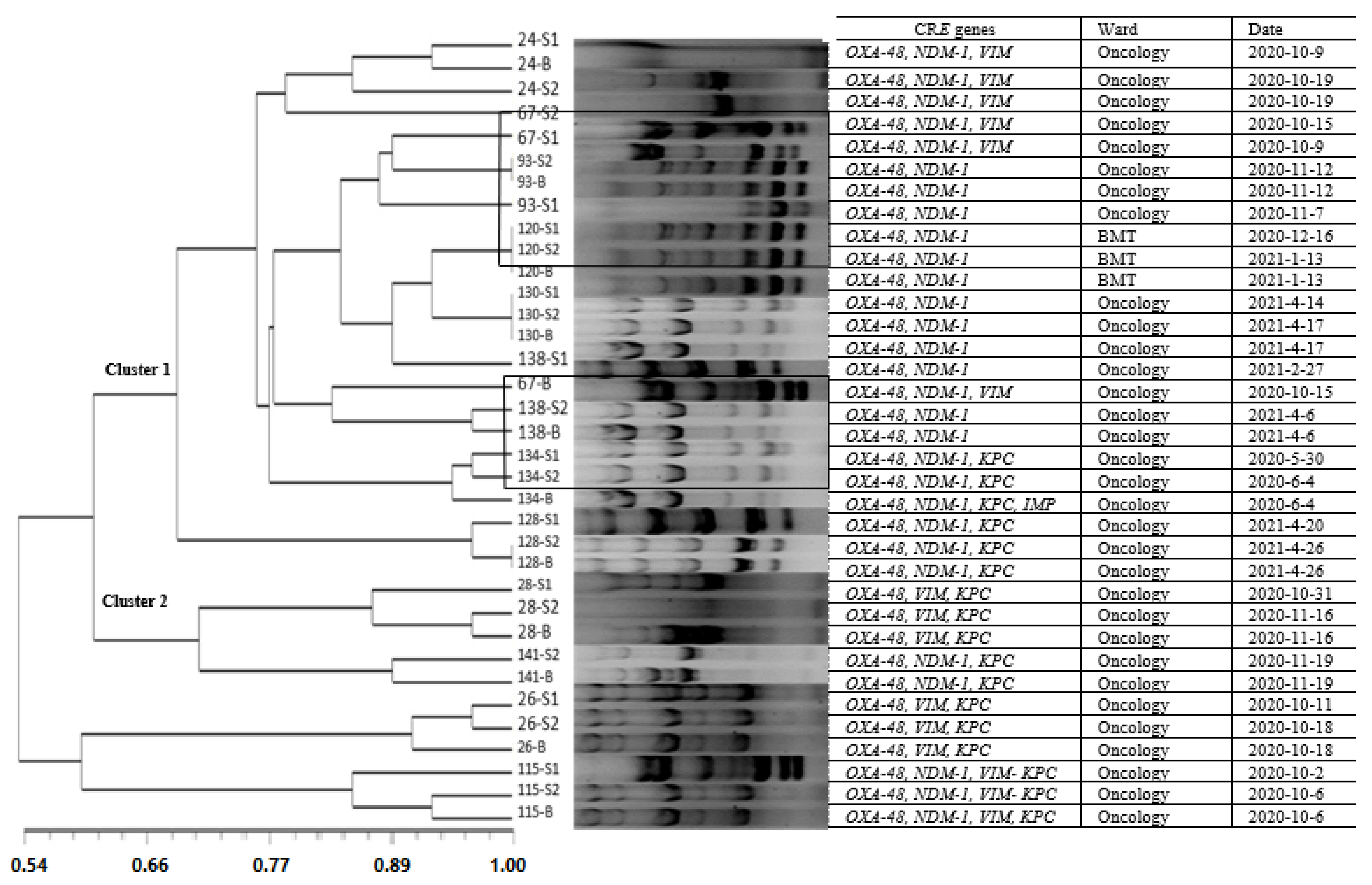
3.5. Molecular Typing of CRE in the Blood Samples and Their Similarity with the Stool Isolates

As shown in Figure 1, among the patients from whom the same species of CRE were isolated from the blood and stool samples, molecular identity was detected in 33.33% (4/12) of them. Bacteremia with related or similar strains from the primary and second stool samples was found in 50% (6/12) of the patients, and bacteremia with similar strains or unrelated fecal strains from both the primary and secondary stool samples were detected in 8.33% (1/12) and 8.33% (1/12), respectively. No homology among the CRE isolates between the studied wards was shown in this study. More details are shown in Figure 1.

4. Discussion

One of the problems of CRE bacteremia is the unknown source of the infection, which can be either endogenous or exogenous. Immunocompromised pediatrics are very susceptible to endogenous infections due to the loss of their natural defense barrier [33]. In this study, we detected the source of endogenous bacteremia caused by CRE in immunocompromised children for the first time in the world. In addition, the correlation between gender, age, fever days, length of stay, underlying diseases, wards of the hospital, prophylaxis antibiotics, alternative antibiotics, chemotherapy drugs, death of patients, and CRE bacteremia was evaluated. Our findings showed significant correlations between length of hospital stay, fever days, death rate, and CRE bacteremia among the children. Consistent with our results, the correlation of mortality rate, length of hospital stay, and CRE bacteremia among children was shown in two studies in India and Spain [20,34]. Our results showed a high rate of mortality in children with CRE bacteremia that underwent chemotherapy or those who were prescribed immunosuppressive drugs, which was similarly presented in a study of seven Italian pediatric centers [9,35]. In our study, the most common species isolated from primary fecal samples was E. coli (63.63%); 28.57% of them were carbapenem-resistant. Based on a study of stool samples of children admitted to the outpatient clinic of a hospital in Shanghai in 2019, a carriage rate of 3.6% was shown for CRE. The most common CRE isolates belonged to E. coli (37.5%) and K. pneumonia (37.5%), while E. cloacae (18.8%), Enterobacter aerogenes (3.1%), and Raoultella planticola (3.1%) were also characterized [36]. In our study, E. coli was present at the highest frequency among strains isolated from secondary fecal samples during fever onset (62.22%), of which 35.71% of them were CRE. Despite numerous studies that have been performed on the intestinal colonization of CRE in patients with different clinical situations, according to our knowledge, there are no follow-up studies to show the risk of this colonization in bacteremia, especially among children during the hospitalization period. In the present study, members of Enterobacteriaceae constituted 31.11% of the blood isolates in febrile children, of which 100% of them were carbapenem-resistant. The most common Enterobacteriaceae strains belonged to K. pneumoniae (50%) and E. coli (21.42%) in the blood samples. Similar to our findings, in a study in India that was performed in Enterobacteriaceae in the blood samples of children, K. pneumoniae (76%) and E. coli (23%) were the most common strains, of which 97% of the strains were carbapenem-resistant [20]. It seems that CRE bacteremia is not only limited to immunocompromised children or those with cancer. In a study of adult cancer patients in Iran, 51% of E. coli and K. pneumoniae isolates in blood samples were resistant to carbapenems [37].
According to the antimicrobial susceptibility patterns of the strains isolated from fecal and blood samples of patients with bacteremia, the highest and the lowest resistance in primary fecal samples was observed against cefotaxime (75%) and tigecycline (6%), respectively. In the isolates from secondary stool samples, the highest and the lowest resistance phenotypes were against ceftazidime (86.6%) and tigecycline (15.5%), respectively. While resistance to third-generation cephalosporins was common in the strains isolated from stool samples, the highest resistance patterns of the blood samples were observed against carbapenems (28.88%) and cefotaxime (28.88%). Similarly, tigecycline showed the lowest resistance among the blood isolates (6.66%). The high rate of intestinal colonization with CRE in pediatric patients could be explained through their history of multiple admissions in different parts of hospitals, including surgery, respiratory, Pediatric Intensive Care Unit (PICU), and infectious units, plus a history of a variety of broad-spectrum antimicrobial drug usage, including carbapenems. In support of this finding, the results of a study on critically ill adult patients showed that CRE colonization at the time of ICU admission, previous hospitalization, or ICU admission are predictors of 90-day mortality [38].
The most frequent carbapenemase was OXA-48 (100%), which was similar to previous reports from the Middle East and Iran. In a study on immunocompromised patients conducted in Iran, 96% of CRE carried OXA-48 [39]. In another study in Egypt, 58% of Enterobacteriaceae strains isolated from immunocompromised children with cancer carried the OXA-48 gene [40]. The high percentage of OXA-48 in clinical isolates of Enterobacteriaceae indicates its widespread distribution, which is important epidemiologically. The second most common carbapenemase gene in the present study was NDM-1 (74.81%). This gene is located on a plasmid that can harbor resistance genes to other antibiotics. NDM-1 plays an important role in HAI, so it is known as one of the most important carbapenemase enzymes that can endanger public health [41,42,43]. While the presence of two carbapenemase genes was reported among Enterobacteriaceae, the co-carriage of three carbapenemase genes in different combinations among different genera, including OXA-48, VIM, and KPC in K. oxytoca, OXA-48, VIM, KPC and NDM-1 in K. pneumonia, OXA-48, VIM and NDM-1 in K. pneumonia and OXA-48, VIM, and NDM-1 in E. coli, was reported for the first time.
Based on the molecular epidemiology results, identical patterns with 100% homology between the blood and the stool samples were detected among 33.33% of the patients. Changes in the CRE molecular types occurred in a few patients during the two sampling periods, which highlighted the importance of intestinal colonization at the time of admission and its risk for bacteremia and mortality. The presence of common source of infection with CRE was shown in this study through observed similarity in the ERIC-PCR molecular patterns of the bacterial isolates in fecal and blood samples between two patients at oncology and BMT wards. The patients were hospitalized for similar periods with similar genotypes and antibiotic resistance patterns. Our results also showed similar ERIC-PCR patterns between two patients in the oncology ward that were hospitalized in a similar time period; however, the isolates did not have identical CRE genes.
There are pros and cons about the current study. According to our knowledge, this is the first study in immunocompromised children that has investigated the primary intestinal colonization of CRE and its impact on bacteremia during the hospitalization. Moreover, the association of clinical data, such as antibiotic regimens, treatment periods, and underlying diseases, with the microbiological findings were analyzed in this study, which highlighted the link between CRE bacteremia and chemotherapy, the length of hospital stays, and death in this population. However, the lack of some information, such as the history of previous hospitalization and infection with CRE, and inability to perform the comparison of patients in the Oncology/chemotherapy and Transplantation wards with those referred to the other wards were among main limitations at the time of this study. The follow-up of the patients and study of the situation of carrier status of CRE could provide valuable data about the impact of hospital stays, and antibiotics and chemotherapeutic regimens for immunocompromised children. The application of high-throughput techniques for molecular typing is recommended instead of ERIC-PCR, which was not possible regarding to the time and budget constraints. Further studies are needed to detect and control sources of CRE in the community and in hospital settings and control its spread.

5. Conclusions

Our findings showed the primary intestinal colonization with CRE in immunocompromised pediatrics and their translocation to blood, which is a risk factor for mortality. The co-carriage of several carbapenem resistance genes in some strains, such as K. oxytoca, K. pneumoniae, and E. coli, indicates the role of mobile genetic elements in the emergence of new life-threatening CRE variants. A history of multiple hospitalization and the length of hospital stay was correlated with CRE colonization and bacteremia in these children, which show the necessity of designing proper prophylactic regimens in oncology and chemotherapy wards.

Supplementary Materials

The following supporting information can be downloaded at: https://www.mdpi.com/article/10.3390/tropicalmed8080402/s1, Figure S1: Diagram of sampling stages from immunocompromised children.

Author Contributions

N.A.T.: Investigation, Methodology, Formal analysis, Writing and editing. L.A.: Investigation, Methodology, Editing. S.A.: Methodology, Conceptualization, Editing. N.S.: Methodology, Editing. F.F.: Conceptualization, Validation. A.K.: Conceptualization, Validation. B.S.S.: methodology. S.N.: Conceptualization, Methodology. M.A.: Conceptualization, Formal analysis, Methodology, Writing and editing. All authors have read and agreed to the published version of the manuscript.

Funding

This study is based on an MSc thesis that was supported by the Pediatric Infections Research Center, Research Institute for Children’s Health, Shahid Beheshti University of Medical Sciences, Tehran, Iran.

Institutional Review Board Statement

The study was conducted in accordance with the Declaration of Helsinki, and approved by the Ethics Committee of the Research Institute for Children’s Health, Shahid Beheshti University of Medical Sciences (IR. SBMU.RICH.1400.029, 5 August 2021).

Informed Consent Statement

Informed consent was obtained from all subjects involved in the study.

Data Availability Statement

The data presented in this study are available upon reasonable request from the corresponding authors.

Acknowledgments

Research reported in this publication was supported by grant number 24201 from Pediatric Infections Research Center, Research Institute for Children’s Health, Shahid Beheshti University of Medical Sciences. Tehran, Iran.

Conflicts of Interest

The authors have no relevant financial or non-financial interest to disclose.

References

  1. Rai, S.; Das, D.; Niranjan, D.K.; Singh, N.P.; Kaur, I.R. Carriage prevalence of carbapenem-resistant Enterobacteriaceae in stool samples: A surveillance study. Australas. Med. J. 2014, 7, 64. [Google Scholar] [CrossRef]
  2. Nagarjuna, D.; Mittal, G.; Dhanda, R.; Verma, P.; Gaind, R.; Yadav, M. Faecal Escherichia coli isolates show potential to cause endogenous infection in patients admitted to the ICU in a tertiary care hospital. New Microbes New Infect. 2015, 7, 57–66. [Google Scholar] [CrossRef] [Green Version]
  3. Habib, I.; Al-Rifai, R.H.; Mohamed, M.-Y.I.; Ghazawi, A.; Abdalla, A.; Lakshmi, G.; Agamy, N.; Khan, M. Contamination Levels and Phenotypic and Genomic Characterization of Antimicrobial Resistance in Escherichia coli Isolated from Fresh Salad Vegetables in the United Arab Emirates. Trop. Med. Infect. Dis. 2023, 8, 294. [Google Scholar] [CrossRef]
  4. Donskey, C.J. The role of the intestinal tract as a reservoir and source for transmission of nosocomial pathogens. Clin. Infect. Dis. 2004, 39, 219–226. [Google Scholar] [CrossRef] [PubMed] [Green Version]
  5. Tokur, M.E.; Korkmaz, P.; Alkan, S.; Yıldız, E.; Arık, Ö.; Renders, D.P.; Balcı, C. Mortality predictors on the day of healthcare-associated Acinetobacter baumanni bacteremia in intensive care unit. J. Infect. Dev. Ctries. 2022, 16, 1473–1481. [Google Scholar] [CrossRef] [PubMed]
  6. Tabone, M.; Moissenet, D.; Leverger, G. Nosocomial infections in immunocompromised children. Pathol.-Biol. 2000, 48, 893–900. [Google Scholar] [PubMed]
  7. Mittal, G.; Gaind, R.; Kumar, D.; Kaushik, G.; Gupta, K.B.; Verma, P.; Deb, M. Risk factors for fecal carriage of carbapenemase producing Enterobacteriaceae among intensive care unit patients from a tertiary care center in India. BMC Microbiol. 2016, 16, 138. [Google Scholar] [CrossRef] [Green Version]
  8. Agyeman, A.A.; Bergen, P.J.; Rao, G.G.; Nation, R.L.; Landersdorfer, C.B. A systematic review and meta-analysis of treatment outcomes following antibiotic therapy among patients with carbapenem-resistant Klebsiella pneumoniae infections. Int. J. Antimicrob. Agents 2020, 55, 105833. [Google Scholar] [CrossRef] [PubMed]
  9. Al-Tonbary, Y.A.; Soliman, O.E.; Sarhan, M.M.; Hegazi, M.A.; El-Ashry, R.A.; El-Sharkawy, A.A.; Salama, O.S.; Yahya, R. Nosocomial infections and fever of unknown origin in pediatric hematology/oncology unit: A retrospective annual study. World J. Pediatr. 2011, 7, 60–64. [Google Scholar] [CrossRef]
  10. Grabowski, M.E.; Kang, H.; Wells, K.M.; Sifri, C.D.; Mathers, A.J.; Lobo, J.M. Provider role in transmission of carbapenem-resistant Enterobacteriaceae. Infect. Control. Hosp. Epidemiol. 2017, 38, 1329–1334. [Google Scholar] [CrossRef]
  11. Montassier, E.; Gastinne, T.; Vangay, P.; Al-Ghalith, G.; Bruley des Varannes, S.; Massart, S.; Moreau, P.; Potel, G.; de La Cochetiere, M.; Batard, E. Chemotherapy-driven dysbiosis in the intestinal microbiome. Aliment. Pharmacol. Ther. 2015, 42, 515–528. [Google Scholar] [CrossRef] [PubMed] [Green Version]
  12. Bavaro, D.F.; Pizzutilo, P.; Catino, A.; Signorile, F.; Pesola, F.; Di Gennaro, F.; Cassiano, S.; Marech, I.; Lamorgese, V.; Angarano, G.; et al. Incidence of infections and predictors of mortality during checkpoint inhibitor immunotherapy in patients with advanced lung cancer: A retrospective cohort study. Open. Forum. Infect. Dis. 2021, 8, ofab187. [Google Scholar] [CrossRef] [PubMed]
  13. Balkan, I.I.; Aygün, G.; Aydın, S.; Mutcalı, S.I.; Kara, Z.; Kuşkucu, M.; Midilli, K.; Şemen, V.; Aras, Ş.; Yemişen, M. Blood stream infections due to OXA-48-like carbapenemase-producing Enterobacteriaceae: Treatment and survival. Int. J. Infect. Dis. 2014, 26, 51–56. [Google Scholar] [CrossRef]
  14. Bower, C.; Adelman, M.; Howard-Anderson, J.; Ansari, U.; Lutgring, J.; Tiga, G.; Jacob, J. Carbapenem-Resistant Enterobacteriaceae Resistant Only to Ertapenem: An Epidemiologically Distinct Cohort, Atlanta, 2016–2018. Infect. Control Hosp. Epidemiol. 2020, 41, s463–s464. [Google Scholar] [CrossRef]
  15. Thomas, S.; Espeisse, O. Antimicrobial resistance. N. Engl. J. Med. 2020, 17, 330–333. [Google Scholar]
  16. Li, Z.; Lin, X.X.; Liu, C.X.; Ye, W.J.; Liu, P.N.; Li, H.Y.; Dong, L. Epidemiological characteristics and risk factors of nosocomial carbapenem-resistant Enterobacteriaceae infections in children. Chin. Med. J. 2020, 133, 2756–2758. [Google Scholar] [CrossRef]
  17. Cherak, Z.; Loucif, L.; Moussi, A.; Bendjama, E.; Benbouza, A.; Rolain, J.-M. Emergence of Metallo-β-Lactamases and OXA-48 Carbapenemase Producing Gram-Negative Bacteria in Hospital Wastewater in Algeria: A Potential Dissemination Pathway Into the Environment. Microb. Drug Resist. 2021, 28, 23–30. [Google Scholar] [CrossRef]
  18. Kim, S.; Covington, A.; Pamer, E.G. The intestinal microbiota: Antibiotics, colonization resistance, and enteric pathogens. Immunol. Rev. 2017, 279, 90–105. [Google Scholar] [CrossRef] [PubMed]
  19. Dethlefsen, L.; Relman, D.A. Incomplete recovery and individualized responses of the human distal gut microbiota to repeated antibiotic perturbation. Proc. Natl. Acad. Sci. USA 2011, 108, 4554–4561. [Google Scholar] [CrossRef] [PubMed]
  20. Nabarro, L.E.; Shankar, C.; Pragasam, A.K.; Mathew, G.; Jeyaseelan, V.; Veeraraghavan, B.; Verghese, V.P. Clinical and bacterial risk factors for mortality in children with carbapenem-resistant Enterobacteriaceae bloodstream infections in India. Pediatr. Infect. Dis. J. 2017, 36, e161–e166. [Google Scholar] [CrossRef]
  21. Rashidi, A.; Kaiser, T.; Graiziger, C.; Holtan, S.G.; Rehman, T.U.; Weisdorf, D.J.; Khoruts, A.; Staley, C. Specific gut microbiota changes heralding bloodstream infection and neutropenic fever during intensive chemotherapy. Leukemia 2020, 34, 312–316. [Google Scholar] [CrossRef] [PubMed]
  22. Tao, X.; Wang, H.; Min, C.; Yu, T.; Luo, Y.; Li, J.; Hu, Y.; Yan, Q.; Liu, W.E.; Zou, M. A retrospective study on Escherichia coli bacteremia in immunocompromised patients: Microbiological features, clinical characteristics, and risk factors for shock and death. J. Clin. Lab. Anal. 2020, 34, e23319. [Google Scholar] [CrossRef] [PubMed] [Green Version]
  23. Murray, P.R.; Rosenthal, K.S.; Pfaller, M.A. Medical Microbiology E-Book; Elsevier Health Sciences: Philadelphia, PA, USA, 2020. [Google Scholar]
  24. Wolter, D.J.; Khalaf, N.; Robledo, I.E.; Vázquez, G.J.; Santé, M.I.; Aquino, E.E.; Goering, R.V.; Hanson, N.D. Surveillance of carbapenem-resistant Pseudomonas aeruginosa isolates from Puerto Rican Medical Center Hospitals: Dissemination of KPC and IMP-18 β-lactamases. Antimicrob. Agents Chemother. 2009, 53, 1660–1664. [Google Scholar] [CrossRef] [PubMed] [Green Version]
  25. Liu, S.; Wang, Y.; Xu, J.; Li, Y.; Guo, J.; Ke, Y.; Yuan, X.; Wang, L.; Du, X.; Wang, Z. Genome sequence of an OXA23-producing, carbapenem-resistant Acinetobacter baumannii strain of sequence type ST75. Am. Soc. Microbiol. 2012, 194, 6000–6001. [Google Scholar] [CrossRef] [Green Version]
  26. Szejbach, A.; Mikucka, A.; Bogiel, T.; Gospodarek, E. Usefulness of phenotypic and genotypic methods for metallo-beta-lactamases detection in carbapenem-resistant Acinetobacter baumannii strains. Med. Sci. Monit. Basic Res. 2013, 19, 32. [Google Scholar] [CrossRef] [Green Version]
  27. Lowings, M.; Ehlers, M.M.; Dreyer, A.W.; Kock, M.M. High prevalence of oxacillinases in clinical multidrug-resistant Acinetobacter baumannii isolates from the Tshwane region, South Africa—An update. BMC Infect. Dis. 2015, 15, 521. [Google Scholar] [CrossRef] [Green Version]
  28. Azimi, L.; Rastegar-Lari, A.; Talebi, M.; Ebrahimzadeh-Namvar, A.; Soleymanzadeh-Moghadam, S. Evaluation of phenotypic methods for detection of Klebsiella pneumoniae carbapenemase-producing K. pneumoniae in Tehran. J. Med. Bacteriol. 2013, 2, 26–31. [Google Scholar]
  29. Brkić, S.; Božić, D.; Stojanović, N.; Vitorović, T.; Topalov, D.; Jovanović, M.; Stepanović, M.; Ćirković, I. Antimicrobial Susceptibility and Molecular Characterization of Carbapenemase-Producing Enterobacter spp. Community Isolates in Belgrade, Serbia. Microb. Drug Resist. 2020, 26, 378–384. [Google Scholar] [CrossRef]
  30. Tümmler, B. Molecular epidemiology in current times. Environ. Microbiol. 2020, 22, 4909–4918. [Google Scholar] [CrossRef]
  31. Van Belkum, A.; Tassios, P.; Dijkshoorn, L.; Haeggman, S.; Cookson, B.; Fry, N.; Fussing, V.; Green, J.; Feil, E.; Gerner-Smidt, P. Guidelines for the validation and application of typing methods for use in bacterial epidemiology. Clin. Microbiol. Infect. 2007, 13, 1–46. [Google Scholar] [CrossRef]
  32. Ghalavand, Z.; Alebouyeh, M.; Ghanati, K.; Azimi, L.; Rashidan, M. Genetic relatedness of the Enterococcus faecalis isolates in stool and urine samples of patients with community-acquired urinary tract infection. Gut Pathog. 2020, 12, 42. [Google Scholar] [CrossRef]
  33. Moore, D.L. Essentials of paediatric infection control. Paediatr. Child Health 2001, 6, 571–579. [Google Scholar] [CrossRef] [PubMed]
  34. Ara-Montojo, M.F.; Escosa-García, L.; Alguacil-Guillén, M.; Seara, N.; Zozaya, C.; Plaza, D.; Schuffelmann-Gutierrez, C.; De la Vega, A.; Fernandez-Camblor, C.; Ramos-Boluda, E. Predictors of mortality and clinical characteristics among carbapenem-resistant or carbapenemase-producing Enterobacteriaceae bloodstream infections in Spanish children. J. Antimicrob. Chemother. 2021, 76, 220–225. [Google Scholar] [CrossRef] [PubMed]
  35. Montagnani, C.; Prato, M.; Scolfaro, C.; Colombo, S.; Esposito, S.; Tagliabue, C.; Lo Vecchio, A.; Bruzzese, E.; Loy, A.; Cursi, L. Carbapenem-resistant Enterobacteriaceae infections in children. Pediatr. Infect. Dis. J. 2016, 35, 862–868. [Google Scholar] [CrossRef]
  36. Pan, F.; Tian, D.; Wang, B.; Zhao, W.; Qin, H.; Zhang, T.; Zhang, H. Fecal carriage and molecular epidemiology of carbapenem-resistant Enterobacteriaceae from outpatient children in Shanghai. BMC Infect. Dis. 2019, 19, 678. [Google Scholar] [CrossRef] [PubMed] [Green Version]
  37. Fazeli, H.; Moghim, S.; Zare, D. Antimicrobial resistance pattern and spectrum of multiple-drug-resistant enterobacteriaceae in Iranian hospitalized patients with cancer. Adv. Biomed. Res. 2018, 7, 69. [Google Scholar]
  38. McConville, T.H.; Sullivan, S.B.; Gomez-Simmonds, A.; Whittier, S.; Uhlemann, A.-C. Carbapenem-resistant Enterobacteriaceae colonization (CRE) and subsequent risk of infection and 90-day mortality in critically ill patients, an observational study. PLoS ONE 2017, 12, e0186195. [Google Scholar] [CrossRef] [Green Version]
  39. Azimi, L.; Nordmann, P.; Lari, A.R.; Bonnin, R.A. First report of OXA-48-producing Klebsiella pneumoniae strains in Iran. GMS Hyg. Infect. Control 2014, 9, Doc07. [Google Scholar]
  40. Kamel, N.A.; El-Tayeb, W.N.; El-Ansary, M.R.; Mansour, M.T.; Aboshanab, K.M. Phenotypic screening and molecular characterization of carbapenemase-producing Gram-negative bacilli recovered from febrile neutropenic pediatric cancer patients in Egypt. PLoS ONE 2018, 13, e0202119. [Google Scholar] [CrossRef]
  41. Bocanegra-Ibarias, P.; Garza-González, E.; Morfín-Otero, R.; Barrios, H.; Villarreal-Treviño, L.; Rodríguez-Noriega, E.; Garza-Ramos, U.; Petersen-Morfin, S.; Silva-Sanchez, J. Molecular and microbiological report of a hospital outbreak of NDM-1-carrying Enterobacteriaceae in Mexico. PLoS ONE 2017, 12, e0179651. [Google Scholar] [CrossRef]
  42. Hudson, C.M.; Bent, Z.W.; Meagher, R.J.; Williams, K.P. Resistance determinants and mobile genetic elements of an NDM-1-encoding Klebsiella pneumoniae strain. PLoS ONE 2014, 9, e99209. [Google Scholar] [CrossRef] [PubMed] [Green Version]
  43. Das, S.; Bush, K. Success and Challenges Associated with Large Scale Collaborative Surveillance for Carbapenemase Genes in Gram Negative Bacteria. Antimicrob. Agents Chemother. 2021, 66, e02299-21. [Google Scholar] [CrossRef] [PubMed]
Figure 1. ERIC-PCR products and dendrogram of CRE strains isolated from primary and secondary fecal and blood samples of children in oncology and transplantation units. Legend. The scale bar represents the similarity coefficient and distance among the strains.
Tropicalmed 08 00402 g001
Table 1. Nucleotide sequences of the primers used for detection of carbapenemase genes and ERIC-PCR typing.
Carbapenemase GenesPCR Product (bp)Sequence (5′–3′)PCR ConditionsReference
OXA-48392F: CCAAGCATTTTTACCCGCATCKACC
R: GYTTGACCATACGCTGRCTGCG
Initial denaturation at 95 °C for 1 min; 30 cycles of denaturation at 95 °C for 30 s, annealing at 55 °C for 30 s, extension at 75 °C for 1 min; and final extension at 72 °C for 7 min[24]
NDM-1129F: CCCCGCCACACCAGTGACANCTC
R: GTAGTGCTCAGTGTGGGCAT
Initial denaturation at 95 °C for 1 min; 32 cycles of denaturation at 95 °C for 30 s, annealing at 61 °C for 30 s, extension at 7 °C for 1 min; and final extension at 72 °C for 5 min[25]
VIM390F: GATGGTGTTTGGTCGCATA
R: CGAATGCGCAGCACCAG
Initial denaturation at 94 °C for 10 min; 35 cycles of denaturation at 94 °C for 30 s, annealing at 61 °C for 40 s, extension at 72 °C for 1 min; and final extension at 72 °C for 7 min[26]
IMP139F: TTGACACTCCATTTACDG
R: GATYGAGAATTAAGCCACYCT
Initial denaturation at 94 °C for 10 min; 33 cycles of denaturation at 94 °C for 30 s, annealing at 61 °C for 40 s, axtension at 72 °C for 40 s; and final extension at 72 °C for 7 min[27]
KPC636F: CTGTCTTGTCTCTCATGGCC
R: CCTCGCTGTGCTTGTCATCC
Initial denaturation at 94 °C for 5 min; 32 cycles of denaturation at 94 °C for 35 s, annealing at 62 °C for 35 s, extension at 72 °C for 32 s; and final extension at 72 °C for 5 min[28]
ERIC-PCR F: ATGTAAGCTCCTGGGGATTCAC
R: AAGTAAGTGACTGGGGTGAGCG
Initial denaturation at 94 °C for 3 min; 35 cycles of denaturation at 94 °C for 1 min, annealing at 48 °C for 1 min, extension at 72 °C for 2 min; and final extension at 72 °C for 5 min[29]
Table 2. Diversity and frequency of members of Enterobacteriaceae in stool and blood samples from febrile immunocompromised children.
Bacterial SpeciesPrimary Stool a
N = 44 (n, %)
Secondary Stool b
N = 45 (n, %)
Blood c
N = 45 (n, %)
Escherichia coli28, (63.63)28, (62.22)3, (6.66)
Klebsiella pneumoniae9, (20.45)12, (26.66)7, (15.55)
Klebsiella ozaenae1, (2.27)0, (0)-
Enterobacter. agglomerans1, (2.27)0, (0)-
Escherichia herminie1, (2.27)1, 2.22)-
Klebsiella oxytoca1, (2.27)1, (2.22)2, (4.44)
Enterobacter cloacae1, (2.27)1, (2.22)1, (2.22)
Serratia rubidaea1, (2.27)1, (2.22)1, (2.22)
a Primary stool sample: the stool sample collected during the first 24 h of admission. The primary stool sample was not provided for 1 of the febrile patients (44/45). b Secondary stool sample: the stool samples were collected after the patients developed a fever. c Blood sample: blood samples collected from febrile children.
Table 3. Antimicrobial resistance patterns of Enterobacteriaceae strains isolated from fecal and blood samples in febrile children.
Antibiotics aPrimary Stool Sample b
N = 44 (n, %)
Secondary Stool Samples
N = 45 (n, %)
Blood Samples
N = 45 (n, %)
IMP (10 µg)12, (27.27)19, (42.2)13, (28.88)
MEM (10 µg)14, (31.83)18, (40)13, (28.88)
AN (30 µg)13, (29.95)15, (33)8, (17.77)
GM (10 µg)22, (50)24, (53.3)11, (24.44)
CAZ (30 µg)29, (65.9)39, (86.6)12, (26.66)
LVX (5 µg)9, (20.4)15, (33)4, (8.88)
CTX (30 µg)33, (75)37, (82.2)13, (28.88)
CEF (30 µg)23, (52.27)26, (57.5)12, (26.66)
AZT (30 µg)29, (65.9)32, (71.1)11, (24.44)
TGC (15 µg)3, (6.8)7, (15.5)3, (6.66)
a IMP (Imipenem), MEM (Meropenem), AN (Amikacin), GM (Gentamycin), CAZ (Ceftazidime), LVX (Levofloxacin), CEF (Cefepime), CTX (Cefotaxime), AZT (Aztreonam), TGC (Tigecycline). b The primary stool sample was not provided for 1 of the febrile patients (44/45).
Table 4. Demographic data of immunocompromised children with fever and the frequency of CRE a in a pediatric hospital in Tehran.
VariablesFrequency (n = 45)CRE (n = 14)p-Value
Gender 0.99
  Female20 (20/45, 44.4%)6 (6/20, 30%)
  Male25 (25/45, 55.5%)8 (8/25, 32%)
Age 0.99
  1–5 years old22 (22/45, 48%)7 (7/22, 31.81%)
  6–10 years old11 (11/45, 24%)3 (3/11, 0.27%)
  11–15 years old12 (12/45, 26%)4 (4/12, 0.33%)
Fever day 0.022 b
  7 days (first week)36 (36/45, 80%)8 (8/36, 0.22%)
  14 days (second week)5 (5/45, 11%)3 (3/5, 0.6%)
  21 days (third week)4 (4/45, 8%)3 (3/4, 0.75%)
LOS 0.018 b
  7 days (first week)13 (13/45, 28.8%)2 (2/13, 15.38%)
  14 days (second week)17 (17/45, 37.7%)3 (3/17, 17.64%)
  Over 14 days (third week)15 (15/45, 33.3%)9 (9/15, 0.6%)
Underlying diseases 0.83
  AML12 (12/45, 26%)0
  ALL6 (4/45, 13%)1 (1/6, 16.66%)
  Burkitt’s lymphoma3 (3/45, 6.6%)3 (3/3, 100%)
  Neuroblastoma5 (5/45, 11.1%)2 (2/5, 0.4%)
  CGD2 (2/45, 4.4%)0
  Histiocytic disorders1 (1/45, 2.2%)0
  Lymphoma7 (7/45, 15.5%)3 (3/7, 42.85%)
  PNH1 (1/45, 2.2%)0
  RMS1 (1/45, 2.2%)1 (1/1, 100%)
  Wilms tumor3 (3/45, 6.6%)1 (1/3, 33.33%)
  HLH1 (1/45, 2.2%)0
  YST1 (1/45, 2.2%)1 (1/1, 100%)
  RES1 (1/45, 2.2%)1 (1/1, 100%)
  Aplastic anemia1 (1/45, 2.2%)1 (1/1, 100%)
Ward 0.46
  BMT11 (11/45, 24.44%)2 (2/11, 18.18%)
  Oncology34 (34/45, 75.5%)12 (12/34, 35.29%)
Prophylaxis antibiotics 0.99
  Carbapenem based regime19 (19/45, 42.22%)5 (5/19, 26.31%)
  Non-carbapenem based regime26 (26/45, 57.77%)9 (9/26, 34.61%)
Alternative antibiotic 0.99
  Carbapenem based regime20 (20/45, 44.44%)6 (6/20, 3%)
  Non-carbapenem based regime25 (25/45, 55.55%)8 (8/25, 32%)
Chemotherapy drug 0.03 b
  Cyclophosphamide + Adriamycin + vincristine10 (10/45, 22.25%)1 (1/10, 10%)
  Cytosar8 (8/45, 17.7%)3 (3/8, 37.5%)
  GCSF7 (7/45, 15.5%)2 (2/7, 28.57%)
  Cyclophosphamide + vincristine7 (7/45, 15.5%)4 (4/7, 57.14%)
  Doxorubicin + cyclophosphamide + vincristine5 (5/45, 11.1%)1 (1/5, 20%)
  Fludarabin + cytocar4 (4/45, 8.8%)1 (1/4, 25%)
  6MP4 (4/45, 8.8%)2 (2/14, 50%)
Death 0.02 b
  Yes11 (11/45, 24.4%)7 (7/11, 63.63%)
  No34 (34/45, 75.5%)7 (7/34, 20.58%)
(a) CRE: Carbapenem-resistant Enterobacteriaceae; LOS; Length of stay, AML; Adult acute myeloid leukemia, ALL; Acute lymphocytic leukemia, CGD; Chronic granulomatous disease, PNH; Paroxysmal nocturnal hemoglobinuria, RMS; Rhabdomyosarcoma, HLH; Hemophagocytic lymphohistiocytosis, YST; Yolk sac tumor, RES; Ewing Sarcoma, Alternative antibiotic; the antibiotic alternated after patients got fever. (b) p values represent significant differences between fever onset during the first week of hospitalization compared with longer periods; CRE bacteremia among children who presented >2 weeks LOS; inverse correlation between chemotherapy regimens and bacteremia; and death rate comparison between patients with and without CRE bacteremia. Numbers in bold represent p-Values that are statistically significant.
Disclaimer/Publisher’s Note: The statements, opinions and data contained in all publications are solely those of the individual author(s) and contributor(s) and not of MDPI and/or the editor(s). MDPI and/or the editor(s) disclaim responsibility for any injury to people or property resulting from any ideas, methods, instructions or products referred to in the content.

