Article

Characterization of Beta-Lactamase and Fluoroquinolone Resistance Determinants in Escherichia coli, Klebsiella pneumoniae, and Pseudomonas aeruginosa Isolates from a Tertiary Hospital in Yola, Nigeria

by
Diane E. Kawa
1,*,
Isabella A. Tickler
2,
Fred C. Tenover
3 and
Shuwaram A. Shettima
4
1
Department of Medical and Scientific Affairs, Cepheid, Sunnyvale, CA 94089, USA
2
Department of Medical and Scientific Affairs, Cepheid, 20090 Milan, Italy
3
College of Arts and Sciences, University of Dayton, Dayton, OH 45469, USA
4
Department of Medical Microbiology, Parasitology and Immunology, Modibbo Adama University Teaching Hospital, Yola 640001, Adamawa State, Nigeria
*
Author to whom correspondence should be addressed.
Trop. Med. Infect. Dis. 2023, 8(11), 500; https://doi.org/10.3390/tropicalmed8110500
Submission received: 3 October 2023 / Revised: 9 November 2023 / Accepted: 10 November 2023 / Published: 16 November 2023

Abstract

:
Infections due to antimicrobial resistant gram-negative bacteria cause significant morbidity and mortality in sub-Saharan Africa. To elucidate the molecular epidemiology of antimicrobial resistance in gram-negative bacteria, we characterized beta-lactam and fluoroquinolone resistance determinants in Escherichia coli, Klebsiella pneumoniae, and Pseudomonas aeruginosa isolates collected from November 2017 to February 2018 (Period 1) and October 2021 to January 2022 (Period 2) in a tertiary medical center in north-eastern Nigeria. Whole genome sequencing (WGS) was used to identify sequence types and resistance determinants in 52 non-duplicate, phenotypically resistant isolates. Antimicrobial susceptibility was determined using broth microdilution and modified Kirby–Bauer disk diffusion methods. Twenty sequence types (STs) were identified among isolates from both periods using WGS, with increased strain diversity observed in Period 2. Common ESBL genes identified included blaCTX-M, blaSHV, and blaTEM in both E. coli and K. pneumoniae. Notably, 50% of the E. coli in Period 2 harbored either blaCTX-M-15 or blaCTX-M-1 4 and phenotypically produced ESBLs. The blaNDM-7 and blaVIM-5 metallo-beta-lactamase genes were dominant in E. coli and P. aeruginosa in Period 1, but in Period 2, only K. pneumoniae contained blaNDM-7, while blaNDM-1 was predominant in P. aeruginosa. The overall rate of fluoroquinolone resistance was 77% in Period 1 but decreased to 47.8% in Period 2. Various plasmid-mediated quinolone resistance (PMQR) genes were identified in both periods, including aac(6)-Ib-cr, oqxA/oqxB, qnrA1, qnrB1, qnrB6, qnrB18, qnrVC1, as well as mutations in the chromosomal gyrA, parC and parE genes. One E. coli isolate in Period 2, which was phenotypically multidrug resistant, had ESBL blaCTX-M-15, the serine carbapenemase, blaOXA-181 and mutations in the gyrA gene. The co-existence of beta-lactam and fluoroquinolone resistance markers observed in this study is consistent with widespread use of these antimicrobial agents in Nigeria. The presence of multidrug resistant isolates is concerning and highlights the importance of continued surveillance to support antimicrobial stewardship programs and curb the spread of antimicrobial resistance.

1. Introduction

The development and spread of antimicrobial resistance (AMR) among bacterial isolates continue to pose therapeutic challenges globally. In 2019, an estimated 4.95 million deaths were associated with AMR, with 1.27 million attributable deaths [1]. The highest burden of AMR is in western sub-Saharan Africa [1,2], where it is driven by inappropriate use (misuse and overuse) of antimicrobial agents [2,3].
Gram-negative bacteria, such as Escherichia coli, Klebsiella pneumoniae, Enterobacter spp., Pseudomonas aeruginosa, and Acinetobacter baumannii, are leading causes of antimicrobial-resistant infections [1] with resistance to fluoroquinolones and β-lactam drugs being widely reported [1,2]. Treatment of infections is often problematic due to the high level of intrinsic and acquired antimicrobial resistance genes. Plasmid-mediated transfer of genes encoding extended-spectrum beta-lactamases (ESBLs) and carbapenemases are increasingly reported in Africa and Asia [4,5,6,7,8]. Fluoroquinolone resistance is primarily driven by mutations in the quinolone resistance-determining regions (QRDR) of the DNA gyrase (gyr) and topoisomerase IV (par) encoding genes, overexpression of quinolone efflux pumps, and porin inhibition [9,10]. However, the emergence of plasmid-mediated quinolone resistance (PMQR) is particularly concerning and increasingly reported in the Enterobacterales [11,12].
Since the introduction of fluoroquinolones to Nigeria in 2004, there has been intense use of this class of broad-spectrum antibiotics both in and out of the hospital setting. Common indicators for use include urinary tract infections (UTIs), sepsis, enteric fever, acute bacterial gastroenteritis, acute otitis media, pelvic inflammatory disease, and perioperative antimicrobial prophylaxis. Consequently, over the last decade, there have been several reports of fluoroquinolone-resistant bacteria from human and animal sources [13,14,15,16]. Beta-lactam antibiotics are also widely used to treat UTIs caused by Enterobacterales and Pseudomonaceae [6] and reports of multidrug resistance associated with CTX-M-type ESBL-producing E. coli and K. pneumoniae have also increased [17].
At our hospital in Yola, Nigeria, the 2019 cumulative antimicrobial susceptibility test data (i.e., antibiogram), which guides empiric therapy, revealed that the percent susceptibility among gram-negative bacteria commonly isolated from clinical samples, was 2–64% for the beta-lactam drugs including meropenem, and 6–16% for ciprofloxacin, respectively (unpublished data). This prompted widespread clinician education about the appropriate use of these antimicrobial agents, and an improved percent susceptibility for the beta-lactam agents (22–76%) and for ciprofloxacin (28–59%) was observed in the 2021 antibiogram (unpublished data). Until recently, there was little information about the underlying mechanisms for antimicrobial resistance among gram-negative isolates at our institution and their prevalence. Using whole genome sequencing (WGS), we determined that the most common carbapenem-resistance mechanisms in gram-negative bacterial isolates were mediated by Class B metallo-beta-lactamases, including the New Delhi metallo-beta-lactamase blaNDM-7 and blaNDM-1 genes, the Verona integron-encoded metallo-beta-lactamase, blaVIM-5, and Class D beta-lactamase blaOXA-181 [18,19]. This information was used to supplement the new 2021 antibiogram and further educate clinicians about antibiotic use.
The goal of this study was to compare the molecular epidemiology and emergence of beta-lactam and fluoroquinolone resistance determinants in E. coli, K. pneumoniae, and P. aeruginosa isolates, the most common gram-negative isolates at our hospital, across two time periods (November 2017 to February 2018 and from October 2021 to January 2022).

2. Materials and Methods

2.1. Bacterial Isolates

This study utilized non-duplicate E. coli, K. pneumoniae, and P. aeruginosa isolates from clinical specimens in the Medical Microbiology Laboratory at the Modibbo Adama University Teaching Hospital in Yola, Nigeria. A total of four E. coli, one K. pneumoniae, and 8 P. aeruginosa isolates collected from November 2017 to February 2018 (Period 1), previously characterized as carbapenem non-susceptible [18], and 19 E. coli, 12 K. pneumoniae, and 8 P. aeruginosa isolates collected between October 2021 to January 2022 (Period 2) were evaluated. The isolates were obtained from various clinical specimens; the most common source was urine (Table 1). The gram-negative isolates were identified to the species level using manual biochemical methods [20]. Susceptibility to antimicrobial agents was determined using the modified Kirby–Bauer disk diffusion method on Mueller–Hinton agar as described by the Clinical and Laboratory Standards Institute (CLSI) document M02-A13 [21]. The disk diffusion results were interpreted according to CLSI recommendations M100-S32 [22]. Gram-negative isolates that were non-susceptible/resistant to beta-lactam antimicrobial agents and fluoroquinolones were stored in physiological saline containing 25% glycerol in cryovials at −20 °C and shipped to a central laboratory for further analysis (described in Section 2.2). Ethical approval to collect bacterial isolates was obtained from the Federal Medical Centre, Yola Health Research Ethics Committee (HREC).

2.2. Bacterial Identification and Antimicrobial Susceptibility Testing

Identification of the bacterial isolates was performed at a central laboratory using MALDI-TOF MS (Bruker Daltonics GmbH, Bremen, Germany) according to the manufacturer’s instructions. Antimicrobial susceptibility testing was conducted using the Neg MIC 56 panel on the MicroScan WalkAway 40 SI Plus system (Beckman Coulter, Inc., West Sacramento, CA, USA) as described by the manufacturer, and MIC results were interpreted according to CLSI recommendations [22]. The antimicrobial agents were aztreonam (ATM), ceftazidime (CAZ), cefepime (FEP), cefotaxime (CTX), ceftriaxone (CRO), cefiderocol (FDC), piperacillin-tazobactam (TZP), ceftolozane-tazobactam (C/T), ceftazidime-avibactam (CZA), meropenem-avibactam (MVB), ertapenem (ETP), imipenem (IPM), meropenem (MEM), ciprofloxacin (CIP), levofloxacin (LVX), moxifloxacin (MXF). The concentrations of the antimicrobial agents included in the Neg MIC56 panel can be found at https://www.beckmancoulter.com/en/products/microbiology/-/media/63adeb88ba294285874b037c2ad875e4.ashx (accessed on 9 November 2023). Quality control organisms included P. aeruginosa ATCC 27853, E. coli ATCC 25922 and ATCC 35218, and K. pneumoniae ATCC 700603 and ATCC BAA-1705. The bacterial isolates were also tested for susceptibility to 12 antimicrobial agents using the modified Kirby–Bauer disk diffusion method on Mueller–Hinton agar (Hardy Diagnostics, Santa Maria, CA, USA) as described in CLSI M02-A13 [21]. Antimicrobial agents tested with disk diffusion included ATM (30 μg), CAZ (30 μg; with and without clavulanic acid), FEP (30 μg), CTX (30 μg; with and without clavulanic acid), CRO (30 μg), FDC (30 μg), CZA (30/20 μg), ETP (10 μg), IPM (10 μg), and MEM (30 μg); Zone diameters and ESBL detection were interpreted using the criteria described in CLSI M100-S32 [22]. Quality control organisms included P. aeruginosa ATCC 27853, E. coli ATCC 25922 and ATCC 35218, and K. pneumoniae ATCC 700603 and ATCC BAA-1705.

2.3. Phenotypic Detection of Carbapenemase Production

The modified carbapenem inactivation method (mCIM) was used in conjunction with the EDTA-modified carbapenem inactivation method (eCIM) to determine carbapenemase production and to differentiate metallo-beta-lactamases from serine carbapenemases, according to CLSI guidelines [22].

2.4. Whole Genome Sequencing

Genomic DNA was extracted from pure cultures of organisms grown overnight on blood agar plates (Hardy Diagnostics) using the Qiagen DNeasy blood and tissue kit on the Qiacube (Qiagen, Valencia, CA, USA). Genomic libraries were prepared for each isolate using the Illumina DNA Prep Kit (Illumina, San Diego, CA USA), and sequencing was performed on the Illumina Miseq system using Miseq Reagent Kit v2 (Illumina, San Diego, CA) according to the manufacturer’s instructions. De novo assemblies, multi-locus sequence typing (MLST), detection of acquired antimicrobial resistance genes, and detection of point mutations were performed with the CLC Genomics Workbench version 22.0.2 and CLC Microbial Genomics Module version 22.1.1 (QIAGEN Bioinformatics, Aarhus, Denmark). All nucleic acid sequence data from this study have been deposited in the NCBI BioProject database (https://www.ncbi.nlm.nih.gov/bioproject/, accessed on 9 November 2023) with links to BioProject Accession # PRJNA701275 (2017–2018 isolates) and # PRJNA962793 (2021–2022).

3. Results

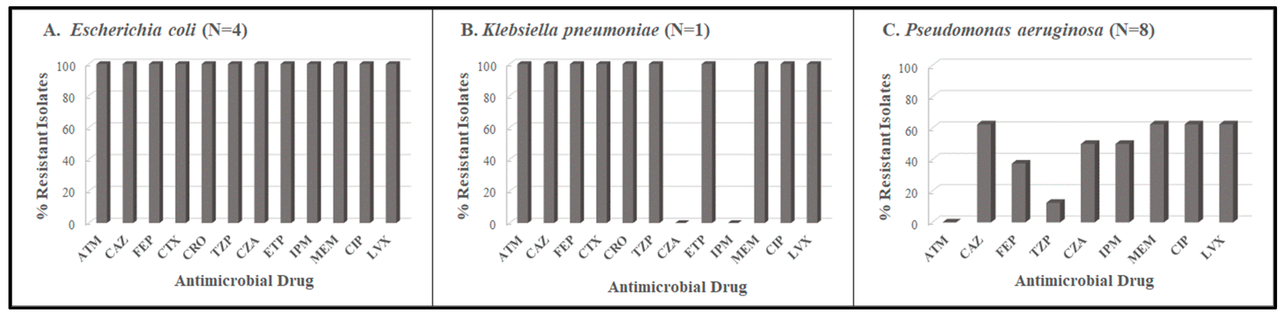
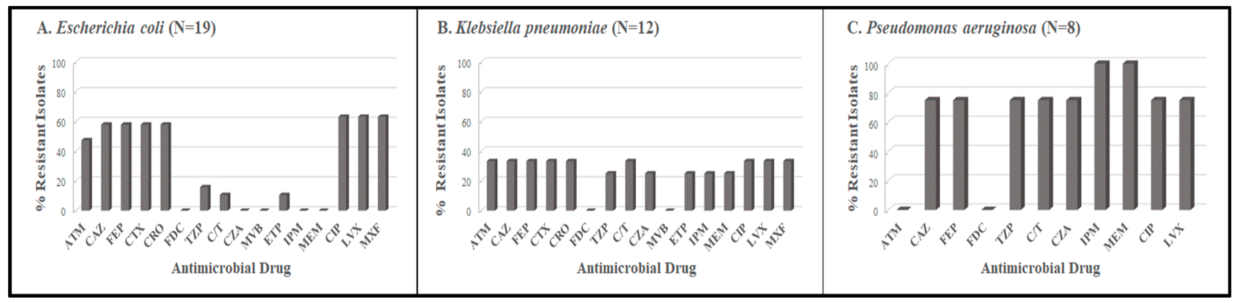
3.1. Antimicrobial Resistance Profiles

The beta-lactam and fluoroquinolone resistance profiles of the E. coli, K. pneumoniae, and P. aeruginosa isolates from Period 1, based on antimicrobial suscpetibility testing performed at the central laboratory, are shown in Figure 1. The overall prevalence of antimicrobial resistance among the 13 isolates was 76.9%. The highest rate was in E. coli, which demonstarted 100% resistance to all classes of antimicrobial drugs tested. The only Klebsiella pneumoniae isolate 16047, was characterized as phenotypically positive for ESBL production (Table S1). Ten isolates were non-susceptible to one or more carbapenems and of these, nine isolates (4 E. coli and 5 Pseudomonas aeruginosa) produced a metallo-beta-lactamase (Table S1).
In contrast to Period 1, the overall resistance rate among the 39 isolates from Period 2 was 53.8% for beta-lactams, 33.8% for carbapenems, and 56.4% for fluoroquinolones (Figure 2). The highest rate was for P. aeruginosa, with 100% resistance to the carbapenems. A total of 12 isolates produced ESBLs. Of the 12 isolates that were non-susceptible to the carbapenems, three Klebsiella pneumoniae and six Pseudomonas aeruginosa produced a metallo-beta-lactamase (Table S2). Notably, one E. coli isolate (17781) produced an ESBL and a serine carbapenemase as determined by mCIM/eCIM (Table S2). There was 100% concordance in antimicrobiaol susceptibility test results between the broth microdilution and disk diffusion methods for the isolates.

3.2. Strain Types and Resistance Determinants among E. coli Isolates

The multi-locus sequence type (MLST) and resistance determinants for the E. coli isolates from Period 1 are presented in Table 2. All four E. coli ST692 isolates harbored ESBL blaCTX-M-15 gene and metallo-beta-lactamase blaNDM-7 gene. These genetic profiles were consistent with the beta-lactam and carbapenem resistance phenotypes observed (Table 2). The isolates also had the PMQR aac(6′)Ib-cr gene and mutations in the parC and parE genes, respectively. Three isolates had mutations in gyrA consistent with the fluoroquinolone-resistant phenotype of the isolates (Table S1).
In Period 2, there were multiple sequence types for E. coli, with ST2 being dominant (Table 3). ESBL-encoding genes blaCTX-M-15 and blaCTX-M-14 were identified in 57.9% of the isolates and all (100%) were phenotypically ESBL producers (Table S2). The distribution of PMQR genes and mutations in the gyrA, parC and parE genes also varied. Five E. coli isolates (26.3%) had one or more PMQR genes and 14 (73.6%) had at least one mutation in gyrA, parC, or parE. Of the 12 isolates that were fluoroquinolone resistant (Table S2), 11 (91.6%) harbored two mutations in gyrA, suggesting this was the main mechanism of resistance in these isolates. One E. coli isolate (17781; ST692) harbored multiple resistance markers including blaCTX-M-15, blaOXA-181, and mutations in gyrA (Table 3). Notably, this E. coli isolate produced an ESBL, a serine carbapenemase, and was phenoptypically non-susceptible to ertapenem and the fluoroquinolones (Table S2).

3.3. Strain Types and Resistance Determinants in K. pneumoniae Isolates

The sole K. pneumoniae isolate (16047; ST147) from Period 1, had blaCTX-M-15, four PMQR genes, and mutations in the gyrA gene (Table 2), was an ESBL-producer and was phenotypically non-susceptible to the carbapenems (Table S1). In contrast, the 12 K. pneumoniae isolates from Period 2 represented seven distinct sequence types (Table 3). Almost all the K. pneumoniae isolates (83.3%) from this period had at least one beta-lactamase gene present. Although only one K. pneumoniae isolate (17785; ST45) had blaCTX-M-15 and was an ESBL producer, three isolates (25%) harbored blaNDM-7, produced a metallo-beta-lactamase and were phenotypically non-susceptible to carbapenems (Table S2). The prevalence of PMQR genes oqxA and oqxB among K. pneumoniae in Period 2 was 100%. No QRDR mutations were detected in these isolates; however, all were resistant to the fluoroquinolones (Table S2), suggesting that the PMQR determinants were associated with the resistant phenotype observed in Period 2.

3.4. Strain Types and Resistance Determinants in P. aeruginosa Isolates

The P. aeruginosa isolates from Period 1 represent an array of sequence types (Table 2). The overall prevalence of carbapenemase genes among these isolates was 62%. Four isolates (50%) harbored blaVIM-5, one had blaNDM-1, and all five were phenotypically non-susceptible to the carbapenems via production of metallo-beta-lactamases (Table S1). Interestingly, these same five P. aeruginosa isolates also had qnrVC1, identical mutations in the gyrA and parC genes (Table 2), and were all resistant to fluoroquinolones (Table S1). It is unclear which fluoroquinole-resistance deteminants were directly responsible for the resistant phenotype observed.
The majority of the P. aeruginosa isolates (75%) from Period 2 were ST773 and had identical resistance determinants including, blaNDM-1, qnrVC1 genes, and mutations in gyrA and parC (Table 3). Not surprisingly, these six isolates showed a corresponding phenotype for carbapenem non-susceptibility and fluoroquinolone resistance (Table S2). Although the remaining two P. aeruginosa isolates were also carbapenem-resistant, they lacked a known carbapenemase gene.

4. Discussion

As part of our antimicrobial resistance surveillance efforts, we characterized beta-lactam and fluoroquinolone resistance determinants in E. coli, K. pneumoniae, and P. aeruginosa isolates collected within two time periods from November 2017 to February 2018 and from October 2021 to January 2022, at the Modibbo Adama University Teaching Hospital in Yola, which is located in the north-eastern region of Nigeria. The findings of this study will be used to supplement our annual antibiograms that guide clinician decisions for empiric treatment for gram-negative bacterial infections, especially UTIs.
There was considerable diversity in sequence types among the gram-negative isolates across the two study periods. Notably, E. coli ST131, which is a highly virulent strain associated with multidrug resistance [23] and has recently been reported in Central Nigeria [24], was not identified at our institution. The high prevalence of the blaCTX-M-15 and blaCTX-M-14 ESBLs among the E. coli isolates in our study is consistent with the regional and global distribution of these resistance mechanisms. For instance, in a study characterizing multidrug resistant E. coli isolates in Abuja, Nigeria (Central region), Medugu et al., reported that blaCTX-M-15 and expression of a ESBL phenotype were detected in 70.1% and 50% of the isolates, respectively [24]. Similarly, in a study characterizing multidrug resistant uropathogenic Enterobacterales, and Pseudomonaceae at their institution in south-west Nigeria, Ogbolu et al., found that blaCTX-M-15 was the dominant CTX-M gene (83.3%) and positivity for blaCTX-M-14 was 33.3%. Some E. coli and K. pneumoniae isolates carried both genes and although CTX-M genes are not typically found in P. aeruginosa, two isolates were also shown to carry blaCTX-M-15 [17]. In a recent study in the United States of America, ESBL genes were identified in 66.2% of gram-negative bacteria isolated from urine and blood specimens, with blaCTX-M-15 being the most common [25].
We observed a shift in the dominant metallo-beta-lactamase from blaNDM-7 and blaVIM-5 in Period 1 to primarily the blaNDM-1 gene in Period 2, although the occurrence of this carbapenemase in several ST 773 P. aeruginosa isolates suggests it was possibly related to clonal expansion. A mutation in the oprD gene was identified in one carbapenem-non-susceptible P. aeruginosa that lacked a carbapenemase gene; however, no mutations associated with porin alterations or overexpression of efflux pumps that could lead to a resistance phenotype [9], were observed in the other isolate. Various carbapenemase genes, including blaNDM-7, blaNDM-1 and blaOXA-181 have been identified in Nigeria [26] and other parts of Africa [7].
The high rate of fluoroquinolone resistance among E. coli, K. pneumoniae, and P. aeruginosa isolates in Period 1 (77%) and Period 2 (47.8%) was not surprising, given the widespread use of fluoroquinolones in Nigeria. Consistent with our findings, a study at a tertiary hospital in southern Nigeria revealed that 93.3% of E. coli isolates harboured at least one fluoroquinolone resistance gene [27]. Similar patterns of high levels of fluoroquinolone resistance have been reported in other parts of sub-Saharan Africa including South Africa and Kenya [10,28,29,30,31]. In our study, fluoroquinolone resistance in E.coli and P. aeruginosa was primarily conferred by double or triple mutations in the gyrA gene. While high-level fluoroquinolone resistance is not typically associated with PMQR genes, their presence in some isolates suggests a likely role in decreasing fluoroquinolone susceptibility. Co-existence of PMQR determinants and mutations in the QRDR regions of the gyrA and parC housekeeping genes has been documented as mediating higher levels of resistance [32,33]. In their study in Poland, Pierkaska et al., suggest that PMQR genes may contribute to promoting the mutations of the QRDR leading to increased fluoroquinolone non-susceptibility [32].
In our study, several isolates co-harbored blaCTX-M-15 and blaCTX-M-14 ESBL genes, and blaNDM-7 and blaNDM-1 carbapenemase genes together with various combinations of PMQR genes. The possibility that ESBL and PMQR genes may co-exist on the same plasmid is worrisome because of the potential for rapid horizontal transfer of resistance between bacterial strains. One E. coli isolate (17781; ST692) from the second study period carried the blaOXA-181 carbapenemase gene and exhibited a serine carbapenemase phenotype. This isolate, which was recovered from a urine specimen, is of particular concern because it co-harbored blaCTXM-15, a Class D AmpC beta-lactamase gene, blaCMY-2, the PMQR qnrS1 gene, and had multiple mutations in the gyrA, parC, and parE genes. Not surprisingly, the isolate was non-susceptible to all fluoroquinolone and beta-lactam antimicrobials except cefiderocol. The potential spread of this multidrug resistant E. coli strain poses a significant threat to the treatment of urinary tract infections, which are common in our region.
This study had some limitations. First, the analysis was performed with E. coli, K. pneumoniae and P. aeruginosa isolates, and does not represent all the gram-negative bacterial organisms isolated at our institution that may contain fluoroquinolone and beta-lactam resistance markers. Secondly, the E. coli, K. pneumoniae, and P. aeruginosa isolates from Period 1 were a subset of carbapenem- non-susceptible isolates from our hospital and this may have introduced a pre-selection bias in favour of isolates with a high level of resistance to carbapenems and other antimicrobial drugs. However, several isolates in Period 2, which had much broader selection criteria, also had multiple resistant determinants and demonstrated a resistant or non-susceptible phenotype for the beta-lactam and fluoroquinolone antimicrobials.
In conclusion, our study showed significant diversity in the sequence types and co-existence of beta-lactam and fluoroquinolone resistance determinants in E. coli, K. pneumoniae and P. aeruginosa isolates at our institution. Due to the high mortality and morbidity associated with antimicrobial resistance in gram-negative bacteria, our findings underscore the importance of continued molecular surveillance for existing and emerging resistant organisms that can inform therapeutic decisions and antimicrobial stewardship programs in healthcare settings, especially in sub-Saharan Africa.

Supplementary Materials

The following supporting information can be downloaded at: https://www.mdpi.com/article/10.3390/tropicalmed8110500/s1, Table S1: Beta-lactam and fluoroquinolone resistant phenotypes in E. coli, K. pneumoniae and P. aeruginosa isolates collected during Period 1; Table S2: Beta-lactam and fluoroquinolone resistant phenotypes in E. coli, K. pneumoniae and P. aeruginosa isolates collected during Period 2.

Author Contributions

Conceptualization, F.C.T., S.A.S., I.A.T. and D.E.K.; methodology, F.C.T., S.A.S., I.A.T. and D.E.K.; validation, S.A.S. and I.A.T.; formal analysis, I.A.T.; investigation, F.C.T.; resources, F.C.T.; data curation, I.A.T.; writing—original draft preparation, D.E.K.; writing—review and editing, F.C.T., S.A.S. and I.A.T.; visualization, I.A.T.; supervision, F.C.T.; project administration, D.E.K.; funding acquisition, F.C.T. All authors have read and agreed to the published version of the manuscript.

Funding

This research received no external funding.

Institutional Review Board Statement

The study protocol was approved by Ethics Committee of the Federal Medical Centre, Yola Health Research Ethics Committee (protocol FMCY/HREC/21/129; date of approval 4 August 2021), for the collection of bacterial isolates.

Informed Consent Statement

Not applicable.

Data Availability Statement

Data are contained within the article and supplementary materials.

Conflicts of Interest

Diane E. Kawa and Isabella A. Tickler are employees of Cepheid, and Fred C. Tenover is a former employee of Cepheid. Shuwaram A. Shettima is a current employee at the Modibbo Adama University Teaching Hospital.

References

  1. Murray, C.J.; Ikuta, K.S.; Sharara, F. Global burden of bacterial antimicrobial resistance in 2019: A systematic analysis. Lancet 2022, 399, 629–655. [Google Scholar] [CrossRef]
  2. Kariuki, S.; Kering, K.; Warimu, C. Antimicrobial Resistance Rates and Surveillance in Sub-Saharan Africa: Where Are We Now? Infect. Drug Resist. 2022, 15, 3589–3609. [Google Scholar] [CrossRef]
  3. Godman, B.; Egwuenu, A.; Wesangula, E. Tackling antimicrobial resistance across sub-Saharan Africa: Current challenges and implications for the future. Expert Opin. Drug Saf. 2022, 21, 1089–1111. [Google Scholar] [CrossRef]
  4. Saka, H.K.; García-Soto, S.; Dabo, N.T. Molecular detection of extended spectrum β-lactamase genes in Escherichia coli clinical isolates from diarrhoeic children in Kano, Nigeria. PLoS ONE 2020, 15, e0243130. [Google Scholar] [CrossRef]
  5. Kazmierczak, K.M.; Rabine, S.; Hackel, M. Multiyear, multinational survey of the incidence and global distribution of metallo-β-lactamase-producing Enterobacteriaceae and Pseudomonas aeruginosa. Antimicrob. Agents Chemother. 2016, 60, 1067–78. [Google Scholar] [CrossRef]
  6. Ogbolu, D.O.; Webber, M.A. High-level and novel mechanisms of carbapenem resistance in Gram-negative bacteria from tertiary hospitals in Nigeria. Int. J. Antimicrob. Agents. 2014, 43, 412–417. [Google Scholar] [CrossRef]
  7. Manenzhe, R.I.; Zar, H.J.; Nicol, M.P. The spread of carbapenemase-producing bacteria in Africa: A systematic review. J. Antimicrob. Chemother. 2015, 70, 23–40. [Google Scholar] [CrossRef] [PubMed]
  8. Nordmann, P.; Poirel, L. Epidemiology and diagnostics of carbapenem resistance in Gram-negative bacteria. Clin. Infect Dis. 2019, 69 (Suppl. S7), S521–S528. [Google Scholar] [CrossRef] [PubMed]
  9. De Oliveira, D.M.P.; Forde, B.M. Antimicrobial Resistance in ESKAPE Pathogens Clin. Microbiol. Rev. 2020, 33, e00181-19. [Google Scholar] [CrossRef] [PubMed]
  10. Chattaway, M.A.; Aboderin, A.O. Fluoroquinolone-resistant enteric bacteria in sub-Saharan Africa: Clones, implications and research needs. Front. Microbiol. 2016, 7, 1–20. [Google Scholar] [CrossRef] [PubMed]
  11. Salah, F.D.; Soubeiga, S.T.; Ouattara, A.K. Distribution of quinolone resistance gene (qnr) in ESBL-producing Escherichia coli and Klebsiella spp. in Lomé, Togo. Antimicrob Resist. Infect. Control. 2019, 8, 104. [Google Scholar] [CrossRef] [PubMed]
  12. Kuo, P.Y.; Lo, Y.T.; Chiou, Y.J. Plasmid-mediated quinolone resistance determinants in fluoroquinolone-nonsusceptible Escherichia coli isolated from patients with urinary tract infections in a university hospital, 2009–2010 and 2020. J. Glob. Antimicrob. Resist. 2022, 30, 241–248. [Google Scholar] [CrossRef] [PubMed]
  13. Monárrez, R.; Braun, M.; Coburn-Flynn, O. A large self-transmissible resistance plasmid from Nigeria contains genes that ameliorate a carrying cost. Sci. Rep. 2019, 9, 19624. [Google Scholar] [CrossRef]
  14. Eghieye, M.O.; Nkene, I.H.; Abimuku, R.H. Molecular detection of plasmid-mediated quinolone resistance in ciprofloxacin-resistant Escherichia coli from urine of patients attending Garki Hospital, Abuja, Nigeria. Eur. J. Biol. Biotechnol. 2020, 1, 1–7. [Google Scholar] [CrossRef]
  15. Adekanmbi, A.O.; Usidamen, S.; Akinlabi, O.C. Carriage of plasmid-mediated qnr determinants and quinolone efflux pump (qepA) by ciprofloxacin-resistant bacteria recovered from urinary tract infection (UTI) samples. Bull. Natl. Res. Cent. 2022, 46, 27. [Google Scholar] [CrossRef]
  16. Raufu, I.A.; Fashae, K.; Ameh, J.A. Persistence of fluoroquinolone-resistant Salmonella enterica serovar Kentucky from poultry and poultry sources in Nigeria. J. Infect. Dev. Ctries. 2014, 8, 384–388. [Google Scholar] [CrossRef]
  17. Ogbolu, D.O.; Alli, O.A.T.; Webber, M.A. CTX-M-15 is Established in Most Multidrug-Resistant Uropathogenic Enterobacteriaceae and Pseudomonaceae from Hospitals in Nigeria. Eur. J. Microbiol. Immunol. (Bp) 2018, 8, 20–24. [Google Scholar] [CrossRef] [PubMed]
  18. Shettima, S.A.; Tickler, I.A.; Dela Cruz, C.M.; Tenover, F.C. Characterization of carbapenem-resistant Gram-negative organisms from clinical specimens in Yola. Nigeria. J. Glob. Antimicrob. Resist. 2020, 21, 42–5. [Google Scholar] [CrossRef]
  19. Tickler, I.A.; Shettima, S.A.; Dela Cruz, C.M. Characterization of Carbapenem-Resistant Gram-Negative Bacterial Isolates From Nigeria by Whole Genome Sequencing. Diagn Microbiol. Infect. Dis. 2021, 101, 115422. [Google Scholar] [CrossRef]
  20. Jorgensen, J.H.; Pfaller, M.A. Manual of Clinical Microbiology, 11th ed.; ASM Press: Washington, DC, USA, 2015; pp. 685–790. [Google Scholar]
  21. Clinical and Laboratory Standards Institute. Performance Standards for Antimicrobial Disk Susceptibility Tests; Approved Standard-Thirteenth Edition M2-A13; Clinical and Laboratory Standards Institute: Wayne, PA, USA, 2018. [Google Scholar]
  22. Clinical and Laboratory Standards Institute. Performance Standards for Antimicrobial Susceptibility Testing; Thirty-second Edition; CLSI supplement M100-S32; Clinical and Laboratory Standards Institute: Wayne, PA, USA, 2022. [Google Scholar]
  23. Castanheira, M.; Simner, P.J.; Bradford, P.A. Extended-spectrum β-lactamases: An update on their characteristics, epidemiology and detection. JAC Antimicrob. Resist. 2021, 3, dlab092. [Google Scholar] [CrossRef]
  24. Medugu, N.; Aworh, M.K.; Iregbu, K. Molecular characterization of multi drug resistant Escherichia coli isolates at a tertiary hospital in Abuja, Nigeria. Sci. Rep. 2022, 12, 14822. [Google Scholar] [CrossRef]
  25. Tickler, I.A.; Kawa, D.; Obradovich, A.E. The Healthcare Associated Infections Consortium. Characterization of Carbapenemase- and ESBL-Producing Gram-Negative Bacilli Isolated from Patients with Urinary Tract and Bloodstream Infections. Antibiotics 2023, 12, 1386. [Google Scholar] [CrossRef]
  26. Medugu, N.; Tickler, I.A.; Duru, C. Phenotypic and molecular characterization of beta-lactam resistant Multidrug-resistant Enterobacterales isolated from patients attending six hospitals in Northern Nigeria. Sci. Rep. 2023, 13, 10306. [Google Scholar] [CrossRef] [PubMed]
  27. Nsofor, C.M.; Tattfeng, M.Y.; Nsofor, C.A. High prevalence of qnrA and qnrB genes among fluoroquinolone-resistant Escherichia coli isolates from a tertiary hospital in Southern Nigeria. Bull. Natl. Res. Cent. 2021, 45, 26. [Google Scholar] [CrossRef]
  28. Essack, S.Y.; Desta, A.T.; Abotsi, R.E. Antimicrobial resistance in the WHO African region: Current status and roadmap for action. J. Public. Health. 2017, 39, 8–13. [Google Scholar] [CrossRef]
  29. Sekyere, J.O.; Amoako, D.G. Genomic and phenotypic characterisation of fluoroquinolone resistance mechanisms in Enterobacteriaceae in Durban, South Africa. PLoS ONE 2017, 12, e0178888. [Google Scholar] [CrossRef]
  30. Tadesse, G.; Tessema, T.S.; Beyene, G. Molecular epidemiology of fluoroquinolone resistant Salmonella in Africa: A systematic review and meta-analysis. PLoS ONE 2018, 13, e0192575. [Google Scholar] [CrossRef]
  31. Kariuki, K.; Diakhate, M.M.; Musembi, S. Plasmid-mediated quinolone resistance genes detected in Ciprofloxacin non-susceptible Escherichia coli and Klebsiella isolated from children under five years at hospital discharge, Kenya. BMC Microbiol. 2023, 23, 129. [Google Scholar] [CrossRef]
  32. Piekarska, K.; Wołkowicz, T.; Zacharczuk, K. Co-existence of plasmid-mediated quinolone resistance determinants and mutations in gyrA and parC among fluoroquinolone-resistant clinical Enterobacteriaceae isolated in a tertiary hospital in Warsaw, Poland. Int. J. Antimicrob. Agents. 2015, 45, 238–43. [Google Scholar] [CrossRef]
  33. Hamed, S.M.; Elkhatib, W.F.; El-Mahallawy, H.A. Multiple mechanisms contributing to ciprofloxacin resistance among Gram negative bacteria causing infections to cancer patients. Sci. Rep. 2018, 8, 12268. [Google Scholar] [CrossRef]
Figure 1. Beta-lactam and fluoroquinolone resistance profiles for E. coli, K. pneumoniae, and P. aeruginosa isolates collected during Period 1. ATM: aztreonam; CAZ: ceftazidime; FEP: cefepime; CTX: cefotaxime; CRO: ceftriaxone; TZP: piperacillin/tazobactam; CZA: ceftazidime/avibactam; ETP: ertapenem; IPM: imipenem; MEM: meropenem; CIP: ciprofloxacin; LVX: levofloxacin.
Tropicalmed 08 00500 g001
Figure 2. Beta-lactam and fluoroquinolone resistance profiles for E. coli, K. pneumoniae and P. aeruginosa isolates collected during Period 2. ATM: aztreonam; CAZ: ceftazidime; FEP: cefepime; CTX: cefotaxime; CRO: ceftriaxone; FDC: cefiderocol; TZP: piperacillin/tazobactam; C/T: ceftolozane-tazobactam; CZA: ceftazidime/avibactam; MVP: meropenem-avibactam; ETP: ertapenem; IPM: imipenem; MEM: meropenem; CIP: ciprofloxacin; LVX: levofloxacin.
Tropicalmed 08 00500 g002
Table 1. Select antimicrobial-resistant Escherichia coli, Klebsiella pneumoniae and Pseudomonas aeruginosa isolates recovered from clinical specimens from November 2017 to February 2018 (Period 1) and October 2021 to January 2022 (Period 2).
Collection PeriodOrganismBlood CultureEar SwabEndocervical SwabEye SwabStoolVaginal SwabSputumUrethral SwabUrineWoundTotal
Period 1 November 2017–February 2018Escherichia coli 1 3 4
Klebsiella pneumoniae 1 1
Pseudomonas aeruginosa 1 2 328
Period 2 October 2021–January 2022Escherichia coli 3 12419
Klebsiella pneumoniae1 1 413212
Pseudomonas aeruginosa 2 1 1 318
Total1311137125952
Table 2. Beta-lactamase and fluoroquinolone resistance determinants and strain types among E. coli, K. pneumoniae, and P. aeruginosa isolates collected during Period 1.
IDOrganismSourceMLSTBeta-Lactamase GenesFluoroquinolone PMQR GenesFluoroquinolone QRDR Mutations
gyrAparCparE
15949Escherichia coliear swabST692blaCMY-59, blaCTX-M-15, blaNDM-7, blaOXA-1, blaTEM-1, ampHaac(6′)Ib-crS83L, D87NS80IS458A
16020Escherichia coliurineST692blaCMY-59, blaCTX-M-15, blaNDM-7, blaOXA-10, blaOXA-140, blaTEM-1, ampH, ampCaac(6′)Ib-crWTS80IS458A
16029Escherichia coliurineST692blaCMY-59, blaCTX-M-15, blaNDM-7, blaTEM-1, ampH, ampCaac(6′)Ib-crS83L, D87NS80IS458A
16032Escherichia coliurineST692blaCTX-M-15, blaNDM-7, blaOXA-1, blaCMY-59, blaTEM-1, ampH, ampCaac(6′)Ib-crS83L, D87NS80IS458A
16047Klebsiella pneumoniaeurineST147blaCTX-M-15, blaOXA-1, blaSHV-187, K. pneumoniae OmpK37, E. coli ampHaac(6′)Ib-cr oqxA, oqxB qnrA1, qnrB1S83Y, D87AWTWT
15958Pseudomonas aeruginosasputumST2935blaOXA-50, blaPDC-10NoneS912del, E913delF254V, S331T, A346QND
15964Pseudomonas aeruginosawoundST1203blaGES-9, blaOXA-21, blaOXA-50, blaPDC-1, blaVIM-5aac(6′)Ib-cr, qnrVC1T83I, S912del, E913delS87L, F254V, A346QND
15965Pseudomonas aeruginosaurineST773blaNDM-1, blaOXA-50, blaPDC-1qnrVC1T83IS87L, F254V, A346QND
15966Pseudomonas aeruginosaurineST1203blaGES-9, blaOXA-21-like, blaOXA-50, blaPDC-1, blaVIM-5aac(6′)Ib-cr, qnrVC1T83I, S912del, E913delS87L, F254V, A346QND
15986Pseudomonas aeruginosawoundST654blaOXA-10, blaOXA-50, blaPDC-3, blaVIM-5aac(6′)Ib-cr, qnrVC1T83I, S912del, E913delS87L, F254V, A346QND
16014Pseudomonas aeruginosastoolST244blaOXA-486, blaPDC-1NoneWTF254V, A346QND
16018Pseudomonas aeruginosaurineST654blaOXA-10, blaOXA-50, blaPDC-3, blaTEM-1, blaVIM-5, blaSCO-1aac(6′)Ib-cr, qnrVC1T83I, S912del, E913delS87L, F254V, A346QWT
16048Pseudomonas aeruginosasputumST1555blaOXA-50, blaPDC-10NoneS912del, E913delF254V, S331T, A346QND
MLST, multi-locus sequence type; PMQR, plasmid-mediated quinolone resistance; QRDR, Quinolone Resistance Determinant Region; WT, wild type; ND, not determined.
Table 3. Beta-lactamase and fluoroquinolone resistance determinants and strain types among E. coli, K. pneumoniae, and P. aeruginosa isolates collected during Period 2.
IDOrganismSourceMLSTBeta-Lactamase GenesFluoroquinolone PMQR GenesFluoroquinolone QRDR Mutations
gyrAparCparE
17757Escherichia coliurineAmbiguous (ST506, ST566)blaCTX-M-14, blaTEM-1BNoneS83L, D87GS80IS458A, I529L
17758Escherichia coliVSAmbiguous (ST27, ST129)blaTEM-1BNoneS83LWTWT
17762Escherichia coliurineAmbiguous (ST27, ST129)blaTEM-1BNoneS83LWTWT
17771Escherichia coliurineST2blaTEM-1BNoneWTWTWT
17772Escherichia coliurineST2blaCTX-M-15, blaOXA-1, blaTEM-1Baac(6′)-Ib-crS83L, D87NS80IS458A
17773Escherichia coliVSST83NoneqnrB7WTWTWT
17775Escherichia coliurineInconclusive (ST721, ST662, ST472)blaCTX-M-15, blaOXA-1, blaTEM-1Baac(6′)-Ib-crS83L, D87NS80IS458A
17776Escherichia coliurineInconclusive (ST466, ST210, ST132)blaTEM-1BNoneS83L, D87NS80IS458A
17781Escherichia coliurineST692blaCMY-2, blaCTX-M-15, blaOXA-181, blaTEM-1BqnrS1S83L, D87NS80IS458A
17782Escherichia coliurineST471blaCTX-M-15, blaOXA-1NoneS83L, D87NS80IS458A
17786Escherichia coliwoundST132blaTEM-1BNoneS83L, D87NS80IS458A
17789Escherichia coliurineInconclusive (ST500, ST437)blaTEM-1BqnrS1WTWTWT
17792Escherichia coliwoundST2632blaCTX-M-15, blaOXA-1, blaTEM-1Baac(6′)-Ib-crS83L, D87NS80IS458A
17795Escherichia coliurineST86blaCTX-M-15, blaTEM-1BqnrS1S83L, D87NS80I
17796Escherichia coliurineAmbiguous (ST566, ST506)blaCTX-M-14, blaTEM-1BNoneS83L, D87GS80IS458A, I529L
17803Escherichia coliwoundST2blaCTX-M-15, blaOXA-1, blaSHV-187, blaTEM-1Baac(6′)-Ib-cr, oqxA, oqxBS83L, D87NS80IS458A
17804Escherichia coliwoundST471blaCTX-M-15aac(6′)-Ib-crWTWTWT
17862Escherichia coliVSInconclusive (ST500, ST437)blaTEM-1BqnrS1WTWTWT
17864Escherichia coliurineST58blaCTX-M-15, blaOXA-1, blaOXA-320/534, blaTEM-1BNoneS83L, D87NS80IS458T
17759Klebsiella pneumoniaeurineST340blaCTX-M-27, blaNDM-7, blaSHV-187aac(6′)-Ib-cr, oqxA, oqxB, qnrB6WTWTWT
17765Klebsiella pneumoniaesputumST86blaOXA-1, blaSHV-187aac(6′)-Ib-cr, oqxA, oqxBWTWTWT
17766Klebsiella pneumoniaesputumST20blaSHV-187oqxA, oqxBWTWTWT
17767Klebsiella pneumoniaeUSST392blaSHV-11aac(6′)-Ib-cr, oqxA, oqxB qnrB6WTWTWT
17768Klebsiella pneumoniaeurineST340blaCTX-M-27, blaNDM-7, blaSHV-187aac(6′)-Ib-cr, oqxA, oqxB, qnrB6WTWTWT
17770Klebsiella pneumoniaeBCST86blaSHV-187oqxA, oqxBWTWTWT
17785Klebsiella pneumoniaewoundST45blaCTX-M-15, blaSHV-187,
blaTEM-1B
oqxA, oqxBWTWTWT
17787Klebsiella pneumoniaesputumST2632blaSHV-93oqxA, oqxBWTWTWT
17790Klebsiella pneumoniaewoundST86blaSHV-187oqxA, oqxBWTWTWT
17793Klebsiella pneumoniaesputumST661blaSHV-187oqxA, oqxBWTWTWT
17861Klebsiella pneumoniaeurineST86blaSHV-187oqxA, oqxBWTWTWT
17863Klebsiella pneumoniaeESST340blaCTX-M-27, blaNDM-7, blaSHV-187aac(6′)-Ib-cr, oqxA, oqxB, qnrB18, qnrB6WTWTWT
17760Pseudomonas aeruginosasputumST274blaOXA-486, blaPDC-24NoneSilentF254V, A346QND
17761Pseudomonas aeruginosaear swabInconclusive (nearest ST244)blaOXA-847, blaPDC-423NoneSilentF254V, A346QND
17763Pseudomonas aeruginosaurineST773blaNDM-1, blaOXA-395, blaPDC-385qnrVC1T83IS87L, F254V, A346QND
17777Pseudomonas aeruginosaurineST773blaNDM-1, blaOXA-395, blaPDC-385qnrVC1T83IS87L, F254V, A346QND
17778Pseudomonas aeruginosaear swabST773blaNDM-1, blaOXA-395, blaPDC-385qnrVC1T83IS87L, F254V, A346QND
17779Pseudomonas aeruginosaurineST773blaNDM-1, blaOXA-395, blaPDC-385qnrVC1T83IS87L, F254V, A346QND
17791Pseudomonas aeruginosaeye swabST773blaNDM-1, blaOXA-395, blaPDC-385qnrVC1T83IS87L, F254V, A346QND
17794Pseudomonas aeruginosawoundST773blaNDM-1, blaOXA-395, blaPDC-385qnrVC1T83IS87L, F254V, A346QND
VS, vaginal swab; ES, endocervical swab; US, urethral swab; BC, blood culture; MLST, multi-locus sequence type; PMQR, plasmid-mediated quinolone resistance; QRDR, Quinolone Resistance Determinant Region; WT, wild type; ND, not determined; Silent, synonymous mutation with no change in amino acid sequence.
Disclaimer/Publisher’s Note: The statements, opinions and data contained in all publications are solely those of the individual author(s) and contributor(s) and not of MDPI and/or the editor(s). MDPI and/or the editor(s) disclaim responsibility for any injury to people or property resulting from any ideas, methods, instructions or products referred to in the content.

