Case Report

Visceral Leishmaniasis after Anti-Interleukin 17A (IL-17A) Therapy in a Patient Affected by Psoriatic Arthritis

by
Tommaso Lupia
1,*,
Silvia Corcione
2,3,
Valentina Fornari
2,
Barbara Rizzello
2,
Roberta Bosio
2,
Maria Teresa Brusa
1 and
Francesco Giuseppe De Rosa
1,2
1
Unit of Infectious Diseases, Cardinal Massaia, 14100 Asti, Italy
2
Department of Medical Sciences, Infectious Diseases, University of Turin, 10126 Turin, Italy
3
School of Medicine, Tufts University, Boston, MA 02111, USA
*
Author to whom correspondence should be addressed.
Trop. Med. Infect. Dis. 2022, 7(10), 319; https://doi.org/10.3390/tropicalmed7100319
Submission received: 22 August 2022 / Revised: 24 September 2022 / Accepted: 28 September 2022 / Published: 20 October 2022
(This article belongs to the Special Issue Advances in Cell Biology and Immunology of Leishmania)

Abstract

:
The reactivation of latent Leishmania infection in chronic diseases and immunocompromised hosts is a broad and heterogeneous field in medicine and infectious diseases. We reported one of the first cases of Visceral Leishmaniasis occurring in a Caucasian middle-aged man living in an endemic country (Italy) for Leishmania infantum infection following secukinumab treatment for psoriatic arthritis. The patient was cured with a Liposomal Amphotericin B (L-AmB, 3 mg/Kg on days 1–5, followed by a dose on days 10, 17, 24, 31 and 38) regimen, after which his anti-interleukin 17 treatment was restarted—without recurrence in the follow-up.

1. Introduction

Interleukin-17 (IL-17) is a proinflammatory cytokine that plays a critical role in the human immune response. It is a crucial factor in the pathogenesis of many autoimmune diseases, mainly psoriasis and psoriatic arthritis (PA) [1].
Despite it being thought for several years to be produced by Th17 cells, better understanding of cellular sources of IL-17 in psoriasis and PA has recently led to the conclusion that macrophages, dendritic cells, mast cells, γδ T cells, αβ T cells and innate lymphoid cells are also involved in its release [2].
The IL-17 group includes five members, IL-17A to F, and is part of the IL-23/IL-17 immunologic pathway, which is responsible for initiating and feeding the progression of the whole disease pattern. By up-regulating downstream gene transcription, this cytokine axis, in synergy with TNF-α, not only promotes differentiation, activation, proliferation and survival of Th17 cells, but also directly targets skin and joint tissues, triggering inflammation, coagulation, and bone/joint damage [3,4].
Among its various activities, IL-17 can also increase the expression of antimicrobial factors (i.e., lipocalin 2, S100A, beta-defensins) and psoriasis autoantigen that promotes the production of proinflammatory cytokines, empowering the proinflammatory cascade [5,6]. Therapies targeting IL-17 (secukinumab, ixekizumab, brodalumab) are broadly used as a biological treatment of psoriasis and PA, but its role in mediating immune response against infections has yet to be understood. Probably because of its immunologic pattern of disease, leishmaniasis is currently being investigated as a possible factor linked to psoriasis and PA [4,5]. The link between visceral leishmaniasis and IL-7 is with regards to its role in controlling Leishmania spp. replication by inducing Th1 response and parasite clearance; indeed, it has been demonstrated that stimulation with Leishmania spp. antigens may induce IL-17 and IL-23 production by peripheral-blood mononucleated cells [5,6].
Leishmaniasis is a protozoal infection that might be caused by over 20 Leishmania species, transmitted to humans by infected female phlebotomine sandfly bites. We know about three main forms of leishmaniasis: cutaneous leishmaniasis, mucocutaneous leishmaniasis and VL [7]. Many names can be used to popularly refer to Leishmaniasis i: Aleppo boil, Aleppo button, and Aleppo evil; Baghdad boil; Biskra button and Biskra nodule; Calcutta ulcer; chiclero ulcer; Delhi boil; Jericho button; Kandahar sore; Lahore sore; Oriental button and Oriental sore; Pian bois; Uta for cutaneous leishmania; black fever; dum-dum fever; and Kala-azar for visceral leishmania [8].
It has a worldwide distribution. L. infantum is the most common aetiological agent of VL in Europe and Mediterranean territories and, along with other species, represents a global burden of disease, since endemic areas have become more extended over recent decades. Adaptation phenomena of vectors to urban environments and currently-occurring climate changes contribute to the phlebotomine sandfly’s fitness in new settings, including broader areas in Northern and Eastern Europe [9,10]. Despite an estimated prevalence of 900,000 to 1.3 million new cases occurring annually, 20,000 to 30,000 deaths, and 350 million people at risk of infection, Leishmaniasis is still considered one of the most neglected diseases [9].
Leishmaniasis frequently occurs subclinically in immunocompetent subjects [7]. Moreover, within the leishmaniasis disease spectrum, VL is well known to be at greater risk of reactivation in immunocompromised hosts, most notably in PLWHA and patients undergoing immunosuppressive regimens [11,12,13]. Indeed, such cases have been increasingly reported in the literature lately, although none of them was found to be associated with IL-17 inhibitors [11].
Our following case report consists of the first described VL reactivation anti-IL-17 secukinumab treatment for psoriatic arthritis.

2. Case Report

In March 2022, a 67-year-old Caucasian male diagnosed with psoriatic arthritis in 1996 was admitted to the Internal Medicine ward of our hospital in North-West Italy. His past rheumatologic history was characterized by steroids and disease-modifying antirheumatic (i.e., methotrexate and cyclosporine) drug failures. Moreover, in September 2021, anti-IL-17 secukinumab treatment was started with a significant clinical benefit due to the inefficacy of a two-year course of anti-TNF therapy with infliximab (Figure 1). However, from January 2022, secukinumab had to be prematurely discontinued due to a persistent fever, pancytopenia and a four-kilo weight loss.
Before starting secukinumab therapy between July and August 2021, he repeated a comprehensive microbiological screening with Mantoux test, QuantiFERON-TB Gold, HIV Ab, HCV Ab, HbsAg, HbsAb and HbcAg, which all yielded negative results. He also complained of arterial hypertension and was a former smoker in his past medical history. He was a retired office worker with a hobby of agriculture. He was living in the countryside with his wife, and he had had a dog two years previously that passed away in 2019 in unclear circumstances. Moreover, the patient had not travelled outside Italy in the previous five years.
At the time of admission, laboratory findings showed 8.2 g/dL haemoglobin, 53,000 platelets per μL, 1440 white cells per μL (respectively 620 neutrophils and 530 lymphocytes) and an IgG hypergammaglobulinemia (Table 1).
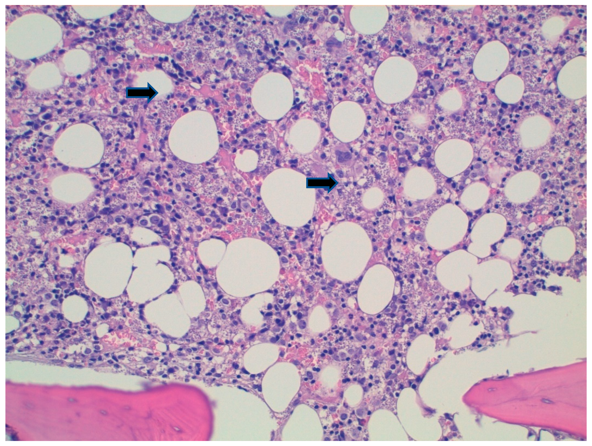
A plain chest X-ray was negative. Echocardiography was performed, and found to be negative for endocarditic vegetations or pericardial involvement. Furthermore, a Total body Computed Tomography showed enlarged liver and concomitant splenomegaly of 20 cm. Due to persistent fever, further microbiological analysis was requested (Table 2), and in a positive serology, a Western Blot for Leishmania spp. was found. Due to the high suspicion of visceral leishmaniasis (VL), an osteomedullary bone biopsy was performed, showing multiple amastigotes (Figure 2), and a confident diagnosis of VL was made.
The patient started therapy with Liposomal Amphotericin B (L-AmB, 3 mg/Kg on days 1–5, followed by a dose on days 10, 17, 24, 31 and 38) with an immunocompromised patient regimen. The treatment was well tolerated, and laboratory and clinical values returned to normal. A second osteomedullary bone biopsy was performed which showed normal cellularity. However, since the patient presented with an exacerbation of pre-existent arthropathic psoriasis in May, secukinumab was re-started four weeks after the end of the L-AmB regimen without complications, and currently, the patient has reached the third month after the start of anti-IL17, with no clinical or laboratory signs of reactivation.

3. Discussion

VL is usually characterized by prolonged fever, weight loss, splenomegaly and hepatomegaly, progressive anemia or pancytopenia and hypergammaglobulinemia [7]. Moreover, the reactivation of latent Leishmania infection in chronic diseases and immunocompromised hosts is a broad and heterogeneous field in medicine and infectious diseases [11,12,13]. In particular, among patients with psoriasis, with or without psoriatic arthritis, reported cases of Leishmania infections and reactivations are growing in the literature [12].
Interestingly, the link between psoriasis and Leishmania spp. has historically been complicated and yet to be fully understood. On the one hand, some authors have argued that psoriasis might represent a protective factor against Leishmania infection [6]. On the other, a first-generation polyvalent vaccine manufactured with proteins from several cultured Leishmania species has proved effective in treating psoriasis [5].
Recently, Colomba et al. reported a brief literature review of patients with psoriasis (and psoriatic arthritis) and leishmaniasis [12]. Moreover, their literature review, which included cases from 2004 to 2020, showed that no patients undergoing immunosuppressive treatment with anti-interleukin-17 were reported [12].
We describe the first case of Leishmania reactivation in a patient who underwent secukinumab treatment.
Interleukin-17 (IL-17) inhibitors (i.e., ixekizumab, secukinumab and brodalumab) are used successfully in the biological treatment of psoriasis and psoriatic arthritis, but at the same time mediate the immune response against bacteria and fungi [14]. IL-17 is a well-studied cytokine and an essential player in the mammalian immune system. IL-17 was initially thought to be produced by CD4 T Helper 17 (Th17), but is now known to be produced by various other cells also, including macrophages, dendritic cells, CD8 Tc17, lymphoid tissue inducer, and γδ-T [15]. Although this cytokine plays an essential role in many infectious diseases, it promotes inflammatory pathology in autoimmunity and in other settings [16].
Previous research has shown that a Th1 immune response is protective against cutaneous variants of the disease, while a Th1/Th2 response is more typically reported against visceral infections [4]. The pro-inflammatory IL-17 cytokine family plays a significant role in the elimination of intracellular parasites in addition to the Th1/Th2 cytokine families. IL-17, which is produced by Th17 cells, is demonstrated to worsen illness in Leishmania-induced skin lesions [4,5]. This finding suggests a role for IL-17 in the pathophysiology of this condition [5]. Growth of IL-17 generating cells during vaccine-induced immunity, however, suggests a protective function for IL-17 [6]. Further evidence that IL-17 is involved in vaccine induced protective immunity comes from studies on humans with visceral leishmaniasis (VL), where it was found that IL-17 and IL-22 were linked to protection against re-exposure to Leishmania [4,5,6].
Furthermore, our patient underwent methotrexate, cyclosporine and subsequently infliximab for a long time, despite no clinical or laboratory signs of Leishmania spp. reactivation. According to Colomba et al., in 22 cases collected between 2004 and 2020, 14 and 9 patients had had ongoing treatment with methotrexate or infliximab, respectively [12].
Anti-TNF treatment-related immunosuppression seems to have a wider spectrum of interactions in the cytokine cascade than anti-IL-17 regimens, and also involves T helper cells (Th17) [15,16,17]. Recently, the functional role of IL-17-producing Th17 in psoriasis has been suggested by its reduction during successful anti-TNF treatment [15,16,17].
Moreover, Maritati et al. found a higher prevalence of subclinical leishmaniasis in patients with inflammatory rheumatic diseases receiving biological drugs than in those treated with other immunosuppressive drugs [18]. In addition, Kurizky et al. conducted a cross-sectional screening study in an area endemic to leishmaniasis with a cohort of 311 patients undergoing treatment with immune suppressive regimens [19]. They found that a total of 29 patients (seven serology alone, thirteen conventional PCR and nine real-time PCR) were positive for Leishmania spp. screening, but no patients using IL-17A inhibitors tested positive for Leishmania spp. [19].
Regarding previous cases reported of VL in psoriatic patients, middle-aged men with arthritic involvement were the most represented in the literature, and these clinical characteristics were also present in our patient [12].
Recently, Kurizky and colleagues published a systematic review that tried to assess the real burden of Leishmaniasis in the whole immunosuppressed population [19]. In their work, the mean age at the time of leishmaniasis development was 51.63 years and the mean duration of immunosuppression was 5.75 years [19].
They observed how the problem of patients treated with immunosuppressive drugs is mostly reported in European countries around the Mediterranean Sea. In Italy, the most common diseases are systemic lupus erytematous, rheumatic arthritis, Wegener disease and psoriatic arthritis, while the most frequent species of Leishmania are L. infantum and L. donovani [19].
Their systematic review showed unexpected results in the geographical distribution of the included cases. The countries that most often reported leishmaniasis in patients treated with immunosuppressive drugs were considerably different from the global picture of this disease in the general population. Normally, Leishmaniasis in Europe accounts for only 2% of all leishmaniasis cases in the world, but they reported that most of the cases of leishmaniasis in immunosuppressed individuals were in Europe (almost 80%) [20]. Chronic rheumatic conditions are more common in developed countries than in developing countries [21], and, for that reason, European countries around the Mediterranean coast have more access to modern immunosuppressive treatments than countries of South America and the Middle East, in which leishmaniasis is endemic [20].
The diagnosis of the reactivation of VL in our study, rather than acute infection, is related to different factors. First, the patient had contact with dogs and the countryside; moreover, his dog died precociously three years before diagnosis. Second, the patient lived in an area in which Leishmania is endemic. Third, splenomegaly and cytopenia showed a subacute or chronic infection that resembled a long-lasting or reactivated infection. In Leishmania spp. infections, past medical history, habits and hobbies, contact with animals and living in endemic countries may increase the risk of infection and reactivation under certain circumstances [21]. Fourth, the suspected reactivation of Leishmania could be due to infliximab or immunosuppressive agents that the patient took for many years before secukinumab.
Our patient’s response to L-AmB treatment was evaluated with clinical and laboratory follow-up, completed by an osteomedullary biopsy that turned negative six weeks after starting treatment. Several direct and indirect biomarkers for Leishmania spp. response after treatment have been proposed in the past, including PCR for Leishmania, which was unfortunately negative from admission [22].
Treatment restart with secukinumab for exacerbation of psoriatic arthritis was discussed with a rheumatologist and a senior infectious disease consultant. The patient was followed as an outpatient in both clinics with monthly follow-up through clinical and laboratory markers (including PCR for Leishmania spp.) that at the time of this report have steadily remained within normal limits.

4. Conclusions

Increasing awareness of Leishmania reactivation risk in endemic countries was testified by case reports and case series available in the literature, although a more standardized approach for screening populations at risk has yet to be proposed to reduce the risk of premature suspension of treatment and high mortality if left untreated for VL. Furthermore, of interest is the role of IL-17 in the elimination of intracellular parasites of Leishmania and in the pathways of immune-response against this microorganism.

Author Contributions

Conceptualization, T.L.; investigation, V.F.; data curation, B.R., R.B. and V.F.; writing—original draft preparation, T.L.; writing—review and editing, S.C. and M.T.B.; supervision, F.G.D.R. All authors have read and agreed to the published version of the manuscript.

Funding

This research received no external funding.

Institutional Review Board Statement

The study was conducted in accordance with the Declaration of Helsinki, and Ethical Committee approval was waived for this study due to the format of the study (case report).

Informed Consent Statement

Informed consent was obtained from all subjects involved in the study.

Data Availability Statement

Not applicable.

Conflicts of Interest

The authors declare no conflict of interest.

References

  1. Blauvelt, A.; Chiricozzi, A. The Immunologic Role of IL-17 in Psoriasis and Psoriatic Arthritis Pathogenesis. Clin. Rev. Allergy Immunol. 2018, 55, 379–390. [Google Scholar] [CrossRef] [PubMed] [Green Version]
  2. Keijsers, R.R.M.C.; Joosten, I.; van Erp, P.E.J.; Koenen, H.J.P.M.; van de Kerkhof, P.C.M. Cellular sources of IL-17 in psoriasis: A paradigm shift? Exp. Dermatol. 2014, 23, 799–803. [Google Scholar] [CrossRef] [PubMed]
  3. Lande, R.; Botti, E.; Jandus, C.; Dojcinovic, D.; Fanelli, G.; Conrad, C.; Chamilos, G.; Feldmeyer, L.; Marinari, B.; Chon, S.; et al. The antimicrobial peptide LL37 is a T-cell autoantigen in psoriasis. Nat. Commun. 2014, 5, 5621. [Google Scholar] [CrossRef] [PubMed] [Green Version]
  4. Pitta, M.G.; Romano, A.; Cabantous, S.; Henri, S.; Hammad, A.; Kouriba, B.; Argiro, L.; El Kheir, M.; Bucheton, B.; Mary, C.; et al. IL-17 and IL-22 are associated with protection against human kala azar caused by Leishmania donovani. J. Clin. Investig. 2009, 119, 2379–2387. [Google Scholar] [CrossRef] [PubMed] [Green Version]
  5. Banerjee, A.; Bhattacharya, P.; Joshi, A.B.; Ismail, N.; Dey, R.; Nakhasi, H.L. Role of pro-inflammatory cytokine IL-17 in Leishmania pathogenesis and in protective immunity by Leishmania vaccines. Cell Immunol. 2016, 309, 37–41. [Google Scholar] [CrossRef] [PubMed]
  6. Nascimento, M.S.; Carregaro, V.; Lima-Júnior, D.S.; Costa, D.L.; Ryffel, B.; Duthie, M.S.; de Jesus, A.; de Almeida, R.P.; da Silva, J.S. Interleukin 17A acts synergistically with interferon gamma to promote protection against Leishmania infantum infection. J. Infect. Dis. 2015, 211, 1015–1026. [Google Scholar] [CrossRef] [PubMed] [Green Version]
  7. Torres-Guerrero, E.; Quintanilla-Cedillo, M.R.; Ruiz-Esmenjaud, J.; Arenas, R. Leishmaniasis: A review. F1000Reserch 2017, 26, 750. [Google Scholar] [CrossRef] [PubMed]
  8. Nazzaro, G.; Rovaris, M.; Veraldi, S. Leishmaniasis: A Disease With Many Names. JAMA Dermatol. 2014, 150, 1204. [Google Scholar] [CrossRef] [PubMed]
  9. World Health Organization Regional Office for the Americas (WHO PAHO). Leishmaniasis. Washington: WHO PAHO. Available online: https://www.paho.org/hq/dmdocuments/2017/2017-cha-leishmaniasis-factsheet-work.pdf (accessed on 24 September 2022).
  10. Ferroglio, E.; Battisti, E.; Zanet, S.; Bolla, C.; Concialdi, E.; Trisciuoglio, A.; Khalili, S.; Biglino, A. Epidemiological evaluation of Leishmania infantum zoonotic transmission risk in the recently established endemic area of Northwestern Italy. Zoonoses Public Health 2018, 65, 675–682. [Google Scholar] [CrossRef] [PubMed]
  11. Colomba, C.; Saporito, L.; Bonura, S.; Campisi, G.; Di Carlo, P.; Panzarella, V.; Caputo, V.; Cascio, A. Leishmania infection in psoriasis. J. Infect. 2020, 80, 578–606. [Google Scholar] [CrossRef] [PubMed]
  12. Pagliano, P.; Esposito, S. Visceral leishmaniosis in immunocompromised host: An update and literature review. J. Chemother. 2017, 29, 261–266. [Google Scholar] [CrossRef] [PubMed]
  13. Lupia, T.; Corcione, S.; Boglione, L.; Cariti, G.; DE Rosa, F.G. Visceral leishmaniasis in a patient with active HBV/HDV co-infection. J. Infect. Public. Health 2020, 13, 306–308. [Google Scholar] [CrossRef] [PubMed]
  14. Ghoreschi, K.; Balato, A.; Enerbäck, C.; Sabat, R. Therapeutics targeting the IL-23 and IL-17 pathway in psoriasis. Lancet 2021, 397, 754–766. [Google Scholar] [CrossRef]
  15. O’Daly, J.A.; Lezama, R.; Gleason, J. Isolation of Leishmania amastigote protein fractions which induced lymphocyte stimulation and remission of psoriasis. Arch. Dermatol. Res. 2009, 301, 411–427. [Google Scholar] [CrossRef] [PubMed]
  16. Gonçalves-de-Albuquerque, S.D.C.; Pessoa-E-Silva, R.; Trajano-Silva, L.A.M.; de Goes, T.C.; de Morais, R.C.S.; da COliveira, C.N.; De Lorena, V.M.B.; De Paiva-Cavalcanti, M. The Equivocal role of Th17 cells and neutrophils on immunopathogenesis of leishmaniasis. Front. Immunol. 2017, 8, 1437. [Google Scholar] [CrossRef] [PubMed] [Green Version]
  17. McGeachy, M.J.; Cua, D.J.; Gaffen, S.L. The IL-17 Family of Cytokines in Health and Disease. Immunity 2019, 50, 892–906. [Google Scholar] [CrossRef] [PubMed]
  18. Deng, Y.; Chang, C.; Lu, Q. The inflammatory response in psoriasis: A comprehensive review. Clin. Rev. Allergy Immunol. 2016, 50, 377–389. [Google Scholar] [CrossRef] [PubMed]
  19. Kurizky, P.S.; Gomes, C.M.; Cesetti, M.V.; Martins, G.A.; Regattieri, N.A.T.; Marianelli, F.F.; Sevilha Santos, L.; Medeiros Silva, V.; de Paula, N.A.; Frade, M.A.C.; et al. Cross-sectional screening study for Leishmania DNA and antibodies in biologic-treated patients with psoriasis living in an area endemic for leishmaniasis. Br. J. Dermatol. 2019, 81, 1337–1339. [Google Scholar] [CrossRef] [PubMed]
  20. Surveillance of Leishmaniasis in the WHO European Region, 2016 and Global Leishmaniasis Surveillance Update, 1998–2016. Available online: https://www.who.int/publications/i/item/who-wer9340 (accessed on 19 August 2022).
  21. Maritati, M.; Trentini, A.; Michel, G.; Bellini, T.; Almugadam, S.; Hanau, S.; Govoni, M.; Marty, P.; Contini, C. Subclinical Leishmania infection in patients with rheumatic diseases under biological drugs. Infection 2018, 46, 801–809. [Google Scholar] [CrossRef] [PubMed]
  22. Akhoundi, M.; Downing, T.; Votýpka, J.; Kuhls, K.; Lukeš, J.; Cannet, A.; Ravel, C.; Marty, P.; Delaunay, P.; Kasbari, M.; et al. Leishmania infections: Molecular targets and diagnosis. Mol. Asp. Med. 2017, 57, 1–29. [Google Scholar] [CrossRef] [PubMed]
Figure 1. Timeline of treatment of psoriasis until diagnostic of visceral leishmaniasis. Abbreviations: MTX: metothrexate.
Tropicalmed 07 00319 g001
Figure 2. Hematoxylin-eosine stain on osteomedullary biopsy showing amastigotes (black arrows). Caption: Hematoxylin-eosin shows histiocytes containing innumerable hematoxylinophilic corpuscles compatible with Leishmania amastigotes (arrows), also present in the interstitium (20×).
Tropicalmed 07 00319 g002
Table 1. Timeline from admission to first 3-month follow-up.
Day 0 Day 5 Day 10 Day 38 1 Month PT 3 Months PT
Total WBC count (103/uL) 1.44 2.35 3.0 3.27 6.07 5.40
Lymphocytes (absolute) 530 770 770 940 1370 1550
Neutrophils (absolute) 620 1.220 1710 1880 3990 3220
Hb (g/dL) 8.2 8.4 8.5 9.6 11.0 11.8
PLTS (103/uL) 53 98 134 197 191 161
LDH (U/L) 351 NA NA NA NA NA
IgG (g/L) 24 NA NA 21 22.6 NA
Albumin (g/L) 26 NA NA NA NA NA
Ferritin (ng/mL) 1075 NA NA NA NA NA
GOT 19 24 14 16 NA 12
GPT 20 29 9 13 NA 8
Bilirubin Total (mg/dL) 0.9 NA 0.6 NA NA NA
Creatinine (mg/dL) 1.45 2.5 1.65 1.42 NA 1.4
Protein C-reactive (mg/dL) 93.3 15.7 29.7 43.5 33.5 10.7
Procalcitonin (ng/mL) 0.8 0.18 0.08 NA 0.10 0.04
Protrombin Time 66% NA NA NA NA NA
Abbreviations: PT: post-treatment; WBC: White Blood Cell; Hb: Hemoglobin; PLTS: platelets; LDH: lactate dehydrogenase; GOT: glutamic-oxaloacetic transaminase; GPT: glutamic-pyruvic transaminase; NA: not available.
Table 2. Microbiological Screening for Pancytopenia and Fever.
Microbiological Screening on Admission
HIV ½ Ab and p24 Negative
HBsAg, antiHbs, anti-HBc Negative
HCV Ab Negative
Treponema total Ab Negative
CMV IgM negative, IgG positive
EBV VCA IgG, Positive, EBNA IgG positive
HSV 1 & 2 IgM negative, IgG positive
HHV-6 IgM negative, IgG positive
Adenovirus IgM negative, IgG negative
Parvovirus B19 IgM negative, IgG negative
Mycobacterium tuberculosis QuantiferonNegative
Galactomannan Ag Negative
Beta D-glucan Ag Negative
Leishmania spp.WB p14 positive, p16 positive. PCR: negative
COVID-19 Negative
Abbreviations: HIV: human immunodeficiency virus; HB: hepatitis B; HCV: hepatitis C virus; CMV: cytomegalovirus; EBV: Epstein–Barr virus; HSV: herpes virus; HHV-6: human herpes virus.
Publisher’s Note: MDPI stays neutral with regard to jurisdictional claims in published maps and institutional affiliations.