Article

Assessing the Presence of Wuchereria bancrofti Infections in Vectors Using Xenomonitoring in Lymphatic Filariasis Endemic Districts in Ghana

by
Sellase Pi-Bansa
1,2,3,*,
Joseph H. N. Osei
3,4,
Worlasi D. Kartey-Attipoe
3,
Elizabeth Elhassan
5,
David Agyemang
5,
Sampson Otoo
3,
Samuel K. Dadzie
3,
Maxwell A. Appawu
3,
Michael D. Wilson
3,
Benjamin G. Koudou
6,7,
Dziedzom K. de Souza
3,
Jürg Utzinger
1,2 and
Daniel A. Boakye
3
1
Swiss Tropical and Public Health Institute, CH-4002 Basel, Switzerland
2
University of Basel, CH-4003 Basel, Switzerland
3
Noguchi Memorial Institute for Medical Research, College of Health Sciences, University of Ghana, LG 581 Legon, Ghana
4
Department of Animal Biology and Conservation Science, University of Ghana, LG 67 Legon, Ghana
5
SightSavers International, Ghana Office, Accra, Ghana
6
Liverpool School of Tropical Medicine, Liverpool L3 5QA, UK
7
Centre Suisse de Recherches Scientifiques en Côte d’Ivoire, 01 BP 1303, Abidjan 01, Côte d’Ivoire
*
Author to whom correspondence should be addressed.
Trop. Med. Infect. Dis. 2019, 4(1), 49; https://doi.org/10.3390/tropicalmed4010049
Submission received: 17 January 2019 / Revised: 10 March 2019 / Accepted: 13 March 2019 / Published: 17 March 2019
(This article belongs to the Special Issue Spatial Epidemiology of Neglected Tropical Diseases (NTDs))

Abstract

:
Mass drug administration (MDA) is the current mainstay to interrupt the transmission of lymphatic filariasis. To monitor whether MDA is effective and transmission of lymphatic filariasis indeed has been interrupted, rigorous surveillance is required. Assessment of transmission by programme managers is usually done via serology. New research suggests that xenomonitoring holds promise for determining the success of lymphatic filariasis interventions. The objective of this study was to assess Wuchereria bancrofti infection in mosquitoes as a post-MDA surveillance tool using xenomonitoring. The study was carried out in four districts of Ghana; Ahanta West, Mpohor, Kassena Nankana West and Bongo. A suite of mosquito sampling methods was employed, including human landing collections, pyrethrum spray catches and window exit traps. Infection of W. bancrofti in mosquitoes was determined using dissection, conventional and real-time polymerase chain reaction and loop mediated isothermal amplification assays. Aedes, Anopheles coustani, An. gambiae, An. pharoensis, Culex and Mansonia mosquitoes were sampled in each of the four study districts. The dissected mosquitoes were positive for filarial infection using molecular assays. Dissected An. melas mosquitoes from Ahanta West district were the only species found positive for filarial parasites. We conclude that whilst samples extracted with Trizol reagent did not show any positives, molecular methods should still be considered for monitoring and surveillance of lymphatic filariasis transmission.

1. Introduction

Lymphatic filariasis is a disease that occurs in tropical and subtropical parts of the world. The aim of the Global Programme to Eliminate Lymphatic Filariasis (GPELF), launched by the World Health Organization (WHO) in 2000, is to interrupt the transmission of lymphatic filariasis caused by Wuchereria bancrofti and Brugia species, and to manage morbidity and disability in affected individuals [1,2]. By 2011, guidelines had been developed and mass drug administration (MDA) was scaled up in 53 of the 73 lymphatic filariasis endemic countries [3], including Ghana. The Ghana Filariasis Elimination Programme (GFEP) was established in 2000 [4]. The inception was governed by preliminary data, indicating that lymphatic filariasis was endemic in 49 out of 110 districts. Microfilariae (mf) and immunochromatographic test (ICT) prevalence ranged between 19.8% and 29.6% and between 33.1% and 45.4%, respectively [4]. This led to the commencement of MDA in 2001 in 10 districts and the subsequent scaling up to the remaining endemic districts by 2006 [4,5]. Monitoring and evaluation (M&E) of the impact of MDA usually does not involve the detection of filarial larvae in mosquito vectors. Hence, xenomonitoring has not been officially part of WHO recommendations for lymphatic filariasis surveillance.
WHO put forth rigorous procedures for documenting interruption of lymphatic filariasis transmission in endemic countries [1]. These include mapping for the identification of endemic regions, followed by at least five rounds of annual MDA with periodic M&E. A transmission assessment survey (TAS) is conducted after the cessation of MDA and a 5-year post-validation to confirm that no recrudescence of lymphatic filariasis occurred [6]. Measuring progress of any lymphatic filariasis control programme is, however, dependent on the effectiveness of M&E post-MDA [7,8], among other issues. Monitoring of lymphatic filariasis transmission by programme managers mainly involves mf assays and antigen tests in the human populations. A challenge with this monitoring approach is the reluctance of individuals to provide samples [9] and its inability to provide a “real-time” estimate of the disease [3,7]. Xenomonitoring, which detects infection in vectors, could serve as a complementary diagnostic tool to serology. Xenomonitoring is convenient, non-invasive [7,9] and can be used to assess the progress of lymphatic filariasis control activities [3,10,11]. Dorkenoo and colleagues, in a study in Togo, demonstrated the possibility of using molecular xenomonitoring for post-lymphatic filariasis validation surveillance [6]. In their study, the feasibility of using large-scale xenomonitoring was demonstrated. Furthermore, the absence of W. bancrofti infections in Anopheles gambiae was observed during a post-validation molecular xenomonitoring survey in Togo. In the southern part of Ghana, a recent study revealed 0.9% W. bancrofti infection and 0.5% infectivity rates in An. gambiae following several rounds of MDA in endemic districts [12].
The purpose of the current study was to evaluate lymphatic filariasis transmission in vectors using dissection and molecular xenomonitoring as diagnostic tools. The study was implemented in four districts; two districts in northern Ghana and two districts in southern Ghana. The results complement existing information on W. bancrofti infections in vector mosquitoes, and provide additional evidence of the feasibility of using xenomonitoring for M&E and surveillance activities post-MDA.

2. Materials and Methods

2.1. Study Sites

The study was conducted in eight communities, selected from four districts in the Western and Upper East regions of Ghana. Two communities were selected from each district. In the Upper East region, Badunu and Navio Central were selected from Kassena Nankana West district, and Atampiisi Bongo and Balungu Nabiisi from the Bongo district. In the Western region, Antseambua and Asemkow were selected from Ahanta West district, while Ampeasem and Obrayebona were selected from Mpohor district. A map showing the study districts has been published elsewhere [13]. These sites were selected based on lymphatic filariasis prevalence data stemming from monitoring activities by the Ghana National Neglected Tropical Disease Programme unit of the Ghana Health Service (Table 1).

2.2. Mosquito Collection and Identification

Mosquito sampling spanning both the dry and rainy seasons was done for 13 months (from July 2015 to July 2016) in the four study districts. A detailed explanation of the three mosquito sampling methods (i.e., human landing collections, pyrethrum spray catches and window exit traps) used by community vector collectors (CVCs) has been described by Pi-Bansa et al. [14]. Mosquitoes sampled were morphologically and molecularly identified. In short, morphological identification of mosquitoes involved the observation of mosquitoes under a microscope and separation into various genera [15,16]. Deoxyribonucleic acid (DNA) extracted from the legs of An. gambiae was used for the identification of sibling species [17] and molecular forms within the An. gambiae complex [18,19].

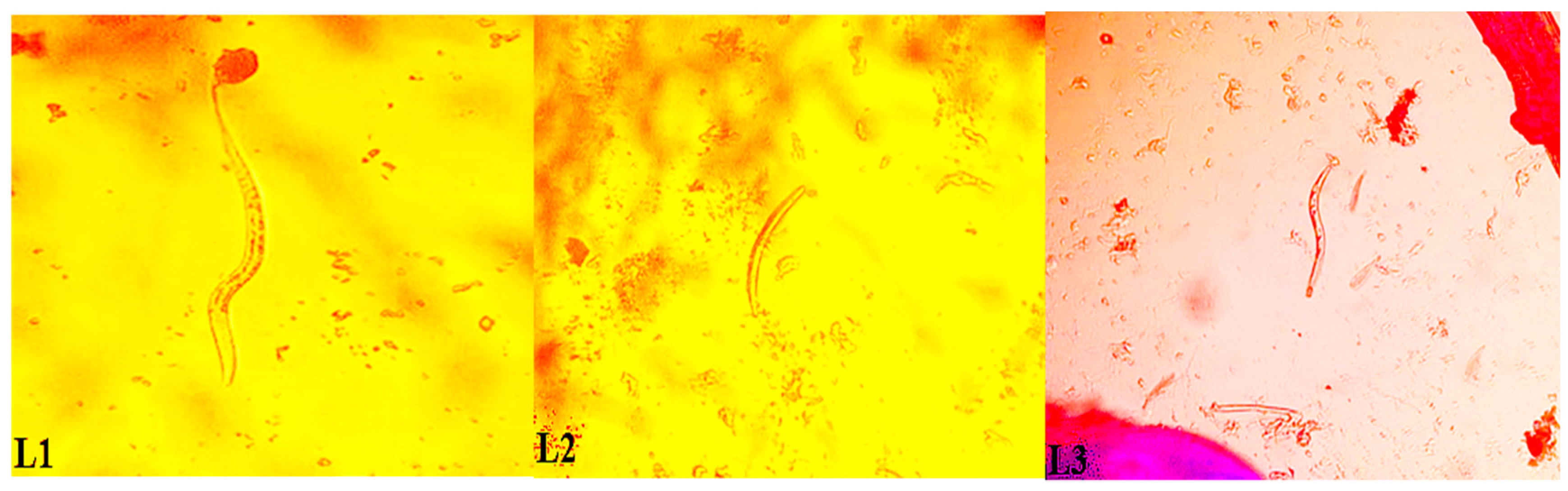
2.3. Mosquito Dissection

The sample size of An. gambiae mosquitoes for dissection was specifically calculated for the various districts, as described by Naing et al. [20]. Mosquitoes were placed on a glass slide. A pair of dissecting pins was used to separate the head, thorax and abdomen. Then, a drop of normal saline was added to each segment. Dissection of mosquitoes and identification of the W. bancrofti larval stages was done under a microscope [21].

2.4. Extraction and Detection of W. bancrofti in Dissected Mosquitoes

All W. bancrofti negative and positive mosquitoes were scraped into Eppendorf tubes, pending further molecular analyses. The various mosquito species were grouped into pools ranging from 1–25. DNA was extracted from pooled mosquitoes using the Qiagen DNeasy tissue kit (Qiagen CA) extraction method, adhering to the manufacturer’s instructions. Extraction was followed by identification of parasite DNA in pooled mosquitoes using a loop-mediated isothermal amplification (LAMP) assay [11,22], conventional polymerase chain reaction (PCR) [23] and real-time (RT)-PCR [24]. These assays were performed using standard protocols described elsewhere [11,22,23,24]. Positive and negative controls were included in all reactions.

2.5. Extraction of Nucleic Acids from Pooled Mosquitoes with a TRIzol Reagent

Mosquitoes were randomly selected for the extraction of DNA and RNA using a TRIzol reagent (Life Technologies; Carlsbad, CA, USA). In order to estimate an infection rate of 1% with a power of 0.80, the estimated total number of mosquitoes required for each district was 2000 [6,25]. The protocol for determining infectivity required that samples were stored in RNAlater so as to enable RNA extraction from mosquitoes. An. gambiae, Mansonia and Culex species sampled by human landing catches and stored in RNAlater reagent (Life Technologies; Carlsbad, CA, USA) were pooled (range: 5–20). The determination of the number of mosquitoes in a pool was based on prior research pursued by Boakye et al., which tested different mosquito pool sizes (i.e., 25, 50, 100 and 200) [26]. Several additional studies had pools of mosquitoes of up to 30 specimens [9,11,27]. Extraction of DNA and RNA on pooled mosquitoes was done to assess both W. bancrofti infection and infectivity rates, respectively [27]. Detection of both infection and infectivity in pooled mosquitoes followed the protocols of Rao et al. [24] and Laney et al. [27]. Furthermore, quality control was done for the detection of infection in An. gambiae complex by extracting DNA from pooled Kisumu mosquitoes (laboratory reared susceptible An. gambiae strains, n = 20) spiked with 5–20 µL of W. bancrofti mf positive blood samples (57 mf/mL), which showed amplification for the parasite. The extraction protocol was replicated for this study (see Supplementary File).

2.6. Statistical Analysis

Data were entered into Microsoft Excel (Microsoft Corporation; Redmond, WA, USA). The Poolscreen 2.0 software (University of Alabama; Birmingham, USA) was used to calculate the maximum likelihood estimate of infection in the vector populations, along with the 95% confidence interval (CI) [28]. The various entomological indices assessed included vector biting density, infection and infectivity rates, annual/monthly transmission potentials and worm load in mosquitoes [29,30].

2.7. Ethical Approval

This study was approved by the institutional review board of the Noguchi Memorial Institute for Medical Research (Accra, Ghana; reference no. CPN 077/13-14, 7 May 2014) and the institutional research commission of the Swiss Tropical and Public Health Institute (Basel, Switzerland; reference no. FK 122a, 24 November 2015). All CVCs consented orally to participate in the study. Albendazole and ivermectin were administered to CVCs before mosquito sampling commenced. Arrangement was also made with the nurses at the community-based health planning and services compound to provide treatment for CVCs who reported at their facility and tested positive for malaria.

3. Results

3.1. Mosquito Abundance and Composition

A total of 31,064 mosquitoes were collected during the 13-month study period: 27,739 (89.3%) by human landing catches, 2687 (8.7%) by pyrethrum spray collections and 638 (2.1%) by window exit traps. The numbers of mosquitoes sampled from all districts using the various sampling techniques are summarised in Table 2. An. gambiae sensu lato (s.l.) (n = 23,102; 83.3%), the main lymphatic filariasis vector in Ghana, had the highest number collected using human landing catches. Other species collected included Mansonia spp. (n = 2474; 8.9%), Culex spp. (n = 2056; 7.4%), Aedes spp. (n = 92; 0.3%), An. coustani (n = 11; 0.04%) and An. pharoensis (n = 4; 0.01%). For pyrethrum spray collections, 1884 (70.1%) An. gambiae, 720 (26.8%) Culex spp., 40 (1.5%) An. pharoensis, 26 (1.0%) Mansonia spp., 10 An. coustani and 7 Aedes spp. were collected. A total of 562, 10, 3 and 1 mosquitoes were reported for An. gambiae, An. pharoensis, Aedes spp. and An. coustani, respectively, using window exit traps. Culex and Mansonia spp. had the same number (n = 31) sampled for window exit traps.

3.2. Molecular Identification of An. gambiae and W. bancrofti

A total of 320, 368, 217 and 211 An. gambiae s.l. from Ahanta West, Mpohor, Kassena Nankana West and Bongo districts, respectively, were identified at the molecular level. Results shown in Table 3 indicate high numbers of the sibling species An. melas in Ahanta West district. Relatively high numbers of An. coluzzii, formerly known as M form of the An. gambiae complex, were obtained from Mpohor, Kassena Nankana West and Bongo districts. Eight mosquitoes observed to be infected with W. bancrofti by dissection tested positive when pool screened using PCR.

3.3. Transmission Indices of An. gambiae Complex from Ahanta West District

The average vector biting density for An. gambiae, sampled using human landing collections from Ahanta West, Mpohor, Kassena Nankana West and Bongo districts, were 43.8, 9.9, 1.0 and 0.8 bites/person/night, respectively. W. bancrofti infections were reported only in An. melas, a sibling species within the An. gambiae complex from Ahanta West district for this study. Eight An. melas mosquitoes were found infected (harbouring any of the developmental stage(s) of the parasite: mf, larval stages 1 (L1), 2 (L2) or 3 (L3), of which two mosquitoes were infective, harbouring only L3, as shown in Figure 1. The total numbers of L1, L2 and L3 counted from all the slides were 10, 2 and 2, respectively. The monthly infective biting rates (MIBR) and the annual infective biting rate (AIBR) were 8.0 and 95.9 infective bites/person, respectively. The annual transmission potential (ATP) due to An. gambiae in the Ahanta West district was 7.4 (Table 4).

3.4. Detection of W. bancrofti Using Molecular Techniques

A total of 2000 An. gambiae s.l. from Ahanta West and Mpohor districts, 253 from Kassena Nankana West and 225 from Bongo districts were screened for W. bancrofti infections and infectivity using RT-PCR. None of the 4478 An. gambiae processed in 214 pools from all study districts were found positive for W. bancrofti. Screening was also done for both Mansonia and Culex species from the four districts, though very few numbers were sampled from Mpohor, Kassena Nankana West and Bongo, compared to Ahanta West. Both Mansonia and Culex species were found negative for W. bancrofti in all districts (Table 5). All dissected mosquitoes from the four districts that were negative for W. bancrofti parasite and further screened by LAMP, conventional PCR and RT-PCR tested negative.

4. Discussion

Rigorous monitoring of W. bancrofti infections in mosquito vectors after several rounds of MDA is recommended to provide information on the progress of control and elimination activities. Indeed, such monitoring activities are necessary for making programmatic decisions that will eventually lead to certification of lymphatic filariasis elimination in previously endemic regions [26]. The current study, which was part of an operational research project to determine reasons for persistent lymphatic filariasis transmission in selected districts of Ghana after more than 10 rounds of MDA, investigated the feasibility and usefulness of a xenomonitoring approach for post-MDA surveillance to assess filarial infections in vectors [22,31]. Our study also provides information on the lymphatic filariasis infection status in vectors after multiple rounds of MDA in previously endemic districts.
The sampling methods used for this study included human landing collections, pyrethrum spray catches and window exit traps. These collection methods have been used before for sampling mosquitoes for xenomonitoring activities [14,32,33,34]. Recently, the Ifakara tent trap has been reported as an alternative to human landing collections and it was emphasised that it exhibits an improved ethical profile [35,36]. However, at the time our study was implemented, we did not have access to the Ifakara tent trap. Results from our study revealed high numbers of An. gambiae complex, the primary lymphatic filariasis vector in Ghana [9,37], in all four districts. The highest density was observed in Ahanta West district. The high densities of vectors and observed infections (L1, L2 and L3) in Ahanta West district might explain the presence of W. bancrofti infection in the An. gambiae complex from this district. A relatively higher density of An. gambiae was recorded in Kassena Nankana West district, compared to Bongo district. Both districts are in the dry Guinea savannah ecological zone [29], whilst the Ahanta West and Mpohor districts are situated in the rain forest ecological zone [38]. In the year 2000, high baseline mf prevalences of 19.5% and 29.4% were reported in Ahanta West and Kassena Nankana West districts, while considerably lower mf prevalences were observed in 2014; 2.7% and 1.3%, respectively (Table 1), after multiple rounds of MDA. The present study recorded W. bancrofti infection rates of 0.025 and nil for Ahanta West and Kassena Nankana West districts, respectively (Table 4). These very low infection rates observed in mosquitoes from this study correspondingly reflect the low lymphatic filariasis prevalence rates in the human population. Moreover, the availability of efficient vectors (An. melas and/or Mansonia spp.) in all four study districts can lead to picking up W. bancrofti infections, even at low parasitaemia, as seen in the Ahanta West district. Despite the large numbers of efficient vectors in a given district, the very low rates or the absence of W. bancrofti infections in the human population is likely to result in the absence of infections in vectors. Hence, there should be enough W. bancrofti parasites in the blood of human population for vectors to successfully ingest after a blood meal, since at very low mf levels, vectors are unlikely to ingest parasites. This may explain the absence of infections in the large number of An. gambiae vectors collected and examined in Kassena Nankana West, Mpohor and Bongo districts.
Furthermore, results from molecular species identification of the An. gambiae complex showed a high proportion of An. coluzzii (formally the M form of An. gambiae complex) in almost all districts (Table 3). This could be associated with the fact that An. coluzzii, which prefer breeding in ephemeral sites like run-off and flood water, are mostly found in the northern and coastal savannah areas of Ghana where this study was conducted [39]. Kassena Nankana West district recording the highest number of An. arabiensis could possibly be due to its location in the northern part of Ghana where the climate is arid, which represents the preferred breeding condition for this mosquito species [19]. An. melas, which is a sibling species within the An. gambiae complex, was mostly found in the Ahanta West district, corroborating previous findings by Dunyo et al. [40]. Anopheles mosquitoes are known to exhibit “facilitation”, which makes it possible for these mosquito species to pick up W. bancrofti parasites at high mf rates in the human population and develop them to the infective stage [41,42,43]. However, An. melas exhibits “limitation”, and hence this species can ingest and develop mf to the infective stage, even at low parasite densities [42,43]. In view of the high numbers of An. melas recorded in Ahanta West district, it is conceivable that this species is responsible for the observed W. bancrofti parasites (L1, L2 and L3).
The ABR for An. gambiae complex was highest in the Ahanta West district (15,987 bites/person/year). Finding W. bancrofti infections in Ahanta West district may be due to the high number of lymphatic filariasis vectors, specifically from the An. gambiae complex with a reported prevalence of 2.7% in this district. In the Kassena Nankana West district, before the commencement of our study, the reported prevalence of 1.3% was indicative of low persistent lymphatic filariasis transmission. A possible reason for the absence of infections in Kassena Nankana West is the relatively low level of infection in the human population. Another factor is the lower ABR (376.3 bites/person/year) in this district. There were no W. bancrofti infections recorded in the Mpohor and Bongo districts. This may have been due to the zero mf prevalence reported for these two districts (Table 1) before the onset of this study. In a previous study, Appawu et al. [29] investigated the entomological role played by the two lymphatic filariasis vectors An. gambiae and An. funestus at irrigation project sites in the Upper East region of Ghana. The authors recorded W. bancrofti infections in all study districts. Their results indicated that for irrigated communities like Tono and Vea, higher vector densities resulted in more infective feeds compared to Azoka, a non-irrigated community. The 7.4 ATP of An. gambiae in the Ahanta West district was due to the observed L3 in An. melas and reported mf-positive individuals from this district. The ATP value of An. gambiae obtained in spite of the low infectivity rate might be explained by the large number of An. gambiae collected in this district. In our study, the vector observed having W. bancrofti infections (L1, L2 and L3) was only An. melas belonging to the An. gambiae complex. Additionally, identification of Mansonia species in Ahanta West district suggests that these vectors could take up mf and successfully develop them to the infective stage, even at low parasitaemia [12,44].
Mosquito vector control activities reduce vector densities and human-vector contact [45,46]. This in turn decreases the likelihood of vectors picking up W. bancrofti parasites in endemic areas that have undergone several rounds of MDA. We found considerably higher bednet usage in Mpohor and Bongo districts, compared to Ahanta West and Kassena Nankana West districts. Hence, there was higher human–vector contact in the latter two districts. This could have contributed to the high ABR recorded for Ahanta West leading to W. bancrofti infections in the vectors due to infections in the human population. Though Mpohor district had recorded a relatively high ABR (3604), the reported prevalence of zero may explain the absence of infections in this district.
Additionally, mosquito species previously considered as non-vectors might be acting as vectors of lymphatic filariasis as in the case of Mansonia in Ghana [47] and Culex in Nigeria [48,49]. This observation, together with the fact that parasite DNA can be detected in both vector and non-vector mosquitoes [6], led to the investigation of both species in this study. No positive result was recorded for culicines using molecular assays. Furthermore, molecular assays run on DNA and RNA extracted from selected An. gambiae complex from the various districts was negative for filarial infections. The absence of infections in An. gambiae complex could have been as a result of PCR inhibition due to the masking of parasite DNA by mosquito DNA due to extraction of pooled mosquito samples.

5. Conclusions

Our study, employing xenomonitoring as a post-MDA surveillance tool, revealed that, at low parasitaemia, infections are usually found and sustained in vectors that exhibit limitation as seen in Ahanta West district. Additionally, An. melas emerges as an important vector for xenomonitoring along the coastal communities of the Western region in the southern part of Ghana. Moreover, effective vector control activities like high coverage of bednets can decrease ABR values in any endemic foci. As revealed in our previous work, vector control activities (bednet usage) in Mpohor and Bongo districts were relatively high. The reported zero prevalence of human infections and reduction in the human vector contact due to bednet usage might be responsible for the absence of infections in mosquito vectors from these districts. Presently in Ghana, only little emphasis is placed on the inclusion of xenomonitoring in decision-making processes during lymphatic filariasis programmatic activities. As shown here, data from xenomonitoring could be used by programme managers and other stakeholders to support decisions of stopping or continuing MDA. Additionally, complementing vector control activities with MDA during lymphatic filariasis control activities could reduce W. bancrofti infections in mosquitoes.

Supplementary Materials

The following are available online at https://www.mdpi.com/2414-6366/4/1/49/s1, Figure S1. Amplification of Wuchereria bancrofti DNA from pooled laboratory reared susceptible Kisumu mosquitoes. The diagram shows amplification curves for four different pools (n = 20) of susceptible Kisumu mosquitoes and a positive control. The four pools were spiked with 5–20 µL of Wuchereria bancrofti microfilariae positive blood having a concentration of 57 mf/mL.

Author Contributions

S.P.-B., J.H.N.O., D.K.d.S. and D.A.B. contributed towards the design and conception of the study; S.P.-B., J.H.N.O., W.D.K.-A. and S.O. collected data. S.P.-B., J.H.N.O. and W.D.K.-A. analysed the data with assistance from M.A.A. and D.K.d.S., S.P.-B. wrote the first draft of this manuscript. E.E., D.A., S.K.D., M.A.A., M.D.W., B.G.K., D.K.d.S., J.U. and D.A.B. revised the manuscript. All authors approved the final version of the manuscript.

Funding

This work was funded by the Department for International Development (DFID) through the Liverpool Centre for Neglected Tropical Disease (CNTD) and SightSavers. SPB is grateful to the “Amt für Ausbildungsbeiträge” of the Canton of Basel-Stadt for a PhD fellowship.

Acknowledgments

The authors would like to thank the chief, elders, opinion leaders, assembly members and residents of the various communities for allowing us to work in their districts. We would also like to exhibit our appreciation to the heads of the various district health directorates for granting us permission to work in the respective districts. Furthermore, we extend our profound gratitude to Sandra Laney and Charles R. Katholi for providing us with the protocol for simultaneous extraction of DNA and RNA, and the Poolscreen 2.0 software, respectively. We thank the Parasitology Department and head of the United States Naval Medical Research Unit #3 (NAMRU-3) at the Noguchi Memorial Institute for Medical Research for their contribution in the conduct of this study by providing us with work station and equipment.

Conflicts of Interest

The authors declare no conflict of interest.

References

  1. WHO. Monitoring and Epidemiological Assessment of Mass Drug Administration in Global Programme to Eliminate Lymphatic Filariasis: A Manual for National Elimination Programmes; World Health Organization: Geneva, Switzerland, 2011. [Google Scholar]
  2. Jones, C.; Ngasala, B.; Derua, Y.A.; Tarimo, D.; Reimer, L.; Bockarie, M.; Malecela, M.N. Lymphatic filariasis transmission in Rufiji district, southeastern Tanzania: infection status of the human population and mosquito vectors after twelve rounds of mass drug administration. Parasit. Vectors 2018, 11, 588. [Google Scholar] [CrossRef]
  3. Okorie, P.N.; de Souza, D.K. Prospects, drawbacks and future needs of xenomonitoring for the endpoint evaluation of lymphatic filariasis elimination programs in Africa. Trans. R. Soc.Trop. Med. Hyg. 2016, 110, 90–97. [Google Scholar] [CrossRef] [PubMed]
  4. Biritwum, N.K.; de Souza, D.K.; Marfo, B.; Odoom, S.; Alomatu, B.; Asiedu, O.; Yeboah, A.; Hervie, T.E.; Mensah, E.O.; Yikpotey, P.; et al. Fifteen years of programme implementation for the elimination of lymphatic filariasis in Ghana: impact of MDA on immunoparasitological indicators. PLoS Negl. Trop. Dis. 2017, 11, e0005280. [Google Scholar] [CrossRef]
  5. Kanamitie, J.N.; Ahorlu, C.S.; Otchere, J.; Aboagye-Antwi, F.; Kwansa-Bentum, B.; Boakye, D.A.; Biritwum, N.-K.; Wilson, M.D.; de Souza, D.K. Twelve-month longitudinal parasitological assessment of lymphatic filariasis-positive individuals: Impact of a biannual treatment with ivermectin and albendazole. Trop. Med. Int. Health 2017, 22, 1451–1456. [Google Scholar] [CrossRef]
  6. Dorkenoo, M.A.; de Souza, D.K.; Apetogbo, Y.; Oboussoumi, K.; Yehadji, D.; Tchalim, M.; Etassoli, S.; Koudou, B.; Ketoh, G.K.; Sodahlon, Y.; et al. Molecular xenomonitoring for post-validation surveillance of lymphatic filariasis in Togo: No evidence for active transmission. Parasit. Vectors 2018, 11, 52. [Google Scholar] [CrossRef] [PubMed]
  7. Goodman, D.S.; Orelus, J.N.; Roberts, J.M.; Lammie, J.; Streit, T.G. PCR and mosquito dissection as tools to monitor filarial infection levels following mass treatment. Filaria J. 2003, 2, 11. [Google Scholar] [CrossRef]
  8. Plichart, C.; Sechan, Y.; Davies, N.; Legrand, A.M. PCR and dissection as tools to monitor filarial infection of Aedes polynesiensis mosquitoes in French Polynesia. Filaria J. 2006, 5, 2. [Google Scholar] [CrossRef]
  9. Owusu, I.O.; de Souza, D.K.; Anto, F.; Wilson, M.D.; Boakye, D.A.; Bockarie, M.J.; Gyapong, J.O. Evaluation of human and mosquito based diagnostic tools for defining endpoints for elimination of Anopheles transmitted lymphatic filariasis in Ghana. Trans. R. Soc. Trop. Med. Hyg. 2015, 109, 628–635. [Google Scholar] [CrossRef] [PubMed]
  10. Bockarie, M.J. Molecular xenomonitoring of lymphatic filariasis. Am. J. Trop. Med. Hyg. 2007, 77, 591–592. [Google Scholar] [CrossRef] [PubMed]
  11. Kouassi, B.L.; de Souza, D.K.; Goepogui, A.; Narh, C.A.; King, S.A.; Mamadou, B.S.; Diakité, L.; Dadzie, S.K.; Boakye, D.A.; Utzinger, J.; et al. Assessing the presence of Wuchereria bancrofti in vector and human populations from urban communities in Conakry, Guinea. Parasit. Vectors 2015, 8, 492. [Google Scholar] [CrossRef]
  12. de Souza, D.; Otchere, J.; Ahorlu, C.; Adu-Amankwah, S.; Larbi, I.; Dumashie, E.; McCarthy, F.; King, S.; Otoo, S.; Osabutey, D.; et al. Low microfilaremia levels in three districts in coastal Ghana with at least 16 years of mass drug administration and persistent transmission of lymphatic filariasis. Trop. Med. Infect. Dis. 2018, 3, 105. [Google Scholar] [CrossRef] [PubMed]
  13. Pi-Bansa, S.; Osei, J.H.N.; Frempong, K.K.; Elhassan, E.; Akuoko, O.K.; Agyemang, D.; Ahorlu, C.; Appawu, M.A.; Koudou, B.G.; Wilson, M.D.; et al. Potential factors influencing lymphatic filariasis transmission in “hotspot” and “control” areas in Ghana: The importance of vectors. Infect. Dis. Poverty 2019, 8, 9. [Google Scholar] [CrossRef] [PubMed]
  14. Pi-Bansa, S.; Osei, J.H.N.; Joannides, J.; Woode, M.E.; Agyemang, D.; Elhassan, E.; Dadzie, S.K.; Appawu, M.A.; Wilson, M.D.; Koudou, B.G.; et al. Implementing a community vector collection strategy using xenomonitoring for the endgame of lymphatic filariasis elimination. Parasit. Vectors 2018, 11, 672. [Google Scholar] [CrossRef] [PubMed]
  15. Gillies, M.T.; De Meillon, B. The Anophelinae of Africa south of the Sahara (Ethiopian zoogeographical region). Publ. S. Afr. Inst. Med. Res. 1968, 54, 1–343. [Google Scholar]
  16. Gillies, M.T.; Coetzee, M. A supplement to the Anophelinae of Africa south of the Sahara (Afrotropical region). Publ. S. Afr. Inst. Med. Res. 1987, 55, 1–143. [Google Scholar]
  17. Scott, J.A.; Brogdon, W.G.; Collins, F.H. Identification of single specimens of the Anopheles gambiae complex by the polymerase chain reaction. Am. J. Trop. Med. Hyg. 1993, 49, 520–529. [Google Scholar] [CrossRef] [PubMed]
  18. Fanello, C.; Santolamazza, F.; Della Torre, A. Simultaneous identification of species and molecular forms of the Anopheles gambiae complex by PCR-RFLP. Med. Vet. Entomol. 2002, 16, 461–464. [Google Scholar] [CrossRef] [PubMed]
  19. Coetzee, M.; Hunt, R.H.; Wilkerson, R.; Torre, A.D.; Coulibaly, M.B.; Besansky, N.J. Anopheles coluzzii and Anopheles amharicus, new members of the Anopheles gambiae complex. Zootaxa 2013, 3619, 246–274. [Google Scholar] [CrossRef]
  20. Naing, L.; Winn, T.; Rusli, B.N. Practical issues in calculating the sample size for prevalence studies. Arch. Orofac. Sci. 2006, 1, 9–14. [Google Scholar]
  21. WHO. Lymphatic Filariasis: A Handbook of Practical Entomology for National Elimination Programmes; World Health Organization: Geneva, Switzerland, 2013. [Google Scholar]
  22. de Souza, D.K.; Sesay, S.; Moore, M.G.; Ansumana, R.; Narh, C.A.; Kollie, K.; Rebollo, M.P.; Koudou, B.G.; Koroma, J.B.; Bolay, F.K.; et al. No evidence for lymphatic filariasis transmission in big cities affected by conflict related rural-urban migration in Sierra Leone and Liberia. PLoS. Negl. Trop. Dis. 2014, 8, e2700. [Google Scholar] [CrossRef] [PubMed]
  23. Ramzy, R.M.R.; Farid, H.A.; Kamal, I.H.; Ibrahim, G.H.; Morsy, Z.S.; Faris, R.; Weil, G.J.; Williams, S.A.; Gad, A.M. A polymerase chain reaction-based assay for detection of Wuchereria bancrofti in human blood and Culex pipiens. Trans. R. Soc.Trop. Med. Hyg. 1997, 91, 156–160. [Google Scholar] [CrossRef]
  24. Rao, R.U.; Atkinson, L.J.; Ramzy, R.M.R.; Helmy, H.; Farid, H.A.; Bockarie, M.J.; Susapu, M.; Laney, S.J.; Williams, S.A.; Weil, G.J. A real-time PCR-based assay for detection of Wuchereria bancrofti DNA in blood and mosquitoes. Am. J. Trop. Med. Hyg. 2006, 74, 826–832. [Google Scholar] [CrossRef] [PubMed]
  25. WHO. The Role of Polymerase Chain Reaction Techniques for Assessing Lymphatic Filariasis Transmission; World Health Organization: Geneva, Switzerland, 2009. [Google Scholar]
  26. Boakye, D.A.; Baidoo, H.A.; Glah, E.; Brown, C.; Appawu, M.; Wilson, M.D. Monitoring lymphatic filariasis interventions: adult mosquito sampling, and improved PCR-based pool screening method for Wuchereria bancrofti infection in Anopheles mosquitoes. Filaria J. 2007, 6, 13. [Google Scholar] [CrossRef]
  27. Laney, S.J.; Ramzy, R.M.R.; Helmy, H.H.; Farid, H.A.; Ashour, A.A.; Weil, G.J.; Williams, S.A. Detection of Wuchereria bancrofti L3 larvae in mosquitoes: A reverse transcriptase PCR assay evaluating infection and infectivity. PLoS. Negl. Trop. Dis. 2010, 4, e602. [Google Scholar] [CrossRef] [PubMed]
  28. Katholi, C.R.; Toe, L.; Merriweather, A.; Unnasch, T.R. Determining the prevalence of Onchocerca volvulus infection in vector populations by polymerase chain reaction screening of pools of black flies. J. Infect. Dis. 1995, 172, 1414–1417. [Google Scholar] [CrossRef]
  29. Appawu, M.A.; Dadzie, S.K.; Baffoe Wilmot, A.; Wilson, M.D. Lymphatic filariasis in Ghana: entomological investigation of transmission dynamics and intensity in communities served by irrigation systems in the Upper East region of Ghana. Trop. Med. Int. Health 2001, 6, 511–516. [Google Scholar] [CrossRef]
  30. Coulibaly, Y.; Dembele, B.; Diallo, A.; Kristensen, S.; Konate, S.; Dolo, H.; Dicko, I.; Sangare, M.; Keita, F.; Boatin, B.A.; et al. Wuchereria bancrofti transmission pattern in southern Mali prior to and following the institution of mass drug administration. Parasit. Vectors 2013, 6, 247. [Google Scholar] [CrossRef]
  31. Opoku, M.; Minetti, C.; Kartey-Attipoe, W.D.; Otoo, S.; Otchere, J.; Gomes, B.; de Souza, D.K.; Reimer, L.J. An assessment of mosquito collection techniques for xenomonitoring of anopheline-transmitted lymphatic filariasis in Ghana. Parasitology 2018, 145, 1783–1791. [Google Scholar] [CrossRef]
  32. Govella, N.J.; Chaki, P.P.; Geissbühler, Y.; Kannady, K.; Okumu, F.; Charlwood, J.D.; Anderson, R.A.; Killeen, G.F. A new tent trap for sampling exophagic and endophagic members of the Anopheles gambiae complex. Malar J. 2009, 8, 157. [Google Scholar] [CrossRef]
  33. Wong, J.; Bayoh, N.; Olang, G.; Killeen, G.F.; Hamel, M.J.; Vulule, J.M.; Gimnig, J.E. Standardizing operational vector sampling techniques for measuring malaria transmission intensity: evaluation of six mosquito collection methods in western Kenya. Malar J. 2013, 12, 143. [Google Scholar] [CrossRef]
  34. Pam, D.D.; de Souza, D.K.; D’Souza, S.; Opoku, M.; Sanda, S.; Nazaradden, I.; Anagbogu, I.N.; Okoronkwo, C.; Davies, E.; Elhassan, E.; et al. Is mass drug administration against lymphatic filariasis required in urban settings? The experience in Kano, Nigeria. PLoS. Negl. Trop. Dis. 2017, 11, e0006004. [Google Scholar] [CrossRef] [PubMed]
  35. WHO. Training Module on Malaria Control: Malaria Entomology and Vector Control. Guide for Tutors; World Health Organization: Geneva, Switzerland, 2013. [Google Scholar]
  36. Briët, O.J.T.; Huho, B.J.; Gimnig, J.E.; Bayoh, N.; Seyoum, A.; Sikaala, C.H.; Govella, N.; Diallo, D.A.; Abdullah, S.; Smith, T.A.; et al. Applications and limitations of Centers for Disease Control and Prevention miniature light traps for measuring biting densities of African malaria vector populations: A pooled-analysis of 13 comparisons with human landing catches. Malar J. 2015, 14, 247. [Google Scholar] [CrossRef] [PubMed]
  37. Boakye, D.A.; Wilson, M.D.; Appawu, M.A.; Gyapong, J. Vector competence, for Wuchereria bancrofti, of the Anopheles populations in the Bongo district of Ghana. Ann. Trop. Med. Parasitol. 2004, 98, 501–508. [Google Scholar] [CrossRef] [PubMed]
  38. Gyapong, J.O.; Webber, R.H.; Morris, J.; Bennett, S. Prevalence of hydrocele as a rapid diagnostic index for lymphatic filariasis. Trans. R. Soc. Trop. Med. Hyg. 1998, 92, 40–43. [Google Scholar] [CrossRef]
  39. de Souza, D.; Kelly-Hope, L.; Lawson, B.; Wilson, M.; Boakye, D. Environmental factors associated with the distribution of Anopheles gambiae s.s. in Ghana; an important vector of lymphatic filariasis and malaria. PLoS ONE 2010, 5, e9927. [Google Scholar] [CrossRef] [PubMed]
  40. Dunyo, S.K.; Appawu, M.; Nkrumah, F.K.; Baffoe-Wilmot, A.; Pedersen, E.M.; Simonsen, P.E. Lymphatic filariasis on the coast of Ghana. Trans. R. Soc. Trop. Med. Hyg. 1996, 90, 634–638. [Google Scholar] [CrossRef]
  41. Southgate, B.A.; Bryan, J.H. Factors affecting transmission of Wuchereria bancrofti by anopheline mosquitoes. 4. Facilitation, limitation, proportionality and their epidemiological significance. Trans. R. Soc. Trop. Med. Hyg. 1992, 86, 523–530. [Google Scholar] [CrossRef]
  42. Amuzu, H.; Wilson, M.D.; Boakye, D.A. Studies of Anopheles gambiae s.l. (Diptera: Culicidae) exhibiting different vectorial capacities in lymphatic filariasis transmission in the Gomoa district, Ghana. Parasit. Vectors 2010, 3, 85. [Google Scholar] [CrossRef] [PubMed]
  43. de Souza, D.K.; Koudou, B.; Kelly-Hope, L.A.; Wilson, M.D.; Bockarie, M.J.; Boakye, D.A. Diversity and transmission competence in lymphatic filariasis vectors in West Africa, and the implications for accelerated elimination of Anopheles-transmitted filariasis. Parasit. Vectors 2012, 5, 259. [Google Scholar] [CrossRef] [PubMed]
  44. Southgate, B.A. The significance of low density microfilaraemia in the transmission of lymphatic filarial parasites. J. Trop. Med. Hyg. 1992, 95, 79–86. [Google Scholar] [PubMed]
  45. Bockarie, M.J.; Pedersen, E.M.; White, G.B.; Michael, E. Role of vector control in the Global Program to Eliminate Lymphatic Filariasis. Annu. Rev. Entomol. 2009, 54, 469–487. [Google Scholar] [CrossRef] [PubMed]
  46. Kelly-Hope, L.A.; Molyneux, D.H.; Bockarie, M.J. Can malaria vector control accelerate the interruption of lymphatic filariasis transmission in Africa; capturing a window of opportunity? Parasit. Vectors 2013, 6, 39. [Google Scholar] [CrossRef] [PubMed]
  47. Ughasi, J.; Bekard, H.; Coulibaly, M.; Adabie-Gomez, D.; Gyapong, J.; Appawu, M.; Wilson, M.; Boakye, D. Mansonia africana and Mansonia uniformis are vectors in the transmission of Wuchereria bancrofti lymphatic filariasis in Ghana. Parasit. Vectors 2012, 5, 89. [Google Scholar] [CrossRef] [PubMed]
  48. Anosike, J.C.; Nwoke, B.E.; Ajayi, E.G.; Onwuliri, C.O.; Okoro, O.U.; Oku, E.E.; Asor, J.E.; Amajuoyi, O.U.; Ikpeama, C.A.; Ogbusu, F.I.; et al. Lymphatic filariasis among the Ezza people of Ebonyi state, eastern Nigeria. Ann. Agric. Environ. Med. 2005, 12, 181–186. [Google Scholar] [PubMed]
  49. Agi, P.; Ebenezer, A. Observations on filarial infection in Amassoma community in the Niger Delta, Nigeria. J. Appl. Sci. Environ. Manag. 2010, 13, 15–19. [Google Scholar] [CrossRef]
Figure 1. Three larval stages of W. bancrofti parasites from Ahanta West district in Ghana.
Tropicalmed 04 00049 g001
Table 1. Number of mass drug administration (MDA) rounds and prevalence of microfilariae in the four districts of Ghana where the current study was conducted between July 2015 and July 2016.
DistrictCommunityNumber of MDA RoundsMicrofilariae Prevalence in 2000 (%)Microfilariae Prevalence in 2014 (%)Number of An. gambiae Dissected
Ahanta WestAsemkow
Antseambua
1619.52.7320
MpohorObrayebona
Ampeasem
110.00.0368
Kassena Nankana WestNavio Central
Badunu
1529.41.3217
BongoAtampiisi Bongo
Balungu Nabiisi
1316.70.0211
Table 2. Mosquitoes sampled using three different sampling methods from four study districts in Ghana during a 13-month sampling period between July 2015 and July 2016.
MethodCommunityDistrictAn. gambiaeCulex speciesMansonia speciesAedes speciesAn. pharoensisAn. coustaniTotal Collected
Human landing catchesAsemkow
Antseambua
Ahanta West18,21312002386804
Obrayebona
Ampeasem
Mpohor41096672603
Badunu
Navio Central
Kassena Nankana West426489114224
Atampiisi Bongo
Balungu Nabiisi
Bongo35430153620
Pyrethrum spray catchesAsemkow
Antseambua
Ahanta West2714191360
Obrayebona
Ampeasem
Mpohor375147010
Badunu
Navio Central
Kassena Nankana West8013840119
Atampiisi Bongo
Balungu Nabiisi
Bongo4373180521
Window exit trapAsemkow
Antseambua
Ahanta West3961729000
Obrayebona
Ampeasem
Mpohor11911190
Badunu
Navio Central
Kassena Nankana West1261110
Atampiisi Bongo
Balungu Nabiisi
Bongo3570101
Total 25,54828072531102542231,064
Table 3. Distribution of members of the An. gambiae complex in four study districts, Ghana, collected between July 2015 and July 2016.
DistrictSibling Species of the Anopheles gambiae Complex
An. gambiae s. s.An. arabiensisAn. melasAn. coluzzii
n%n%n%n%
Ahanta West30.9113.427585.9123.8
Mpohor22661.40010.312233.2
Kassena Nankana West5726.32511.50012457.1
Bongo5425.6000014267.3
Table 4. Entomological indices showing relevant parameters for the estimation of the annual transmission potential (ATP).
DistrictAverage Number of An. gambiae Sampled per MonthVector Biting Density (MBR)Annual Biting Rate (ABR)Average Number of An. gambiae Dissected per MonthAverage Infection per MonthAverage Infectivity per MonthInfection Rate (%)Infectivity Rate (%)Annual Infective Biting Rate (AIBR)Average Worm Load per MonthAnnual Transmission Potential (ATP)
Ahanta West140143.815,987250.6200.1500.025 (2.5)0.006 (0.6)95.9220.0777.386
Mpohor3169.93614280000000
Kassena Nankana West331.0365170000000
Bongo270.8292160000000
Table 5. Number of mosquito pools processed per study district from July 2015 to July 2016.
SpeciesDistrictNumber of PoolsAverage Pool SizeNumber of Mosquitoes ProcessedPositive (Infection/Infectivity)95% CI
An. gambiaeAhanta West9720.6200000–0.00095
Mpohor9122.0200000–0.00095
Kassena Nankana West1319.525300–0.00756
Bongo1317.322500–0.00849
Mansonia speciesAhanta West8321.1175400–0.00109
Mpohor225.05000–0.03767
Kassena Nankana West114.01400–0.12815
Bongo15.0500–3.18868
Culex speciesAhanta West6320.0126100–0.00152
Mpohor219.03800–0.04927
Kassena Nankana West1919.436900–0.00518
Bongo816.313300–0.01433

