Review

Melioidosis in Myanmar

1
Department of Medical Research, Yangon, Myanmar
2
Myanmar Oxford Clinical Research Unit, Yangon, Myanmar
3
Centre for Tropical Medicine and Global Health, Nuffield Department of Clinical Medicine, Old Road Campus, University of Oxford, Oxford OX3 7FZ, UK
4
Department of Microbiology, Yangon General Hospital, University of Medicine 1, Yangon, Myanmar
5
Pathology Department, Microbiology Section, North Okkapala General Hospital, University of Medicine 2, Yangon, Myanmar
6
Shoklo Malaria Research Unit, Mahidol-Oxford Tropical Medicine Research Unit, Faculty of Tropical Medicine, Mahidol University, Mae Sot 63110, Thailand
7
Mandalay General Hospital, University of Medicine, Mandalay, Myanmar
8
Pathology Department, Microbiology section, Thingungyun Hospital, University of Medicine 1, Yangon, Myanmar
9
Department of Preventive & Social Medicine, Defence Services Medical Academy, Yangon, Myanmar
10
Lao-Oxford-Mahosot Hospital-Wellcome Trust Research Unit, Microbiology Laboratory, Mahosot Hospital, Vientiane, Laos
11
Faculty of Infectious and Tropical Diseases, London School of Hygiene and Tropical Medicine, London WC1E 7HT, UK
12
Medical Action Myanmar, Yangon, Myanmar
*
Author to whom correspondence should be addressed.
Trop. Med. Infect. Dis. 2018, 3(1), 28; https://doi.org/10.3390/tropicalmed3010028
Submission received: 9 February 2018 / Revised: 20 February 2018 / Accepted: 23 February 2018 / Published: 1 March 2018
(This article belongs to the Special Issue Global Burden and Challenges of Melioidosis)

Abstract

:
Sporadic cases of melioidosis have been diagnosed in Myanmar since the disease was first described in Yangon in 1911. Published and unpublished cases are summarized here, along with results from environmental and serosurveys. A total of 298 cases have been reported from seven states or regions between 1911 and 2018, with the majority of these occurring before 1949. Findings from soil surveys confirm the presence of Burkholderia pseudomallei in the environment in all three regions examined. The true epidemiology of the disease in Myanmar is unknown. Important factors contributing to the current gaps in knowledge are lack of awareness among clinicians and insufficient laboratory diagnostic capacity in many parts of the country. This is likely to have led to substantial under-reporting.

1. Introduction and History of Melioidosis in Myanmar

All historical accounts of melioidosis start with Myanmar (formerly Burma), since the disease was first recognized in 1911 in Rangoon (now Yangon) General Hospital by Alfred Whitmore, a British pathologist who worked in Burma between 1906 and 1924 [1]. In 1913, Whitmore reported 38 post-mortem cases of a ‘glanders-like disease’, the majority of whom were male and bore stigmata of morphine injection. Krishnaswamy, Whitmore’s assistant, reported in 1917 that he had personally seen more than 200 cases in Rangoon over six years, during which the disease accounted for approximately one in every 20 autopsies he had conducted [2]. In the ensuing years, reported case numbers in the country dwindled, and after the end of World War II, the disease disappeared from sight for more than 50 years. This hiatus corresponded firstly with the transition from colonial rule to independence (in 1948) and later with a prolonged period of military rule in Myanmar (1962–2011), during which the country became closed off from the rest of the world, investment in healthcare slowed, and research stagnated. Microbiologists working in larger centres inside Myanmar have reported sporadic cases over the last 20 years, but the true epidemiology of the disease is unknown. Most health facilities in Myanmar do not have the capacity to identify B. pseudomallei, and most health staff are not aware of this disease. It is therefore likely that melioidosis is significantly underdiagnosed in Myanmar, and the projected annual number of cases (more than 6000) and deaths (more than 3000) from a recent modelling study could well be an accurate reflection of the true situation [3].

2. Review of Melioidosis Cases and Presence of B. pseudomallei in the Country

We searched the PubMed database for cases of melioidosis or evidence of B. pseudomallei using the following search terms: ((Myanmar) OR Burma)) AND ((melioid*) OR pseudomallei) OR glanders*)). A separate search was performed for ‘Parotid abscess AND Myanmar’ (no hits). We also consulted the Myanmar medical literature and the melioidosis global database [4], and contacted consultant microbiologists in the biggest centres in the country (public sector).

2.1. Results

Evidence for the presence of B. pseudomallei in Myanmar comes from reports of clinical cases, environmental sampling and serosurveys. Melioidosis has never been diagnosed in animals in Myanmar although, by analogy with neighbouring countries, infections in animals are almost certainly occurring undiagnosed.

2.1.1. Clinical Cases

We found a total of 298 published and unpublished cases of melioidosis in Myanmar since 1913 (Table 1). The vast majority (257) were published before the end of 1948. The next report of a case diagnosed inside the country came about half a century later in 2000 in Mandalay [5]. Of the remaining cases, several were diagnosed outside Myanmar, and the remainder came from a very small number of centres. Eighty-five were known to have been culture-confirmed. The report by Krishnaswamy in which he mentions 200 cases does not state explicitly that these were culture-confirmed; however, given his position as Whitmore’s assistant and the descriptions of the first 38 cases in which the diagnosis was confirmed by culture, it is likely that these too were confirmed cases. Of note, only five cases (~2%) were in females and three in children. A total of 276/298 (93%) cases were diagnosed in patients from the Yangon area with six from Kayin state, two from Rakhine state, one from Bago region, two from Ayeyarwady, two from Magway, two from Pyay and four not specified (Table 1 and Figure 1).
In 2014, the results of all the blood cultures processed by the laboratory of Yangon General Hospital between 2005 and 2013 were published [26]. B. pseudomallei was not isolated from any of the 3865 cultures; however, the authors note that bacterial isolation and sub-optimal identification techniques may have been one explanation. In addition, the number of cultures requested was low relative to the number of admissions in this busy hospital, which exceeds 50,000 per year.

2.1.2. Serological Evidence for B. pseudomallei in Myanmar

The gold standard for the diagnosis of melioidosis is bacterial isolation. Serology is difficult to interpret in endemic countries due to background positivity rates, but gives some indication of population exposure. A cross-sectional sero-survey of 968 migrant workers from Myanmar was undertaken in Thailand, close to the western border, in 2005 using a haemagglutination assay [27]. The majority of participants (99%) came from Kayin or Mon States and Bago or Yangon Regions. The median [IQR] population IHA titre was 1:20 [1:10 to 1:40], with no regional variation observed. Sixty-nine (7%) participants had a titre ≥1:160, which is a recommended cut-off in Thailand used to support a clinical diagnosis of melioidosis. Only 8% of surveyed adults were agricultural workers. Between 2016 and 2017, blood samples were taken for serological testing using a hemolysin co-regulated protein 1 (Hcp1)-based ELISA from 265 febrile patients presenting to a three outpatient clinics in a large suburb of Yangon as part of a research study [28]. All patients tested negative (optical density cut-off 1.16); median [interquartile range] of 0.02 [0.01–0.04] [29].

2.1.3. Evidence from Environmental Surveys

Myanmar has a variable landscape, ranging from the delta region in the south and west, to the drier central plains, and ending at the Himalayan foothills in the north. Using a mathematical modelling approach and taking into account known cases, climate and soil type, Myanmar has been predicted to provide a very suitable environment for the causative bacterium [3]. Results of a farm survey performed in two townships in Yangon (Thanlyin and Hmawbi) were reported in 2016. From 120 soil samples and 12 water samples collected and processed according to standard microbiological guidelines for the detection of B. pseudomallei, there were seven positive results (confirmed by molecular methods) [30]. A national survey is underway by the authors, and preliminary testing has detected the bacterium by culture of soil samples from townships in Yangon, Kayin and Mon Regions, which is in agreement with the provenance of some of the reported clinical cases (Table 2). Full results should be available by the end of 2018.

3. Current Recommendations and Availability of Measures Against Melioidosis

Currently, there is no requirement to notify cases of melioidosis in the country. The Ministry of Health and Sports has set up a national system for communicable disease surveillance and response. This targets epidemic-prone diseases, specific diseases under national surveillance (DUNS), emerging infectious diseases, climate-related communicable diseases, and vaccine-preventable diseases, but not melioidosis [31]. Under-recognition of the disease is very likely due to a lack of awareness among health staff, under-utilisation of microbiology services by clinicians and inability of many laboratories to identify the organism due to poor availability of necessary reagents, as well as a shortage of microbiologists, particularly in rural areas. There are no standard treatment guidelines for melioidosis, and the few patients identified are managed on a case by case basis.

4. Awareness of Melioidosis

With the exception of a few ‘enthusiasts’, melioidosis is almost entirely unknown among healthcare providers. Raising awareness among health staff, clinicians and laboratory staff is therefore the essential prerequisite to identifying melioidosis patients. When diagnostic and treatment services for suspected melioidosis cases are established, raising awareness among the population working in agriculture should follow in high-risk areas. Microbiology in Myanmar is perceived as a diagnostic rather than a clinical service, and there is limited interaction between microbiologists and clinicians, which means opportunities to raise awareness are missed. This was highlighted in the report by Hlaing et al. in 2004 of melioidosis co-infection in a patient with tetanus. A report that B. pseudomallei had been isolated was duly sent to the ward, but went unnoticed for several days and was acted upon only when the patient deteriorated.

5. Current and Future Challenges

The burden of melioidosis in Myanmar needs to be defined. The evidence suggests that the disease is under-recognised and under-reported. While the majority of cases have been reported from Yangon, evidence from a multicentre abscess survey, routine blood culture surveillance data at a large centre and a recent serosurvey suggests it may not be a leading cause of sepsis in the greater Yangon region, despite the fact that it was clearly an important cause of death in the early 20th century [2,21,26]. Reasons for a possible decline in incidence in areas of Yangon where it was common previously are unclear, although the strong link with injection drug use in the early fatal cases raises the possibility of contamination of the morphine, which is likely to have originated from Upper Burma [32]. An alternative explanation is that this group was relatively immunocompromised and more susceptible to infection following exposure in Yangon itself, and that with increasing urbanization, the risk of exposure has fallen. It is probable that there are unrecognised melioidosis ‘hot-spots’ elsewhere, so accurate data across the whole country are needed. Diabetes mellitus, a well-known risk factor for melioidosis, has moved into the top ten causes of death in the country in the last three years (http://www.healthdata.org/myanmar), which may drive up case numbers in the future. Important contributing factors to the current lack of knowledge are a lack of microbiology laboratory diagnostic capacity, particularly in smaller townships and rural areas, low rates of blood culture requesting, and lack of awareness of the disease among clinicians. Many laboratories at township level have been upgraded as part of the National Health Plan, but are not functional due a shortage of microbiologists. Myanmar has a national reference laboratory (National Health Laboratory), established in 1963, and a system for disease reporting. Addition of B. pseudomallei to the list of notifiable pathogens might yield more information on the distribution of cases in the country, but is unlikely to uncover large numbers until the capacity and usage of diagnostic microbiology increase. Environmental surveys may be useful to help target scarce resources to augment case-based surveillance in areas where it is likely to have the highest yield. This should be accompanied by advocacy meetings with state/regional and township health authorities to raise awareness among clinicians and health providers.

Acknowledgments

Thank you to Yoel Lubell, Thomas Althaus and Narisara Chantratita from the Mahidol Oxford Tropical Medicine Research Unit and Faculty of Tropical Medicine, Mahidol University, Thailand, for sharing unpublished serology data. The soil survey was funded by Medical Action Myanmar. MOCRU, LOMWRU and SMRU are part of the MORU Tropical Health Network, funded by the Wellcome Trust of Great Britain.

Author Contributions

M.M.W. and E.A. wrote the first draft. M.M.W., D.A.B.D., M.M.M.S., K.N.Z., M.T.A., F.N., W.M.T., N.N.Z., M.Y.A., K.M.T. and F.S. searched for published cases in the Myanmar literature and/or provided information on unpublished cases, recent environmental surveys and the current situation in Myanmar. All authors reviewed, edited and approved the submitted version.

Conflicts of Interest

The authors declare no conflict of interest. The funding sponsors had no role in the design of the study; in the collection, analyses, or interpretation of data; in the writing of the manuscript, and in the decision to publish the results.

References

  1. Whitmore, A. An account of a glanders-like disease occurring in Rangoon. J. Hyg. 1913, 13, 1–34. [Google Scholar] [CrossRef] [PubMed]
  2. Krishnaswamy, C.S. Morphia injector’s septicaemia. Indian Med. Gaz. 1917, 52, 296–299. [Google Scholar]
  3. Limmathurotsakul, D.; Golding, N.; Dance, D.A.; Messina, J.P.; Pigott, D.M.; Moyes, C.L.; Rolim, D.B.; Bertherat, E.; Day, N.P.; Peacock, S.J.; et al. Predicted global distribution of Burkholderia pseudomallei and burden of melioidosis. Nat. Microbiol. 2016, 1, 15008. [Google Scholar] [CrossRef] [PubMed]
  4. Melioidosis. Info Online Database. 2018. Available online: http://www.melioidosis.info/ (accessed on 25 February 2018).
  5. Aung, M.K. Case report: Melioidosis—A hidden disease in Myanmar. Myanmar J. Curr. Med. Pract. 2000, 5, 57–59. [Google Scholar]
  6. Knapp, H.H.G. Morphine injector’s septicaemia (‘Whitmore’s disease’). Indian Med. Gaz. 1915, 50, 287–288. [Google Scholar]
  7. Cox, C.D.; Arbogast, J.L. Melioidosis. Am. J. Clin. Pathol. 1945, 15, 567–570. [Google Scholar] [CrossRef] [PubMed]
  8. Harries, E.J.; Lewis, A.A.; et al. Melioidosis treated with sulphonamides and penicillin. Lancet 1948, 1, 363–366. [Google Scholar] [CrossRef]
  9. Sen, S. A case of melioidosis. Indian Med. Gaz. 1948, 83, 186–187. [Google Scholar]
  10. van der Schaaf, A. Veterinary experiences as a Japanese prisoner of war and ex-POW along the Burma railroad from 1942 to January 1946. Vet. Quart. 1979, 1, 212–228. [Google Scholar] [CrossRef] [PubMed]
  11. Wilairatana, P.; Looareesuwan, S. Melioidotic otitis media. Southeast Asian J. Trop. Med. Public Health 1994, 25, 776–777. [Google Scholar] [PubMed]
  12. Hadano, Y. Imported melioidosis in Japan: A review of cases. Infect. Drug Resist. 2018, 11, 163–168. [Google Scholar] [CrossRef] [PubMed]
  13. Kunishima, H.; Seki, R.; Iwabuchi, T.; Nakamura, T.; Ishida, N.; Takagi, T.; Shimada, J. Case of melioidosis associated with acute empyema and cellulitis of the leg. Nihon Naika Gakkai Zasshi J. Jpn. Soc. Intern. Med. 1999, 88, 1101–1103. [Google Scholar]
  14. Lee, S.C.; Ling, T.S.; Chen, J.C.; Huang, B.Y.; Sheih, W.B. Melioidosis with adrenal gland abscess. Am. J. Trop. Med. Hyg. 1999, 61, 34–36. [Google Scholar] [CrossRef] [PubMed]
  15. Htun, Z.T.; Hla, T.; Myat, T.W.; Lin, N.; Wah, T.T. Detection of Burkholderia pseudomallei in patients with suppurative infections attending Yangon General Hospital and New Yangon General Hospital. Myanmar Health Sci. Res. J. 2013, 25, 114–119. [Google Scholar]
  16. Leeuwenburgh, I.; Driessen, J.T.; van Keulen, P.H.; Stijnen, P.J.; Verburg, G.P. Melioidosis. Nederlands Tijdschrift voor Geneeskunde 2002, 146, 723–725. [Google Scholar] [PubMed]
  17. Aung, M.K. Indigenous isolates of Burkholderia pseudomallei—The causative agent of melioidosis. Myanmar J. Curr. Med. Pract. 2004, 9, 13–15. [Google Scholar]
  18. Win, M.M. A case of melioidosis from Yangon General Hospital. Myanmar J. Curr. Med. Pract. 2004, 9, 5–7. [Google Scholar]
  19. Hlaing, S.S. Isolation of Pseudomonas pseudomallei (Burkholderia pseudomallei) from a case of tetanus in YGH. Myanmar J. Curr. Med. Pract. 2004, 9, 8–12. [Google Scholar]
  20. Demar, M.; Ferroni, A.; Dupont, B.; Eliaszewicz, M.; Bouree, P. Suppurative epididymo-orchitis and chronic prostatitis caused by Burkholderia pseudomallei: A case report and review. J. Travel Med. 2005, 12, 108–112. [Google Scholar] [CrossRef] [PubMed]
  21. Aung, M.K.; Mar, T.T. Re-emergence of melioidosis in Myanmar. Transact. Royal Soc. Trop. Med. Hyg. 2008, 102, S10–S11. [Google Scholar] [CrossRef]
  22. Min, T.T. Characterization of Burkholderia pseudomallei Isolates from Melioidosis Cases in Magway Division, Myanmar. Ph.D. Thesis, University of Medicine 1, Yangon, Myanmar, 2012. [Google Scholar]
  23. Chu, C.S.; Winearls, S.; Ling, C.; Torchinsky, M.B.; Phyo, A.P.; Haohankunnathum, W.; Turner, P.; Wuthiekanun, V.; Nosten, F. Two fatal cases of melioidosis on the Thai-Myanmar border. F1000Research 2014, 3, 4. [Google Scholar] [CrossRef] [PubMed]
  24. Kyi, M.M.; Kyi, T.T.; Thit, S.S.; Aung, N.M. A forgotten disease of Myanmar origin. Myanmar Med. J. 2014, 56, 55–58. [Google Scholar]
  25. Brummaier, T.; Ling, C.; Chu, C.S.; Wuthiekanun, V.; Haohankhunnatham, W.; McGready, R. Subcutaneous abscess formation in septic melioidosis, devoid of associated risk factors. IDCases 2016, 4, 23. [Google Scholar] [CrossRef] [PubMed]
  26. Myat, T.O.; Prasad, N.; Thinn, K.K.; Win, K.K.; Htike, W.W.; Zin, K.N.; Murdoch, D.R.; Crump, J.A. Bloodstream infections at a tertiary referral hospital in Yangon, Myanmar. Transact. R. Soc. Trop. Med. Hyg. 2014, 108, 692–698. [Google Scholar] [CrossRef] [PubMed]
  27. Wuthiekanun, V.; Langa, S.; Swaddiwudhipong, W.; Jedsadapanpong, W.; Kaengnet, Y.; Chierakul, W.; Day, N.P.; Peacock, S.J. Short report: Melioidosis in Myanmar: Forgotten but not gone? Am. J. Trop. Med. Hyg. 2006, 75, 945–946. [Google Scholar] [PubMed]
  28. Pumpuang, A.; Dunachie, S.J.; Phokrai, P.; Jenjaroen, K.; Sintiprungrat, K.; Boonsilp, S.; Brett, P.J.; Burtnick, M.N.; Chantratita, N. Comparison of o-polysaccharide and hemolysin co-regulated protein as target antigens for serodiagnosis of melioidosis. PLoS Negl. Trop. Dis. 2017, 11, e0005499. [Google Scholar] [CrossRef] [PubMed]
  29. Lubell, Y.; Mahidol-Oxford Research Unit, Bangkok, Thailand. Unpublished work. 2018.
  30. Win, M.M.; Aung, W.W.; Thu, H.M.; Wah, T.T.; Aye, K.M.; Htwe, T.T.; Htay MT and San, K.K. The environmental study on melioidosis in agricultural farms of Thanlyin and Hmawbi Townships. In Proceedings of the 44th Myanmar Health Research Congress, Yangon, Myanmar, 5–9 January 2016. [Google Scholar]
  31. Ministry of Health. Health in Myanmar 2014; Ministry of Health: Nay Pyi Taw, Myanmar, 2014.
  32. Aye Aye Hlaing. Opium in Myanmar (1885–1948). Ph.D. Thesis, University of Mandalay, Mandalay, Myanmar, 2008. [Google Scholar]
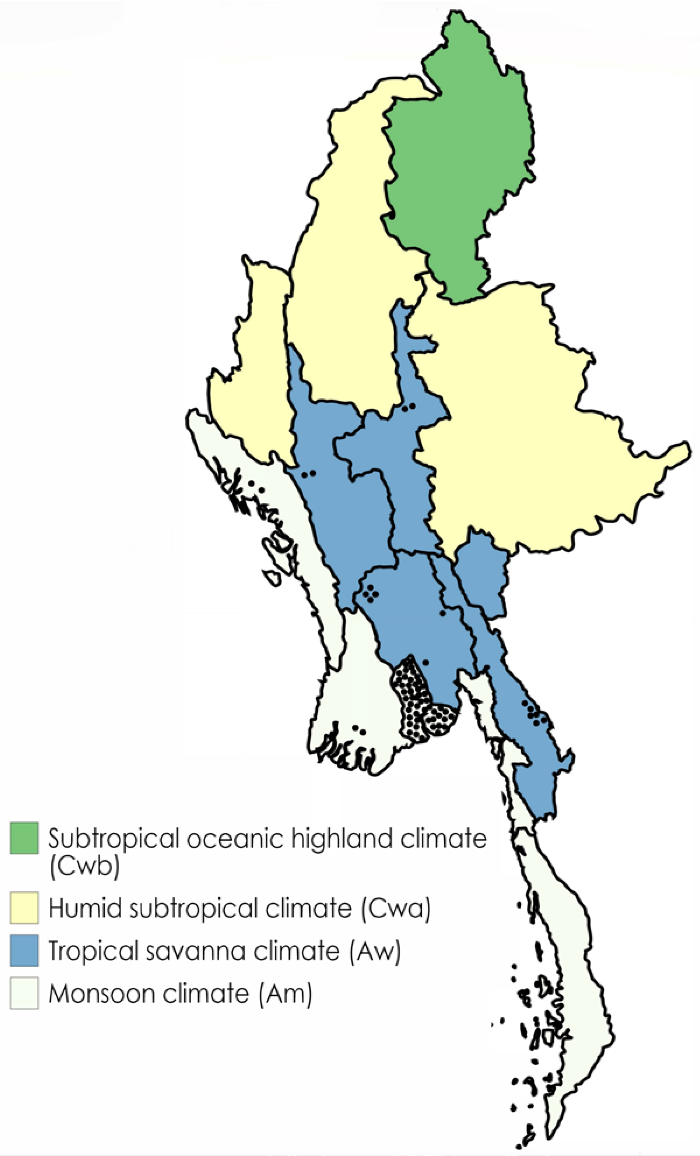
Figure 1. Map of Myanmar showing distribution of reported cases (1912–present) by state or region, categorized by predominant climate type. Each black dot represents a case.
Tropicalmed 03 00028 g001
Table 1. Cases of melioidosis reported from Myanmar.
Year of ReportFirst AuthorN CasesN FatalitiesN Culture ConfirmedLocation in MyanmarRemarks
1913Whitmore [1]383838YangonCase-series identified post-mortem
1915Knapp [6]11111 (remaining NS)YangonCase-series identified post-mortem
1917Krishnaswamy [2]~200~200NSYangonCases of ‘morphia injector’s septicaemia’ identified post-mortem
1945Cox [7]111Not knownUS Army soldier
1947Harries [8]625Between Pyay & Yangon (5), Rakhine (1)West African soldiers serving in Myanmar
1948Sen [9]111YangonCase identified post-mortem
1979van der Schaaf [10]111Not knownEx-Royal Netherlands East Indies Army (KNIL) prisoner of war. Post-mortem identification
1994Wilairatana [11]101Not knownMyanmar national diagnosed in Bangkok
1999Kunishima [12,13]101Not knownReturned traveler (diagnosed in Japan)
1999Lee [14]101YangonReturned traveler (diagnosed in Taiwan)
2000May Kyi Aung [5]101MandalayFemale
2002Than Than Aye; referenced in [15]1NSNSYangonCerebral melioidosis (no information)
2002Leeuwenburgh [16]101Not knownReturned female traveler (diagnosed in Netherlands)
2004May Kyi Aung [17]111Yangon-
2004Mo Mo Win [18]101Yangon (Hmawbi)-
2004Su Su Hlaing [19]101PyayConcomitant tetanus
2005Demar [20]101Shwepyitha (Yangon) or Kyunbin(Bago)Returned traveler (diagnosed in France)
2008May Kyi Aung [21]3NS3YangonSurvey of 133 patients with abscesses in 22 Yangon hospitals
2012Thae Thae Min [22]20 12MagwayStudy of 307 patients hospitalised with infectious diseases
2013Zaw Than Htun [15]3NS3Yangon (Hmawbi, Thanlyin), Ayeyarwady (Mawkyun)Includes one female
2014Chu [23]222Thai-Myanmar border (close to Kayin state) 2Two fatal cases (one female)
2014Mar Mar Kyi [24]101Taungoo (Bago)Healthy student with multiple skin abscesses
2015Mo Mo Win [4]21 32Yangon-
2016Brummaier [25]101Thai-Myanmar border (close to Kayin state) 2Ten-year-old boy with subcutaneous abscesses
2016Mo Mo Win [4]212Yangon-
2016–2017Shoklo Malaria Research Unit [4]303Thai-Myanmar border (close to Kayin state) 2Includes one child
2017Mo Mo Win (pers.comm)717Rakhine(1), Ayeyarwady (1), Yangon (5)Includes one female
2017Ni Ni Zaw (pers.comm.)1NS1Mandalay-
2018Kyaw Myo Tun (pers.comm.)111Yangon-
2018Mo Mo Win (pers.comm)10 41Yangon-
TOTAL298
Cases are adult males unless otherwise stated. 1 One patient survived. The other patient was referred to another centre (no outcome recorded). 2 The cases from the Thai-Myanmar border were diagnosed in a migrant population from Myanmar. 3 Second patient’s outcome not known. 4 Discharged home in pre-terminal condition at request of family; NS = not stated.
Table 2. Preliminary results of a soil survey being conducted in Myanmar.
Site NumberState or RegionTownshipTotal Number of SamplesNumber of Positive Samples
1YangonHmawbi1400
2YangonTontay105
3YangonThanlyin200
4YangonKyauktan203
5YangonThone Gwa200
6YangonKha Yan200
7YangonDala200
8Kayin Kyain Seikgyi600
9Kayin Myawaddy1409
10Kayin Kawkareik501
11Kayin Hpa-an501
12MonKyaikhto400
13MonBilin300
14MonThaton300
15MonYae300
16MonThanbyuzayat405
17MonKyaikmaraw302
18MonMawlamyaing200
19MonChaung Sone200
20MonPaung200
1 Note: an earlier survey found B. pseudomallei in Hmawbi (see text).