Abstract
Leptospirosis is a zoonotic disease of global public health importance caused by bacteria of the genus Leptospira. Small non-flying mammals are important reservoirs of the pathogen. The Brazilian Atlantic Forest is a biodiversity hotspot located in a densely populated area and subject to intense degradation. Although documented through serosurveys and the detection of leptospiral DNA in wild small mammals, no study has performed a genetic characterization of the bacteria in the region. The present study aimed to evaluate the genetic diversity of pathogenic leptospires identified in small non-flying mammals in the Southeast Atlantic Forest and to perform intraspecific genetic inferences with other hosts. The studied area included five different conservation units. Molecular diagnosis was performed based on the lipl32 gene. The SLST typing method was applied based on the secY gene. In total, 56% of samples were lipL32-PCR-positive and identified as L. interrogans, with a high genetic identity among them, distributed in four main haplogroups. The largest haplogroup also included reference sequences from humans, dogs, and urban rats, all belonging to the Icterohaemorrhagiae serogroup. Our results reinforce the role of small mammals as important carriers of L. interrogans and highlight the Atlantic Forest as a significant environment for the circulation and dissemination of spirochetes with zoonotic potential.
1. Introduction
Leptospirosis is a globally neglected, reemerging zoonotic disease of global public health importance caused by pathogenic spirochetal bacteria of the genus Leptospira that circulates among a wide spectrum of hosts, including humans and domestic and wild animals [1]. Leptospires can survive in the environment, particularly in moist conditions, and can be transmitted through different routes: (i) direct contact with the urine of an infected animal; (ii) indirect contact with contaminated water, soil, or feed; and (iii) sexual and vertical transmission in some species (e.g., bovine) [2].
Historically, the etiology of leptospiral infections can be divided into those determined by host-adapted or incidental strains [3]. Adapted strains are observed in the reservoir animal host, resulting from a long process of host–pathogen coevolution, leading to asymptomatic or inapparent disease or mild and subclinical symptoms of a chronic nature [3]. These maintenance hosts are infected asymptomatic animals that shed viable leptospires in their fluids, acting simultaneously as hosts and reservoirs. They play an important role in the epidemiology of leptospirosis, harboring and shedding leptospires in the environment for long periods, becoming important sources of leptospiral infection for non-adapted hosts [4]. Examples of adapted serovars are Bratislava in horses, Pomona in pigs, Hardjo in ruminants, Canicola in dogs, and Icterohaemorrhagiae in rodents [5]. On the other hand, incidental serovars infect hosts other than the maintenance ones, leading to an apparent acute, often severe illness [4]. Importantly, the L. interrogans serogroup Icterohaemorrhagiae is the most associated with acute human disease [6], highlighting the incidental nature of infection in people.
The clinical signs of leptospirosis in humans and animals are diverse and can affect multiple organ systems. In humans, the disease can present with a wide spectrum of symptoms, ranging from mild, flu-like symptoms to severe, life-threatening conditions such as Weil’s disease, characterized by jaundice, renal failure, and hemorrhagic manifestations [7]. The bacteria enter through abrasions or cuts in the skin and mucous membranes, including the conjunctiva [8]. Macerated skin, resulting from prolonged exposure to water, is also a suspected route of infection [9].
Small non-flying mammals, such as rodents and marsupials, play a crucial role in the epidemiology of leptospirosis and act as reservoirs of Leptospira spp. [8]. These animals shed the bacteria in their urine, contaminating the environment, particularly water sources. This can lead to human infection through direct or indirect contact with contaminated water or soil, representing a constant risk, especially in urban and peri-urban environments [10].
Due to its high biodiversity and species endemism in a small remaining area, the Brazilian Atlantic Forest is among the world’s 36 biodiversity hotspots [11]. It harbors a wide range of mammalian hosts, which provide favorable biotic conditions for the bacterial cycle. In addition, it is located in a tropical region and presents ideal environmental abiotic conditions, such as a hot and humid climate, that favor the survival of leptospires [12]. Several wild and domestic animals live in the region, including 72% of the Brazilian population [13]. Leptospirosis is endemic in this region, with an average of 3810 cases reported annually and with higher numbers in urban areas, including the Rio de Janeiro state, located in the southeastern region of the Atlantic Forest, where the disease remains a significant public health concern, with periodic outbreaks, particularly during the rainy season [14]. Poverty and urbanization, among other environmental and socioeconomic factors, influence morbidity and mortality rates [15].
The few studies that have documented the occurrence of Leptospira spp. in small mammals in the Atlantic Forest [16,17,18,19] were based on serosurveys, which present limited value since reservoirs may not be detected by serology [20]. Previous studies conducted by our group in the region have identified the presence of pathogenic leptospiral DNA in wild marsupials and rodents based on conventional PCR [21,22]. However, to date, no study has performed a genetic characterization of the bacteria.
Therefore, the present study aimed to analyze the genetic diversity of pathogenic leptospires identified in small non-flying mammals in the Atlantic Forest of southeastern Brazil, comparing the identified haplotypes with those described in human and domestic animals to assess the risk of zoonotic transmission and provide new epidemiological inferences.
2. Materials and Methods
All experimental procedures were approved by the Ethics Committee on Animal Use of the Oswaldo Cruz Institute (CEUA-IOC, Comissão de Ética no Uso de Animais, Instituto Oswaldo Cruz, protocol number P-70/13-2, license number LW-39/14). Fieldwork was in full compliance with the federal license issued by the Instituto Brasileiro do Meio Ambiente e dos Recursos Naturais Renováveis of the Brazilian Ministry of the Environment (IBAMA/SISBIO, license number 13373-1).
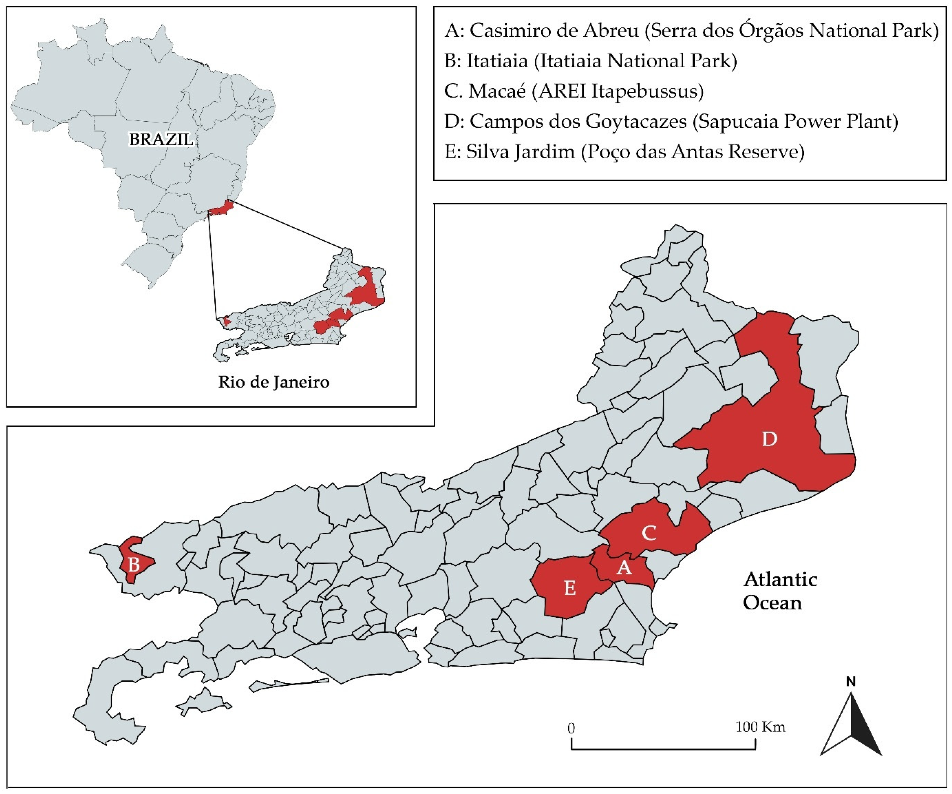
2.1. Animal Capture and Study Areas
The capture and taxonomic identification procedures of the small non-flying mammals are shown in [21,22]. Briefly, specimens were captured with Sherman® and Tomahawk® live traps set in sampling transects, 4–10 in each study area, each with 20 traps, evenly spaced 10–20 m apart. The traps were verified and rebaited daily during seven to ten consecutive sampling nights. In total, 265 kidney samples were obtained, one for each host, including the orders Didelphimorphia (n = 21) and Rodentia (n = 244).
The study area included five different conservation units within the Atlantic Forest in the Rio de Janeiro state, Brazil: (A) Serra dos Órgãos National Park/Casimiro de Abreu; (B) Itatiaia National Park/Itatiaia; (C) Area of Relevant Ecological Interest Itapebussus/Macaé; (D) Sapucaia Power Plant/Campos dos Goytacazes; and (E) Poço das Antas Reserve/Silva Jardim (Figure 1). Areas A, B, and E are strictly protected regions with minimal human interference, while areas C and D are zones with more pronounced anthropogenic activities due to industrial, agricultural, or urban development.
Figure 1.
Map of the State of Rio de Janeiro, Brazil, showing the collection sites (municipality/conservation area) of small non-flying mammals (red). The Atlantic Forest is the biome found along the coast of the Atlantic Ocean.
Samples from areas A (n = 15), B (n = 77) and C (n = 34) had been previously subjected to amplification based on the lipL32 gene. Of these, 63/126 (50%) were positive [21,22]. Samples from areas D (n = 52) and E (n = 87) were processed in the present study, totaling 139 samples. These were subjected to DNA extraction and molecular diagnosis.
2.2. Molecular Diagnosis
All unprocessed small mammal kidney samples (n = 139) were subjected to DNA extraction using a DNeasy® Blood & Tissue Kit (Qiagen, Hilden, Germany following the manufacturer’s instructions. For detecting pathogenic Leptospira spp., lipL32 gene-based PCR was performed using primers and conditions described by Hamond et al. (2014) [23]. Each sample set included ultrapure water as a negative control in all reactions, and 10fg of DNA extracted from Leptospira interrogans serovar Copenhageni (Fiocruz L1–130) served as a positive control. PCR products were analyzed by 2% agarose gel electrophoresis and visualized under ultraviolet light after staining with GelRed®.
2.3. Genetic Characterization of Pathogenic Leptospira spp.
All samples positive for the lipL32 gene were subjected to a nested PCR based on the secY gene, a genetic marker with good taxonomic resolution, whose use is well established for the genetic characterization of leptospires [24]. Reactions were performed using primers and conditions described in Grillová et al., 2020 [25], with the enzyme Platinum Taq DNA Polymerase kit (Invitrogen, Carlsbad, CA, USA). The amplicons were purified with a PCR Clean-Up System Kit (Promega, Madison, WI, USA) according to the manufacturer’s instructions and intended for sequencing. The reactions were performed in both directions using a Big Dye Terminator v. 3.1 Cycle Sequencing Kit (Applied Biosystems, Foster City, CA, USA) on a 3100 automated DNA sequencer according to the manufacturer’s instructions.
2.4. Phylogenetic Analyses
For molecular epidemiology purposes, we used the Single Locus Sequencing Typing (SLST) typing method. For this, sequences of the secY gene were selected from GenBank and subjected to phylogenetic analyses with the sequences obtained in the present study. Very short sequences (<400 bp) were excluded to avoid bias. After aligning the sequences in the ClustalX v 2.0 software [26], a maximum likelihood (ML) tree was constructed in MEGA X software v 11.0.13 [27] using the best-fitting DNA substitution model determined by the Bayesian information criterion. Genetic distances were calculated in MEGA X with the model previously determined by the analysis of the data set.
2.5. Assessment of Genetic Diversity and Determination of Haplotype Circulation Patterns
The identification of unique haplotypes was performed after screening in the DAMBE software v 7.3.32 [28]. To assess the intraspecific genetic diversity of Leptospira spp. populations and evaluate the dynamics of haplotype circulation between different hosts and regions, haplotype networks were constructed from the aligned sequences and scripts in nexus format in the PopART population genetics software using the median-joining inference method [29].
3. Results
3.1. Molecular Identification of Pathogenic Leptospira spp.
Of the 139 samples processed and submitted to molecular diagnosis based on the lipL32 PCR, 78 (56%) were positive. Those 78 plus the 63 previously positive samples [21,22] were subjected to a nested PCR based on the secY genetic marker, totaling 141 DNA samples. It was possible to obtain strong amplicons, suitable for sequencing, from 93 samples.
3.2. Genetic Characterization of Pathogenic Leptospira spp. Identified
Based on the secY gene sequencing, all samples were identified as L. interrogans with >99% identity with reference sequences after searching against the BLAST/NCBI platform (accessed 15 January 2025). The majority of hosts were from the order Rodentia (n = 83), with few from the order Didelphimorphia (n = 10), totaling 10 different genera. From these, Akodon and Mus had the highest number of positive animals with generated sequences (n = 23 and n = 21, respectively) (Table 1). The distribution of genetically characterized samples according to location and hosts is shown in Supplementary Table S1.
Table 1.
Haplogroup distribution of Leptospira interrogans identified in the present study according to collection site and host.
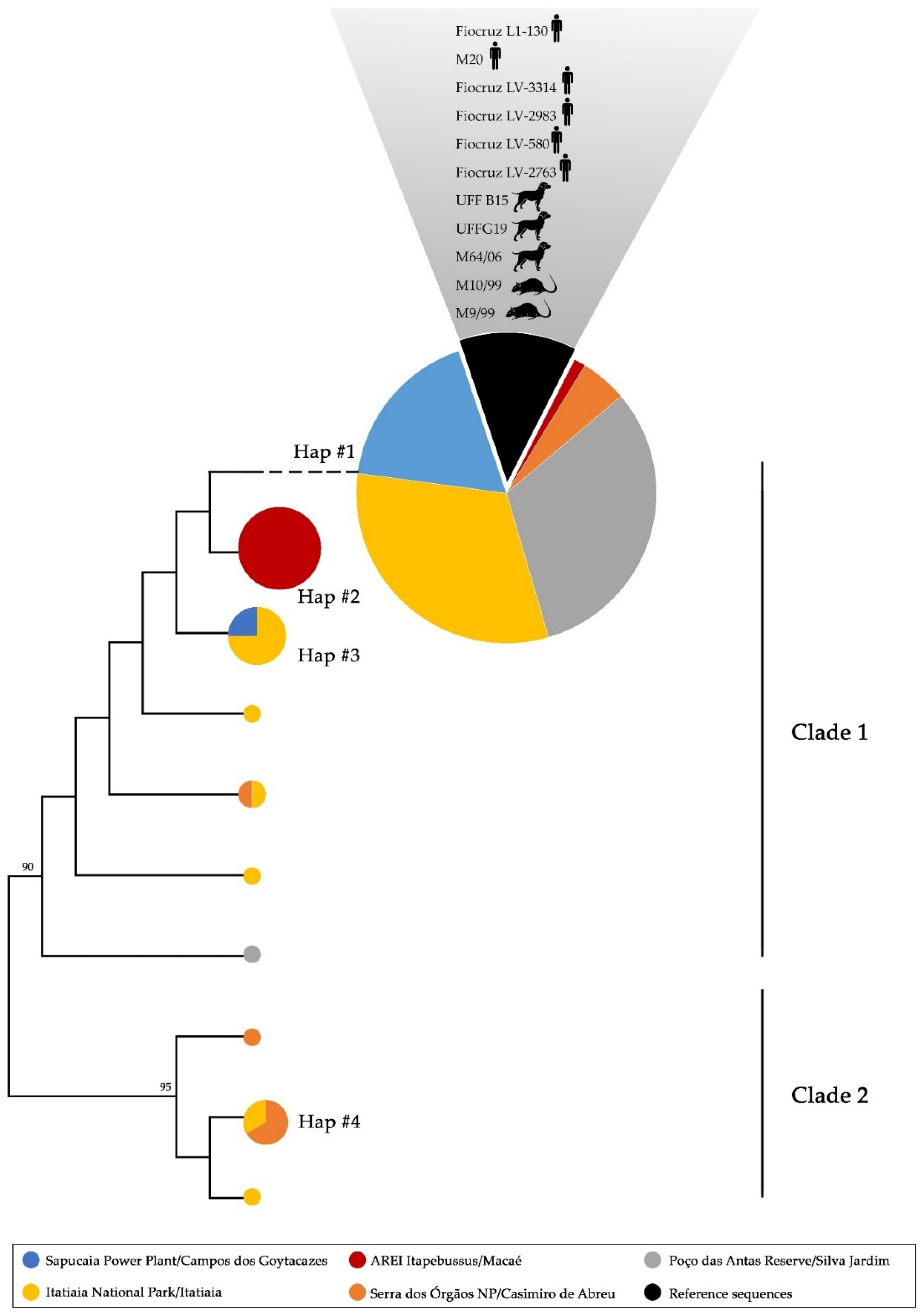
3.3. Phylogenetic Analyses and Haplotype Distribution
The Tamura Nei model (TN92) was the best DNA substitution model determined. Based on this, a high genetic identity was found among the sequences from the present study (TN92 = 0.01 ± 0.01; 99% identity). The phylogenetic analyses and haplotype network demonstrated the presence of four main haplogroups, Hap#1 (n = 68), Hap#2 (n = 9), Hap#3 (n = 5), and Hap#4 (n = 4), included in two main clades, with Hap#1, Hap#2, and Hap#3 included in clade 1 and Hap#4 included in clade 2 (Figure 2). This clade included only the sequences from Serra dos Órgãos National Park and Itatiaia National Park, while clade 1 included sequences from all studied sites. Hap#2 was formed exclusively by sequences from AREI Itapebussus. Seven haplotypes were identified as unique, not forming haplogroups (Table 1, Figure 2). There were no specific haplogroups formed according to hosts (Figure 2).
Figure 2.
Maximum likelihood (ML) phylogenetic tree and haplogroup distribution inferred from partial secY sequences of L. interrogans. The area of the circle is proportional to the number of sequences. Colors indicate the place of origin of the sequence (legend). Reference sequences obtained from GenBank were also included, identified in hosts (identified by a vector) and also from the Atlantic Forest (represented in black). AREI: Area of Relevant Ecological Interest; NP: National Park.
Hap#1 included the vast majority of sequences in the present study, including those from rodents and marsupials from the five regions studied. Furthermore, they presented 100% identity with sequences identified in humans, dogs, and urban rats, all from the Icterohaemorrhagiae serogroup from the same geographical area (Figure 2).
4. Discussion
Herein we performed, for the first time, the genetic characterization of pathogenic leptospires identified in 15 species of small non-flying wild mammals from conservation units of the Brazilian Atlantic Forest. A high occurrence was found (56%), which agrees with studies carried out in the same region [21,22]. Another study conducted in small wild rodents from the southeast Brazilian Atlantic Forest, but in rural areas, revealed a lower prevalence (21.3%) and host diversity (five species) based on qPCR diagnosis [30]. This is not surprising, since conservation areas often support greater diversity and abundance of reservoir hosts, greater environmental persistence of the bacteria, low human impact and less use of control measures, greater interactions between species and trophic networks, and less competition with synanthropic species [31]. A study also conducted in conservation areas of the Brazilian Amazon biome revealed a high prevalence of leptospiral DNA in small mammals (44.7%) from 16 different species [32], reinforcing that conservation areas, with their rich biodiversity, can harbor multiple natural reservoirs for Leptospira.
Most of the hosts were from the order Rodentia (n = 83), with emphasis on the genera Akodon (n = 23) and Mus (n = 21). The genus Akodon, which includes neotropical wild rodents, has been identified as a major carrier of pathogenic Leptospira in the Atlantic Forest region of Brazil. Studies have shown that a substantial proportion of Akodon spp. are PCR-positive for pathogenic Leptospira, highlighting their role as reservoirs in this environment [21,22]. This suggests that Akodon rodents contribute to the maintenance and transmission of leptospires in areas where they are prevalent, although their impact may be more pronounced in rural or forested areas rather than urban settings like Rio de Janeiro. Studies on Akodon and other sigmodontine rodents in other South American regions, such as the Paraná Delta in Argentina, further support their involvement in the transmission of Leptospira. The species Akodon azarae was identified as a host for Leptospira interrogans serovar Icterohaemorrhagiae, suggesting that these rodents can carry and potentially transmit zoonotic Leptospira strains across their geographic range, which includes parts of Brazil [33]. In contrast, the role of Mus musculus in transmitting Leptospira spp. varies by region. While they can be relevant carriers in certain areas, such as urban settlements in Nairobi, Kenya [34], and rural settings in Puerto Rico [35], their role seems to be less pronounced in urban areas in Brazil, like in Rio de Janeiro, where Rattus species are the major reservoirs [36]. Other rodents identified in the present study included Oligoryzomys nigrips, Trinomys marmosa, Brucepattersonius sp., Delomys altimontanus, Delomys dorsalis, Oxymycterus sp., and Necromys lasiurus, previously identified as hosts of Leptospira in the region [21,22,30].
Regarding the marsupials, Didelphis aurita, Monodelphis sp., and Marmosa paraguayana were herein identified as hosts of L. interrogans. Other studies have investigated marsupials, particularly opossums, as hosts of Leptospira spp. In a study performed in Southern Brazil, the white-eared opossum (Didelphis albiventris) was identified as a reservoir of pathogenic Leptospira spp. [37]. Another study examined a broader range of wildlife in Brazil, including opossums, and identified L. santarosai as the prevalent species, with a higher risk of infection observed in adult and male opossums when compared to their younger and female counterparts [19]. Additionally, a study conducted in Rio Grande do Sul, Brazil, detected pathogenic Leptospira spp. DNA in various wild mammals, including the white-eared opossum. The species L. borgpetersenii and L. interrogans were identified [38]. These studies collectively highlight the significance of marsupials, particularly opossums, as hosts of Leptospira in Brazil, with potential contributions to the maintenance of the bacterial transmission cycle.
Regarding the phylogenetic analyses based on the SLST method applied, it was possible to observe a high genetic identity between the sequences from the present study (TN92 = 0.01 ± 0.01; 99% identity) (Figure 2). This can be explained by the geographic proximity between the studied areas, which present shared environmental features such as similar climatic and ecological conditions (e.g., temperature, humidity, and water bodies). Moreover, some Leptospira spp. strains exhibit genetic stability over time, especially if they are well adapted to their host and/or environment [39], which appears to be the case for rodent and marsupial hosts in this region of the Atlantic Forest. Limited selective pressure in certain environments may reduce the rate of genetic divergence [40]. On the other hand, it is important to note that, while in the Atlantic Forest, a low genetic diversity was observed among Leptospira spp., a study conducted in the Western Amazon Forest identified three different species, with high intraspecific genetic variation [32]. This greater genetic diversity of Leptospira in the Amazon region is probably related to the high biodiversity of hosts and less fragmentation of the habitat. The Atlantic Forest, with its high fragmentation and smaller number of reservoir species, may be subject to a genetic bottleneck effect, limiting the diversity of bacterial lineages [41].
Regarding intraspecific analysis, our results showed the presence of four main haplogroups, not related to hosts or geographic locations, except for Hap#2, which was formed exclusively by sequences from AREI Itapebussus (Figure 2). Importantly, Hap#1 included the largest number of sequences in the present study (n = 68) that also comprised L. interrogans reference sequences from humans, dogs, and urban rats from the Icterohaemorrhagiae serogroup. It is well known that the clonal subpopulation Fiocruz L1-130 of L. interrogans serogroup Icterohaemorrhagiae is widely distributed in Brazil and Latin America in different hosts with a major zoonotic role [42,43]. The data from the present study demonstrate that small wild mammals from the Atlantic Forest are also reservoirs of this strain, which seems to be ubiquitarian in that biome. This outcome raises an important public health concern: L. interrogans from the Icterohaemorrhagiae serogroup is the major agent of leptospirosis in humans [8], and rodents in forested areas, particularly in regions like the Atlantic Forest, can act as a direct or indirect link to human infection.
The Atlantic Forest is one of the most biodiverse biomes in the world but is facing severe degradation due to urbanization, agriculture, and logging, with much of its original cover already destroyed [44]. It is the most deforested forest in Brazil, and currently, only 12.4% of the original forest remains [45]. This degradation has a direct impact on local ecology and public health, especially on the transmission of zoonotic diseases such as leptospirosis, which may occur due to different factors: (i) habitat fragmentation and increased human–wildlife interaction; (ii) disruption of ecosystem balance (disturbed trophic interactions and bacterial persistence in altered environments); (iii) increased exposure to contaminated water sources; (iv) changes in biodiversity that may lead to shift in host species; and (v) climate change, which can promote favorable conditions for the spread of the bacteria [46].
5. Conclusions
The present study revealed a high genetic identity among pathogenic leptospires circulating in small non-flying mammals from the southeastern Atlantic Forest biome, highlighting the presence of a larger haplogroup that includes a highly virulent strain also detected in humans, dogs, and urban rats (L. interrogans serogroup Icterohaemorrhagiae, strain Fiocruz L1-130), and evidenced the zoonotic potential of the identified leptospires. Our results reinforce the role of small mammals, such as rodents and marsupials, as important carriers of Leptospira interrogans and draw attention to the Atlantic Forest as an important environment for the circulation and dissemination of the spirochaete. Detecting virulent strains in animals of different species that inhabit forests and peri-urban areas warns of the imminent risk to public health, especially in regions where interactions between humans and wildlife are frequent. This study highlights the urgent need for epidemiological surveillance and control actions in southeast Brazil, aiming to mitigate the transmission of leptospirosis and prevent future outbreaks, especially in highly populated areas from the Atlantic Forest submitted to intense fragmentation and anthropic actions. In this context, a One Health approach is fundamental, integrating human, animal, and environmental health to effectively manage and mitigate the impact of zoonotic diseases such as leptospirosis.
Supplementary Materials
The following supporting information can be downloaded at https://www.mdpi.com/article/10.3390/tropicalmed10030062/s1, Supplementary Table S1: Complementary information regarding the origin and hosts of pathogenic leptospires from the Brazilian Atlantic Forest genetically characterized in the present study based on the secY gene.
Author Contributions
Conceptualization, M.I.N.D.A. and W.L.; Methodology, A.C.d.R.S., C.E. and A.S.V.; Software, M.I.N.D.A.; Validation, M.I.N.D.A., F.A.C.-C., A.S.V. and W.L.; Formal analysis, M.I.N.D.A., A.C.d.R.S., C.E. and A.S.V.; Investigation, C.E., F.A.C.-C. and W.L.; Resources, F.A.C.-C. and W.L.; Data curation, M.I.N.D.A., A.C.d.R.S. and A.S.V.; Writing—original draft, M.I.N.D.A., A.C.d.R.S. and C.E.; Writing—review & editing, M.I.N.D.A., F.A.C.-C., A.S.V. and W.L.; Visualization, A.S.V.; Supervision, W.L.; Project administration, W.L.; Funding acquisition, F.A.C.-C. and W.L. All authors have read and agreed to the published version of the manuscript.
Funding
W.L. is a CNPq and FAPERJ fellow.
Institutional Review Board Statement
All experimental procedures were approved by the Ethics Committee on Animal Use of the Oswaldo Cruz Institute protocol number P-70/13-2, license number LW-39/14.
Informed Consent Statement
Not applicable.
Data Availability Statement
The data that support the findings of this study are openly available in GenBank under accession numbers PQ999001- PQ999093.
Acknowledgments
We would like to immensely thank Paulo Sérgio D’Andrea and his team from Laboratório de Biologia e Parasitologia de Mamíferos Silvestres Reservatórios/Fundação Oswaldo Cruz for their efforts in animal capture and licenses obtained to carry out the pilot project. We also thank the Program for Technological Development in Tools for Health—PDTIS/FIOCRUZ—for use of its facilities.
Conflicts of Interest
The authors declare no conflict of interest.
References
- Karpagam, K.B.; Ganesh, B. Leptospirosis: A neglected tropical zoonotic infection of public health importance—An updated review. Eur. J. Clin. Microbiol. Infect. Dis. 2020, 39, 835–846. [Google Scholar] [CrossRef] [PubMed]
- Barragan, V.; Olivas, S.; Keim, P.; Pearson, T. Critical knowledge gaps in our understanding of environmental cycling and transmission of Leptospira spp. Appl. Environ. Microbiol. 2017, 83, e01190-17. [Google Scholar] [CrossRef]
- Lehmann, J.S.; Matthias, M.A.; Vinetz, J.M.; Fouts, D.E. Leptospiral pathogenomics. Pathogens 2014, 3, 280–308. [Google Scholar] [CrossRef]
- Putz, E.J.; Nally, J.E. Investigating the immunological and biological equilibrium of reservoir hosts and pathogenic Leptospira: Balancing the solution to an acute problem? Front. Microbiol. 2020, 11, 2005. [Google Scholar] [CrossRef]
- Adler, B.; de la Peña Moctezuma, A. Leptospira and leptospirosis. Vet. Microbiol. 2010, 140, 287–296. [Google Scholar] [CrossRef] [PubMed]
- Schulze, M.H.; Raschel, H.; Langen, H.-J.; Stich, A.; Tappe, D. Severe Leptospira interrogans serovar Icterohaemorrhagiae infection with hepato-renal-pulmonary involvement treated with corticosteroids. Clin. Case Rep. 2014, 2, 191–196. [Google Scholar] [CrossRef] [PubMed]
- Wang, S.; Dunn, N. Leptospirosis. In StatPearls; StatPearls Publishing: Treasure Island, FL, USA, 2025. [Google Scholar]
- Haake, D.A.; Levett, P.N. Leptospirosis in humans. Curr. Top. Microbiol. Immunol. 2015, 387, 65–97. [Google Scholar] [CrossRef] [PubMed]
- Leptospirosis|CDC Yellow Book. 2024. Available online: https://wwwnc.cdc.gov/travel/yellowbook/2024/infections-diseases/leptospirosis (accessed on 30 January 2025).
- Soni, N.; Eyre, M.T.; Souza, F.N.; Diggle, P.J.; Ko, A.I.; Begon, M.; Pickup, R.; Childs, J.E.; Khalil, H.; Carvalho-Pereira, T.S.A.; et al. Disentangling the influence of reservoir abundance and pathogen shedding on zoonotic spillover of the Leptospira agent in urban informal settlements. Front. Public Health 2024, 12, 1447592. [Google Scholar] [CrossRef]
- Colombo, A.F.; Joly, C.A. Brazilian Atlantic Forest lato sensu: The most ancient Brazilian forest, and a biodiversity hotspot, is highly threatened by climate change. Braz. J. Biol. 2010, 70, 697–708. [Google Scholar] [CrossRef]
- Correia, L.; Loureiro, A.P.; Lilenbaum, W. Effects of rainfall on incidental and host-maintained leptospiral infections in cattle in a tropical region. Vet. J. 2017, 220, 63–64. [Google Scholar] [CrossRef] [PubMed]
- IBGE-Censo Brasileiro de 2022. Available online: https://www.ibge.gov.br/estatisticas/sociais/trabalho/22827-censo-demografico-2022.html (accessed on 5 January 2025).
- Galan, D.I.; Roess, A.A.; Pereira, S.V.C.; Schneider, M.C. Epidemiology of human leptospirosis in urban and rural areas of Brazil, 2000–2015. PLoS ONE 2021, 16, e0247763. [Google Scholar] [CrossRef] [PubMed]
- Baquero, O.S.; Machado, G. Spatiotemporal dynamics and risk factors for human leptospirosis in Brazil. Sci. Rep. 2018, 8, 15170. [Google Scholar] [CrossRef] [PubMed]
- Santa Rosa, C.A.; Sulzer, C.R.; Giorgi, W.; da Silva, A.S.; Yanaguita, R.M.; Lobao, A.O. Leptospirosis in wildlife in Brazil: Isolation of a new serotype in the Pyrogenes group. Am. J. Vet. Res. 1975, 36, 1363–1365. [Google Scholar] [PubMed]
- Cordeiro, F.; Sulzer, C.R.; Ramos, A.A. Leptospira interrogans in several wildlife species in Southeast Brazil. Rev. Bras. Pesq. Vet. 1981, 1, 1–29. [Google Scholar]
- Medici, E.P.; Mangini, P.R.; Fernandes-Santos, R.C. Health assessment of wild Lowland Tapir (Tapirus terrestris) populations in the Atlantic Forest and Pantanal biomes, Brazil (1996–2012). J. Wildl. Dis. 2014, 50, 817–828. [Google Scholar] [CrossRef]
- Fornazari, F.; Langoni, H.; Marson, P.M.; Nóbrega, D.B.; Teixeira, C.R. Leptospira reservoirs among wildlife in Brazil: Beyond rodents. Acta Trop. 2018, 178, 205–212. [Google Scholar] [CrossRef] [PubMed]
- Libonati, H.; Pinto, P.S.; Lilenbaum, W. Seronegativity of bovines face to their own recovered leptospiral isolates. Microb. Pathog. 2017, 108, 101–103. [Google Scholar] [CrossRef]
- Vieira, A.S.; Di Azevedo, M.I.N.; D’Andrea, P.S.; do Val Vilela, R.; Lilenbaum, W. Neotropical wild rodents Akodon and Oligoryzomys (Cricetidae: Sigmodontinae) as important carriers of pathogenic renal Leptospira in the Atlantic Forest, in Brazil. Res. Vet. Sci. 2019, 124, 280–283. [Google Scholar] [CrossRef]
- Vieira, A.S.; D’Andrea, P.S.; Vilela, R.d.V.; Loretto, D.; Jaeger, L.H.; Carvalho-Costa, F.A.; Lilenbaum, W. Pathogenic Leptospira species are widely disseminated among small mammals in Atlantic Forest Biome. Transbound. Emerg. Dis. 2019, 66, 1195–1201. [Google Scholar] [CrossRef] [PubMed]
- Hamond, C.; Martins, G.; Loureiro, A.P.; Pestana, C.; Lawson-Ferreira, R.; Medeiros, M.A.; Lilenbaum, W. Urinary PCR as an increasingly useful tool for an accurate diagnosis of leptospirosis in livestock. Vet. Res. Commun. 2014, 38, 81–85. [Google Scholar] [CrossRef]
- Lam, J.-Y.; Low, G.K.-K.; Chee, H.-Y. Diagnostic accuracy of genetic markers and nucleic acid techniques for the detection of Leptospira in clinical samples: A meta-analysis. PLoS Negl. Trop. Dis. 2020, 14, e0008074. [Google Scholar] [CrossRef] [PubMed]
- Grillová, L.; Angermeier, H.; Levy, M.; Giard, M.; Lastère, S.; Picardeau, M. Circulating genotypes of Leptospira in French Polynesia: An 9-Year molecular epidemiology surveillance follow-up study. PLoS Negl. Trop. Dis. 2020, 14, e0008662. [Google Scholar] [CrossRef] [PubMed]
- Larkin, M.A.; Blackshields, G.; Brown, N.P.; Chenna, R.; McGettigan, P.A.; McWilliam, H.; Valentin, F.; Wallace, I.M.; Wilm, A.; Lopez, R.; et al. Clustal W and Clustal X version 2.0. Bioinformatics 2007, 23, 2947–2948. [Google Scholar] [CrossRef] [PubMed]
- Kumar, S.; Stecher, G.; Li, M.; Knyaz, C.; Tamura, K. MEGA X: Molecular evolutionary genetics analysis across computing platforms. Mol. Biol. Evol. 2018, 35, 1547–1549. [Google Scholar] [CrossRef] [PubMed]
- Xia, X.; Xie, Z. DAMBE: Software package for data analysis in molecular biology and evolution. J. Hered. 2001, 92, 371–373. [Google Scholar] [CrossRef] [PubMed]
- Leigh, J.W.; Bryant, D. Monte Carlo strategies for selecting parameter values in simulation experiments. Syst. Biol. 2015, 64, 741–751. [Google Scholar] [CrossRef] [PubMed]
- da Silva, E.C.; Fornazari, F.; de Paula Antunes, J.M.A.; de Castro Demoner, L.; de Oliveira, L.H.O.; Peres, M.G.; Megid, J.; Langoni, H. Molecular detection of Leptospira spp. in small wild rodents from rural areas of São Paulo State, Brazil. Rev. Soc. Bras. Med. Trop. 2023, 56, e0160-2023. [Google Scholar] [CrossRef] [PubMed]
- Bradley, E.A.; Lockaby, G. Leptospirosis and the environment: A review and future directions. Pathogens 2023, 12, 1167. [Google Scholar] [CrossRef]
- Medeiros, L.D.S.; Braga Domingos, S.C.; di Azevedo, M.I.N.; Peruquetti, R.C.; de Albuquerque, N.F.; D’Andrea, P.S.; Botelho, A.L.d.M.; Crisóstomo, C.F.; Vieira, A.S.; Martins, G.; et al. Small mammals as carriers/hosts of Leptospira spp. in the Western Amazon Forest. Front. Vet. Sci. 2020, 7, 569004. [Google Scholar] [CrossRef]
- Colombo, V.C.; Gamietea, I.; Loffler, S.G.; Brihuega, B.F.; Beldomenico, P.M. New host species for Leptospira borgpetersenii and Leptospira interrogans serovar Copenhageni. Vet. Microbiol. 2018, 215, 90–92. [Google Scholar] [CrossRef]
- Halliday, J.E.B.; Knobel, D.L.; Allan, K.J.; Bronsvoort, B.M.d.C.; Handel, I.; Agwanda, B.; Cutler, S.J.; Olack, B.; Ahmed, A.; Hartskeerl, R.A.; et al. Urban leptospirosis in Africa: A cross-sectional survey of Leptospira infection in rodents in the Kibera Urban Settlement, Nairobi, Kenya. Am. J. Trop. Med. Hyg. 2013, 89, 1095–1102. [Google Scholar] [CrossRef] [PubMed]
- Benavidez, K.M.; Guerra, T.; Torres, M.; Rodriguez, D.; Veech, J.A.; Hahn, D.; Miller, R.J.; Soltero, F.V.; Ramírez, A.E.P.; Perez de León, A.; et al. The prevalence of Leptospira among invasive small mammals on Puerto Rican cattle farms. PLoS Negl. Trop. Dis. 2019, 13, e0007236. [Google Scholar] [CrossRef] [PubMed]
- Costa, F.; Ribeiro, G.S.; Felzemburgh, R.D.M.; Santos, N.; Reis, R.B.; Santos, A.C.; Fraga, D.B.M.; Araujo, W.N.; Santana, C.; Childs, J.E.; et al. Influence of household rat infestation on Leptospira transmission in the urban slum environment. PLoS Negl. Trop. Dis. 2014, 8, e3338. [Google Scholar] [CrossRef] [PubMed]
- Jorge, S.; Hartleben, C.P.; Seixas, F.K.; Coimbra, M.A.A.; Stark, C.B.; Larrondo, A.G.; Amaral, M.G.; Albano, A.P.N.; Minello, L.F.; Dellagostin, O.A.; et al. Leptospira borgpetersenii from free-living white-eared opossum (Didelphis albiventris): First isolation in Brazil. Acta Trop. 2012, 124, 147–151. [Google Scholar] [CrossRef]
- Ulsenheimer, B.C.; Tonin, A.A.; von Laer, A.E.; Dos Santos, H.F.; Sangioni, L.A.; Fighera, R.; Dos Santos, M.Y.; Brayer, D.I.; de Avila Botton, S. Leptospira borgptersenii and Leptospira interrogans identified in wild mammals in Rio Grande Do Sul, Brazil. Braz. J. Microbiol. 2024, 55, 1941–1948. [Google Scholar] [CrossRef] [PubMed]
- Xu, Y.; Zhu, Y.; Wang, Y.; Chang, Y.-F.; Zhang, Y.; Jiang, X.; Zhuang, X.; Zhu, Y.; Zhang, J.; Zeng, L.; et al. Whole genome sequencing revealed host adaptation-focused genomic plasticity of pathogenic Leptospira. Sci. Rep. 2016, 6, 20020. [Google Scholar] [CrossRef]
- Croix, M.D.S.; Holmes, J.; Wanford, J.J.; Moxon, E.R.; Oggioni, M.R.; Bayliss, C.D. Selective and non-selective bottlenecks as drivers of the evolution of hypermutable bacterial loci. Mol. Microbiol. 2020, 113, 672–681. [Google Scholar] [CrossRef] [PubMed]
- Wein, T.; Dagan, T. The effect of population bottleneck size and selective regime on genetic diversity and evolvability in bacteria. Genome Biol. Evol. 2019, 11, 3283–3290. [Google Scholar] [CrossRef]
- Jaeger, L.H.; Pestana, C.P.; Carvalho-Costa, F.A.; Medeiros, M.A.; Lilenbaum, W. Characterization of the clonal subpopulation Fiocruz L1–130 of Leptospira interrogans in rats and dogs from Brazil. J. Med. Microbiol. 2018, 67, 1361–1367. [Google Scholar] [CrossRef] [PubMed]
- Di Azevedo, M.I.N.; Aymée, L.; Borges, A.L.D.S.B.; Lilenbaum, W. Molecular epidemiology of pathogenic Leptospira spp. infecting dogs in Latin America. Animals 2023, 13, 2422. [Google Scholar] [CrossRef] [PubMed]
- Wagner, F.H.; Sanchez, A.; Aidar, M.P.M.; Rochelle, A.L.C.; Tarabalka, Y.; Fonseca, M.G.; Phillips, O.L.; Gloor, E.; Aragão, L.E.O.C. Mapping Atlantic Rainforest degradation and regeneration history with indicator species using convolutional network. PLoS ONE 2020, 15, e0229448. [Google Scholar] [CrossRef] [PubMed]
- Guedes Pinto, L.F.; Voivodic, M. Reverse the tipping point of the Atlantic Forest for mitigation. Nat. Clim. Change 2021, 11, 364–365. [Google Scholar] [CrossRef]
- Pfenning-Butterworth, A.; Buckley, L.B.; Drake, J.M.; Farner, J.E.; Farrell, M.J.; Gehman, A.-L.M.; Mordecai, E.A.; Stephens, P.R.; Gittleman, J.L.; Davies, T.J. Interconnecting global threats: Climate change, biodiversity loss, and infectious diseases. Lancet Planet. Health 2024, 8, e270–e283. [Google Scholar] [CrossRef] [PubMed]
Disclaimer/Publisher’s Note: The statements, opinions and data contained in all publications are solely those of the individual author(s) and contributor(s) and not of MDPI and/or the editor(s). MDPI and/or the editor(s) disclaim responsibility for any injury to people or property resulting from any ideas, methods, instructions or products referred to in the content. |
© 2025 by the authors. Licensee MDPI, Basel, Switzerland. This article is an open access article distributed under the terms and conditions of the Creative Commons Attribution (CC BY) license (https://creativecommons.org/licenses/by/4.0/).