Review

Non-Coding RNAs as Emerging Biomarkers in Leishmaniasis and Chagas Disease

by
Eduardo Ramos Juárez
1,†,
Eduardo Pérez-Campos Mayoral
2,†,
Laura Pérez-Campos Mayoral
2,
Adriana Moreno Rodríguez
3,
Carlos Romero-Díaz
1,
Miriam Emily Avendaño-Villegas
1,
Tania Sinaí Santiago Ramírez
4,
Margarito Martínez Cruz
1,
José Luis Hernández-Morales
2,
Lilian Guadalupe Bolaños-Hilario
1,
Iam Kevin Suárez Luna
2,
Jesús Elizarrarás-Rivas
5,6,7,
Aldo Abel García González
2,
Hector Alejandro Cabrera-Fuentes
2,8,9,10,
María Teresa Hernández-Huerta
2,11,*,‡ and
Eduardo Pérez-Campos
1,*,‡
1
División de Estudios de Posgrado e Investigación, Tecnológico Nacional de Mexico/Instituto Tecnológico de Oaxaca, Oaxaca 68030, Mexico
2
Centro de Investigación Facultad de Medicina UNAM-UABJO, Facultad de Medicina y Cirugía, Universidad Autónoma “Benito Juárez” de Oaxaca, Oaxaca 68020, Mexico
3
Laboratorio de Estudios Epidemiológicos, Clínicos, Diseños Experimentales e Investigación, Facultad de Ciencias Químicas, Universidad Autónoma “Benito Juárez” de Oaxaca, Oaxaca 68120, Mexico
4
Facultad de Medicina y Cirugía, Benemérita Universidad de Oaxaca, Oaxaca 68020, Mexico
5
Coordinación de Investigación en Salud, IMSS, Oaxaca 68040, Mexico
6
Hospital General de Zona No.1 Dr. Demetrio Mayoral Pardo, IMSS, Oaxaca 68040, Mexico
7
Facultad de Medicina y Cirugía, Universidad Autónoma “Benito Juárez” de Oaxaca, Oaxaca 68020, Mexico
8
División de Estudios de Posgrado e Investigación, Tecnológico Nacional de Mexico/Instituto Tecnológico de Tijuana, Tijuana 22414, Mexico
9
R&D Group, Vice Presidency Scientific Research & Innovation, Imam Abdulrahman Bin Faisal University (IAU), Dammam 31441, P.O. Box. 1982, Saudi Arabia
10
Dirección de la División de Investigación y Desarrollo Científico, Benemérita Universidad de Oaxaca, Oaxaca 68000, Mexico
11
SECIHTI, Facultad de Medicina y Cirugía, Universidad Autónoma “Benito Juárez” de Oaxaca, Oaxaca 68020, Mexico
*
Authors to whom correspondence should be addressed.
These authors contributed equally to this work.
Trop. Med. Infect. Dis. 2025, 10(11), 319; https://doi.org/10.3390/tropicalmed10110319
Submission received: 14 September 2025 / Revised: 1 November 2025 / Accepted: 9 November 2025 / Published: 13 November 2025
(This article belongs to the Section Neglected and Emerging Tropical Diseases)

Abstract

Leishmaniasis and Chagas disease, caused by Leishmania spp. and Trypanosoma cruzi, are neglected tropical diseases with significant global health burden, particularly in resource-limited regions. Despite their impact, diagnosis and treatment remain challenging due to limited diagnostic tools and the toxicity of available therapies. Our objective is to propose the incorporation of markers for the diagnosis of leishmaniasis and Chagas disease using ncRNA. This narrative review evaluates studies published between 2010 and 2024 (PubMed, Scopus, Google Scholar) using the SANRA scale to assess the potential of non-coding RNAs (ncRNAs) as biomarkers for these infections. Both parasites release small RNAs via extracellular vesicles that modulate host–pathogen interactions and gene expression. Although RNA interference machinery is absent in T. cruzi and most Leishmania species, it persists in early-diverging lineages. In leishmaniasis, distinct miRNA expression profiles—including miR-155-5p, miR-5011-5p, miR-6785-5p, and miR-361-3p—demonstrate high diagnostic accuracy for detecting infection (AUC up to 1.0). Serum long ncRNAs such as MALAT1 and NUTM2A-AS1 show potential diagnostic value, though clinical validation remains pending. For Chagas disease, the available evidence on ncRNAs primarily addresses the diagnosis of clinical manifestations rather than initial infection. Host miRNAs, including miR-21, miR-145, miR-146a/b, and miR-19a-3p, correlate with cardiac involvement, immune dysregulation, and inflammation during chronic T. cruzi infection. Circulating miRNAs exhibit modest sensitivity (57–67%) and specificity (57–80%) for diagnosing chronic Chagas cardiomyopathy, indicating their utility in assessing disease progression and organ damage rather than detecting early infection. This review distinguishes between ncRNAs that diagnose infection and those that evaluate disease severity or organ involvement. Altered ncRNA expression profiles represent promising biomarkers for species differentiation, treatment monitoring, and assessing cardiac complications in Chagas disease, with broader diagnostic applications emerging for leishmaniasis.

1. Introduction

Leishmaniasis and American trypanosomiasis are diseases caused by protozoan parasites and are considered neglected tropical diseases. The etiological agent of leishmaniasis is a different species of the genus Leishmania, whereas for Trypanosomiasis, also known as Chagas disease, it is the parasite Trypanosoma cruzi (T. cruzi). According to the World Health Organization (WHO), between 700,000 and 1 million new cases of leishmaniasis are reported annually, with an estimated 12 million infected individuals worldwide. In comparison, Chagas disease affects between 6 and 8 million people, causing 12,000 deaths per year [1].
In Mexico, leishmaniasis, also known as “gum ulcer,” was first recorded in 1912 among individuals who collected chewing gum from the Manilkara zapota tree. The first cases of Chagas disease were reported in 1940 in two patients from Oaxaca [2].
Globally, leishmaniasis and Chagas disease mainly affect developing countries, with a greater impact in rural areas and tropical and subtropical regions. In Africa, Asia, and Latin America, cutaneous and visceral leishmaniasis continues to be a significant public health problem, with many cases undiagnosed or poorly treated. For example, in the Amazon region, Brazil is one of the countries with the highest burden of visceral leishmaniasis, while India, Pakistan, and Afghanistan have high rates of cutaneous leishmaniasis [1].
Chagas disease remains endemic in Latin America, particularly in Brazil, Bolivia, Argentina, and Mexico, where prevalence is highest. Migration patterns have introduced the disease to non-endemic regions, including the United States, Europe, Africa, and Asia. This geographic expansion has transformed Chagas disease from a regional problem into a global health threat, requiring international surveillance and control strategies [3].
The transmission of these diseases can occur through the bite of vector insects (such as bed bugs and sandflies), via blood transfusion, or organ transplant. In the case of Trypanosomiasis, it can also be spread by ingesting food contaminated with parasites. Among the strategies for reducing cases are prevention strategies, which include informing the population about the disease and its vectors, as well as screening blood banks to prevent the use of contaminated blood [4].
Leishmaniasis manifests with diverse clinical symptoms depending on the causative parasite. Cutaneous leishmaniasis presents skin lesions, including ulcers, smooth nodules, and wart-like lesions. In contrast, visceral leishmaniasis primarily presents symptoms such as fever, weight loss, anemia, abdominal distension, splenomegaly, and hepatomegaly [3]. Chagas disease progresses through acute and chronic phases. The acute phase can manifest as chagoma of inoculation, fever, lymphadenopathy, anemia, musculoskeletal pain, heart failure, and meningoencephalitis. The chronic phase typically involves cardiac abnormalities, including heart failure, arrhythmias, and ventricular aneurysms, alongside gastrointestinal issues like megaesophagus and megacolon [4].
For the diagnosis of leishmaniasis, biopsies and cultures are performed on the spleen, bone marrow, liver, lymph nodes, and skin; the latter is a widely used option in cases of cutaneous leishmaniasis. In addition, serological tests such as indirect agglutination and indirect immunofluorescent antibody tests, as well as polymerase chain reaction (PCR) tests for Leishmania, and direct tests for detecting the parasite in blood by microscopy (the fat drop test), are also used [5].
While parasitological methods in blood can detect Trypanosoma cruzi in the acute phase, in the chronic phase, the diagnosis depends on clinical evaluation, serological tests, such as enzyme-linked immunosorbent (ELISA) assays, indirect immunofluorescence, or indirect hemagglutination, and epidemiological history [6,7,8]. In recent years, nucleic acid amplification techniques, such as real-time PCR, have been implemented in some countries, particularly for the early diagnosis of congenital Chagas disease in infants, as well as for treatment monitoring in the context of clinical trials [9,10].
Detection methods for these diseases do not define the progression of infection in patients; however, the use of biomarkers can serve as a more accurate detection method, offering an advantage in the treatment of these diseases by detecting infections during their early stages and determining the status of the infection.
Non-coding RNAs (ncRNAs) have emerged as potential biomarkers for leishmaniasis and Chagas disease, although the available evidence primarily addresses different diagnostic applications. For leishmaniasis, ncRNAs show promise in detecting active infection, while for Chagas disease, current research focuses mainly on identifying cardiac involvement during the chronic phase rather than initial infection. This review evaluates the diagnostic potential of ncRNAs—including microRNAs (miRNAs), long non-coding RNAs (lncRNAs), PIWI-interacting RNAs (piRNAs), short interfering RNAs (siRNAs), and tRNA-derived small RNAs (tRFs)—as biomarkers for infections caused by Leishmania spp. and T. cruzi. We distinguish between ncRNAs that detect infection and those that assess disease progression or organ involvement, particularly cardiac manifestations in chronic Chagas disease. A comprehensive literature search was conducted across PubMed, Scopus, and Google Scholar databases. The search strategy incorporated a combination of keywords and Medical Subject Headings (MeSH) terms, including “Leishmania,” “Leishmaniasis,” “Trypanosoma cruzi,” “Chagas disease,” “ncRNA,” “non-coding RNA,” “lncRNA,” “miRNA,” “piRNA” and “siRNAs”. Inclusion criteria focused on original research and review articles relevant to the stated objective. The quality of included articles was independently assessed using the SANRA (Scale for the Assessment of Narrative Review Articles) quality assessment tool.

2. Clinical Diagnosis of Leishmaniasis and Trypanosomiasis

Multiple laboratory tests are available to diagnose leishmaniasis and trypanosomiasis, as accurate detection often requires a combination of techniques to maximize the probability of a positive result—such as microscopy, molecular methods, and serology—to enhance sensitivity, specificity, and species identification.
The three primary clinical syndromes caused by infection with Leishmania parasites are cutaneous, mucocutaneous, and visceral (also known as kala-azar) leishmaniasis. The diagnosis of visceral leishmaniasis (VL) relies on a combination of clinical signs and parasitological or serological tests. Serological tests are of limited utility in cases of cutaneous and mucocutaneous leishmaniasis, where the diagnosis is confirmed when parasitological analyses support the clinical manifestations [1].
Moreover, many of the regional recommendations issued by the WHO and Pan American Health Organization (PAHO) are based on observational studies, case series, and expert reports. Therefore, the diagnostic approach to leishmaniasis should be based on the combined application of different laboratory tests. Generally, various methods are used for diagnosing leishmaniasis, including the visualization of characteristic amastigotes in smears or tissue samples, the isolation of the parasite through in vitro culture, and the molecular detection of parasitic DNA. Additionally, for individuals with suspected visceral leishmaniasis, serological tests are used when direct diagnostic techniques (such as microscopy, culture, or molecular tests) cannot be performed or when these yield negative results. However, anti-leishmanial antibodies may persist for years after clinically successful therapy or, conversely, may be undetectable or present at low levels in patients with VL who are immunosuppressed due to Human Immunodeficiency Virus/Acquired Immunodeficiency Syndrome (HIV/AIDS) or other causes. This variability, combined with the potential for false-negative results, limits the diagnostic utility of serological assays in this context, Table 1 [11].
The diagnosis of acute Chagas infections is performed through the identification of trypomastigotes in the blood using direct microscopy. This involves preparing and staining thick and thin blood smears to visualize the parasites. However, blood parasite levels decrease rapidly within a few months, becoming undetectable with parasitological methods during the chronic phase of the disease. For the diagnosis of Chagas disease in the chronic phase, the detection of antibodies against the parasite T. cruzi is required using serological tests. No single test is sufficiently sensitive or specific to confirm the diagnosis; therefore, it is recommended to use two or more serological tests that detect antibodies against different antigens. Standard techniques include the indirect hemagglutination, ELISA, and the indirect immunofluorescence antibody (IFA) test. Additionally, assessing the patient’s clinical and epidemiological history helps identify potential risk factors for infection, Table 2 [14].

3. Characteristics of miRNA, piRNA, and lncRNA

The genome contains both coding RNA (cRNA) and ncRNA. NcRNA is broadly categorized into long non-coding RNA (lncRNA) and small non-coding RNA (sncRNA). The main types of sncRNA include microRNA (miRNA), small interfering RNA (siRNA), PIWI-interacting RNA (piRNA), small nucleolar RNA (snoRNA), small nuclear RNA (snRNA), and tRNA-derived fragments (tRFs) [17,18]. Genome-derived microRNAs (miRNAs) and exogenous small interfering RNAs (siRNAs), the two main types of RNA interference (RNAi) molecules, both integrate into RNA-induced silencing complexes to regulate gene expression after transcription [19].
Multiple types of sncRNA regulate gene expression [20]. There are both differences and similarities between siRNA, miRNA, piRNA, and lncRNA. For example, in their nucleotide length, ssiRNAs are 20 to 25 nts, miRNAs are 21 to 23 nts, piRNAs are 26 to 32 nts, and lncRNAs are >200 nts (Table 1).
The existence of siRNAs was first reported in Caenorhabditis elegans in 1998 by Fire et al. [21]. These siRNAs are derived from double-stranded RNA (dsRNA) through a processing step involving Dicer/DCL enzymes [22]. Once processed, siRNAs play a critical role in guiding Argonaute proteins to mediate the precise cleavage of target RNA molecules. In human cells, siRNAs play a crucial role in gene regulation and cellular defense mechanisms. Furthermore, endogenous siRNAs have been identified in Leishmania species that possess an active RNAi pathway [23]. Despite this, the natural presence of an endogenous RNAi pathway in T. cruzi has not yet been established [24].
miRNAs were first reported in C. elegans by Lee et al. in 1993 [25] and were subsequently identified as small RNAs [26]. Although they constitute only 2–3% of the human genome, miRNAs are estimated to regulate the expression of over 60% of genes [27]. Their biogenesis begins with precursor transcripts processed by the RNase III enzymes Drosha and Dicer in animals, or by Dicel-like (DCL) enzymes in plants [28]. In animals, these precursors are transported from the nucleus by Exportin-5 (Exp5) to be loaded into an Argonaute (AGO) protein complex [29,30]. siRNAs and miRNAs are short-duplex molecules that have silencing functions [31].
miRNAs regulate gene expression at the post-transcriptional level through interactions with specific regions of the target RNA, utilizing the RISC complex machinery, which induces RNA destabilization and degradation [32].
Most mature primary piRNAs have a uridine at the 5’ end and a 2’-OH methyl group at the 3’ end [33]. They are transcribed from piRNA gene clusters, most of which are unidirectional in somatic cells and double-stranded in the germline [34]. Mature piRNAs associate with proteins of the PIWI-like family (MILI, MIWI, and MIWI2) to form the piRNA-induced silencing complex (piRISC) for transposon silencing [35]. In humans, these complexes induce gene silencing through the action of the PIWIL1, PIWIL2, PIWIL3, and PIWIL4 proteins. These RNAs are stable and do not degrade in circulation [36].
LncRNA have been defined as transcripts longer than 200 nucleotides that do not encode proteins [37]. LncRNAs are crucial regulators of gene expression, operating at multiple levels. They can guide chromatin-modifying complexes to specific DNA locations, allowing for epigenetic control of gene activity. Furthermore, they influence gene expression transcriptionally and post-transcriptionally by modulating processes like alternative splicing, mRNA stability, and translation [38]. Their functions extend to fundamental cellular activities, including cell differentiation, cell cycle control, and apoptosis [39].
The expression of lncRNAs is often tissue- and cell-type-specific [40]. Consequently, their dysregulation is implicated in a wide range of human diseases, including various cancers and neurodegenerative disorders, highlighting their potential as both biomarkers and therapeutic targets. While their nucleotide sequences tend to be less conserved across species than protein-coding genes, their structural elements may be evolutionarily preserved [38].

4. ncRNA in Kinetoplastids

On the other hand, T. cruzi utilizes ncRNAs to regulate gene expression, releasing various small RNAs through extracellular vesicles (EVs) [39,40]. This regulatory system is critical in many trypanosomatids due to the lack of a functional RNA interference (RNAi) pathway [40]. Genomic analysis confirmed the absence of critical RNAi genes, such as Dicer-like (DCL) enzymes and Argonaute (AGO), in T. cruzi and “higher” Leishmania species (subgenus Leishmania). This explained the lack of RNAi activity in T. cruzi [41]. The composition and abundance of these small RNAs differ significantly between the parasite’s life stages, suggesting diverse biological functions [39]. Among the different types of Small RNAs are the Ribosomal RNA-derived fragments (rRNA-derived, small RNAs or sdrRNAs). These are the most abundant small RNAs found in T. cruzi EVs, constituting 54–74% of the small RNA content in vesicles from epimastigotes (eVes) and metacyclic trypomastigotes (mVes) [42]. In whole-parasite extracts, they represent about 17% of reads in epimastigotes and 25% in metacyclic forms. Under nutritional stress, sdrRNAs become the most prevalent small RNA class in both EVs (46%) and intracellularly (58%). EVs carrying these sdrRNAs can modulate host cell gene expression and invasion [39]. The transfer RNA-derived fragments (tRNA-derived small RNAs or tsRNAs or tRFs), is the second most common class of small RNA, their abundance varies greatly by stage, making up 26–34% in epimastigotes and their vesicles, but increasing to 63% in metacyclic forms, while being lower in metacyclic-derived vesicles (6%) [42]. In epimastigotes, 89% of tsRNAs are 3’ halves with an average length of 38 nucleotides, and 75% have a CCA extension. The dominance of 5’ or 3’ derived fragments changes depending on the parasite’s life stage [39]. The small nucleolar RNA-derived fragments (snoRNA-derived small RNAs or sdRNAs) are primarily observed in metacyclic trypomastigote parental cells (4%) [42]. They have a length of approximately 35 nucleotides [39]. Other small RNAs, originating from coding sequences (CDS), are more prevalent in metacyclic-derived samples (5%) than in epimastigotes (1%). Additionally, small nuclear RNA-derived fragments (snsRNAs) have been identified, mainly from U4 and U5 snRNAs, with a median length of 40 nucleotides. Other researchers at T. cruzi found approximately 95% to 98% ncRNAs. These vary by haplotype and include small RNAs originating from tRNAs, rRNAs, snRNAs, and snoRNAs, Figure 1 [43].
Leishmania species, including L. donovani and L. braziliensis, utilize ncRNAs for gene expression regulation, with exosomal transfer RNA-derived small RNAs (tsRNAs) being frequently observed, primarily from the 5’ arm of tRNA-Asp and tRNA-Gln. Exosomes also contain small nuclear RNA (snRNA)-derived fragments and a small proportion (approximately 1.5%) of small DNA-derived RNAs (sdRNAs). Small ribosomal RNA (srRNA)-derived fragments constitute a significant portion of exosomal in L. donovani (31%) and L. braziliensis (15%), exhibiting distinct peak size distributions [40]. Unlike Trypanosoma cruzi, Leishmania did not exhibit site-specific enrichment for rRNA-derived fragments, with 90% of the mapped fragments mapping to both the 28S and 18S rRNA genes. While some Leishmania subgenera have lost or possess degenerated RNA interference (RNAi) genes, functional RNAi pathways involving AGO and Dicer orthologs are present in early diverging Leishmania subgenera like L. braziliensis and L. guyanensis [41]. In these species, 20–25 nucleotide siRNAs, mainly derived from SLACS and TATE elements, are found in exosomes, suggesting their role in intercellular communication. Experimental confirmation in L. braziliensis revealed small RNAs of the expected sizes that immunoprecipitated with AGO1, indicating a functional involvement similar to that of siRNAs in T. brucei. However, L. braziliensis lacks HEN1, a methyltransferase found in T. brucei that modifies the 3’ end of siRNAs, resulting in significant 3’ heterogeneity in L. braziliensis siRNAs and suggesting mechanistic diversification of the RNAi pathway within trypanosomatids.
On the other hand, in eukaryotes, non-coding RNAs, encompassing small molecules such as miRNA and piRNA, as well as lncRNA, are now recognized as master regulators in the biology of parasitic infections. They play a pivotal role in controlling parasite development, mediating host–pathogen interactions, and facilitating immune evasion, making them valuable as potential biomarkers and therapeutic targets, for example, in kinetoplastids.
Leishmania and Trypanosoma cruzi, lncRNA are key regulators in parasite development, host interaction, and immune modulation [40,44]. In addition, although specific trypanosomatid-specific PIWI-like proteins, such as TcPIWI-tryp, are known to interact with tsRNAs, their roles diverge from the canonical PIWI/Argonaute interactions seen in classical eukaryotic small RNA pathways [43,45].
Also, siRNA currently has no diagnostic application for Chagas disease or leishmaniasis. It is used in experimental settings to study gene function or in therapeutic studies. There is no evidence of its clinical or experimental use as a direct diagnostic marker for Chagas disease or leishmaniasis, and it was used here only for comparison with other ncRNAs in Table 3.

5. ncRNA as Diagnostic Biomarkers in Leishmaniasis

In parasitic infections, the small non-coding RNAs (sncRNAs) can act as “molecular parasites” that influence host–pathogen interactions [49]. A recent comprehensive study has significantly advanced our understanding of leishmaniasis by characterizing ncRNAs across 26 strains from 16 Leishmania spp. [50]. This analysis revealed conserved ncRNAs shared among different Leishmania spp., which proved valuable in differentiating between subgenera and species associated with visceral leishmaniasis, which will be used for diagnosis or therapeutic interventions.
Infections by Leishmania parasites trigger distinct changes in host miRNA expression, which are being explored for their diagnostic potential [51]. Specific miRNAs, such as miR-155 and miR-146a, are capable of distinguishing between the clinical forms of the disease [52]. Masoudsinaki et al. [53] investigated the differential expression of four specific miRNAs (miR-4795-3p, miR-6785-5p, miR-5011-5p, and miR-155-5p) in skin lesions from cutaneous leishmaniasis (CL) patients infected with L. major and L. tropica. They found a significant upregulation of miR-155-5p, miR-5011-5p, and miR-6785-5p in L. tropica-infected patients when compared to healthy controls. Similar, but more pronounced, expression patterns for miR-155-5p and miR-6785-5p were observed in L. major-infected patients, although miR-4795-3p was notably downregulated in this group. Receiver operating characteristic (ROC) analysis revealed that individual miRNAs exhibited high diagnostic potential; for example, miR-155-5p, miR-5011-5p, and miR-6785-5p effectively distinguished L. tropica-infected patients from healthy controls with high accuracy, achieving an Area Under the Curve (AUC) value of up to 1.00 (Table 4). Furthermore, combining these miRNAs further improved diagnostic efficacy, achieving high specificity (over 90%) in differentiating CL patients from healthy individuals and in distinguishing between L. tropica and L. major infections. These findings suggest that distinct miRNA expression profiles in skin lesions could serve as valuable non-invasive diagnostic tools for acute CL. Additionally, significantly elevated levels of miR-361-3p and miR-140-3p have been found in lesions compared to normal skin in CL [52]. Notably, miR-361-3p demonstrated a positive correlation with both therapeutic failure and extended healing times. Further analysis revealed the strong diagnostic potential of miR-361-3p, which accurately identified patients likely to fail initial pentavalent antimonial treatment with high sensitivity and specificity. The authors propose that the observed upregulation of miR-361-3p and miR-140-3p, which target pro-inflammatory genes like TNF and GZMB, may either represent an attempt to mitigate excessive inflammation or, conversely, contribute to tissue damage by inducing these gene expressions. In addition, lncRNAs are being explored for their role in fibrosis regulation and cardiac remodeling, which could offer new diagnostic strategies [54]. Additionally, the development of various molecular typing methods that target multiple copies or multigene families has enhanced the analysis of phylogenetic, taxonomic, and genetic studies [55].

6. ncRNAs as Emerging Biomarkers in the Diagnosis of Chagas Cardiomyopathy

Given that to date there is a lack of evidence on ncRNAs that can be reliably used for the diagnosis of Chagas disease, both acute and chronic, we discuss here the studies on Chagas cardiomyopathy that evaluate the sensitivity and specificity of ncRNAs.
During infection with Trypanosoma cruzi, changes occur in the expression of both coding and non-coding genes of the host cell, as well as in the expression of various ncRNAs, including miRNAs, which have been the most extensively studied. The types of miRNAs that undergo alterations depend on different factors such as the type of cell and the time of infection [57].
Research into circulating miRNAs has identified potential biomarkers for chronic Chagas disease and its associated cardiomyopathy. A study involving the sequencing of plasma samples from patients found higher expression of a group of miRNAs, including miR-Conting-1519, miR-Conting-3244, and miR-148a-3, in individuals with the disease [58]. Furthermore, a review by Gomes Ribeiro et al. compiled available information on the most expressed miRNAs during T. cruzi infection in patients with chagasic cardiomyopathy, identifying miR-21, miR-146b, miR-146a, and miR-145 as key molecules (Table 5). These specific miRNAs were linked to crucial pathological processes, including the immune response, fibrosis, and inflammation of heart tissues [59].
Recent studies on circulating miRNAs have provided strong evidence for their use as diagnostic tools in chronic Chagas cardiomyopathy (CCC). Two key studies, by Nonaka et al. (2019) [60] and Antonietti et al. (2023) [61], highlight the potential of specific miRNA biomarkers. Nonaka et al. (2019) identified miR-19a-3p as a standout diagnostic biomarker. It achieved an area under the ROC curve (AUC) of 0.77, with 67% sensitivity and 80% specificity for differentiating CCC patients from those with indeterminate forms of the disease [60]. The study also validated miR-29b-3p, showing moderate diagnostic performance (AUC = 0.70, 60% sensitivity, 70% specificity), and miR-21-5p, which had measurable but lower utility (AUC = 0.54, 57% sensitivity, 60% specificity). These miRNAs also correlated well with important clinical indicators. Positive correlations with cardiac dysfunction [miR-19a-3p: r = 0.47), NYHA functional class, and cardiac fibrosis (measured by cardiac MRI), and Inverse correlations with ejection fraction and left ventricular strain. In parallel, Antonietti et al. (2023) identified miR-130b-3p as a highly promising diagnostic biomarker, exhibiting superior discriminatory value (AUC = 0.79) [61]. They also found that miR-95-3p exhibited good diagnostic performance (AUC = 0.68). Both studies found that the identified miRNAs were consistently upregulated in CCC patients compared to healthy individuals, Chagas non-cardiomyopathy patients, and ischemic cardiomyopathy controls. This suggests their specificity for CCC diagnosis. Further bioinformatics analysis revealed that these miRNAs regulate genes involved in crucial cardiac processes underlying CCC, such as arrhythmia generation, cardiomegaly, and hypertrophy. Specifically, miR-95 targets CALM1 and miR-130b targets TSC1, both of which are associated with ventricular arrhythmias.
In addition, primary human cardiac fibroblasts have revealed that T. cruzi infection significantly alters the expression profile of piRNAs, suggesting their potential as biomarkers for Chagas disease. A study by Rayford et al. [62] identified 441 unique piRNAs that were differentially expressed during the early phase of parasitic infection. These researchers found that many of these piRNAs, such as hsa_piR_016828 and the novel npiR_17, target genes crucial in the fibrotic and inflammatory processes characteristic of Chagas disease, including ICAM1 and SMAD2. The up- or down-regulation of these piRNAs, e.g., npiR_167, that targets the pro-fibrotic transcription factor EGR1, suggested that these molecules could serve as markers in Chagas disease.

7. Limitations and Future Perspectives

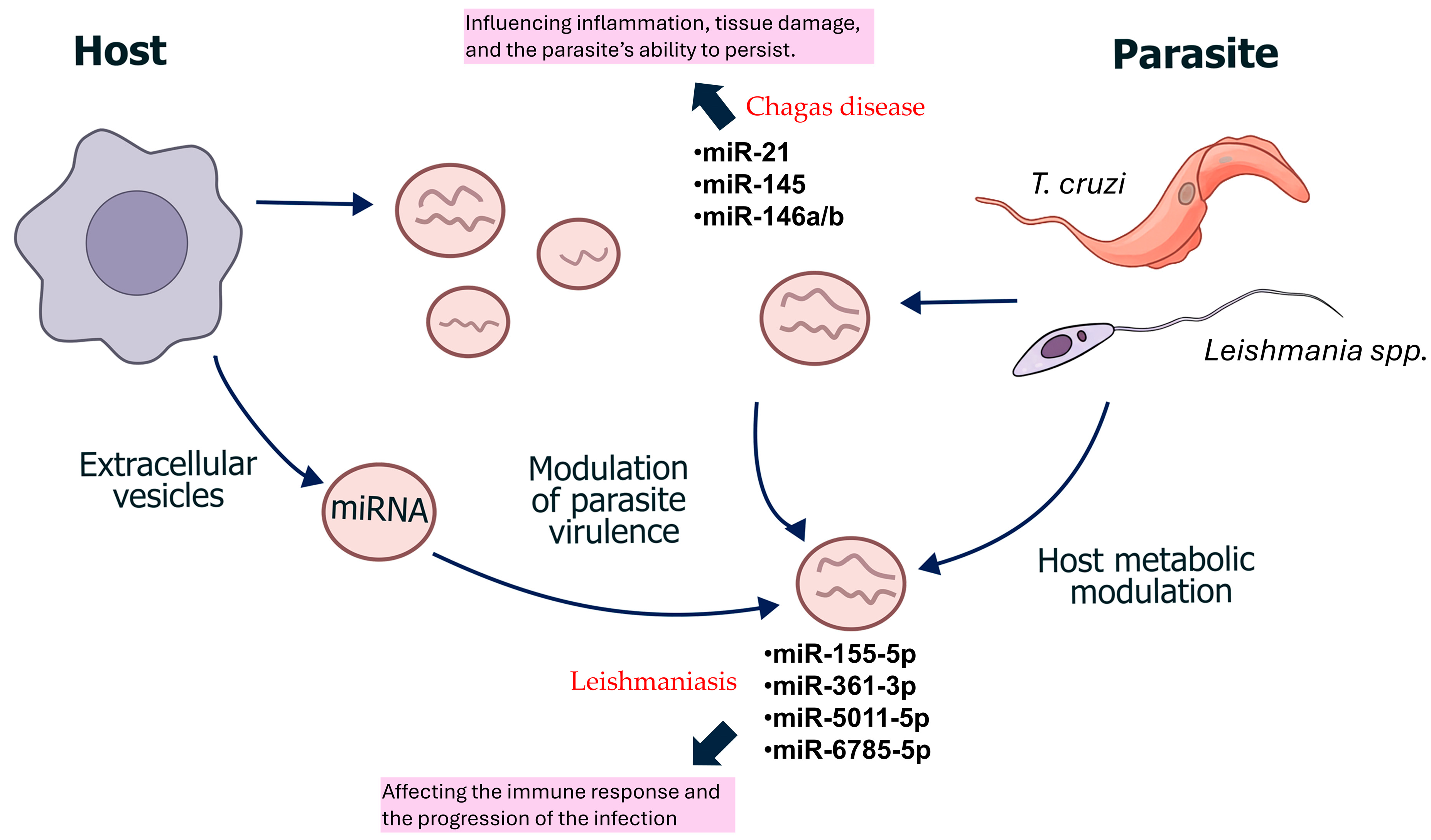
Despite significant advances in understanding the regulatory roles of ncRNAs in Leishmania and Trypanosoma cruzi infections, their clinical translation as reliable biomarkers remains limited, Figure 2. Current studies are often exploratory, based on small sample sizes, heterogeneous experimental designs, and a lack of standardized methodologies for RNA extraction, normalization, and quantification. Furthermore, variations in host species, infection stages, and tissue origins complicate the reproducibility and comparability of results among studies.
A critical knowledge gap exists in differentiating ncRNAs linked to infection detection from those indicative of disease progression or organ-specific pathology [63]. In T. cruzi, most available data relate to cardiac alterations involvement during the chronic phase [64], while information on acute or asymptomatic infections remains scarce. Similarly, for Leishmania, the repertoire of ncRNAs (RNAome) varies between species and clinical forms, complicating their diagnostic application [50].
Future research should prioritize large-scale, multicenter studies using harmonized protocols and clinically relevant cohorts to validate non-coding RNA signatures. The combined use of transcriptomics, proteomics, and metabolomics could elucidate the mechanistic connections between ncRNAs derived from parasites and hosts. Finally, the development of cost-effective, point-of-care assays for ncRNA detection, particularly in endemic and resource-limited settings, represents a crucial step toward their implementation as practical biomarkers for neglected tropical diseases.

8. Conclusions

ncRNAs represent promising molecular biomarkers for both Leishmaniasis and Chagas disease. Current evidence highlights their potential to enhance diagnostic specificity and sensitivity, support disease staging, and predict therapeutic responses. Small RNA species derived from both parasites and host cells play critical roles in modulating pathogenesis, immune responses, and clinical outcomes. Several miRNAs—such as miR-155-5p and miR-361-3p in leishmaniasis, and miR-21, miR-145, and miR-146a/b in Chagas disease—have demonstrated promising diagnostic performance in clinical samples. However, the translation of these findings into routine clinical practice remains constrained by the lack of large-scale, standardized validation studies. Furthermore, lncRNAs represent an underexplored diagnostic resource that requires additional investigation to clarify their potential applications. Also, while small interfering RNAs are valuable research tools for understanding gene regulation and pathogenesis, they are not currently used as biomarkers or direct diagnostic tools for leishmaniasis or Chagas disease. In conclusion, integrating ncRNA biomarker profiling into current diagnostic workflows could revolutionize the clinical management of these neglected diseases, facilitating earlier diagnosis, more accurate prognostic assessment, and targeted therapeutic monitoring. Future research should focus on large, multicenter studies to validate ncRNA signatures, assess cost-effectiveness, and establish standardized methodologies suitable for implementation in endemic regions.

Author Contributions

Conceptualization, H.A.C.-F., M.T.H.-H. and E.P.-C.; writing—original draft preparation, E.R.J., E.P.-C.M., L.P.-C.M., A.M.R., C.R.-D., M.E.A.-V., T.S.S.R. and M.M.C.; writing—review and editing, J.L.H.-M., L.G.B.-H., I.K.S.L., J.E.-R., A.A.G.G., H.A.C.-F., M.T.H.-H. and E.P.-C.; supervision, H.A.C.-F., M.T.H.-H. and E.P.-C. All authors have read and agreed to the published version of the manuscript.

Funding

This research received no external funding.

Institutional Review Board Statement

Not applicable.

Informed Consent Statement

Not applicable.

Data Availability Statement

No new data were created or analyzed in this study.

Acknowledgments

E.R.J., L.G.B.-H., and T.S.S.R. are funded by a scholarship from the Secretaria de Ciencia, Humanidades, Tecnología e Innovación (SECIHTI), México. H.A.C.-F. is a member of the Comité Científico de Salud de los Servicios de Salud de Oaxaca (SSO), México. L.P.-C.M., H.A.C.-F., M.T.H.-H., and E.P.-C. are members of the Comité Oaxaqueño de Trombosis, Hemostasia y Endotelio (COTHE) de los Servicios de Salud de Oaxaca (SSO), México.

Conflicts of Interest

The authors declare no conflicts of interest.

Abbreviations

The following abbreviations are used in this manuscript:
T. cruziTrypanosoma cruzi
miRNAsmicroRNAs
ncRNAsnon-coding RNAs
lncRNAslong non-coding RNAs
PAHOPan American Health Organization
piRNAsPIWI-interacting RNAs
RNAomerepertoire of ncRNAs
SANRAScale for the Assessment of Narrative Review Articles
siRNAsshort interfering RNAs
VLvisceral leishmaniasis
WHOWorld Health Organization

References

  1. World Health Organization (WHO). Leishmaniasis. Available online: https://www.who.int/news-room/fact-sheets/detail/leishmaniasis (accessed on 23 January 2024).
  2. Cruz-Reyes, A.; Pickering-López, J.M. Chagas disease in Mexico: An analysis of geographical distribution during the past 76 years—A review. Mem. Inst. Oswaldo Cruz 2006, 101, 345–354. [Google Scholar] [CrossRef] [PubMed]
  3. Scarpini, S.; Dondi, A.; Totaro, C.; Biagi, C.; Melchionda, F.; Zama, D.; Pierantoni, L.; Gennari, M.; Campagna, C.; Prete, A.; et al. Visceral Leishmaniasis: Epidemiology, Diagnosis, and Treatment Regimens in Different Geographical Areas with a Focus on Pediatrics. Microorganisms 2022, 10, 1887. [Google Scholar] [CrossRef] [PubMed]
  4. Velasco-Castrejón, Ó.; Rivas-Sánchez, B. Apuntes para la historia de la enfermedad de Chagas en México. Bol. Med. Hosp. Infant. Mex. 2008, 65, 57–79. [Google Scholar]
  5. Reithinger, R.; Dujardin, J.C. Molecular diagnosis of leishmaniasis: Current status and future applications. J. Clin. Microbiol. 2007, 45, 21–25. [Google Scholar] [CrossRef]
  6. Lopez-Albizu, C.; Rivero, R.; Ballering, G.; Freilij, H.; Santini, M.S.; Bisio, M.M.C. Laboratory diagnosis of Trypanosoma cruzi infection: A narrative review. Front. Parasitol. 2023, 2, 1138375. [Google Scholar] [CrossRef]
  7. Centers for Disease Control and Prevention (CDC). Clinical Testing and Diagnosis for Chagas Disease. Available online: https://www.cdc.gov/chagas/es/hcp/diagnosis-testing/pruebas-y-diagnostico-clinicos-para-la-enfermedad-de-chagas.html#:~:text=Use%20dos%20o%20m%C3%A1s%20pruebas,de%20anticuerpos%20inmunofluorescentes%20(IFA) (accessed on 3 January 2024).
  8. Pan American Health Organization (PAHO). Chagas Disease. Available online: https://www.paho.org/en/topics/chagas-disease (accessed on 3 January 2024).
  9. Bautista-Lopez, N.; Ndao, M. Usefulness of polymerase chain reaction tests in Chagas disease studies. Front. Parasitol. 2024, 3, 1292143. [Google Scholar] [CrossRef]
  10. Benatar, A.F.; Danesi, E.; Besuschio, S.A.; Bortolotti, S.; Cafferata, M.L.; Ramirez, J.C.; Albizu, C.L.; Scollo, K.; Baleani, M.; Lara, L.; et al. Prospective multicenter evaluation of real time PCR Kit prototype for early diagnosis of congenital Chagas disease. EBioMedicine 2021, 69, 103450. [Google Scholar] [CrossRef]
  11. Aronson, N.; Herwaldt, B.L.; Libman, M.; Pearson, R.; Lopez-Velez, R.; Weina, P.; Carvalho, E.; Ephros, M.; Jeronimo, S.; Magill, A. Diagnosis and Treatment of Leishmaniasis: Clinical Practice Guidelines by the Infectious Diseases Society of America (IDSA) and the American Society of Tropical Medicine and Hygiene (ASTMH). Am. J. Trop. Med. Hyg. 2017, 96, 24–45. [Google Scholar] [CrossRef]
  12. McGwire, B.S.; Satoskar, A.R. Leishmaniasis: Clinical Syndromes and Treatment. QJM 2014, 107, 7–14. [Google Scholar] [CrossRef]
  13. Centers for Disease Control and Prevention (CDC). Diagnosis and Testing—Chagas Disease. Available online: https://www.cdc.gov/chagas/hcp/diagnosis-testing/index.html (accessed on 23 January 2024).
  14. Suárez, C.; Nolder, D.; García-Mingo, A.; Moore, D.A.J.; Chiodini, P.L. Diagnosis and Clinical Management of Chagas Disease: An Increasing Challenge in Non-Endemic Areas. Res. Rep. Trop. Med. 2022, 13, 25–40. [Google Scholar] [CrossRef]
  15. Forsyth, C.J.; Manne-Goehler, J.; Bern, C.; Whitman, J.; Hochberg, N.S.; Edwards, M.; Marcus, R.; Beatty, N.L.; Castro-Sesquen, Y.E.; Coyle, C.; et al. Recommendations for Screening and Diagnosis of Chagas Disease in the United States. J. Infect. Dis. 2022, 225, 1601–1610. [Google Scholar] [CrossRef]
  16. Schijman, A.G.; Alonso-Padilla, J.; Britto, C.; Herrera Bernal, C.P. Retrospect, advances and challenges in Chagas disease diagnosis: A comprehensive review. Lancet Reg. Health Am. 2024, 36, 100821. [Google Scholar] [CrossRef] [PubMed]
  17. Pippadpally, S.; Venkatesh, T. Deciphering piRNA Biogenesis through Cytoplasmic Granules, Mitochondria and Exosomes. Arch. Biochem. Biophys. 2020, 695, 108597. [Google Scholar] [CrossRef]
  18. Russell, S.J.; Zhao, C.; Biondic, S.; Menezes, K.; Hagemann-Jensen, M.; Librach, C.L.; Petropoulos, S. An Atlas of Small Non-Coding RNAs in Human Preimplantation Development. Nat. Commun. 2024, 15, 8634. [Google Scholar] [CrossRef]
  19. Traber, G.M.; Yu, A.M. RNAi-Based Therapeutics and Novel RNA Bioengineering Technologies. J. Pharmacol. Exp. Ther. 2023, 384, 133–154. [Google Scholar] [CrossRef]
  20. Kaikkonen, M.U.; Lam, M.T.; Glass, C.K. Non-Coding RNAs as Regulators of Gene Expression and Epigenetics. Cardiovasc. Res. 2011, 90, 430–440. [Google Scholar] [CrossRef] [PubMed]
  21. Fire, A.; Xu, S.; Montgomery, M.K.; Kostas, S.A.; Driver, S.E.; Mello, C.C. Potent and Specific Genetic Interference by Double-Stranded RNA in Caenorhabditis elegans. Nature 1998, 391, 806–811. [Google Scholar] [CrossRef]
  22. Deerberg, A.; Willkomm, S.; Restle, T. Minimal Mechanistic Model of siRNA-Dependent Target RNA Slicing by Recombinant Human Argonaute 2 Protein. Proc. Natl. Acad. Sci. USA 2013, 110, 17850–17855. [Google Scholar] [CrossRef]
  23. Atayde, V.D.; Shi, H.; Franklin, J.B.; Carriero, N.; Notton, T.; Lye, L.F.; Owens, K.; Beverley, S.M.; Tschudi, C.; Ullu, E. The Structure and Repertoire of Small Interfering RNAs in Leishmania (Viannia) braziliensis Reveal Diversification in the Trypanosomatid RNAi Pathway. Mol. Microbiol. 2013, 87, 580–593. [Google Scholar] [CrossRef]
  24. DaRocha, W.D.; Otsu, K.; Teixeira, S.M.; Donelson, J.E. Tests of Cytoplasmic RNA Interference (RNAi) and Construction of a Tetracycline-Inducible T7 Promoter System in Trypanosoma cruzi. Mol. Biochem. Parasitol. 2004, 133, 175–186. [Google Scholar] [CrossRef] [PubMed]
  25. Lee, R.C.; Feinbaum, R.L.; Ambros, V. The C. elegans Heterochronic Gene lin-4 Encodes Small RNAs with Antisense Complementarity to lin-14. Cell 1993, 75, 843–854. [Google Scholar] [CrossRef]
  26. Bartel, D.P. MicroRNAs: Genomics, Biogenesis, Mechanism, and Function. Cell 2004, 116, 281–297. [Google Scholar] [CrossRef]
  27. Friedman, R.C.; Farh, K.K.; Burge, C.B.; Bartel, D.P. Most Mammalian mRNAs Are Conserved Targets of microRNAs. Genome Res. 2009, 19, 92–105. [Google Scholar] [CrossRef]
  28. Iwakawa, H.O.; Tomari, Y. Life of RISC: Formation, Action, and Degradation of RNA-Induced Silencing Complex. Mol. Cell 2022, 82, 30–43. [Google Scholar] [CrossRef] [PubMed]
  29. Axtell, M.J.; Westholm, J.O.; Lai, E.C. Vive la Différence: Biogenesis and Evolution of microRNAs in Plants and Animals. Genome Biol. 2011, 12, 221. [Google Scholar] [CrossRef]
  30. Hynes, C.; Kakumani, P.K. Regulatory Role of RNA-Binding Proteins in microRNA Biogenesis. Front. Mol. Biosci. 2024, 11, 1374843. [Google Scholar] [CrossRef]
  31. Lam, J.K.; Chow, M.Y.; Zhang, Y.; Leung, S.W. siRNA Versus miRNA as Therapeutics for Gene Silencing. Mol. Ther. Nucleic Acids 2015, 4, e252. [Google Scholar] [CrossRef] [PubMed]
  32. Krützfeldt, J.; Stoffel, M. MicroRNAs: A New Class of Regulatory Genes Affecting Metabolism. Cell Metab. 2006, 4, 9–12. [Google Scholar] [CrossRef] [PubMed]
  33. Clark, J.P.; Lau, N.C. Piwi Proteins and piRNAs Step onto the Systems Biology Stage. Adv. Exp. Med. Biol. 2014, 825, 159–197. [Google Scholar] [CrossRef]
  34. Théron, E.; Dennis, C.; Brasset, E.; Vaury, C. Distinct Features of the piRNA Pathway in Somatic and Germ Cells: From piRNA Cluster Transcription to piRNA Processing and Amplification. Mob. DNA 2014, 5, 28. [Google Scholar] [CrossRef]
  35. Fu, Q.; Wang, P.J. Mammalian piRNAs: Biogenesis, Function, and Mysteries. Spermatogenesis 2014, 4, e27889. [Google Scholar] [CrossRef]
  36. Yang, X.; Cheng, Y.; Lu, Q.; Wei, J.; Yang, H.; Gu, M. Detection of Stably Expressed piRNAs in Human Blood. Int. J. Clin. Exp. Med. 2015, 8, 13353–13358. [Google Scholar] [PubMed]
  37. Mattick, J.S.; Amaral, P.P.; Carninci, P.; Carpenter, S.; Chang, H.Y.; Chen, L.L.; Chen, R.; Dean, C.; Dinger, M.E.; Fitzgerald, K.A.; et al. Long Non-Coding RNAs: Definitions, Functions, Challenges and Recommendations. Nat. Rev. Mol. Cell Biol. 2023, 24, 430–447. [Google Scholar] [CrossRef] [PubMed]
  38. Zhang, X.; Wang, W.; Zhu, W.; Dong, J.; Cheng, Y.; Yin, Z.; Shen, F. Mechanisms and Functions of Long Non-Coding RNAs at Multiple Regulatory Levels. Int. J. Mol. Sci. 2019, 20, 5573. [Google Scholar] [CrossRef] [PubMed]
  39. Bayer-Santos, E.; Lima, F.M.; Ruiz, J.C.; Almeida, I.C.; da Silveira, J.F. Characterization of the Small RNA Content of Trypanosoma cruzi Extracellular Vesicles. Mol. Biochem. Parasitol. 2014, 193, 71–74. [Google Scholar] [CrossRef]
  40. Fort, R.S.; Chavez, S.; Trinidad Barnech, J.M.; Oliveira-Rizzo, C.; Smircich, P.; Sotelo-Silveira, J.R.; Duhagon, M.A. Current Status of Regulatory Non-Coding RNAs Research in the Tritryp. Noncoding RNA 2022, 8, 54. [Google Scholar] [CrossRef]
  41. Lye, L.F.; Dobson, D.E.; Beverley, S.M.; Tung, M.C. RNA Interference in Protozoan Parasites and Its Application. J. Microbiol. Immunol. Infect. 2025, 58, 281–287. [Google Scholar] [CrossRef]
  42. Poloni, J.F.; Oliveira, F.H.S.; Feltes, B.C. Localization Is the Key to Action: Regulatory Peculiarities of lncRNAs. Front. Genet. 2024, 15, 1478352. [Google Scholar] [CrossRef]
  43. Franzén, O.; Arner, E.; Ferella, M.; Nilsson, D.; Respuela, P.; Carninci, P.; Hayashizaki, Y.; Aslund, L.; Andersson, B.; Daub, C.O. The Short Non-Coding Transcriptome of the Protozoan Parasite Trypanosoma cruzi. PLoS Negl. Trop. Dis. 2011, 5, e1283. [Google Scholar] [CrossRef]
  44. Fernandes, J.C.R.; Gonçalves, A.N.A.; Floeter-Winter, L.M.; Nakaya, H.I.; Muxel, S.M. Comparative Transcriptomic Analysis of Long Noncoding RNAs in Leishmania-Infected Human FGs. Front. Genet. 2023, 13, 1051568. [Google Scholar] [CrossRef]
  45. Garcia-Silva, M.R.; das Neves, R.F.; Cabrera-Cabrera, F.; Sanguinetti, J.; Medeiros, L.C.; Robello, C.; Naya, H.; Fernandez-Calero, T.; Souto-Padron, T.; de Souza, W.; et al. Extracellular Vesicles Shed by Trypanosoma cruzi Are Linked to Small RNA Pathways, Life Cycle Regulation, and Susceptibility to Infection of Mammalian Cells. Parasitol. Res. 2014, 113, 285–304. [Google Scholar] [CrossRef]
  46. Peng, R.; Santos, H.J.; Nozaki, T. Transfer RNA-Derived Small RNAs in the Pathogenesis of Parasitic Protozoa. Genes 2022, 13, 286. [Google Scholar] [CrossRef]
  47. Garcia-Silva, M.R.; Sanguinetti, J.; Cabrera-Cabrera, F.; Franzén, O.; Cayota, A. A Particular Set of Small Non-Coding RNAs Is Bound to the Distinctive Argonaute Protein of Trypanosoma cruzi: Insights from RNA-Interference Deficient Organisms. Gene 2014, 538, 379–384. [Google Scholar] [CrossRef]
  48. Ullu, E.; Tschudi, C.; Chakraborty, T. RNA Interference in Protozoan Parasites. Cell Microbiol. 2004, 6, 509–519. [Google Scholar] [CrossRef] [PubMed]
  49. Bayer-Santos, E.; Marini, M.M.; da Silveira, J.F. Non-Coding RNAs in Host-Pathogen Interactions: Subversion of Mammalian Cell Functions by Protozoan Parasites. Front. Microbiol. 2017, 8, 474. [Google Scholar] [CrossRef]
  50. Martinez-Hernandez, J.E.; Aliaga-Tobar, V.; González-Rosales, C.; Monte-Neto, R.; Martin, A.J.M.; Maracaja-Coutinho, V. Comparative and Systems Analyses of Leishmania spp. Non-Coding RNAs through Developmental Stages. PLoS Negl. Trop. Dis. 2025, 19, e0013108. [Google Scholar] [CrossRef]
  51. Paul, S.; Ruiz-Manriquez, L.M.; Serrano-Cano, F.I.; Estrada-Meza, C.; Solorio-Diaz, K.A.; Srivastava, A. Human microRNAs in Host-Parasite Interaction: A Review. 3 Biotech 2020, 10, 510. [Google Scholar] [CrossRef] [PubMed]
  52. Lago, T.S.; Silva, J.A.; Lago, E.L.; Carvalho, E.M.; Zanette, D.L.; Castellucci, L.C. The miRNA 361-3p, a Regulator of GZMB and TNF, Is Associated with Therapeutic Failure and Longer Time Healing of Cutaneous Leishmaniasis Caused by L. (Viannia) braziliensis. Front. Immunol. 2018, 9, 2621. [Google Scholar] [CrossRef]
  53. Masoudsinaki, T.; Hadifar, S.; Sarvnaz, H.; Farahmand, M.; Masoudzadeh, N.; Goyonlo, V.M.; Kerachian, M.; Salim, R.E.; Barhoumi, M.; Gargari, S.L.M.; et al. Altered miRNA Expression in the Lesions of Cutaneous Leishmaniasis Caused by L. major and L. tropica with Insights into Apoptosis Regulation. Sci. Rep. 2025, 15, 20680. [Google Scholar] [CrossRef]
  54. Varikuti, S.; Verma, C.; Natarajan, G.; Oghumu, S.; Satoskar, A.R. MicroRNA155 Plays a Critical Role in the Pathogenesis of Cutaneous Leishmania major Infection by Promoting a Th2 Response and Attenuating Dendritic Cell Activity. Am. J. Pathol. 2021, 191, 809–816. [Google Scholar] [CrossRef] [PubMed]
  55. Akand, S.K.; Rahman, A.; Masood, M.; Tabrez, S.; Jawed, J.J.; Ahmed, M.Z.; Akhter, Y.; Haque, M.M.; Rub, A. Leishmania donovani alters the host sphingolipid biosynthetic pathway regulatory microRNA hsa-miR-15a-5p for its survival. Microbial pathogenesis. 2025, 208, 108019. [Google Scholar] [CrossRef]
  56. Li, Z.; Fang, Y.; Zhang, Y.; Zhou, X. RNA-Seq Analysis of Differentially Expressed lncRNAs from Leishmaniasis Patients Compared to Uninfected Humans. Acta Trop. 2023, 238, 106738. [Google Scholar] [CrossRef]
  57. Rojas-Pirela, M.; Andrade-Alviárez, D.; Medina, L.; Castillo, C.; Liempi, A.; Guerrero-Muñoz, J.; Ortega, Y.; Maya, J.D.; Rojas, V.; Quiñones, W.; et al. MicroRNAs: Master Regulators in Host-Parasitic Protist Interactions. Open Biol. 2022, 12, 210395. [Google Scholar] [CrossRef]
  58. Villar, S.R.; Herreros-Cabello, A.; Callejas-Hernández, F.; Maza, M.C.; Del Moral-Salmoral, J.; Gómez-Montes, M.; Rodríguez-Angulo, H.O.; Carrillo, I.; Górgolas, M.; Bosch-Nicolau, P.; et al. Discovery of Circulating miRNAs as Biomarkers of Chronic Chagas Heart Disease via a Small RNA-Seq Approach. Sci. Rep. 2024, 14, 1187, Erratum in: Sci. Rep. 2024, 4, 18514. [Google Scholar] [CrossRef]
  59. Ribeiro, H.G.; Galdino, O.A.; de Souza, K.S.C.; Rosa Neta, A.P.; Lin-Wang, H.T.; Cunha-Neto, E.; Rezende, A.A.; Silbiger, V.N. Unraveling the Role of miRNAs as Biomarkers in Chagas Cardiomyopathy: Insights into Molecular Pathophysiology. PLoS Negl. Trop. Dis. 2024, 18, e0011865. [Google Scholar] [CrossRef]
  60. Nonaka, C.K.V.; Macêdo, C.T.; Cavalcante, B.R.R.; Alcântara, A.C.; Silva, D.N.; Bezerra, M.D.R.; Caria, A.C.I.; Tavora, F.R.F.; Neto, J.D.S.; Noya-Rabelo, M.M.; et al. Circulating miRNAs as Potential Biomarkers Associated with Cardiac Remodeling and Fibrosis in Chagas Disease Cardiomyopathy. Int. J. Mol. Sci. 2019, 20, 4064. [Google Scholar] [CrossRef] [PubMed]
  61. Antonietti, L.; Mariani, J.; Martínez, M.J.; Santalla, M.; Vensentini, N.; Kyle, D.A.; de Abreu, M.; Tajer, C.; Lacunza, E.; Ferrero, P. Circulating microRNAs as Biomarkers of Chagas Cardiomyopathy. Front. Cardiovasc. Med. 2023, 10, 1250029. [Google Scholar] [CrossRef] [PubMed]
  62. Rayford, K.J.; Cooley, A.; Strode, A.W.; Osi, I.; Arun, A.; Lima, M.F.; Misra, S.; Pratap, S.; Nde, P.N. Trypanosoma cruzi Dysregulates Expression Profile of piRNAs in Primary Human Cardiac Fibroblasts during Early Infection Phase. Front. Cell. Infect. Microbiol. 2023, 13, 1083379. [Google Scholar] [CrossRef] [PubMed]
  63. Nemeth, K.; Bayraktar, R.; Ferracin, M.; Calin, G.A. Non-coding RNAs in disease: From mechanisms to therapeutics. Nat. Rev. Genet. 2024, 25, 211–232. [Google Scholar] [CrossRef]
  64. Cornet-Gomez, A.; O’Valle, F.; Garrido, J.M.; Serrano, F.R.; Nieto, A.I.; Osuna, A. Cardiac alterations induced by Trypanosoma cruzi extracellular vesicles and immune complexes. PLoS Negl. Trop. Dis. 2025, 19, e0013273. [Google Scholar] [CrossRef] [PubMed] [PubMed Central]
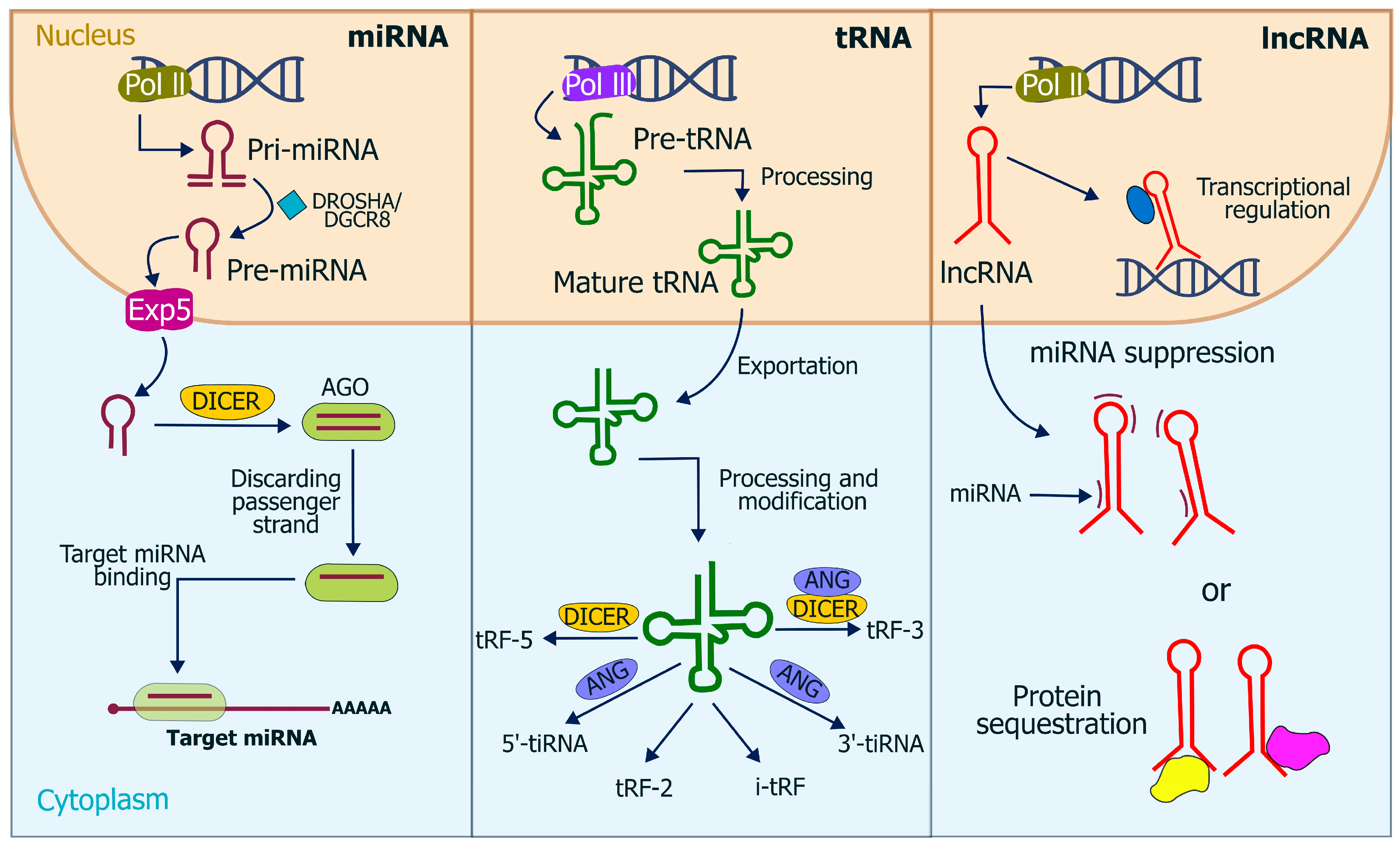
Figure 1. Biogenesis and functions of ncRNAs, including microRNAs (miRNAs), transfer RNAs (tRNAs), and long non-coding RNAs (lncRNAs). In the nucleus, miRNAs are transcribed by RNA polymerase II (Pol II) as primary transcripts (pri-miRNAs) and processed by the DROSHA/DGCR8 complex into precursor miRNAs (pre-miRNAs), which are exported to the cytoplasm via Exportin-5 (Exp5) and further processed by DICER to form Argonaute (AGO) complexes that regulate target mRNAs. tRNAs, transcribed by RNA polymerase III (Pol III), undergo processing and modification to produce mature tRNAs and tRNA-derived fragments (tRFs and tiRNAs) through DICER or ANG activity. lncRNAs, also transcribed by Pol II, participate in transcriptional regulation, miRNA suppression, and protein sequestration. The diagram highlights the nuclear and cytoplasmic localization of these processes.
Tropicalmed 10 00319 g001
Figure 2. ncRNAs represent potential diagnostic biomarkers and regulatory molecules in host–parasite interactions in Leishmania and Trypanosoma cruzi infections.
Tropicalmed 10 00319 g002
Table 1. Diagnostic Comparison of Cutaneous, Mucocutaneous, and Visceral Leishmaniasis [12,13].
CharacteristicCutaneous LeishmaniasisMucocutaneous LeishmaniasisVisceral Leishmaniasis
Causative agentsL. major, L. tropica (Old World); L. mexicana, L. amazonensis, L. guyanensis, L. panamensis, L. braziliensis (Americas)Mainly L. braziliensisL. donovani, L. infantum (Old World); L. infantum/L. chagasi (Americas); occasional cases caused by viscerotropic strains of L. tropica
Endemic regionsOld World: Africa, Middle East, Asia; New World: Central and South AmericaPrimarily South America: Brazil, Peru, Bolivia; also seen in Colombia, Ecuador, Paraguay, and VenezuelaOld World: India, Pakistan, China, Africa, Mediterranean region; New World: mainly Brazil
PathogenesisLocalized skin infection with an inflammatory reaction at the inoculation siteSpread or metastasis of the parasite from cutaneous lesions to nasopharyngeal and/or oropharyngeal mucosal tissuesSystemic dissemination to organs of the reticuloendothelial system (liver, spleen, bone marrow)
Clinical manifestationsSingle or multiple; typically on exposed areas (face, arms, legs)Progressive destruction of mucous membranes and soft tissues: nose, mouth, pharynx, eyelids, can cause severe disfigurement and respiratory/nutritional difficultiesProlonged fever, weight loss, hepatosplenomegaly, anemia, pancytopenia, immunosuppression; HIV coinfection increases severity
Clinical courseUsually self-limiting, but may progress to mucocutaneous disease (especially with L. panamensis and L. braziliensis)Chronic evolution can develop months or years after initial cutaneous lesions; high risk of complicationsPotentially fatal if untreated; immunosuppression increases the risk of opportunistic infections.
DiagnosisVisualization of amastigotes (Microscopic evaluation), culture, PCR (conventional and real-time); biopsy in diffuse casesParasitological and molecular diagnosis (DNA sequencing analysis; also cellulose acetate electrophoresis); evaluation of mucosal involvementCombination of clinical findings, serology (ELISA, DAT), parasite visualization (smear, bone marrow aspirate), PCR (conventional and real-time)
TreatmentLocal therapy in mild cases; systemic therapy in extensive disease or mucocutaneous riskProlonged systemic treatment; limited response, risk of recurrenceRequires immediate systemic therapy; management is more complex in HIV-coinfected patients
PrognosisGood in most cases, especially with early treatmentGuarded; high morbidity and disfigurement; mortality mainly due to secondary infections and malnutritionSevere if untreated; with appropriate therapy, mortality decreases significantly
Abbreviations: PCR, polymerase chain reaction; ELISA, enzyme-linked immunosorbent assay; DAT, direct agglutination test.
Table 2. Diagnostic Methods for Chagas Disease [14,15].
Diagnostic AspectAcute PhaseChronic Phase *
Morphology
  • Detection of trypomastigotes in circulating blood or CSF via direct microscopy.
  • Thick and thin blood smears stained with Giemsa or H&E are used.
  • Amastigotes may also be identified in biopsy specimens.
  • Trypomastigotes rarely found in blood.
  • Serologic tests are preferred for diagnosis.
  • Amastigotes can occasionally be identified in tissue biopsies.
Serology
(Antibody Detection)
  • There are no accurate IgM based methods available for serological diagnosis at the acute phase.
  • Primary diagnostic method due to low parasitemia.
  • Requires ≥ 2 serologic tests targeting different antigens.
  • CDC protocols:
    -
    First line: FDA-cleared enzyme immunoassay (EIA) + TESA immunoblot.
    -
    If discordant: request second sample and repeat testing.
    -
    If still discordant: use immunofluorescence assay (IFA) as a “tie-breaker.”
  • Sensitivity and specificity vary across commercially available tests.
Molecular Testing
  • Recommended in suspected acute infections, congenital cases, transfusion/transplant transmission, or laboratory exposures.
  • CDC method: real-time PCR using two assays (TCZ and MNC).
  • Acceptable specimens: EDTA blood (≥2.2 mL), heart biopsy tissue, and CSF if CNS involvement is suspected.
  • Generally not used routinely for diagnosis but may be performed in reactivation cases linked to immunosuppression (e.g., HIV).
  • Serology remains the preferred method.
Abbreviations: CSF, Cerebrospinal fluid; H&E, Hematoxylin and eosin stain; EIA, Enzyme immunoassay; TESA, Trypomastigote excreted-secreted antigen immunoblot; IFA, Indirect immunofluorescence assay; PCR, Polymerase chain reaction; TCZ, T. cruzi–PCR target; MNC, Minicircle DNA PCR target; EDTA, Ethylenediaminetetraacetic acid. * The WHO advises using at least two assays with different principles to confirm a positive serological result for T. cruzi infection [16].
Table 3. ncRNAs for the diagnosis of Leishmaniasis and Chagas disease [42,43,46,47,48].
FeaturetRNA-Derived Small RNAs in T. cruzitRNA-Derived Small RNAs in LeishmanialncRNAs
Size31–40 nt20–40 nt>200 nt
PrecursorMature tRNAs cleaved at anticodon loop (tRNA halves) or at D loop, T loop, anticodon loop, or 3′ leader (tRFs)Mature tRNAsPrimary transcripts from RNA Pol I, II, or III
Processing enzymesDicer is absent in T. cruziMost Leishmania lack RNAi machineryStandard RNA processing machinery
Associated proteinsTcPIWI-tryp (trypanosomatid-specific PIWI-like protein)AGO/PIWI homolog (present in RNAi-deficient species)Spliceosome, transcriptional regulators, RNA stability proteins
RNAi machineryAbsent (no canonical Dicer or Argonaute)Absent in most species; retained only in L. braziliensis (Viannia subgenus)N/A
Functional notesTcPIWI-tryp binds small RNAs derived from structural RNAs; T. cruzi lost RNAi during evolution.L. major, L. donovani, L. infantum lack RNAi; miRNA-like elements computationally predicted but not validated.Differentially expressed during infection; potential regulatory roles in host–pathogen interactions.
Table 4. Potential ncRNA in the diagnosis of Leishmaniasis.
ncRNA TypeBiomarker(s)Sample/Clinical ContextSensitivitySpecificityAUC
miRNAmiR-155-5p, miR-5011-5p, miR-6785-5p, miR-4795-3p [55]Lesion biopsies (L. major, L. tropica)86–100%100%0.92–1.00
miRNAmiR-361-3p [54]Lesion biopsies (L. braziliensis)81.20%100%Not reported
lncRNAMALAT1, NUTM2A-AS1, LINC00963, others [56]Serum (visceral or cutaneous leishmaniasis patients)Not quantifiedNot quantifiedNot quantified
Table 5. Circulating microRNAs as emerging biomarkers in Chagas Cardiomyopathy.
ncRNA TypeBiomarkerSample/Clinical ContextSensitivitySpecificityAUC (ROC)
miRNA [60]miR-19a-3pSerum67%80%0.77
miR-21-5p57%60%0.54
miR-29b-3p60%70%0.7
miR-199b-5p67%57%0.57
Disclaimer/Publisher’s Note: The statements, opinions and data contained in all publications are solely those of the individual author(s) and contributor(s) and not of MDPI and/or the editor(s). MDPI and/or the editor(s) disclaim responsibility for any injury to people or property resulting from any ideas, methods, instructions or products referred to in the content.