
Changes in the Physicochemical Properties of Reduced Salt Pangasius (Pangasianodon hypophthalmus) Gels Induced by High Pressure and Setting Treatment


Abstract

1. Introduction
2. Materials and Methods
2.1. Materials and Fish Gel Preparation
2.2. High-Pressure and Thermal Treatment of Pangasius Paste
2.3. Proximate Analysis
2.4. Total Plate Count (TPC)
2.5. Color Analysis
2.6. Mechanical Properties
2.7. Protein Solubility
2.8. Water-Holding Capacity (WHC)
2.9. SDS-Polyacrylamide Gel Electrophoresis (SDS-PAGE)
2.10. Fourier-Transform Infrared Spectroscopy (FTIR)
2.11. Scanning Electron Microscopy (SEM)
2.12. Statistical Analysis
3. Results and Discussion
3.1. Proximate Analysis
3.2. TPC
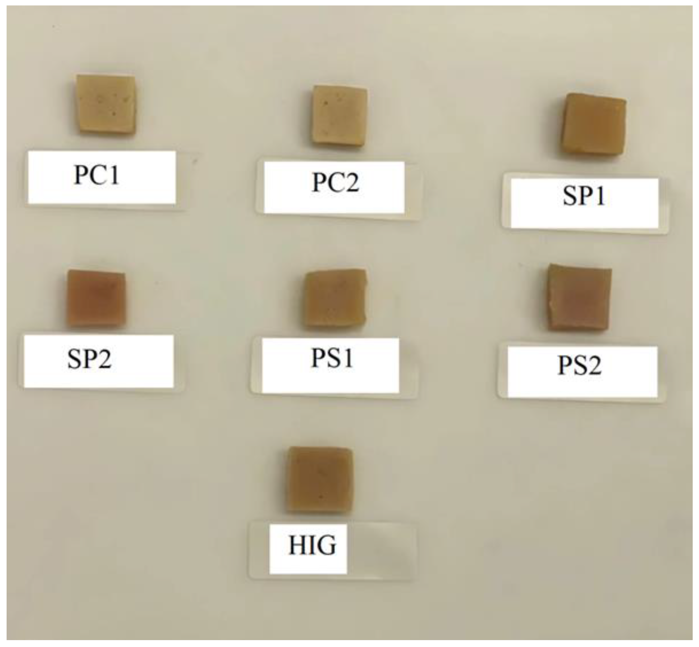
3.3. Color Analysis
3.4. Mechanical Properties
3.5. Protein Solubility
3.6. Water-Holding Capacity
3.7. SDS-Polyacrylamide Gel Electrophoresis (SDS-PAGE)
3.8. Fourier-Transform Infrared Spectroscopy (FTIR)
3.9. Scanning Electron Microscopy (SEM)
4. Conclusions
Supplementary Materials
Author Contributions
Funding
Informed Consent Statement
Data Availability Statement
Conflicts of Interest
Abbreviations
| HPP | High-pressure processing |
| WHC | Water-holding capacity |
| MHC | Myosin heavy chain |
| FTIR | Fourier-transform infrared spectroscopy |
| TGase | Endogenous transglutaminase |
| TPC | Total Plate Count |
| SEM | Scanning Electron Microscopy |
| PMSF | phenylmethylsulfonyl fluoride |
| LSD | least significant difference |
References
- Wang, Q.; Wei, R.; Hu, J.; Luan, Y.; Liu, R.; Ge, Q.; Yu, H.; Wu, M. Moderate pulsed electric field-induced structural unfolding ameliorated the gelling properties of porcine muscle myofibrillar protein. Innov. Food Sci. Emerg. Technol. 2022, 81, 103145. [Google Scholar] [CrossRef]
- Kuraishi, C.; Yamazaki, K.; Susa, Y. Transglutaminase: Its utilization in the food industry. Food Rev. Int. 2001, 17, 221–246. [Google Scholar] [CrossRef]
- Buchert, J.; Ercili Cura, D.; Ma, H.; Gasparetti, C.; Monogioudi, E.; Faccio, G.; Mattinen, M.; Boer, H.; Partanen, R.; Selinheimo, E. Crosslinking Food Proteins for Improved Functionality. Annu. Rev. Food Sci. Technol. 2010, 1, 113–138. [Google Scholar] [CrossRef] [PubMed]
- Motoki, M.; Seguro, K. Transglutaminase and its use for food processing. Trends Food Sci. Technol. 1998, 9, 204–210. [Google Scholar] [CrossRef]
- Wang, Y.; Cui, Q.; Wang, X.; Wu, C.; Xu, X.; Dong, X.; Pan, J. The gelling properties of fish gelatin as improved by ultrasound-assisted phosphorylation. Food Chem. 2024, 449, 139214. [Google Scholar] [CrossRef]
- Li, Z.; Sun, Q.; Zheng, Y.; Wang, J.; Tian, Y.; Zheng, B.; Guo, Z. Effect of two-step microwave heating on the gelation properties of golden threadfin bream (Nemipterus virgatus) myosin. Food Chem. 2020, 328, 127104. [Google Scholar] [CrossRef]
- Shikha, F.H.; Hossain, M.I.; Morioka, K.; Kubota, S.; Itoh, Y. Effect of pH-shifting on the gel forming characteristics of salt-ground meat from walleye pollack. Fish. Sci. 2006, 72, 870–876. [Google Scholar] [CrossRef]
- Wang, W.; Yang, P.; Rao, L.; Zhao, L.; Wu, X.; Wang, Y.; Liao, X. Effect of high hydrostatic pressure processing on the structure, functionality, and nutritional properties of food proteins: A review. Compr. Rev. Food Sci. Food Saf. 2022, 21, 4640–4682. [Google Scholar] [CrossRef]
- Li, H.; Li, C.; Shoaib, M.; Zhang, W.; Murugesan, A. Advances in Non-Thermal Processing of Meat and Monitoring Meat Protein Gels Through Vibrational Spectroscopy. Foods 2025, 14, 1929. [Google Scholar] [CrossRef]
- Yin, J.; Zhang, P.; Fang, Z. Methods to improve the quality of low-salt meat products: A meta-analysis. Food Qual. Saf. 2023, 7, 1–12. [Google Scholar] [CrossRef]
- Herranz, B.; Tovar, C.A.; Borderias, A.J.; Moreno, H.M. Effect of high-pressure and/or microbial transglutaminase on physicochemical, rheological and microstructural properties of flying fish surimi. Innov. Food Sci. Emerg. Technol. 2013, 20, 24–33. [Google Scholar] [CrossRef]
- Cando, D.; Herranz, B.; Borderías, A.J.; Moreno, H.M. Effect of high pressure on reduced sodium chloride surimi gels. Food Hydrocoll. 2015, 51, 176–187. [Google Scholar] [CrossRef]
- Park, J.W. Surimi and Surimi Seafood, 3rd ed.; CRC Press: Boca Raton, FL, USA, 2013. [Google Scholar]
- Pilar, M.; Gomez-Guillen, M.C. High-Pressure Applications on Myosystems, in Novel Food Processing Technologies; CRC Press: Boca Raton, FL, USA, 2004; pp. 311–342. [Google Scholar]
- Liu, H.; Xu, Y.; Zu, S.; Wu, X.; Shi, A.; Zhang, J.; Wang, Q.; He, N. Effects of High Hydrostatic Pressure on the Conformational Structure and Gel Properties of Myofibrillar Protein and Meat Quality: A Review. Foods 2021, 10, 1872. [Google Scholar] [CrossRef] [PubMed]
- Okamoto, M.; Kawamura, Y.; Hayashi, R. Application of High Pressure to Food Processing: Textural Comparison of Pressure-and Heat-induced Gels of Food Proteins. Agric. Biol. Chem. 1990, 54, 183–189. [Google Scholar]
- Okazaki, E.; Fukuda, Y. Effect of water-soluble protein on pressure-induced gelation of Alaska pollack surimi. Prog. Biotechnol. 1996, 13, 363–368. [Google Scholar]
- Truong, B.Q.; Buckow, R.; Nguyen, M.H.; Furst, J. Effect of high-pressure treatments prior to cooking on gelling properties of unwashed protein from barramundi (Lates calcarifer) minced muscle. Int. J. Food Sci. Technol. 2017, 52, 1383–1391. [Google Scholar] [CrossRef]
- Xu, M.; Ni, X.; Liu, Q.; Chen, C.; Deng, X.; Wang, X.; Yu, R. Ultra-high pressure improved gelation and digestive properties of Tai Lake whitebait myofibrillar protein. Food Chem. X 2024, 21, 101061. [Google Scholar] [CrossRef]
- Tyre, C.L.; Jirawat, Y.; Patricio, C.-R. Surimi Gelation Chemistry. In Surimi and Surimi Seafood, 3rd ed.; CRC Press: Boca Raton, FL, USA, 2013; pp. 101–140. [Google Scholar]
- Niwa, E. Chemistry of Surimi Gelation. In Surimi Technology; Lanier, T.C., Lee, C.M., Eds.; Marcel Dekker: New York, NY, USA, 1992; pp. 389–427. [Google Scholar]
- Murthy, L.N.; Phadke, G.G.; Jeyakumari, A.; Ravishankar, C.N. Effect of added calcium and heat setting on gel forming and functional properties of Sardinella fimbriata surimi. J. Food Sci. Technol. 2021, 58, 427–436. [Google Scholar] [CrossRef]
- Tran, T.M.H.; Kohyama, K.; Watanabe, N.; Osako, K.; Okazaki, E. Relationship between the Physical Properties and Perceived Saltiness of Various Surimi Gels Prepared by Different Setting Conditions. J. Exp. Food Chem. 2017, 3, 1000124. [Google Scholar]
- Xiong, Z.; Shi, T.; Jin, W.; Bao, Y.; Monto, A.R.; Yuan, L.; Gao, R. Gel performance of surimi induced by various thermal technologies: A review. Crit. Rev. Food Sci. Nutr. 2024, 64, 3075–3090. [Google Scholar] [CrossRef]
- Walayat, N.; Blanch, M.; Moreno, H.M. Surimi and Low-Salt Surimi Gelation: Key Components to Enhance the Physicochemical Properties of Gels. Gels 2025, 11, 142. [Google Scholar] [CrossRef]
- Nguyen, H.T.; Bao, H.N.D.; Dang, H.T.T.; Tómasson, T.; Arason, S.; Gudjónsdóttir, M. Protein Characteristics and Bioactivity of Fish Protein Hydrolysates from Tra Catfish (Pangasius hypophthalmus) Side Stream Isolates. Foods 2022, 11, 4102. [Google Scholar] [CrossRef]
- Dang, H.T.T.; Gudjónsdóttir, M.; Tómasson, T.; Van Nguyen, M.; Karlsdóttir, M.G.; Arason, S. Influence of processing additives, packaging and storage conditions on the physicochemical stability of frozen Tra catfish (Pangasius hypophthalmus) fillets. J. Food Eng. 2018, 238, 148–155. [Google Scholar] [CrossRef]
- Thong, N.T.; Ankamah-Yeboah, I.; Bronnmann, J.; Nielsen, M.; Roth, E.; Schulze-Ehlers, B. Price transmission in the pangasius value chain from Vietnam to Germany. Aquac. Rep. 2020, 16, 100266. [Google Scholar] [CrossRef]
- Rathod, N.; Pagarkar, A.; Pujari, K.; Shingare, P.; Satam, S.; Phadke, G.; Gaikwad, B. Status of Valuable Components from Pangasius: A Review. Int. J. Curr. Microbiol. Appl. Sci. 2018, 7, 2106–2120. [Google Scholar] [CrossRef]
- AOAC. Official Methods of Analysis; AOAC International: Gaithersburg, MD, USA, 2000. [Google Scholar]
- Mailoa, M.N.; Tapotubun, A.M.; Matrutty, T.E. Analysis Total Plate Counte (TPC) On Fresh Steak Tuna Applications Edible Coating Caulerpa sp During Stored at Chilling Temperature. IOP Conf. Ser. Earth Environ. Sci. 2017, 89, 012014. [Google Scholar] [CrossRef]
- Ramirez-Suarez, J.C.; Morrissey, M.T. Morrissey. Effect of high pressure processing (HPP) on shelf life of albacore tuna (Thunnus alalunga) minced muscle. Innov. Food Sci. Emerg. Technol. 2006, 7, 19–27. [Google Scholar] [CrossRef]
- Cruz-Romero, M.; Kelly, A.; Kerry, J. Effects of high-pressure and heat treatments on physical and biochemical characteristics of oysters (Crassostrea gigas). Innov. Food Sci. Emerg. Technol. 2007, 8, 30–38. [Google Scholar] [CrossRef]
- Pérez-Mateos, M.; Montero, P. High-pressure-induced gel of sardine (Sardina pilchardus) washed mince as affected by pressure-time-temperature. J. Food Sci. 1997, 62, 1183–1188. [Google Scholar] [CrossRef]
- Truong, B.Q.; Buckow, R.; Nguyen, M. Mechanical and Functional Properties of Unwashed Barramundi (Lates calcarifer) Gels as Affected by High-Pressure Processing at three Different Temperatures and Salt Concentrations. J. Aquat. Food Prod. Technol. 2020, 29, 373–382. [Google Scholar] [CrossRef]
- Uresti, R.M.; Velazquez, G.; Vázquez, M.; Ramírez, J.A.; Torres, J.A. Effect of sugars and polyols on the functional and mechanical properties of pressure-treated arrowtooth flounder (Atheresthes stomias) proteins. Food Hydrocoll. 2005, 19, 964–973. [Google Scholar] [CrossRef]
- Pazos, M.; Méndez, L.; Vázquez, M.; Aubourg, S.P. Proteomics analysis in frozen horse mackerel previously high-pressure processed. Food Chem. 2015, 185, 495–502. [Google Scholar] [CrossRef] [PubMed]
- Iwasaki, T.; Washio, M.; Yamamoto, K.; Nakamura, K. Rheological and Morphological Comparison of Thermal and Hydrostatic Pressure-Induced Filamentous Myosin Gels. J. Food Sci. 2005, 70, e432–e436. [Google Scholar] [CrossRef]
- Guimarães, C.F.M.; Mársico, E.T.; Monteiro, M.L.G.; Lemos, M.; Mano, S.B.; Conte Junior, C.A. The chemical quality of frozen Vietnamese Pangasius hypophthalmus fillets. Food Sci. Nutr. 2015, 4, 398–408. [Google Scholar] [CrossRef]
- Yu, L.; Muralidharan, S.; Lee, N.; Lo, R.; Stokes, J.; Fitzgerald, M.; Turner, M. The impact of variable high pressure treatments and/or cooking of rice on bacterial populations after storage using culture-independent analysis. Food Control 2018, 92, 232–239. [Google Scholar] [CrossRef]
- Oliveira, F.A.d.; Neto, O.C.; Santos, L.M.R.d.; Ferreira, E.H.R.; Rosenthal, A. Effect of high pressure on fish meat quality—A review. Trends Food Sci. Technol. 2017, 66, 1–19. [Google Scholar] [CrossRef]
- Uresti, R.M.; Velazquez, G.; Ramírez, J.A.; Vázquez, M.; Torres, J.A. Effect of high-pressure treatments on mechanical and functional properties of restructured products from arrowtooth flounder (Atheresthes stomias). J. Sci. Food Agric. 2004, 84, 1741–1749. [Google Scholar] [CrossRef]
- Truong, B.Q.; Buckow, R.; Nguyen, M.H.; Furst, J. Gelation of barramundi (Lates calcarifer) minced muscle as affected by pressure and thermal treatments at low salt concentration. J. Sci. Food Agric. 2017, 97, 3781–3789. [Google Scholar] [CrossRef]
- Cui, Y.; Zhu, L.; Shang, H.; Xuan, X.; Lin, X. Effects of Combined ε-Polylysine and High Hydrostatic Pressure Treatment on Microbial Qualities, Physicochemical Properties, Taste, and Volatile Flavor Profile of Large Yellow Croaker (Larimichthys crocea). Food Bioprocess Technol. 2024, 18, 3610–3627. [Google Scholar] [CrossRef]
- Hwang, J.-S.; Lai, K.-M.; Hsu, K.-C. Changes in textural and rheological properties of gels from tilapia muscle proteins induced by high pressure and setting. Food Chem. 2007, 104, 746–753. [Google Scholar] [CrossRef]
- Angsupanich, K.; Edde, M.; Ledward, D. Effects of high pressure on the myofibrillar proteins of cod and Turkey muscle. J. Agric. Food Chem. 1999, 47, 92–99. [Google Scholar] [CrossRef]
- Kaewudom, P.; Benjakul, S.; Kijroongrojana, K. Properties of surimi gel as influenced by fish gelatin and microbial transglutaminase. Food Biosci. 2013, 1, 39–47. [Google Scholar] [CrossRef]
- Chanarat, S.; Benjakul, S.; H-Kittikun, A. Comparative study on protein cross-linking and gel enhancing effect of microbial transglutaminase on surimi from different fish. J. Sci. Food Agric. 2012, 92, 844–852. [Google Scholar] [CrossRef] [PubMed]
- Kunnath, S.; Jaganath, B.; Panda, S.K.; Balange, A.K.; Gudipati, V. Effect of high pressure and setting condition on physico-chemical, structural and functional characteristics of transglutaminase mediated fish gels. Food Sci. Technol. Int. 2020, 27, 608–618. [Google Scholar] [CrossRef] [PubMed]
- Angsupanich, K.; Edde, M.; Ledward, D. Effects of High Pressure on Textural Characteristics of Cod (Gadus morhua) Muscle. In Advances in High Pressure Bioscience and Biotechnology; Ludwig, H., Ed.; Springer: Berlin/Heidelberg, Germany, 1999; pp. 405–408. [Google Scholar]
- Zuraida, I.; Raharjo, S.; Hastuti, P.; Indrati, R. Effect of Setting Condition on the Gel Properties of Surimi from Catfish (Clarias gariepinus). J. Biol. Sci. 2018, 18, 223–230. [Google Scholar] [CrossRef]
- Ma, X.-S.; Yi, S.-M.; Yu, Y.-M.; Li, J.-R.; Chen, J.-R. Changes in gel properties and water properties of Nemipterus virgatus surimi gel induced by high-pressure processing. LWT-Food Sci. Technol. 2015, 61, 377–384. [Google Scholar] [CrossRef]
- Tabilo-Munizaga, G.; Barbosa-Canovas, G. Ultra high pressure technology and its use in surimi manufacture: An overview. Food Sci. Technol. Int. 2004, 10, 207–222. [Google Scholar] [CrossRef]
- Truong, B.Q.; Buckow, R.; Nguyen, K.N.; Nguyen, L.T.; Huynh, T.N.; Hoang, V.C. High-Pressure Processing of Reduced Salt Pangasius Catfish (Pangasianodon hypophthalmus) Minced Muscle: The Effects on Selected Quality Properties of Its Gels. Appl. Sci. 2025, 15, 4727. [Google Scholar] [CrossRef]
- Kong, J.; Yu, S. Fourier Transform Infrared Spectroscopic Analysis of Protein Secondary Structures. Acta Biochim. Biophys. Sin. 2007, 39, 549–559. [Google Scholar] [CrossRef]
- Ojagh, S.; Núñez-Flores, R.; López-Caballero, M.; Montero, M.; Gómez-Guillén, M. Lessening of high-pressure-induced changes in Atlantic salmon muscle by the combined use of a fish gelatin–lignin film. Food Chem. 2011, 125, 595–606. [Google Scholar] [CrossRef]
- Hernández, B.; Pflüger, F.; Adenier, A.; Kruglik, S.G.; Ghomi, M. Vibrational Analysis of Amino Acids and Short Peptides in Hydrated Media. VIII. Amino Acids with Aromatic Side Chains: L-Phenylalanine, l-Tyrosine, and l-Tryptophan. J. Phys. Chem. B 2010, 114, 15319–15330. [Google Scholar] [CrossRef]
- Sadat, A.; Joye, I.J. Peak Fitting Applied to Fourier Transform Infrared and Raman Spectroscopic Analysis of Proteins. Appl. Sci. 2020, 10, 5918. [Google Scholar] [CrossRef]
- Venyaminov, S.Y.; Kalnin, N.N. Quantitative IR spectrophotometry of peptide compounds in water (H2O) solutions. II. Amide absorption bands of polypeptides and fibrous proteins in α-, β-, and random coil conformations. Biopolymers 1990, 30, 1259–1271. [Google Scholar] [CrossRef] [PubMed]
- Chen, Y.; Xu, A.; Yang, R.; Jia, R.; Zhang, J.; Xu, D.; Yang, W. Myofibrillar Protein Structure and Gel Properties of Trichiurus Haumela Surimi Subjected to High Pressure or High Pressure Synergistic Heat. Food Bioprocess Technol. 2020, 13, 589–598. [Google Scholar] [CrossRef]
- Heremans, K.; Smeller, L. Protein structure and dynamics at high pressure. Biochim. Biophys. Acta (BBA)-Protein Struct. Mol. Enzymol. 1998, 1386, 353–370. [Google Scholar] [CrossRef]
- Bolumar, T.; Middendorf, D.; Toepfl, S.; Heinz, V. Structural Changes in Foods Caused by High-Pressure Processing. In High Pressure Processing of Food; Balasubramaniam, V., Barbosa-Cánovas, G., Lelieveld, H., Eds.; Springer: New York, NY, USA, 2016; pp. 509–537. [Google Scholar]
- Moreno, H.; Bargiela, V.; Tovar, C.A.; Cando, D.; Borderías, A.J.; Herranz, B. High pressure applied to frozen flying fish (Parexocoetus brachyterus) surimi: Effect on physicochemical and rheological properties of gels. Food Hydrocoll. 2015, 48, 127–134. [Google Scholar] [CrossRef]
- Chen, H.; Zhou, A.; Benjakul, S.; Zou, Y.; Liu, X.; Xiao, S. The mechanism of low-level pressure coupled with heat treatment on water migration and gel properties of Nemipterus virgatus surimi. LWT 2021, 150, 112086. [Google Scholar] [CrossRef]
- Ogawa, M.; Kanamaru, J.U.N.; Miyashita, H.; Tamiya, T.; Tsuchiya, T. Alpha-Helical Structure of Fish Actomyosin: Changes during Setting. J. Food Sci. 1995, 60, 297–299. [Google Scholar] [CrossRef]
- Cao, Y.; Xia, T.; Zhou, G.; Xu, X. The mechanism of high pressure-induced gels of rabbit myosin. Innov. Food Sci. Emerg. Technol. 2012, 16, 41–46. [Google Scholar] [CrossRef]


| Treatment | Salt Concentration | Conditions | |
|---|---|---|---|
| 1% | 2% | ||
| HIG | 0.1 MPa/90 °C/30 min. | ||
| Pressurized–Cooking (PC) | PC1 | PC2 | 500 MPa/10 °C/10 min then heating at 0.1 MPa/90 °C/30 min. |
| Pressurized–Setting (PS) | PS1 | PS2 | 500 MPa/10 °C/10 min then heating at 0.1 MPa/40 °C/2 h |
| Setting–Pressurized (SP) | SP1 | SP2 | 0.1 MPa/40 °C/2 h then heating at 500 MPa/<10 °C/10 min. |
| Pressurized–Cooking (PC) | PC1 | PC2 | 500 MPa/10 °C/10 min then heating at 0.1 MPa/90 °C/30 min. |
| Treatment | Total Microbial Count (log CFU/g) |
|---|---|
| HIG | 2.33 ± 0.33 a |
| PC1 | 1.58 ± 0.26 b |
| PC2 | 1.51 ± 0.30 b |
| PS1 | 2.32 ± 0.29 a |
| PS2 | 2.36 ± 0.28 a |
| SP1 | 2.41 ± 0.43 a |
| SP2 | 2.23 ± 0.26 a |
| Treatment | Whiteness | ∆E |
|---|---|---|
| HIG | 60.48 ± 0.40 a | |
| PC1 | 63.62 ± 0.93 b | 7.77 ± 0.39 a |
| PC2 | 63.48 ± 0.47 b | 7.68 ± 1.18 a |
| PS1 | 61.12 ± 0.76 a | 2.65 ± 1.07 b |
| PS2 | 61.13 ± 0.55 a | 2.18 ± 0.83 b |
| SP1 | 58.29 ± 0.25 c | 2.51 ± 1.14 b |
| SP2 | 59.26 ± 0.41 d | 2.86 ± 0.98 b |
| Treatments | Gel Strength (N.mm) | Hardness (N) | Springiness (mm) |
|---|---|---|---|
| HIG | 391.24 ± 21.44 a | 7.36 ± 0.36 ae | 0.87 ± 0.02 a |
| PC1 | 496.72 ± 41.84 b | 9.62 ± 1.24 b | 0.87 ± 0.03 a |
| PC2 | 501.26 ± 47.38 b | 10.14 ± 1.27 b | 0.89± 0.02 ac |
| PS1 | 436.74 ± 24.35 a | 8.33 ± 0.88 ad | 0.91 ± 0.01 bc |
| PS2 | 443.52 ± 32.06 a | 9.02 ± 0.72 bd | 0.96 ± 0.01 b |
| SP1 | 319.79 ± 50.25 c | 5.87 ± 0.67 c | 0.86 ± 0.01 a |
| SP2 | 338.34 ± 17.25 c | 6.31 ± 0.39 ce | 0.87 ± 0.02 a |
| Treatment | α-Helix (%) | β-Sheet (%) | β-Turn (%) | Random Coil Structure (%) | Aromatic Side Chain |
|---|---|---|---|---|---|
| HIG | 23.61 ± 0.83 a | 27.76 ± 3.26 a | 20.37 ± 2.83 a | 9.47 ± 1.48 a | 18.75 ± 1.53 a |
| PS1 | 24.99 ± 2.42 ab | 26.17 ± 4.59 a | 22.22 ± 2.12 a | 7.04 ± 0.54 b | 19.54 ± 0.21 a |
| PS2 | 25.08 ± 3.03 ab | 25.69 ± 3.57 a | 23.40 ± 3.04 a | 6.78 ± 0.99 b | 19.04 ± 1.74 a |
| PC1 | 28.04 ± 0.91 b | 25.49 ± 0.92 a | 20.69 ± 2.17 a | 6.69 ± 0.92 b | 19.05 ± 4.18 a |
| PC2 | 28.09 ± 0.51 b | 26.13 ± 3.31 a | 20.54 ± 3.46 a | 7.38 ± 0.67 b | 17.83 ± 4.71 a |
| SP1 | 25.37 ± 3.76 ab | 26.97 ± 4.20 a | 21.49 ± 2.30 a | 6.62 ± 0.49 b | 19.54 ± 2.22 a |
| SP2 | 25.96 ± 3.12 ab | 25.99 ± 6.30 a | 22.79 ± 1.90 a | 7.38 ± 0.77 b | 17.86 ± 4.04 a |
Disclaimer/Publisher’s Note: The statements, opinions and data contained in all publications are solely those of the individual author(s) and contributor(s) and not of MDPI and/or the editor(s). MDPI and/or the editor(s) disclaim responsibility for any injury to people or property resulting from any ideas, methods, instructions or products referred to in the content. |
© 2025 by the authors. Licensee MDPI, Basel, Switzerland. This article is an open access article distributed under the terms and conditions of the Creative Commons Attribution (CC BY) license (https://creativecommons.org/licenses/by/4.0/).
Share and Cite
Truong, B.Q.; Vo, B.T.T.; Buckow, R.; Hoang, V.C. Changes in the Physicochemical Properties of Reduced Salt Pangasius (Pangasianodon hypophthalmus) Gels Induced by High Pressure and Setting Treatment. Sci 2025, 7, 99. https://doi.org/10.3390/sci7030099
Truong BQ, Vo BTT, Buckow R, Hoang VC. Changes in the Physicochemical Properties of Reduced Salt Pangasius (Pangasianodon hypophthalmus) Gels Induced by High Pressure and Setting Treatment. Sci. 2025; 7(3):99. https://doi.org/10.3390/sci7030099
Chicago/Turabian StyleTruong, Binh Q., Binh T. T. Vo, Roman Buckow, and Van Chuyen Hoang. 2025. "Changes in the Physicochemical Properties of Reduced Salt Pangasius (Pangasianodon hypophthalmus) Gels Induced by High Pressure and Setting Treatment" Sci 7, no. 3: 99. https://doi.org/10.3390/sci7030099
APA StyleTruong, B. Q., Vo, B. T. T., Buckow, R., & Hoang, V. C. (2025). Changes in the Physicochemical Properties of Reduced Salt Pangasius (Pangasianodon hypophthalmus) Gels Induced by High Pressure and Setting Treatment. Sci, 7(3), 99. https://doi.org/10.3390/sci7030099