Review

The Role of Additive Manufacturing in Dental Implant Production—A Narrative Literature Review

Faculty of Manufacturing Technologies with a Seat in Prešov, Technical University of Košice, Bayerova 1, 080 01 Presov, Slovakia
*
Author to whom correspondence should be addressed.
Sci 2025, 7(3), 109; https://doi.org/10.3390/sci7030109
Submission received: 25 April 2025 / Revised: 8 July 2025 / Accepted: 26 July 2025 / Published: 3 August 2025

Abstract

This narrative review explores the role of additive manufacturing (AM) technologies in the production of dental implants, focusing on materials and key AM methods. The study discusses several materials used in implant fabrication, including porous titanium, trabecular tantalum, zirconium dioxide, polymers, and composite materials. These materials are evaluated for their mechanical properties, biocompatibility, and suitability for AM processes. Additionally, the review examines the main AM technologies used in dental implant production, such as selective laser melting (SLM), electron beam melting (EBM), stereolithography (SLA), selective laser sintering (SLS), and direct metal laser sintering (DMLS). These technologies are compared based on their accuracy, material limitations, customization potential, and applicability in dental practice. The final section presents a data source analysis of the Web of Science and Scopus databases, based on keyword searches. The analysis evaluates the research trends using three criteria: publication category, document type, and year of publication. This provides an insight into the evolution and current trends in the field of additive manufacturing for dental implants. The findings highlight the growing importance of AM technologies in producing customized and efficient dental implants.

1. Introduction

This article presents a comprehensive review of the materials and technologies applicable to the fabrication of dental implants through additive manufacturing (AM). Specifically, it analyzes materials such as porous titanium, trabecular metals (e.g., tantalum), zirconia, polymers, and composite materials. Each of these materials exhibits distinct characteristics in terms of mechanical strength, biocompatibility, and compatibility with AM processes. Particular emphasis is placed on the assessment of various AM technologies currently employed in dental applications. Key technologies include selective laser melting (SLM), electron beam melting (EBM), direct metal laser sintering (DMLS), stereolithography (SLA), and selective laser sintering (SLS). These techniques vary in terms of processing precision, material suitability, and the degree of implant customization they enable. Furthermore, the article addresses current research trends within the field and outlines the potential directions for future technological advancements.
The intersection of advanced technology and healthcare has spurred significant progress across various fields, with additive manufacturing—also known as 3D printing—emerging as an innovative solution in the production of medical devices. This advancement is particularly evident in the field of dentistry, where the ability to create components tailored to individual patient needs enhances not only functionality but also the aesthetic quality of the final products. Additive manufacturing streamlines the dental implant production process by enabling rapid prototyping, minimizing waste, and realizing complex designs that are difficult to achieve through traditional methods. The significance of these innovations in dental implant manufacturing cannot be overstated, as they promise not only to improve patient outcomes but also to optimize the operational efficiency of dental practices—fundamentally transforming the delivery of dental care [1].
The existing literature identifies several key themes related to the application of additive manufacturing in the fabrication of dental implants. Studies highlight the materials and processes that contribute most to successful outcomes, with biocompatibility and mechanical strength cited as primary factors influencing the long-term durability of implants [2]. Research also emphasizes the software tools used during the digital design phase, noting the impact of advanced modelling techniques on the accuracy and quality of implant fit [3]. In particular, the benefits of individualized designs and the ability to integrate complex geometries to create tailored solutions—leading to improved surgical outcomes—are receiving increasing attention [4]. Additionally, the regulatory environment surrounding additive manufacturing in healthcare raises important concerns regarding safety, efficiency, and the standardization of industry practices [5]. However, despite the clear potential of additive manufacturing, current research also highlights significant knowledge gaps, particularly regarding its long-term clinical efficacy and patient acceptance in routine dental practice.
Despite growing interest in this area, several unanswered questions remain in the literature. For example, although many studies highlight the immediate benefits of 3D-printed implants, there is limited longitudinal research tracking their performance and reliability over time [6]. Similarly, the impact of additive manufacturing technology on cost-effectiveness and access to healthcare across different demographic contexts remains understudied [7]. There is also a notable lack of interdisciplinary research that incorporates the perspectives of dentists, engineers, and patients to comprehensively assess the broader implications of this technology [8]. As the body of literature continues to expand, identifying these research gaps is essential for guiding future studies that can support the practical application of knowledge and enhance clinical practice.
Additive manufacturing technologies, commonly known as 3D printing, are transforming the production of dental implants. Continuous advancements in equipment and materials are contributing to their growing adoption in contemporary dental practice. These technologies enable the fabrication of patient-specific implants which have excellent mechanical performance and aesthetic qualities. One of their main advantages lies in the ability to design intricate geometries, allowing for precise customization to match the anatomical features of each individual patient.
This article provides a comprehensive overview of the materials and types of additive manufacturing technologies currently employed in dental implant production. The most commonly used materials include titanium-based materials, tantalum, zirconium dioxide, stainless steel, polymers, and composite materials, offering distinct benefits in terms of mechanical strength, biocompatibility, and visual appeal. Core additive technologies in this domain include stereolithography (SLA), selective laser sintering (SLS), electron beam melting (EBM), and direct metal laser sintering (DMLS). Each technique presents specific advantages, depending on the required precision, mechanical properties, and production speed. The aim of this article is to provide a comprehensive overview of the current materials and technologies employed in the additive manufacturing of dental implants, while also highlighting the potential of this technology to transform the field of dentistry.

2. Theoretical Background

The exploration of additive manufacturing in the fabrication of dental implants has evolved significantly in recent decades, marking a transformative shift in the practice of dentistry. Early research into this innovation highlighted its potential to enhance the accuracy and customization of implant design. Foundational studies emphasized the benefits of computer-aided design (CAD) and computer-aided manufacturing (CAM) technologies, which paved the way for subsequent advances in the field [1,2]. As these technologies gained traction, researchers turned their attention to the materials used in additive manufacturing. Improvements in the biocompatibility and mechanical properties of materials such as titanium were demonstrated, shifting the focus toward their application in clinical settings [3].
During the 2010s, the body of literature expanded significantly, revealing a broad spectrum of uses for 3D printing in the fabrication of dental implants. Several reviews have highlighted a growing trend toward the integration of advanced materials like ceramics and polymers to optimize the performance characteristics of dental implants [4,5]. Additionally, the adoption of more complex 3D-printing techniques has further transformed implant manufacturing, with studies reporting increased efficiency and reduced costs [6,7]. This trend has continued in recent years, as research has begun to examine the long-term success rates of 3D-printed implants, which are showing promising initial results, while also emphasizing the need for larger clinical trials to validate these findings [8,9].
In conclusion, the literature on additive manufacturing in dental implant production demonstrates not only the chronological progression of technological innovation, but also a growing recognition of its clinical implications and potential benefits. This ongoing evolution continues to shape the future of dental care [10,11,12]. As research advances, challenges such as regulatory hurdles and the need for standardization remain key areas of focus [13,14,15].
The exploration of additive manufacturing in dental implant production reveals both significant advancements and persistent challenges. A central theme emerging from the literature is the enhanced precision and customization that this technology enables. Numerous studies highlight its capacity to produce complex geometries unattainable through traditional methods [1,2]. This level of precision not only improves the anatomical fit of implants but also contributes to better patient outcomes, underscoring the growing importance of personalized medicine in modern dentistry [3,4].
Furthermore, the materials used in additive manufacturing have evolved significantly, with biocompatible and durable options enabling the development of implants that may reduce rejection rates and improve integration with bone tissue [5,6]. The literature has also explored the impact of various 3D-printing technologies like selective laser sintering and PolyJet printing, with each providing specific benefits based on its purpose of use [7,8]. By comparing these technologies, researchers provide a comprehensive perspective on their influence on the efficiency and cost-effectiveness of the manufacturing process [9].
Another important aspect under discussion is the regulatory challenges associated with the introduction of the application of 3D printing for dental implant production. Defining the standards and protocols necessary to ensure safety and efficacy remains a significant concern among practitioners, with increasing calls for stronger guidelines that support innovation while safeguarding patient well-being [10,11]. The interplay between technological advancement and regulatory frameworks underscores the complexity of integrating additive manufacturing into clinical practice, highlighting the need for ongoing research and interdisciplinary dialogue in this rapidly evolving field [12,13]. Overall, the literature suggests that while additive manufacturing holds substantial promise for dental implant production, the successful implementation of these innovations depends on strategically addressing the technological, material, and regulatory challenges.
Research on additive manufacturing in the production of dental implants reveals a variety of methodological approaches that significantly influence research outcomes. Several studies highlight how different approaches including stereolithography and SLS yield varying levels of implant accuracy and biocompatibility. For example, recent work suggests that stereolithography can enhance surface morphology, thereby improving osseointegration, making it an improved method for fabricating titanium implants [1]. In contrast, research on selective laser melting has demonstrated its ability to create intricately designed structures that enhance structural integrity under load [2].
Comparative analyses of different methodologies show that while traditional subtractive methods are still predominantly used, there is an increasing shift toward additive techniques due to their ability to reduce material waste and enable customization [3]. This shift is supported by empirical studies that reveal additive manufacturing not only reduces fabrication time but also enhances the adaptability of implants to the unique anatomical requirements of patients [4]. Additionally, advances in materials science, particularly the development of bioactive ceramics and polymers, have expanded the capabilities of additive processes, suggesting that an interdisciplinary approach can yield significant benefits [5].
However, challenges remain, as irregularities in the standardization of various manufacturing protocols complicate regulatory approvals and clinical applications. This issue is highlighted by research into the mechanical properties and reliability of different materials, which calls for further investigation to establish universally accepted benchmarks [7,8]. Overall, the methodological variability in additive manufacturing demonstrates its potential to revolutionize the production of dental implants, though this potential hinges on the development of standardized protocols to enhance its practical application in dentistry.
A survey of 3D printing in the field of dental implants reveals a broad range of theoretical approaches, each offering unique insights and critical perspectives. For example, theories from materials engineering emphasize the importance of biocompatibility and mechanical properties as key factors in the efficacy of 3D-printed dental implants, demonstrating that material selection can significantly impact treatment outcomes [1,2]. In contrast, some authors highlight technological innovations that enable an unprecedented degree of implant customization, arguing that these advancements lead to a more individualized approach in dental care, thereby enhancing the overall effectiveness of treatment [3,4].
Additionally, concerns regarding regulatory frameworks and the ethical implications of additive manufacturing have been raised from techno-economic perspectives. These concerns highlight potential barriers to the widespread adoption of this technology, such as stringent market regulations and quality assurance requirements [5,6]. Such discussions are often grounded in interprofessional frameworks, which suggest that the integration of additive manufacturing into dental practice is not only a technical challenge but also closely tied to cultural perceptions of technology in healthcare [7,8]. Moreover, some researchers offer a critical perspective on the environmental impact of additive manufacturing processes, emphasizing the need for a comprehensive assessment of sustainability within dental practices [9,10].
In conclusion, various theoretical perspectives converge to create a multidimensional understanding of the current state of dental implant manufacturing using additive manufacturing. This overview highlights not only the potential benefits but also the challenges and ethical issues that necessitate further research in this rapidly evolving field.

3. Materials Utilized in the Manufacture of Dental Implants

3.1. Porous Dental Implants

Biomedical metals are extensively utilized in orthopedic surgery and dental implantology due to their excellent anti-corrosive properties, strength, and stiffness. Commonly used materials include traditional alloys such as Ti6Al4V, CoCrMo, and CoNiCrMo, which are primarily employed for the replacement of bone structures exposed to substantial mechanical loads, including those in joints, bones, and teeth [16].
In most clinical applications, fully dense metallic implants are used. These implants typically possess a modulus of elasticity significantly greater than that of natural bone. Consequently, mechanical loads are not evenly transferred to the surrounding tissue, leading to a phenomenon known as stress shielding, which can adversely impact the integration of the implant with the bone [17].
Furthermore, the uniform surface finish of compact metal implants impairs their ability to form a stable connection with the recipient bone. Although surface modification methods, including spraying, sandblasting, or sintering, enhance the implant’s surface texture, expand its overall surface area [18] and improve adhesion between the implant and bone tissue to some extent, the actual condition of the bone tissue surrounding the implant remains unchanged, so the problem of low bond strength is not completely resolved by this method. The modulus of elasticity of porous metals falls below that observed in dense metallic materials. By optimizing the porosity factors, this modulus can be tailored to better match the values of bone tissue [19].
The open, interconnected pores in the structure of porous metals create space and channels for bone tissue ingrowth, promoting its integration from the outer layer to the interior of the implant and enhancing the biological connection intermediate to the implant and the bone. Due to these unique properties, an increasing focus is being placed on the development of porous metal manufacturing technologies and related research [20].
Currently, methods such as the space holder technique, polymer-foam dipping, spontaneous high-temperature synthesis, or chemical vapor deposition are primarily used to prepare porous metal implants [21]. However, when designing implants for bone reconstruction, individual differences between patients must be considered—the size of bone defects varies, and their shapes are often complex and include fine details. This variability significantly complicates the flexible control of the porous structure, and the fabrication of implants tailored to different types of bone defects.
Three-dimensional-printing technologies such as selective laser melting (SLM) and electron beam melting (EBM) represent an appropriate approach for producing porous metal implants and are a key factor in meeting the specific needs of individual patients. These technologies not only allow the creation of metal implants with adjustable elastic modules and open, interconnected porous structures but also enable the rapid and precise fabrication of complex internal and external implant geometries. Nevertheless, the application of EBM and SLM technologies in the additive manufacturing of metal porous implants is still in the initial stage of progress.
A review of research developments, identification of current challenges, and exploration of future research directions in the field of porous implants could provide valuable insights and inspire further advancements in the development of porous metal implants fabricated using additive technologies [22].
The structure of pores in implants, including their size, shape, distribution, spatial arrangement, and interconnections, is crucial for the ingrowth of both soft and hard tissues into the implant. This enhances the interaction and biocompatibility between the implant and human tissues. Additionally, the presence of a porous structure reduces the elastic modulus of metal implants, which is an important factor in ensuring mechanical and biological compatibility between the implant and the patient’s bone.
However, research that comprehensively addresses the design of pore architecture in terms of both mechanical and biocompatibility is limited. Previous studies on porous metals fabricated by SLM or EBM technologies have mostly focused on simple models, such as honeycomb-type structures [23] or crystalline lattices [24], without delving deeper into the design principles and criteria for forming the porous structure.
Beyond the porosity of the implant itself, it is crucial to consider how it connects with the bone, especially when dealing with a heterogeneous porous structure. Even if the entire implant is porous and has a modulus of elasticity similar to that of bone, this does not necessarily guarantee sufficient strength in load-bearing regions or ensure stable integration with bone tissue.
A porous implant surface encourages bone tissue ingrowth, reduces the mismatch in stiffness modulus intermediate to the implant and surrounding bone, and helps minimize the stress distribution imbalance. Simultaneously, a solid internal core in the implant provides the strength needed in highly stressed areas and contributes to a stable bond with the bone.
In research carried out by the training team and co-authors at the University of Chieti-Pescara (Italy), a Ti6Al4V dental implant with a porous outer layer and a dense core produced by selective laser melting (SLM) was investigated (see Figure 1) [25]. The findings demonstrated that the roughened surface of the implant supports bone formation, thereby enhancing the strength of the integration of the implant with the alveolar bone.
Additionally, Stamp and co-authors [26] introduced a technique to regulate the porosity of metallic porous materials by modifying the overlap of laser paths during the selective laser melting (SLM) process. Using this technique, they successfully produced a metal porous implant with 71% porosity, an average pore diameter of 440 μm, and a compressive strength of 70 MPa.
It is also important to highlight the work of Heinl et al. [27] from Friedrich-Alexander University Erlangen–Nuremberg (FAU), Germany, who created a Ti6Al4V porous implant with a stiffness modulus comparable to human bone through the use of electron beam melting (EBM) technology on an Arcam S12 machine (Mölndal, Sweden).
Drawing on porous structure modeling and the use of SLM for producing porous implants, Dr. Xiao [28] from South China University of Technology (SCUT) suggested an approach that utilizes implicit surfaces defined by implicit functions describing porous units. By adjusting the parameters of these functions according to the mechanical requirements and the specific bone-defect location, different porous structures can be designed.
When these porous elements are incorporated into the implant design space, design rules for the porous structure are established that account for the specific characteristics of SLM technology. Li et al. [29] from Shanghai Jiao Tong University constructed porous Ti6Al4V implants with a honeycomb structure using an EBM system under a vacuum below 0.5 Pa. The electron beam had a power of 4 kW, the layer thickness was 0.07 mm, and the scanning speed reached 1 km/s. Scanning electron microscope (SEM) analysis showed that the resulting titanium-based porous alloy exhibited a three-dimensional interconnected porous structure consistent with the intended geometry. It was also found that the samples exhibited approximately 15% shrinkage.
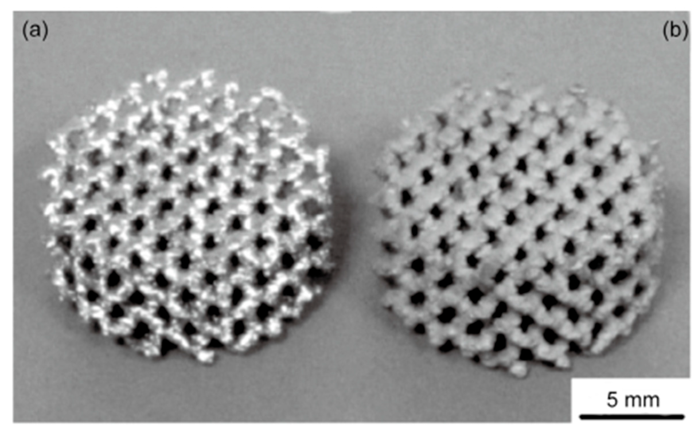
In another study, a titanium porous implant with a diamond-like atomic arrangement was produced using EBM in collaboration between Shanghai Jiao Tong University and the Institute of Metal Research, Chinese Academy of Sciences. The implant surface was subsequently covered with a tantalum layer through chemical vapor deposition (CVD) (see Figure 2). The resulting implant combined excellent mechanical and biological properties. The Ti6Al4V implant had a porosity of 70.5 ± 0.6%, an average pore diameter of 700 ± 50 μm, and an elastic modulus of 11.3 ± 0.4 GPa. This elastic modulus is particularly significant because it is equivalent to that of human cortical bone, ensuring good mechanical compatibility between the implant and the host tissue.

3.2. Trabecular Metal Implants

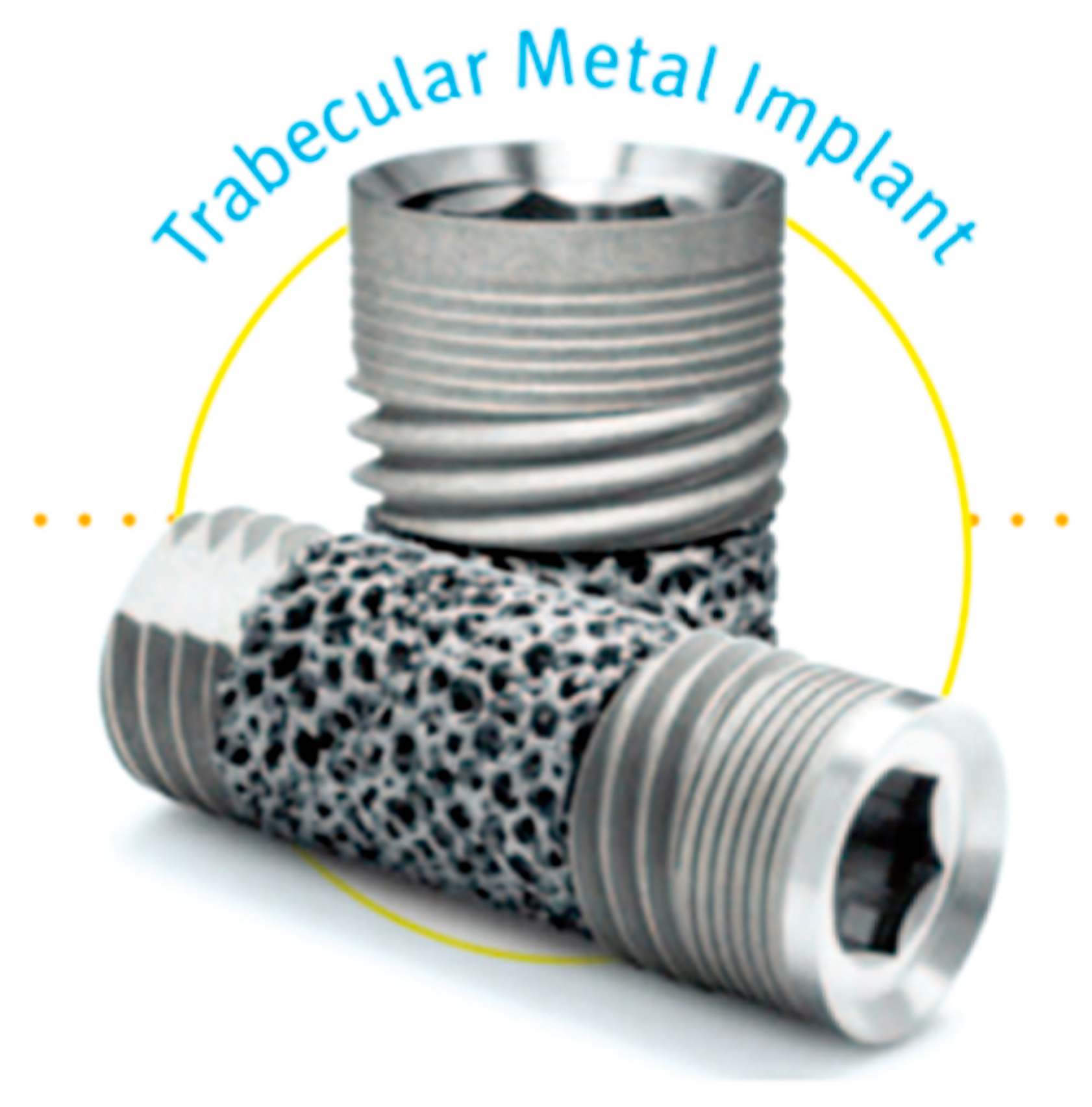
Zimmer (USA) has produced a porous tantalum dental implant using chemical vapor deposition (CVD) (see Figure 3). The implant parameters are as follows: porosity ranges from 15% to 85%, pore size from 400 to 600 μm, and elastic modulus from 1.5 to 3 GPa [31].
Cobalt–chromium alloys, titanium-based materials, and corrosion-resistant steel rank among the most prevalent materials used in orthopedic implant manufacturing. Various advancements—such as surface treatments and the development of porous configurations—have contributed to their strong clinical performance. Despite this, they present certain drawbacks, including limited bulk porosity, a relatively high elastic modulus, and suboptimal frictional characteristics [32].
The emergence of porous tantalum (Ta) has led to the creation of implants that are not only stronger but also exhibit superior biocompatibility, making them well-suited for orthopedic, craniofacial, and dental applications. Tantalum’s porous framework offers substantial volumetric porosity, a reduced elastic modulus, and a relatively elevated coefficient of friction [32].
Although tantalum (Ta) is characterized by its high biocompatibility, inertness, and outstanding anti-corrosive properties, its application in orthopedic implants was historically constrained by the difficulty of processing solid tantalum. In the past, dense materials like titanium and porous substances such as tricalcium phosphate (TCP) or hydroxyapatite (HA) were commonly used for bone replacement. Attempts were also made to coat alloy surfaces, including those of cobalt–chromium and titanium, with HA or TCP. However, these methods failed to replicate the trabecular bone microarchitecture typical of the inner layer of bone tissue. Additionally, the coating materials lacked sufficient elasticity and plasticity, leading to mechanical failures. Only in the early 1990s was the PTTM material introduced [33].
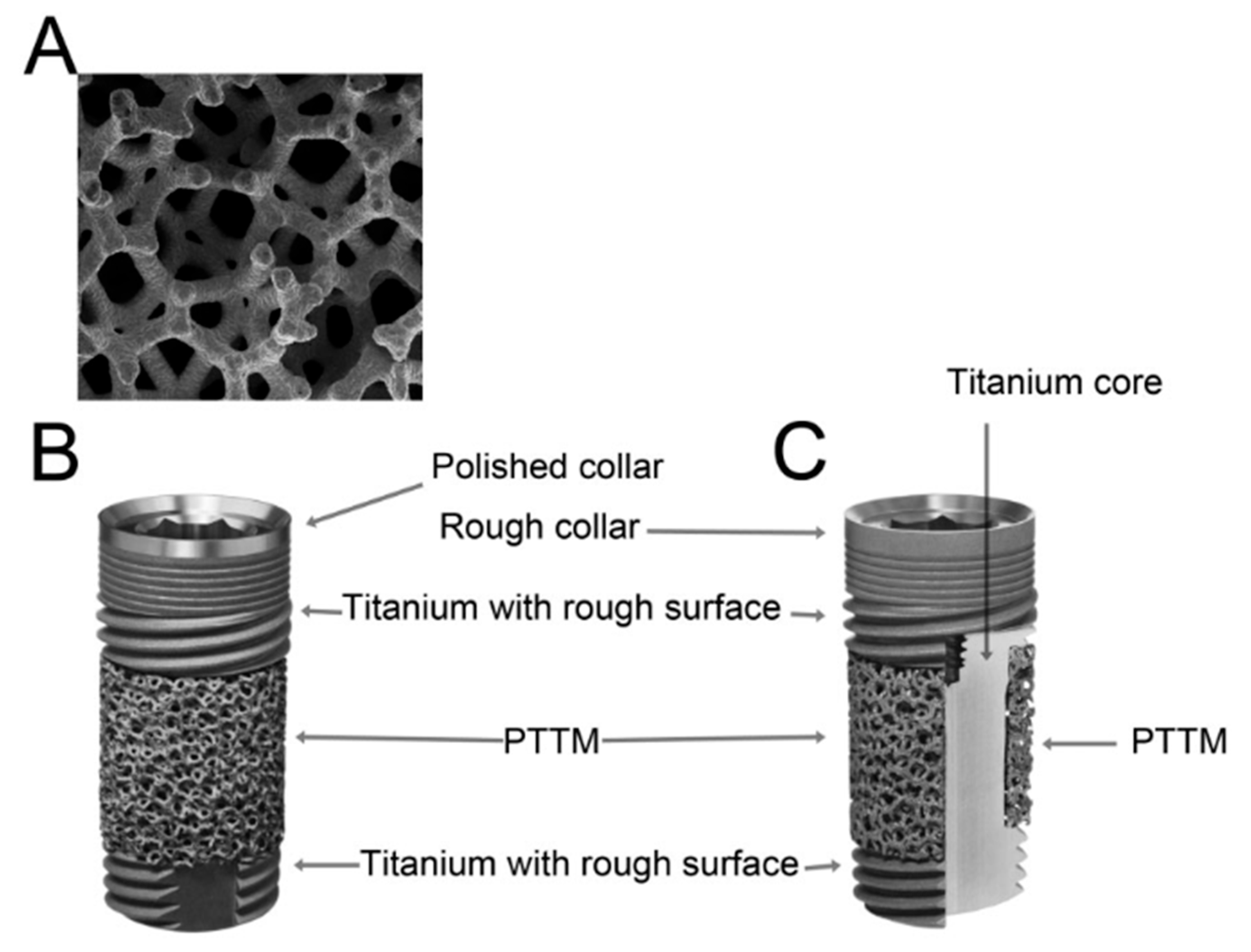
PTTM, sold under the trade name trabecular metal material (Zimmer, Trabecular Metal Technology, Inc., Parsippany, NJ, USA), is a porous, structural biomedical material with an open pore network that resembles the trabecular structure of bone and is made up of three-dimensional repeating dodecahedral units (see Figure 4A). These open dodecahedrons are formed using a glassy carbon foam framework [34,35], which acts as the initial base structure and ultimately forms the internal structure of the PTTM implant.
Figure 3. Porous tantalum dental implant [35].
Sci 07 00109 g003
Figure 4. Titanium dental implants reinforced with porous tantalum trabecular metal (PTTM). (A) PTTM configuration; (B) complete design of a titanium dental implant incorporating PTTM, featuring a smooth titanium collar at the cervical region; (C) design of a titanium implant with a fully roughened surface and PTTM integration, illustrating a cross-sectional view of the middle third, where a porous PTTM layer surrounds a solid titanium core [33].
Sci 07 00109 g004
The glassy carbon framework is subsequently inserted into a sealed chamber. Notably, PTTM is produced using recycled tantalum (Ta), rather than tantalum mined from natural sources. This recycled tantalum typically comes from industrial waste, like condensers found in computers and mobile phones. The tantalum coating is applied to the framework through a vapor-phase chemical diffusion involving chlorine and hydrogen. In this process, chlorine evaporates as TaCl2, and tantalum molecules subsequently deposit onto the framework [34]. This approach gives PTTM an advantage over other metal implant technologies, such as titanium, due to its high porosity [35].
The carbon glassy framework that forms the trabecular bone-like structure of the implant can be adjusted and modified [17], enabling the production of various PTTM designs primarily used for orthopedic implants [36]. The literature reviews on PTTM as a material for orthopedic devices indicate that it demonstrates excellent biocompatibility, osteoconductivity, promotion of bone growth, and vascularization. PTTM improves the implant surface not only for osteogenesis but also for the integration of bone tissue into the implant itself. Its porosity-based design supports the angiogenesis and bone inside the implant. This phenomenon is known as “osteoincorporation” [37].
Although PTTM technology has been widely and successfully applied in orthopedics for nearly twenty years, its use in dental implants is still relatively recent (see Figure 4B). Designs for root-form endoskeletal implants existed even before P. I. Brånemark’s discovery of osseointegration in the 1970s and 1980s [38]. Brånemark made a revolution in surgical protocols by emphasizing the control of heat produced during implant site preparation, utilizing root-shaped implants, and allowing a healing period without load. He recognized the critical importance of temperature regulation during surgery and tested numerous implant designs before settling on a root-shaped titanium implant due to its ease of placement. Furthermore, if a root-shaped implant failed, the resulting bone defect was usually minor and easily corrected. Root-shaped implants also achieved close contact with surrounding bone, which facilitated better healing outcomes. While Brånemark’s foundational principles of osseointegration remain relevant, the practice of delayed loading of implants has been increasingly questioned over time.
Since the adoption of osseointegration concepts, implant design has seen two major advancements compared to Brånemark’s original models: the development of internal connections and modifications to the implant surface. Brånemark’s initial implants featured an external hexagonal design, which was prone to issues with screw loosening and breakage [38]. Today, nearly all implants incorporate some form of internal connection. Just as importantly, Brånemark’s first implants had a machined, relatively smooth surface. Like the external hex connection, smooth surfaces have largely disappeared from the market, as they were often linked to peri-implant bone loss—a phenomenon Brånemark’s team originally considered physiological [39].
Today, all modern implants feature surface designs that incorporate elements to enhance roughness, thereby expanding the intersection of the implant and adjacent bone and promoting better osseointegration. Research has demonstrated that improving surface roughness effectively minimizes bone resorption around the implant, an issue noted with the original Brånemark implant models.
The desired surface roughness or texture can be achieved through various methods, including abrasive jet treatment, plasma spraying, acid etching, or combinations of these methods [40]. Additionally, by adjusting surface roughness or applying specialized coatings, it is possible to enhance the hydrophilicity of the implant surface or decrease the contact angle of liquids [41]. These surface modification approaches improve bone-to-implant contact at both the micro- and nanoscale, while also reducing the surface free energy.
Recently, PTTM technology has been used to develop a three-dimensional matrix that facilitates bone ingrowth around dental implants (see Figure 4B,C). The PTTM material is incorporated into the core of a self-tapping endosseous, titanium multi-threaded dental implant (Fan-type Tapered Screw Implant, Zimmer Dental Inc., Carlsbad, CA, USA).
The lower (mandibular) and cervix-related parts belonging to the titanium implant reinforced with PTTM maintain a helical design with a roughened surface produced by treating with a blast of hydroxyapatite (HA) particles (MTX surface, Zimmer Dental Inc.). The titanium-based material (Ti6Al4V, Grade 5) and PTTM assemblies are manufactured apart. The cervical portion and titanium core are machined as a single unit, while the part located at the tip is produced individually. The PTTM cylindrical covering, roughly 2 mm in diameter, consists of a glassy carbon core (2%) coated with tantalum (98%). This sleeve is inserted into the titanium central core, which is subsequently joined to the apical part using laser welding [34].

3.3. Zirconia Implants

Although additive manufacturing (AM) offers many benefits, it has not yet received official certification as a standard production method for zirconia-based ceramic restorations. Research on the additive technologies used in dental zirconia ceramic production is in its initial phase, but existing studies suggest strong potential for the application of additively manufactured zirconia in dentistry [42]. So far, some investigations have explored the application of AM techniques for fabricating zirconia using commercially available technologies. These efforts generally fall into three main categories: dental restorations, zirconia implants, and bone tissue regeneration [43].
The earliest phase of ceramic dental implants was based on aluminum oxide [44]. Several systems using this material were developed, including monolithic alumina implants. Although these implants demonstrated the ability to integrate with bone, their biomechanical limitations—particularly in terms of fracture resistance—proved to be inadequate. Clinical investigations reported long-term survival rates ranging from 65% to 92% [45]. However, the variability in outcomes prevented the establishment of clear clinical guidelines for routine use, which ultimately led to the discontinuation of alumina-based implants in the early 1990s. As a response to these shortcomings, zirconia ceramics with superior characteristics were introduced. Initially, zirconia found application in crowns and implant abutments (Figure 5) [46]. Today, the preferred ceramic material for dental implant production is tetragonal zirconia polycrystal, especially the 3 mol% yttria-stabilized variant (3Y-TZP) [47]. Its opaque white colour, coupled with promising biocompatibility and low bacterial adhesion, has drawn considerable attention in biomedical research [48,49]. In vitro analyses have not revealed any carcinogenic or mutagenic effects [50]. Zirconia also offers advantageous mechanical and physical features, such as high flexural strength (900–1200 MPa), low thermal conductivity, excellent resistance to wear and corrosion, and favorable fracture resistance. One of the key phenomena contributing to its mechanical robustness is transformation toughening. This process involves a stress-induced phase transition from a phase transformation from a tetragonal to monoclinic structure, leading to a 4% increase in volume, which generates compressive stresses that inhibit crack propagation and enhance the material’s durability [51,52]. Nonetheless, zirconia is subject to deterioration at low temperatures or material aging. When exposed to moisture or water vapor, a gradual transformation to the monoclinic phase occurs, increasing surface roughness and eventually compromising the structural integrity of the implant [53]. This aging process results from the accumulation of compressive stresses and microcracks, with its extent being influenced by their relative balance. Furthermore, manufacturing parameters such as implant shape and surface texture can also impact this phenomenon, although these influences are not yet fully understood. At present, most zirconia implants are created as single units [54]. While they offer some structural simplicity, these designs come with inherent limitations. For example, the surgical placement may not always correspond to ideal prosthetic positioning, and the absence of angled abutments makes correcting deviations difficult. Post-operative adjustments like grinding are discouraged, as they significantly weaken the implant’s fracture resistance. Additionally, one-piece implants are subject to immediate mechanical forces from functional movements such as chewing and tongue pressure, even when provisional prosthetics are in use [55]. Cementation remains the predominant method for securing prosthetic components to single-unit zirconia implants. Meanwhile, the elimination of a micro-gap at the junction of the implant and abutment can be advantageous [56] and accurate vertical positioning becomes more challenging [57]. In aesthetic regions, implants are often placed lower to conceal the edge of the crown, which enhances the likelihood of excess cement accumulating in the submucosal area [58]. This residual cement is often undetectable radiographically [59] and can lead to localized infections or even serious tissue complications [60]. A recent systematic review [61] found that restorations attached using cement were associated with a substantial increase in mechanical and biological issues, compared to those fixed with screws. Currently, only a limited number of ceramic-based structures support dual-component implant configurations. According to two clinical trials [62], pre-manufactured zirconia-based abutments have been affixed to implants using dual-component resin cement. In two other studies [63], customizable fiberglass abutments were bonded to implants using adhesive methods. A key difficulty in these systems is ensuring a reliable and long-lasting connection between the implant and the abutment. In all reported studies, no cases of abutment loosening or detachment were observed. In addition, no fractures were observed in studies by Brüll et al. [61] or Payer et al. [60]. However, Becker et al. [61] documented a case where a fiberglass abutment fractured 23 months post-loading, corresponding to a 2.1% rate of technical complications. Similarly, Cionca et al. [59] reported two abutment fractures in two separate patients—one occurring after 10 days and the other after 8 months—resulting in a rate of adverse outcomes of 4%. Additional concerns associated with this form of abutment–implant link include proper closure and complete elimination of excess adhesive. A single study [60] reported using a rubber dam during the abutment–implant junction process.

3.4. Polymers and Composite Materials

Nearly all early experiments involving polymer implants based on methyl methacrylate (MMA) were unsuccessful. However, in 1969, M. Chodosh and colleagues [64] reported the successful development of polymethacrylate implants for dental restorations. These implants proved to be biocompatible, marking the beginning of further advancements in the use of polymers for dental implants. This material was employed to replace missing teeth with precise replicas, demonstrating its effectiveness in restoring both function and aesthetics [65].
Polymers offer several advantages, including the potential to modify physical characteristics by altering their structure (for instance, creating softer or more porous structures), ease of processing, high reproducibility, improved connective tissue attachment, and simpler microscopic evaluation compared to metal implants. They also possess excellent aesthetic qualities [66]. Among the most accessible polymers is polylactic acid (PLA), which is derived from renewable and biodegradable raw materials like potatoes, corn, rice, or starch [67]. In dental implantology, PLA is notable for its biodegradability and ease of curing. Its biomechanical characteristics are comparable to engineering polymers like poly(lactic-co-glycolic acid), but PLA stands out due to its lower cost and greater availability.
Studies have evaluated porous PLA composites combined with recombinant bone growth factor protein 2 (rhBMP2), demonstrating their capacity to promote bone formation within two weeks through effective protein delivery [68].
Additionally, PLA/octadecylamine nanocomposites with nanodiamond additives have been developed, offering improved mechanical properties (including hardness and Young’s modulus), non-cytotoxicity, and enhanced biocompatibility due to robust intermolecular interactions between the filler and polymer matrix [69].
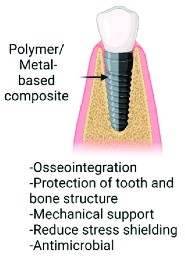
In bone regeneration applications, implant components produced from PLA-PGA (polyglycolic acid) copolymers and their blends are commonly used, as they enable control over degradation rates while remaining non-toxic and biologically compatible [70]. These features make PLA a promising candidate for fabricating frameworks for dental implants (Figure 6), enhancing the mechanical performance of natural polymer materials.
Selecting appropriate functional groups in polymeric materials enables the design of structures with specific properties, allowing the development of synthetic polymers with consistent, predictable, and tunable characteristics. By modifying their chemical composition, these materials can be tailored for applications [71].
Comparative studies of natural and synthetic polymers used in dentistry reveal that synthetic polymers typically exhibit lower bioactivity and osteoconductivity and lack sites that are recognized by cells. However, the breakdown rate of synthetic polymers is able to be precisely managed by adjusting factors such as chemical composition, crystallinity, and molecular weight. Considering these findings, researchers are exploring various ceramic and polymer coatings to enhance surfaces and promote bone regeneration. Among synthetic polymers applied in dentistry, the most common are aliphatic polyesters like PCL [72], PDLA, PLLA [73], and PLGA [74], as well as materials such as polyetherketoneketone (PEKK), polyetheretherketone (PEEK), TEGDMA, Bis-GMA and UDMA. This section discusses recent progress in the development of polyetheretherketone, Bis-GMA, and polyetherketoneketone-based composites for dental implant applications.
The Bis-GMA/TEGDMA system is a resin matrix that cures under visible light. It is composed of bisphenol-A-glycidyl methacrylate (Bis-GMA) mixed with triethyleneglycol dimethacrylate (TEGDMA) and reinforced with fillers including quartz, zirconia, barium, and silica. Due to their excellent aesthetics, sufficient strength, and cost-effectiveness compared to ceramics, Bis-GMA/TEGDMA-based resin composites are commonly utilized in dental practice. They also exhibit strong adhesion to dental tissues [75]. These thermosetting materials are well-suited for high-stress applications, such as dental implants, particularly when supported by E-glass fibers. The three-dimensional polymer network of Bis-GMA facilitates the formation of durable physical bonds, while its low volatility allows it to easily infiltrate 3D-printed structures, where it solidifies into a strong material upon polymerization. These features make Bis-GMA an ideal matrix material for resin composites in the fabrication of dental implants [76].
A review of current and past literature on polymers and polymer composites as materials for dental implants shows that their application in dental implants has not been extensively explored. Despite their relatively low mechanical strength, these materials have garnered significant attention in dentistry due to their processing flexibility, adjustable properties, and cytocompatibility. As biotechnology and healthcare continue to advance, the dental industry is gradually adopting these materials to enhance treatment outcomes for patients. Polymers are affordable, easy to process, and can be adapted for intricate designs and combined with other materials in dental implants. In recent decades, the rise in dental diseases has become a notable public health concern [77]. Meanwhile, traditional solutions such as metal implants and autografts have several drawbacks, including resorption, the risk of immune rejection, or tissue encapsulation. This underscores the need for new and effective strategies in tissue engineering to address these medical challenges. Today, researchers are focused on optimizing the material properties to satisfy biomechanical demands. Essential attributes of ideal implant materials include suitable porosity, specific mechanical and chemical characteristics, and biodegradability—qualities that provide support like the extracellular matrix in polymer composites [78]. Incorporating hybrid reinforcing elements into polymer biomaterials has greatly enhanced their biomechanical performance, potentially enabling them to replace conventional metal and polymer materials. Furthermore, these materials promote bone regeneration and cell growth by facilitating calcium phosphate formation [79].

3.5. Evaluation of the Properties of Selected Materials

This comparative table provides a detailed overview of the structural characteristics of selected components applied in the fabrication of dental implants via additive manufacturing (see Table 1). It focuses on three key parameters: tensile strength, fatigue resistance, and elastic modulus—properties that significantly influence the functionality, durability, and biological compatibility of implants during long-term application in the human body.
Porous titanium implants exhibit a tensile strength ranging from 550 to 900 MPa, which is sufficient for most clinical applications. Due to their open-porous structure, their strength is slightly reduced compared to solid titanium; however, they offer high fatigue resistance, which is critical under repetitive mechanical loading in the oral cavity. Their elastic modulus, ranging between 10 and 30 GPa, is closer to that of bone tissue, which reduces stress shielding and supports osseointegration [80].
Trabecular metal, commonly represented by tantalum, has a lower tensile strength (200–300 MPa), but its excellent biocompatibility and structure mimicking cancellous bone make it suitable for implants in low-load regions. It demonstrates exceptionally high fatigue resistance and a very low elastic modulus (3–5 GPa), contributing to favorable load transfer to the surrounding bone [81].
Zirconia implants (zirconium dioxide) reach very high tensile strength values (900–1200 MPa), making them suitable for high-load applications. However, their fatigue resistance is moderate, due to the brittle nature of ceramics and susceptibility to microcrack formation. Their elastic modulus, ranging from 200 to 210 GPa, is significantly higher than that of bone, which ensures structural rigidity but may lead to undesirable stress shielding [82].
Polymers used in dental implantology offer low tensile strength (50–100 MPa) and limited fatigue resistance. However, their low elastic modulus (1–5 GPa) makes them suitable for applications where shock absorption and reduced stiffness are desired. Their use is typically limited to temporary or auxiliary implant components [82].
Composite materials, combining the benefits of different constituents, offer tensile strength between 100 and 300 MPa and an elastic modulus of 5–20 GPa. Their fatigue resistance ranges from moderate to high, depending on the specific composition. These materials represent a versatile alternative with tunable mechanical properties, making them adaptable to specific clinical requirements [82].
Overall, the table demonstrates that the selection of suitable materials for dental implants depends on mechanical load requirements, biocompatibility, and the intended implant location. When combined with appropriate additive manufacturing technologies, these materials can enable the production of individualized solutions with a high success rate.

4. Additive Technologies Utilized in the Manufacture of Dental Implants

This chapter provides a detailed description of selected additive manufacturing technologies that are currently regarded as the most promising—or are already clinically applied—in the fields of dentistry and implantology. Specifically, it focuses on selective laser melting (SLM), electron beam melting (EBM), direct metal laser sintering (DMLS), stereolithography (SLA), and selective laser sintering (SLS).
These technologies were chosen based on their demonstrated ability to process materials suitable for dental applications—like titanium, cobalt–chromium-based materials, and high-performance polymers—and their relevance in the fabrication of precise, patient-specific prosthetic components, implants, bridges, crowns, and other restorations.
The chapter concludes with a comparative table summarizing the key characteristics of these technologies, including energy type, processable materials, positioning accuracy, surface quality, and principal limitations. This table provides a concise overview of the relative advantages and challenges of SLM, EBM, DMLS, SLA, and SLS, particularly in terms of their suitability for dental applications and practical use in clinical or laboratory settings.
This chapter aims to provide a comprehensive, accessible, and technically accurate overview of additive manufacturing methods relevant to current and future practice in the field of personalized implantology and prosthodontics.
Additive manufacturing technology, often referred to as 3D printing or rapid prototyping, operates on the principle of layer-by-layer material deposition and fusion. This method involves sequentially applying layers of material to build complex three-dimensional objects, with the entire process guided by computer programs based on CAD models and data. In contrast to traditional manufacturing methods such as machining or milling, which remove excess material, additive manufacturing creates products “from the bottom up” [83].
This approach significantly reduces production time by eliminating the need for cutting tools, fixtures, and multi-step molding processes. Furthermore, the greater the design complexity, the more pronounced the time savings in production [84]. Additive manufacturing technology first emerged toward the end of the 1980s, and since then, around 20 different methods have been created. Some of the notable techniques include the following:
  • Stereolithography (SL);
  • Laminated object manufacturing (LOM);
  • Laser cladding (LCF);
  • Selective laser sintering (SLS);
  • Selective laser melting (SLM);
  • Electron beam melting (EBM).
These technologies find applications across a variety of industries, comprising automation, consumer medicine, electronics and dentistry, industrial engineering, aerospace, and more.

4.1. SLM and EBM

SLM (selective laser melting) and EBM (electron beam melting) are two advanced 3D-printing technologies that are well-suited for manufacturing metal components with complex geometries.
Selective laser melting (SLM) is regularly employed in the fabrication of metal dental implants, particularly those made from titanium alloys, owing to its high precision of dimensions and the superior load-bearing capacity of the manufactured components. The technology enables the creation of customized implants tailored to individual patients with tailored geometry and controlled porosity, thereby enhancing osseointegration and biological integration with the surrounding bone tissue.
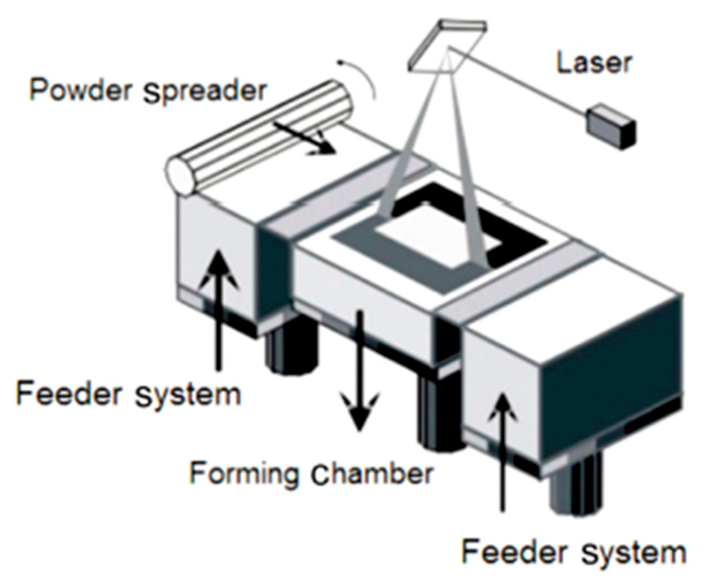
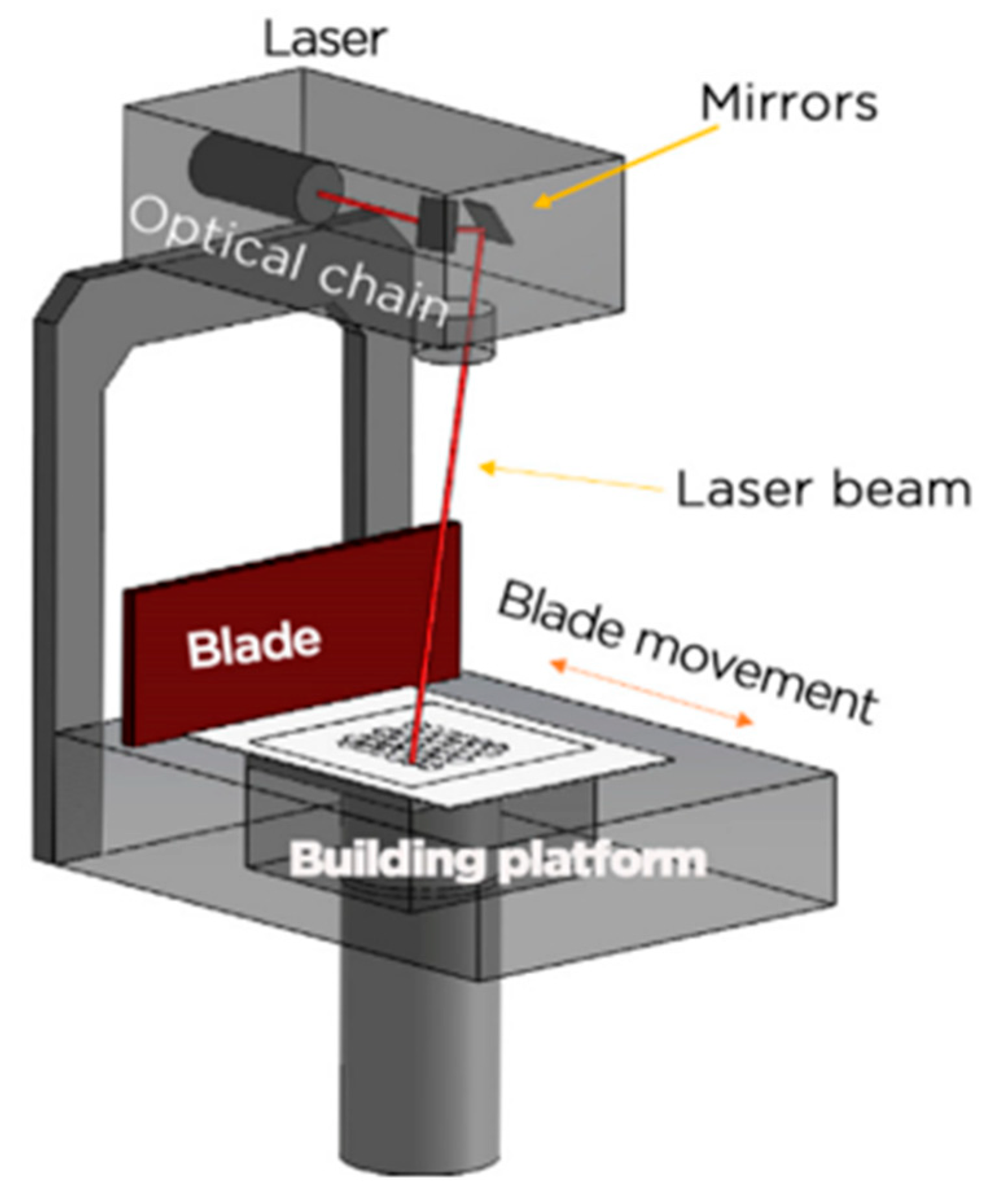
In the SLM process, the forming occurs within an inert atmosphere chamber, where a laser beam scans a layer of metal powder, which is successively deposited by a powder spreader along a predefined path. The solid object is created by metallurgically bonding the layers of metal powder, effectively melting each layer one by one (see Figure 7) [85].
In contrast, the EBM (electron beam melting) process occurs in a vacuum environment, in which a powdered metal is fused using an electron beam. The principle of layered formation in EBM is like that of SLM, with the object being fabricated layer upon layer (see Figure 8).
The primary difference between these two technologies is based on their heat source: SLM employs a laser, whereas EBM utilizes an electron beam. Their advantages and disadvantages are as follows:
SLM:
  • Higher forming accuracy due to smaller laser beam diameter and finer powder particle size.
  • Cheaper equipment and more accessible technology.
  • Disadvantage: lower production speed.
EBM:
  • Significantly higher production speed of up to 80 cm3/h, which is four to five times faster than SLM, because of the high energy output (up to 3000 W—ten times the power of SLM).
  • Using larger powder particles in this technology results in lower molding precision compared to SLM.
Both methods have distinct advantages and are chosen based on the specific requirements for accuracy, production speed, and product geometry complexity. Selective laser melting (SLM) technology was initially introduced by the Fraunhofer Institute in 1995, and the first functional SLM technology was created by the German company MCP in 2003. Subsequently, several German companies, including EOS, Concept Laser, and ILT, have produced commercialized SLM systems. Recently, EOS launched the EOSINT M270, featuring a 200 W ytterbium (Yb) solid-state fiber laser. Key specifications of the EOSINT M270 include a powder layer thickness ranging from 20 to 100 μm, laser beam focal point diameters between 70 and 200 μm, a standard scanning rate of 750 mm/s, part accuracy between 20 and 80 μm, and a surface texture of 10 to 15 μm. SLM-manufactured components have been successfully applied in automotive, medical, aerospace, plastic injection molding, and other technical industries. In 2003, the Rapid Manufacturing Center at Huazhong University of Science and Technology began developing SLM systems using a 150 W YAG 100 W fiber laser and diode laser, initiating SLM research in China. In 2007, South China University of Technology introduced the DiMetal-280 prototype—an upgrade of the DiMetal-240—equipped with an ytterbium fiber laser. DiMetal-280 specifications include a laser spot size of 50 to 200 μm, scanning speeds between 200 and 600 mm/s, manufacturing precision ranging from 20 to 100 μm, and surface roughness between 20 and 30 μm. The Beijing Institute of Aeronautical Manufacturing Engineering developed China’s largest SLM system, the LSF-M360, capable of producing parts up to 350 × 350 × 400 mm. These developments highlight China’s significant progress in SLM equipment and technology R&D. Nonetheless, crucial components such as lasers, optical focusing assemblies, and high-speed galvanometer scanners still depend on imports, which remain a substantial technological challenge. Research on electron beam melting (EBM) began in the early 2000s. In 2003, Sweden’s Arcam developed the world’s first commercial EBM system. This system featured maximum build dimensions of 200 × 200 × 160 mm, a powder layer thickness between 50 and 200 μm, scanning speeds over 1000 mm/s, and a laser spot size ranging from 300 to 500 μm. The process precision ranged from 200 to 400 μm [86]. Numerous research institutions and universities in the US, Japan, the UK, and elsewhere have acquired Arcam equipment to conduct studies across various fields. Currently, more than 100 EBM systems are in operation worldwide [87]. In 2004, Tsinghua University developed China’s first domestically designed EBM prototype (EBSM150) featuring a top scanning speed of 2 m/s [88]. In 2008, the academic institution unveiled the revised model of the EBSM250 system, which offered maximum build dimensions of 230 × 230 × 250 mm. This study addressed several important aspects of the EBM process, including powder preheating, scanning path planning, and analysis of the mechanical properties of the finished components. Data show that load-bearing parts produced from the aerospace alloy Ti6Al4V using EBM exhibit excellent mechanical properties, comparable to those of forged components [89]. Research on porous titanium alloys produced by EBM has also garnered considerable attention, particularly at the North-West Research Institute of Non-Ferrous Metals, which has accumulated significant expertise in this area [90]. Advances in 3D-printing technologies like SLM and EBM have been driven largely by innovations in equipment, improvements in processing methods, and the development of metal powders. Within this framework, the preparation of metal powders has a key function in technological progress. Additive manufacturing imposes strict requirements on metal powders: they must have narrow size distribution, good flow characteristics, fine particle size, high sphericity, and low oxygen content [91]. Currently, the primary technologies for producing metal powders for 3D printing include plasma spheroidization, plasma atomization, electrode induction gas atomization (EIGA) and vacuum induction gas atomization (VIGA). Nevertheless, the variety of available powdered metallic material remains limited. Currently, only a handful of alloys—such as Ti6Al4V, CP-Ti, In625, and CoCrMo—are commonly used in production.
Electron beam melting (EBM) is primarily utilized for processing biocompatible metals, such as titanium, and is well-suited for the production of implants requiring high mechanical strength and minimal residual stress. Due to its operation in a vacuum environment and the resulting relatively rough surface finish, EBM is often applied in cases where enhanced integration with bone tissue is desired.
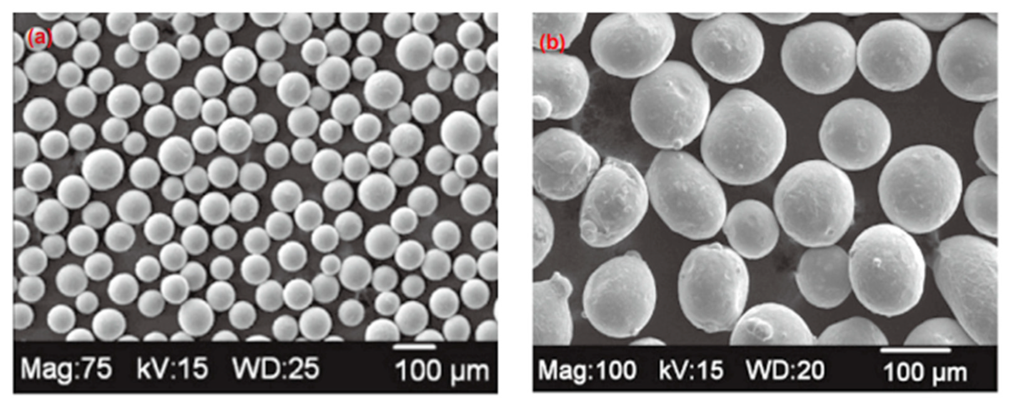
The AP&C Metal Powder Division of Raymor Industries (Canada) concentrates on plasma atomization and holds a prominent position in the metal powder supply market. AP&C’s products feature high particle sphericity, uniform particle size distribution, low oxygen content, exceptional purity, and high bulk density. This company supplies metal powders used by the Swedish firm Arcam. In the United States, Ametek employs plasma spheroidization, hydrogen dehydrogenation, and gas atomization techniques to produce spherical metal powders with mean particle measurements of roughly 100 μm in diameter (see Figure 9). The German company ALD Vacuum Technologies utilizes VIGA and EIGA methods to manufacture top-grade metallic powders with fine particles and a tight size distribution. Even so, the production of high-quality metal powders for additive manufacturing in China still largely depends on imports.

4.2. DMLS

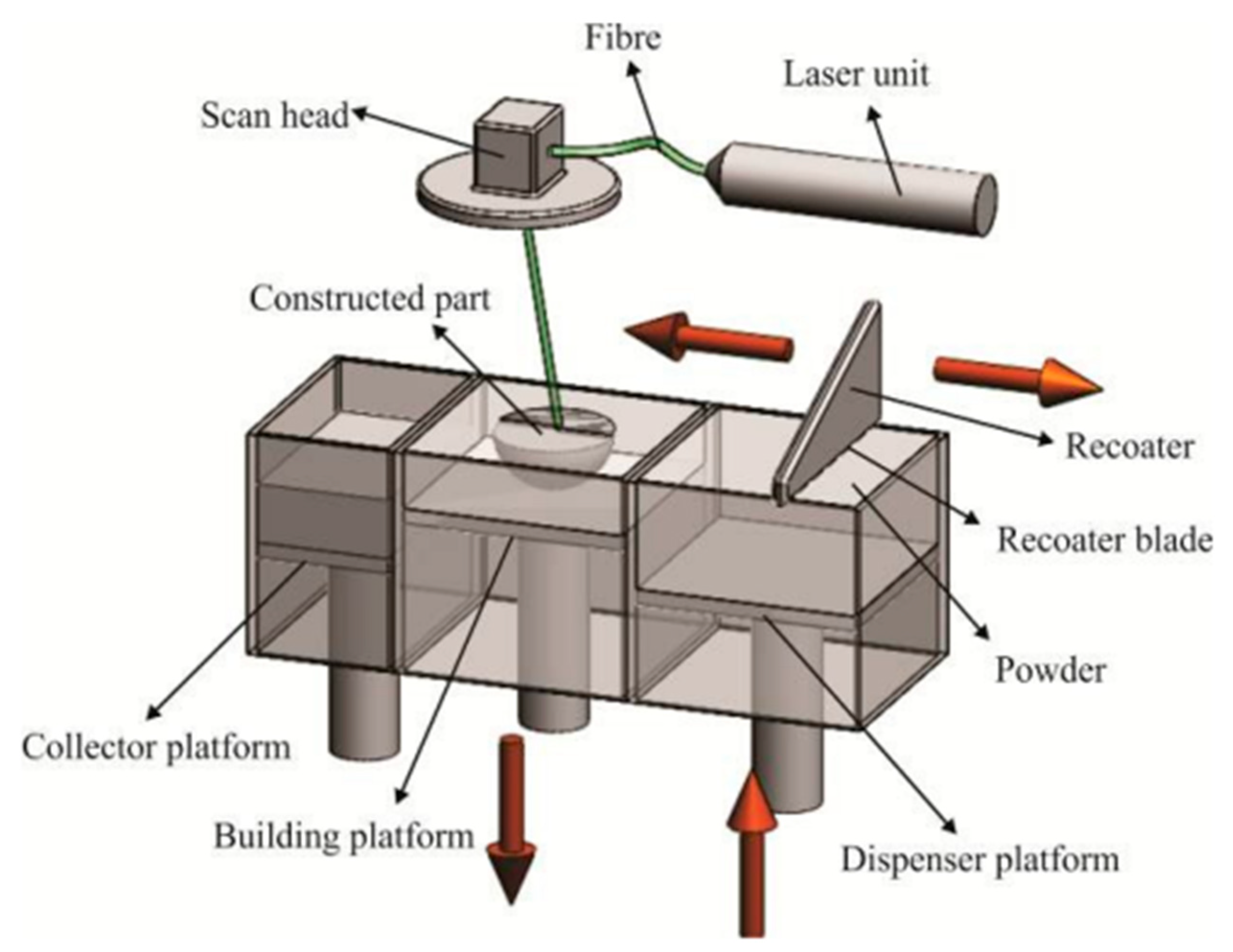
A schematic representation of the DMLS system is presented in Figure 10. To produce a part, the machine follows these steps: first, the build and feed platforms are reduced by the thickness of one layer, allowing the powder application mechanism to move freely. When the distributor (coater) is in its starting position, the feed platform rises, supplying the required amount of powder for the next layer. The distributor then moves from right to left, evenly spreading the metal powder from the feed area onto the build platform, while excess powder is collected in a dedicated container. Next, the scanning head moves into position, directing the laser beam to trace the 2D contour of the layer. The laser switches on and off precisely to expose only the designated areas. As the metal powder absorbs the laser energy, it sinters and fuses with the previously solidified layers. This sequence repeats gradually, layer upon layer, until the entire structure is formed. In this way, complex three-dimensional parts with high precision can be produced in just a few hours. Additionally, many of the part’s functional properties are established throughout the sintering stage itself. However, depending on the intended use of the part, further processing such as annealing or surface finishing may be required [92].
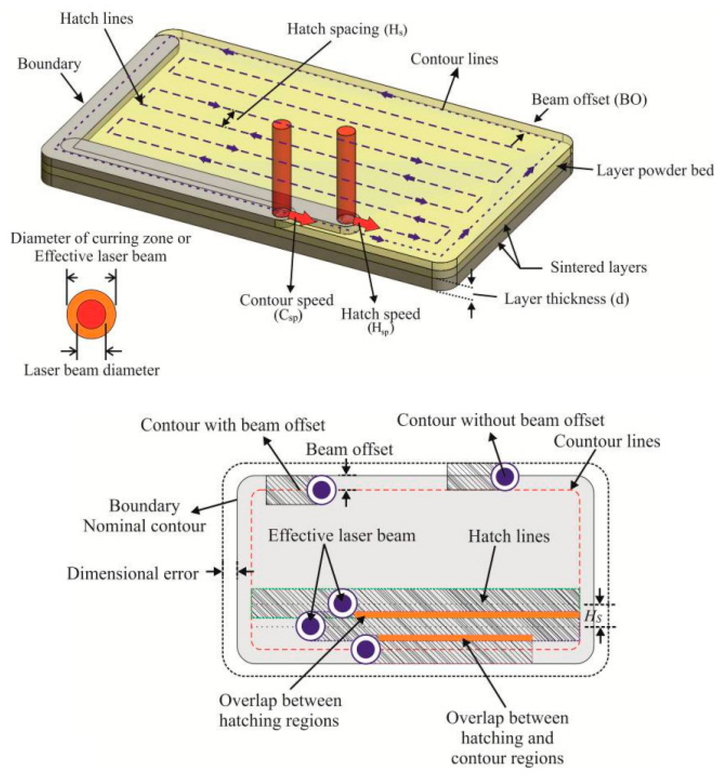
Figure 11 illustrates the process of scanning by laser across the upper layer of a slender powder layer to create a region defined by the model’s cross-sections. Initially, the entire layer contour is scanned using the designated laser power (Lpw) and contour scanning speed (Csp). Because the diameter of the sintered area is generally larger than the laser beam itself, it is necessary to consider the effective laser diameter [93] or the melt pool size [92] to account for potential dimensional deviations. As a result, the laser beam is offset inward by half the width of the sintered zone—this adjustment is known as the beam offset (BO).
During the hatching process, the laser makes multiple passes along parallel lines to ensure complete sintering and to sustain elevated temperatures over time. The distance between these lines, referred to as the hatch spacing (Hs), is typically adjusted to approximately 25% of the beam’s diameter. The beam offset is likewise determined relative to the contour’s edge (see Figure 11). If the offset deviates from the optimal value—whether too high or too low—the powder particles in the exposed area may be insufficiently or excessively sintered, potentially affecting the precision and overall quality of the final part [94].
Another critical characteristic that can lead to part deformation or disruptions in the process can be caused by inappropriate layer thickness. If the thickness is too great, proper interlayer cohesion may not be achieved because the sintering depth is insufficient. Additionally, mechanical stresses can develop within the layer, potentially causing it to separate from the underlying layer [94].
Direct metal laser sintering (DMLS) is a widely adopted method for the fabrication of dental implants from metallic materials such as titanium and cobalt–chromium, owing to its high resolution and the excellent mechanical strength of the printed components. It enables the efficient production of complex geometries with detailed surface features, thereby enhancing implant precision and customization to meet individual patient-specific requirements.
Conversely, if the layer thickness is too small, the structure may be damaged during the application of the next layer, as sintered particles can become trapped between the part and the recoater blade, disrupting the process or damaging the forming part. Therefore, precise adjustment of layer thickness is essential for the successful and high-quality fabrication of 3D parts using DMLS [95].

4.3. SLA

Stereolithography (SLA) marked the introduction of additive manufacturing (AM) technologies being introduced into the medical field, with its initial use reported in 1994 for creating surgical models used in alloplastic implant procedures [96]. SLA works by selectively polymerizing a photosensitive resin under a light source, such as a laser or LED, building the component gradually, one layer at a time. The process typically includes photopolymer mixtures, often combined with ceramic powders [97]. During fabrication, light is directed onto the surface of the resin bath to cure each layer. After curing, the build platform gradually lowers to allow the next layer to form. This cycle repeats until the entire structure is complete. A schematic of the SLA system is shown in Figure 12. A crucial parameter affecting SLA productivity and resolution is the depth of cure. SLA is recognized for its precision, excellent surface quality, and ability to create intricate shapes [98], without requiring high-power lasers. These features have made SLA one of the most employed additive manufacturing techniques, particularly for producing zirconia-based components [99]. In ceramic-based SLA processes, manufacturing begins by dispersing fine ceramic particles, from microns down to nanometers in size, into a photosensitive resin [96]. To achieve a stable suspension, surfactants and dispersants are used to evenly distribute the particles. Since ceramic particles do not react to light, photopolymerization takes place within the organic monomer matrix, which encapsulates the ceramic particles and forms the desired layer shape. This process continues until the full 3D object is printed. The resulting “green” parts require post-processing, typically involving pyrolysis to remove organic binders, followed by high temperature sintering to achieve the required density and mechanical strength [97]. The composition and behavior of the ceramic suspension are critical to the success of SLA. Factors such as light absorption and suspension rheology have a significant impact on print quality. Producing zirconia parts with minimal defects and good structural properties requires a suspension with low viscosity (ideally below 3000 cps), a high solids’ content, and uniform particle dispersion. However, increasing ceramic content raises viscosity, which complicates the printing process. Most commercial suspensions contain less than 40% solids by volume to maintain flowability and prevent phase separation, but this can lead to greater shrinkage and lower density in the final part [98]. Thus, there is a trade-off: lower ceramic content improves processing but reduces density and mechanical properties, while higher content does the opposite. Recent work by Zhang et al. [99] introduced a photosensitive zirconia suspension with a solids’ content of 55%, paving the way for producing high-performance zirconia components using SLA technology.
An additional challenge in ceramic stereolithography (SLA) is attributed to the pronounced effect of light dispersion resulting from the ceramic particles suspended in the medium. Even when these particles do not absorb incident light, scattering can hinder light penetration and consequently reduce the dimensional fidelity of the printed component [97]. Parameters such as particle size, volume fraction, refractive index mismatch between components, and light intensity all play key roles in determining the depth of cure, which directly affects the thickness and uniformity of each printed layer [95].
There is a pronounced mismatch in refractive indices between the zirconia particles and the photopolymer matrix. Zirconia, possessing a higher refractive index, absorbs more ultraviolet radiation upon exposure, which diminishes the depth of cure and attenuates the efficiency of the photopolymerization process [101]. Once the “green” (unfired) parts are printed, they undergo a debinding process aimed at removing residual organic materials. This stage depends on variables like the resin’s composition., the solids’ loading, and the geometry of the printed part [102]. Cracking may occur during debinding, likely because of internal stresses induced by thermally initiated polymerization reactions. The final step is sintering, during which porosity is eliminated, which yields fully densified zirconia parts with improved functional properties [103]. The resulting mechanical strength and density of the sintered components depend largely on the sintering temperature and the thermal gradient applied [101].
Among the various additive manufacturing technologies available, SLA is particularly favored for dental applications because of its high resolution, dimensional precision, and high-grade surface texture [104]. Several recent studies have evaluated the physical and mechanical characteristics of zirconia fabricated via SLA for dental use. In the study conducted by Revilla-León et al. [105], zirconia’s bending strength rods produced by SLA were compared to milled zirconia under simulated aging conditions. The findings revealed that SLA-fabricated zirconia demonstrated lower flexural strength than conventionally milled zirconia, and that simulated mastication significantly reduced bending strength and fracture resistance in both groups.
In a subsequent investigation, Revilla-León et al. [106] assessed the dimensional accuracy and volumetric shrinkage of SLA-fabricated zirconia samples with varying intended porosity levels (0%, 20%, and 40%). Using photopolymerizable ceramic slurries and digital reference models, the samples were sintered at varying temperatures—1450 °C for the 0% porosity group, and between 1225 °C and 1450 °C for the higher porosity groups. Dimensional measurements were performed using a digital caliper, and shrinkage percentages were calculated by comparing final sample dimensions to those of the original digital model. Although none of the groups achieved a perfect match with the virtual design, the one with 40% porosity demonstrated the greatest dimensional precision and minimal volumetric deformation, with the 20% and 0% porosity groups showing progressively lower performance in these aspects.
Nakai et al. [107] explored the microstructure, crystallographic structure, and flexural performance of SLA-fabricated square zirconia specimens, comparing them with milled 3Y-TZP zirconia. The study examined several AM zirconia materials, including LithaCon 3Y 230 (Lithoz) printed at a 90° orientation, 3D Mix Zirconia (3DCeram Sinto) at a 0° orientation, and alumina-toughened zirconia (ATZ) also printed at 0°, in comparison with milled 3Y-TZP. Their results showed that the crystalline phases and remaining porosity in the zirconia samples produced by SLA closely resembled those of milled zirconia. Flexural strength tests revealed comparable performance between AM and milled 3Y-TZP samples. Among the additively manufactured variants, the ATZ group exhibited the highest strength, followed by SLA-fabricated zirconia printed perpendicular to the load (0° orientation), while DLP-fabricated zirconia printed at a 90° orientation (aligned with the loading direction) exhibited the lowest strength measurements [107].

4.4. SLS

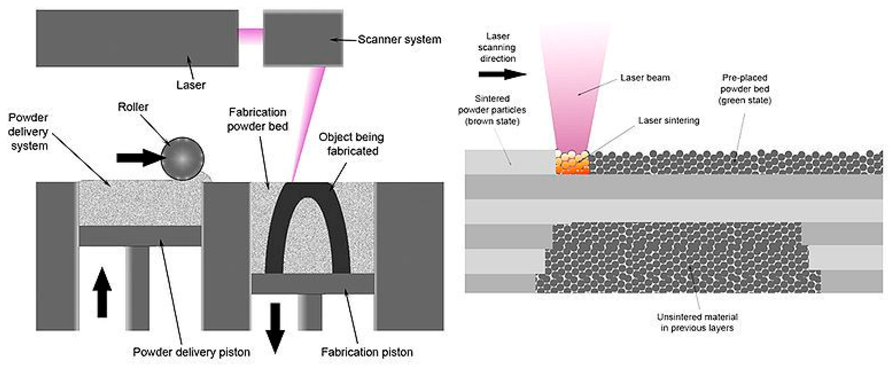
Selective laser sintering (SLS) is an additive manufacturing (AM) method that constructs three-dimensional structures through the sequential layering of ceramic-based material [108]. As the technology’s name implies, this method uses a high-power laser beam to selectively scan the surface of each powder layer. The localized application of energy heats the powder particles, initiating the sintering process—where particles fuse together through mass-transfer mechanisms—without fully melting them (see Figure 13). Once a layer is sintered, a new layer of powder is evenly distributed across the surface, and the laser scanning and sintering process is repeated. This sequence proceeds layer upon layer until the complete three-dimensional structure is built [109].
One of the advantages of selective laser sintering (SLS) technology is that no extra support structures are needed, since overhangs and undercuts are inherently stabilized by the unsintered powder surrounding them in the build area [109]. In ceramic manufacturing, the SLS process is generally categorized into two main approaches: immediate and mediate SLS. In the indirect method, ceramic particles are blended with a polymer-based binding agent. During the build, the laser selectively liquefies the binder, which holds the ceramic components together. Once the desired geometry is achieved, the binder is removed through a degassing step, followed by sintering to consolidate the ceramic structure and enhance its mechanical properties [110].
Selective laser sintering (SLS) is primarily used in dentistry for the fabrication of prototypes, surgical guides, and temporary implants from polymer-based materials. Although it is not intended for the direct production of permanent metal implants, SLS serves as a valuable tool in the planning and preparation of implantological procedures.
In contrast, direct SLS involves the laser-based fusion of ceramic particles without the use of any binder. Here, the ceramic particles are fused directly under the influence of the laser beam. When sufficient laser energy intensity is applied, strong interactions at the particle level facilitate sintering and bonding. In this case, additional thermal post-production stages, such as debinding or furnace sintering, may not be required [111].

4.5. Comparison of Selected Technologies

This comparative table provides an overview of five major 3D-printing technologies used in the fields of medicine and dentistry—namely SLM (selective laser melting), EBM (electron beam melting), DMLS (direct metal laser sintering), SLA (stereolithography), and SLS (selective laser sintering) (see Table 2). Each of these technologies possesses specific characteristics that determine its suitability for materials, displacement accuracy, surface quality, and inherent limitations.
SLM employs an infrared laser to specifically melt metal powders such as titanium, aluminum, stainless steel, and various alloys. It is known for its high printing accuracy (20–50 μm) and moderate to high surface quality. One of its primary limitations includes the presence of residual stresses within the material and the need for support structures during printing, which must be removed post-processing.
EBM uses an electron beam in a vacuum environment and is commonly applied to the processing of titanium alloys. It offers moderate to high precision (50–100 μm), but the resulting surface tends to be rough and typically requires post-treatment. The necessity of operating under vacuum conditions increases the technical complexity and cost of the process.
DMLS, like SLM, utilizes an infrared laser to sinter metal powders like cobalt–chromium, stainless steel, and titanium. It provides high printing accuracy (20–50 μm) and excellent part density. Surface quality is comparable to that achieved with SLM. However, it shares similar constraints, such as the need for support structures and high processing temperatures.
SLA, or stereolithography, relies on UV laser or LED light to cure photosensitive resins, which are often mixed with ceramic particles. This technology achieves very high accuracy (10–50 μm) and excellent surface smoothness, making it ideal for prototyping and fine-detail applications. Its disadvantages include a limited range of compatible materials, sensitivity to resin properties, and the need for post-curing and sintering.
SLS processes polymers or composites using a CO2 laser to sinter powder material. While it offers lower precision (80–150 μm), it does not require additional support structures, as unsintered powder naturally supports the printed object. The resulting surface is moderately rough, and the mechanical strength tends to be lower unless the part undergoes further sintering or post-processing.
Overall, this table allows the clear comparison of individual technologies in terms of their application potential, advantages, and disadvantages. The choice of a specific technique depends on material requirements, precision, mechanical properties of the final product, and the possibility of subsequent processing. In dentistry and implantology, the selection of the appropriate additive manufacturing technology is critical for achieving successful clinical outcomes [112,113].

5. Additive Manufacturing in Dental Implant Production—Data Source Analysis

To emphasize the significance of the research topic, two data sources with approximately the same number of scientific outputs were analyzed: the Web of Science and Scopus databases, both of which are multidisciplinary and open access. The search for publications was conducted using the keywords “additive manufacturing” and “dental implant,” which are closely related to the topic at hand. Three main aspects were analyzed: the year of publication (2015–2024), publication categories based on keywords, and the types of published documents. Based on these data, graphs were created to illustrate the importance of the topic, distribution by publication categories, and the trend in the number of publications over time. The results of these analyses are discussed below and supplemented with the corresponding graphical representations.
The largest proportion of publications falls into the following categories: Dentistry Oral Surgery Medicine (30.1%); Materials Science, Multidisciplinary (24%); Materials Science, Biomaterials (17.3%); and Engineering, Biomedical (17%) (see Figure 14). The most prevalent category in the Web of Science database is Dentistry, Oral Surgery, Medicine, which includes a total of 242 publications.
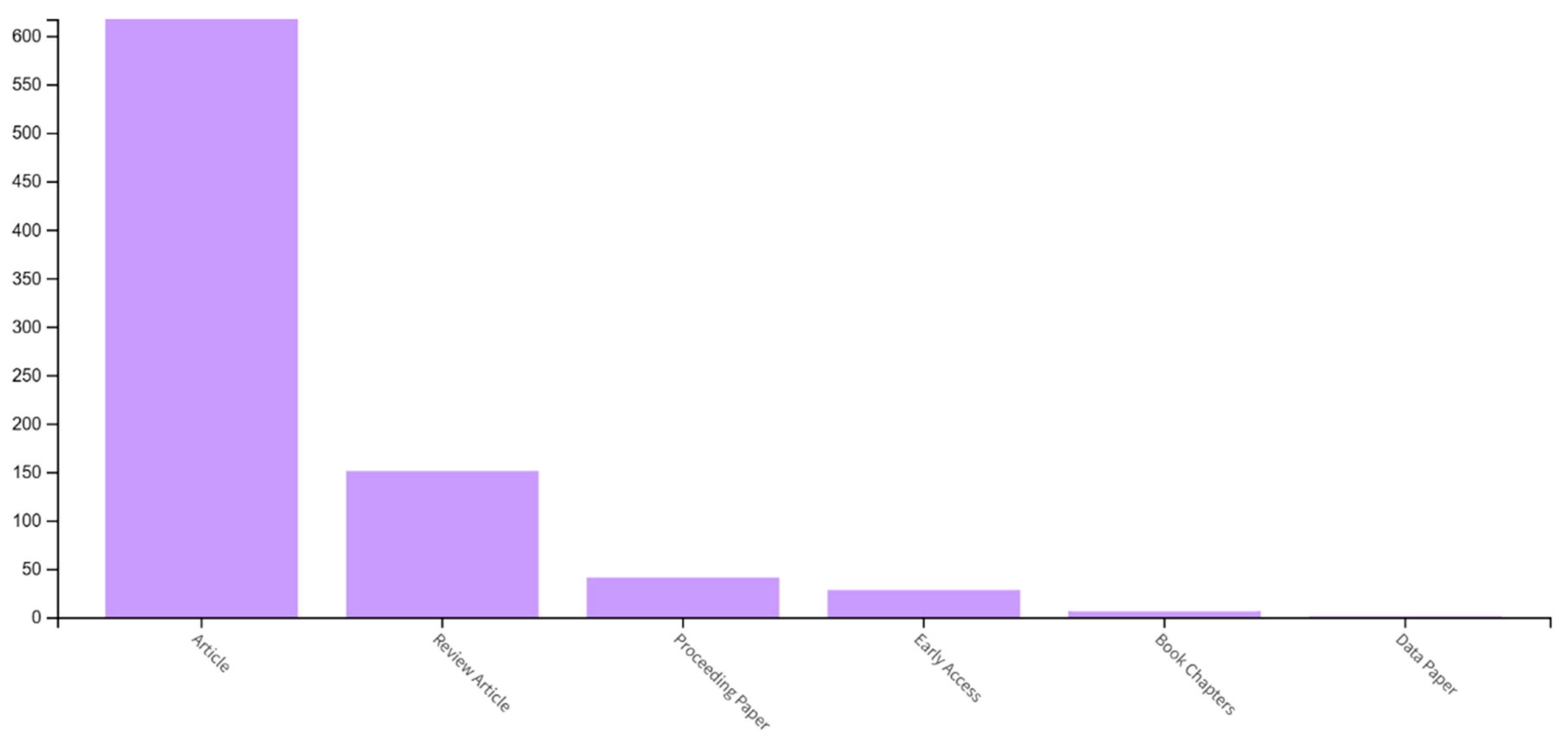
Based on the analysis of the metadata from the Web of Science database, the highest number of publications were recorded in the following formats: articles (76.7%), review articles (18.78%), conference proceedings contributions (5.1%), and pre-access articles (3.5%) (see Figure 15). The most frequently published document type is the scientific article, with a total of 617 records.
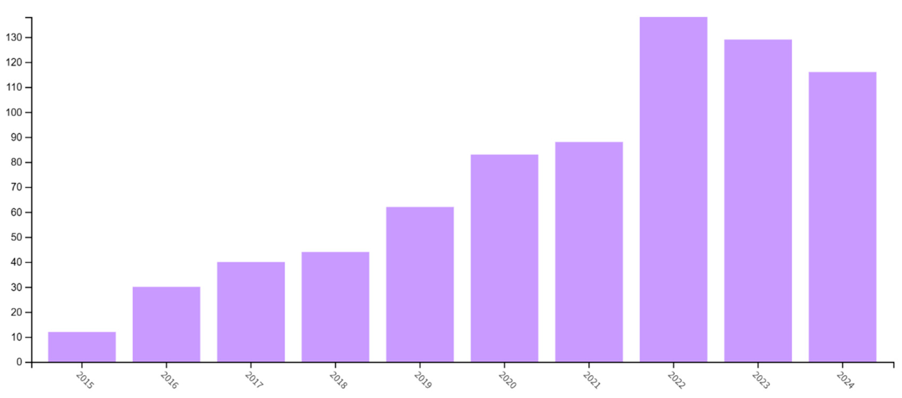
In accordance with the established criteria, 804 scientific papers of various types were published during the specified period. The data obtained from the search and analysis of publications indicate a continuous increase in their number over time, reflecting the growing popularity, importance, and interest in this topic (see Figure 16). A slight decrease was recorded in the years 2023 and 2024. This upward trend is primarily driven by the improved availability of new materials and the expanding research opportunities in the field of dental implant manufacturing.
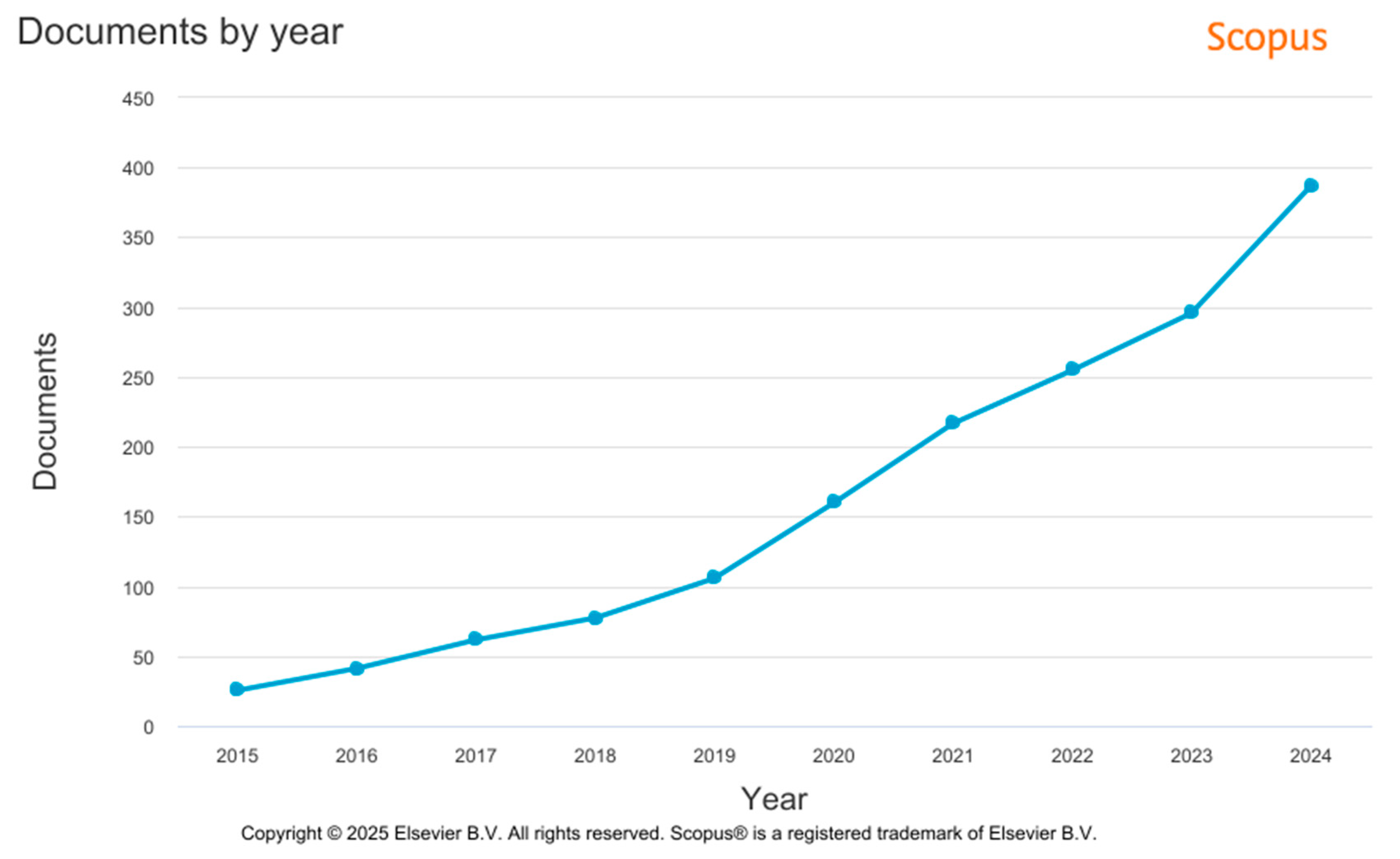
Like the analysis of the Web of Science database metadata, the analysis of Scopus metadata confirmed a dynamic increase in the number of publications between 2015 and 2024 (see Figure 17). Based on the established criteria, 1809 documents of various types were published during this period. The main factors contributing to the increase in publication activity include the improved availability of new materials and the expanded research opportunities in the field of engineering.
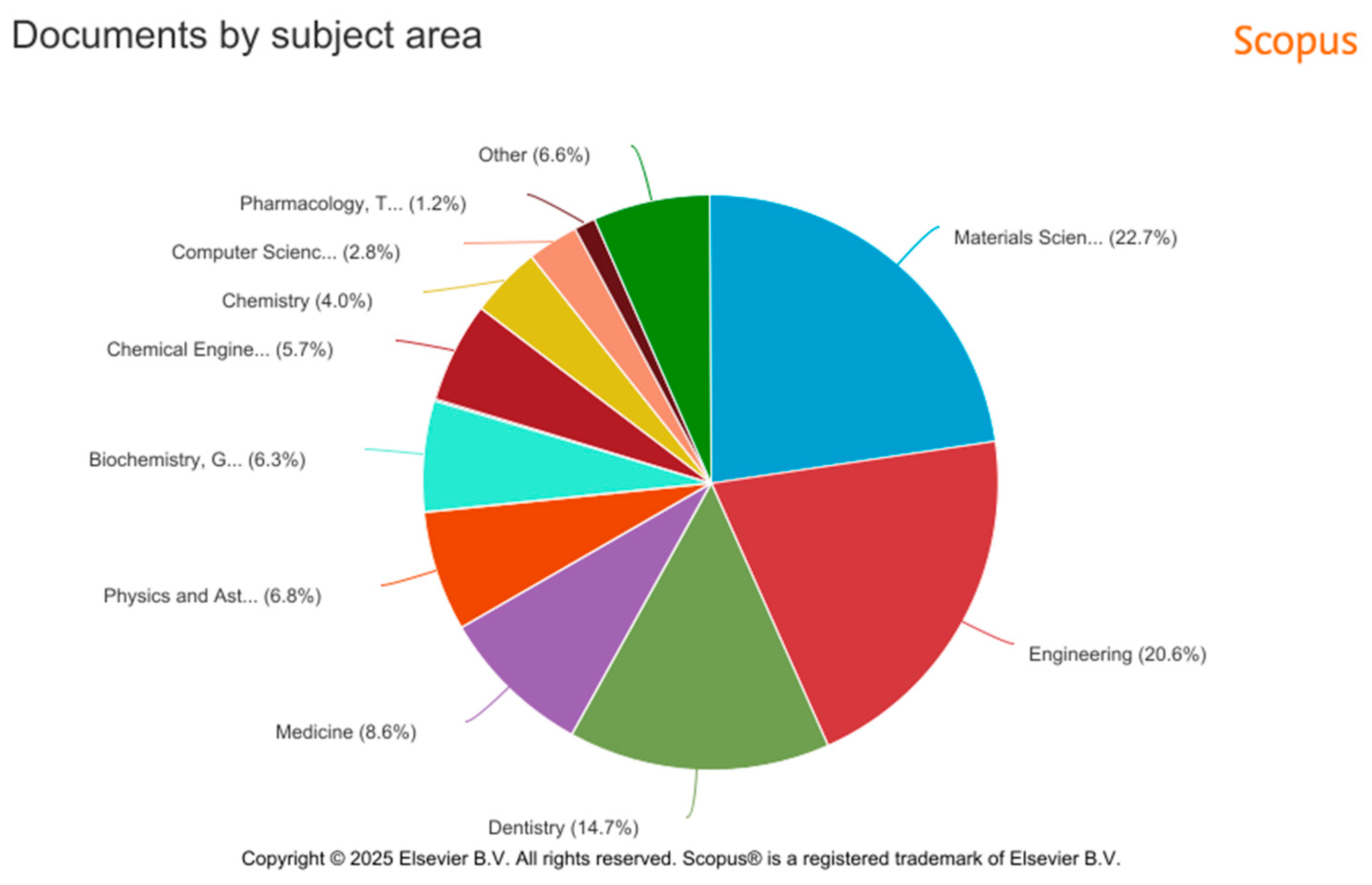
The highest number of publications was recorded in the following categories: Materials Science (22.7%), Engineering (20.6%), Dentistry (14.7%), and Medicine (6.6%) (see Figure 18). The most prominent category within the Scopus metadata is Materials Science, which comprises a total of 247 publications.
The results of the metadata analysis in the Scopus database indicate a greater diversity in the types of published documents. Compared to the Web of Science database, Scopus includes a broader range of documents based on the selected keywords (Figure 19). The most frequently represented document types are articles (63.8%, 1155 documents), review studies (21.1%, 381 documents), book chapters (7.1%, 126 documents), and conference papers (6.6%, 119 documents).

6. Conclusions

Additive manufacturing (AM) represents a revolutionary advancement in dental implant production, enabling the customization and optimization of implants to address the individual requirements of patients. This study extensively examined various materials used in the additive manufacturing of dental implants, including porous titanium, trabecular tantalum, zirconium dioxide, polymers, and composite materials. Each material was evaluated in terms of its mechanical properties, biocompatibility, and suitability for AM processes. Materials such as porous titanium and trabecular tantalum were found to be highly suitable due to their mechanical strength and biological compatibility, while polymers and composites offer flexibility and reduced weight, making them advantageous in certain clinical applications.
Additionally, the study analyzed a broad spectrum of AM technologies used in dental implant production, including selective laser melting (SLM), electron beam melting (EBM), stereolithography (SLA), selective laser sintering (SLS), and direct metal laser sintering (DMLS). These technologies vary in terms of precision, material limitations, and customization capabilities, each with specific advantages and limitations. All these methods enable the production of highly detailed, customized implants, which improve clinical outcomes and patient comfort.
The final section presented a data source analysis of the research in the field of 3D printing for dental implant production, using the Web of Science and Scopus databases. The analysis provided insights into research trends, evaluating publications based on publication category, document type, and year of publication. In both databases, a continuous enhancement in the volume of published works over the years is evident. Additionally, the most popular area of publication is Dentistry Oral Surgery Medicine (30.1%) in Web of Science, and Materials Science (22.7%) in Scopus. The most common type of published documents are articles (76.7%) in Web of Science and 63.8% in Scopus. In total, 2613 publications of various types were published using the selected keywords between 2015 and 2024. The results indicate a growing interest in the field, particularly in the development of new materials and technologies, suggesting that additive manufacturing will remain a growing key factor in dental implantology.
In conclusion, additive manufacturing holds significant potential in the development of dental implants, facilitating custom-made implants with improved mechanical and biological properties, as well as enhancing the overall manufacturing process. With continued research and innovation in materials and technologies, further advancements in this field are expected, contributing to improved patient quality of life and reduced dental care costs.
Future research should prioritize the investigation of the long-term biosafety and clinical efficacy of dental implants produced via additive manufacturing (AM), alongside the standardization of testing protocols. A notable gap exists in the form of systematic clinical studies comparing various AM technologies with respect to their success rates in real-world clinical settings. Further studies are required to enhance the understanding of the interactions between novel implant materials, particularly polymers and composites, and human tissues. Additionally, research should emphasize the optimization of implant design through the application of artificial intelligence and biomechanical load simulations. From a clinical perspective, the development of evidence-based guidelines for selecting appropriate AM technologies and materials based on patient-specific factors and clinical indications is essential. As part of a comprehensive implementation strategy, the translation of research outcomes into clinical practice should be facilitated through interdisciplinary collaboration between researchers, healthcare professionals, and industry stakeholders. Moreover, the regulatory and the ethical implications of applying additive manufacturing in healthcare must be systematically addressed. In summary, continued innovation and research are strongly recommended to improve the accessibility, effectiveness, and safety of personalized dental implants.

Author Contributions

Conceptualization, M.Y. and J.D.; methodology, J.D. and S.M.; formal analysis, D.D.; investigation, M.Y., D.D. and J.T.; resources, J.D.; writing—original draft preparation, M.Y.; writing—review and editing, D.D. and M.Y. All authors have read and agreed to the published version of the manuscript.

Funding

This research was supported by the Scientific Grant Agency of the Ministry of Education, Research, Development and Youth of the Slovak Republic and Slovak Academy of Sciences, grant VEGA 1/0258/24 and by the Cultural and Educational Grant Agency of the Ministry of Education, Research, Development and Youth of the Slovak Republic, grant no. KEGA 024TUKE-4/2023. This work was supported by the Slovak Research and Development Agency under contract No. APVV-21-0293.

Institutional Review Board Statement

Not applicable.

Informed Consent Statement

Not applicable.

Data Availability Statement

Not applicable.

Acknowledgments

This article is supported by project DRP0200194, Moving PLastics and mAchine iNdustry towards Circularity (PLAN-C) under the Interreg Danube Region Program, Co.-funded by the European Union. This article was also funded by the EU NextGenerationEU through the Recovery and Resilience Plan for Slovakia under project No. 09I05-03-V02-00042.

Conflicts of Interest

The authors declare no conflicts of interest.

References

  1. Gao, J.; Pan, Y.; Gao, Y.; Pang, H.R.; Sun, H.; Cheng, L.; Liu, J. Research Progress on the Preparation Process and Material Structure of 3D-Printed Dental Implants and Their Clinical Applications. Coatings 2024, 14, 781. [Google Scholar] [CrossRef]
  2. Hossain, N.; Islam, M.A.; Ahmed, M.M.; Chowdhury, M.A.; Mobarak, M.H.; Rahman, M.M.; Hossain, M.H. Advances and signifcances of titaniumin dental implant applications. Results Chem. 2024, 7, 101394. [Google Scholar] [CrossRef]
  3. Mobarak, M.H.; Islam, M.A.; Hossain, N.; Al Mahmud, M.Z.; Rayhan, M.T.; Nishi, N.J.; Chowdhury, M.A. Recent advances of additive manufacturing in implant fabrication—A review. Appl. Surf. Sci. Adv. 2023, 18, 100462. [Google Scholar] [CrossRef]
  4. Celik, H.K.; Koc, S.; Kustarci, A.; Caglayan, N.; Rennie, A.E. The state of additive manufacturing in dental research–A systematic scoping review of 2012–2022. Heliyon 2023, 9, e17462. [Google Scholar] [CrossRef]
  5. Pagac, M.; Hajnys, J.; Ma, Q.P.; Jancar, L.; Jansa, J.; Stefek, P.; Mesicek, J. A Review of Vat Photopolymerization Technology: Materials, Applications, Challenges, and Future Trends of 3D Printing. Polymers 2021, 13, 598. [Google Scholar] [CrossRef]
  6. Aimar, A.; Palermo, A.; Innocenti, B. The Role of 3D Printing in Medical Applications: A State of the Art. J. Healthcare Eng. 2019, 2019, 5340616. [Google Scholar] [CrossRef]
  7. Yan, Q.; Dong, H.; Su, J.; Han, J.; Song, B.; Wei, Q.; Shi, Y. A Review of 3D Printing Technology for Medical Applications. Engineering 2018, 4, 729–742. [Google Scholar] [CrossRef]
  8. Rezaie, F.; Farshbaf, M.; Dahri, M.; Masjedi, M.; Maleki, R.; Amini, F.; Wirth, J.; Moharamzadeh, K.; Weber, F.E.; Tayebi, L. 3D Printing of Dental Prostheses: Current and Emerging Applications. J. Compos. Sci. 2023, 7, 80. [Google Scholar] [CrossRef]
  9. Andreucci, C.A.; Fonseca, E.M.; Jorge, R.N. 3D Printing as an Effcient Way to Prototype and Develop Dental Implants. BioMedInformatics 2022, 2, 671–679. [Google Scholar] [CrossRef]
  10. Chaudhary, R.; Fabbri, P.; Leoni, E.; Mazzanti, F.; Akbari, R.; Antonini, C. Additive manufacturing by digital light processing: A review. Prog. Addit. Manuf. 2022, 8, 331–351. [Google Scholar] [CrossRef]
  11. Kennedy, S.M.; Raghav, G.R.; Jeen Robert, R.B.; Manikandaraja, g.; Selvakumar, M. PEEK-based 3D printing: A paradigm shift in implant revolution for healthcare. Polym.-Plast. Technol. Mater. 2024, 63, 680–702. [Google Scholar] [CrossRef]
  12. Liang, W.; Zhou, C.; Zhang, H.; Bai, J.; Jiang, B.; Jiang, C.; Ming, W.; Zhang, H.; Long, H.; Huang, X.; et al. Recent advances in 3D printing of biodegradable metals for orthopaedic applications. J. Biol. Eng. 2023, 17, 56. [Google Scholar] [CrossRef]
  13. Kechagias, J.D.; Zaoutsos, S.P. Optimising fused flament fabrication surface roughness for a dental implant. Mater. Manuf. Process. 2023, 38, 954–959. [Google Scholar] [CrossRef]
  14. Bakkal, B.; Topsakal, K.G.; Duran, G.S. New horizons in dentistry and orthodontics with 4D printing technologies 4D printing and orthodontics. Australas. Orthod. J. 2025. Available online: https://www.semanticscholar.org/paper/9135bb33b12d2d7d8a11d927835302be1783a7e3 (accessed on 14 February 2025).
  15. Aura Sium-Abel, L.; Keilig, I.; Dörsam, C. Bourauel Accuracy of template-based guided dental implant placement-An in vitro comparison of different manufacturing methods. Clin. Oral Implant. Res. 2024. Available online: https://www.semanticscholar.org/paper/204ec1b965411b7e494c57989320909d29efa67e (accessed on 23 March 2025).
  16. Li, B. The Investigation of Pore Characteristic and Mechanical Property of Porous Ti. Master’s Dissertation, Dalian Jiao Tong University, Dalian, China, 2005. [Google Scholar]
  17. Hu, H.B.; Liu, H.Q.; Wang, J.E.; Yi, D.Q.; Fu, S.; Sun, W.L. Research Progress of Biomedical Porous Titanium and Its Alloys. Mater. Rev. 2012, 26, 262–270. [Google Scholar]
  18. Li, Q.; Wang, H.; Zhang, L.X. The Surface Treatment of Artificial Joint Metal Materials. Chin. J. Med. Instrum. 2002, 26, 44–47. [Google Scholar]
  19. Agrawal, C.M. Reconstructing the Human-Body Using Biomaterials. JOM 1998, 50, 31–35. [Google Scholar] [CrossRef]
  20. Ryan, G.; Pandit, A. Fabrication methods of porous metals for use in orthopaedic applications. Biomaterials 2006, 27, 2651–2670. [Google Scholar] [CrossRef] [PubMed]
  21. Wang, X.H.; Li, S.J.; Jia, M.T.; Hao, Y.L.; Yang, R.; Guo, Z.X. Porous Ti-24Nb-4Zr8Sn alloy for biomedical applications fabricated by space-holder method. Chin. J. Nonferrous Met. 2010, 20, 967–971. [Google Scholar]
  22. Zhao, J.; Lu, X.; Weng, J. Macro-porous Ti-Based Composite Scaffold Prepared by Polymer Impregnating Method with Calcium Phosphate Coatings. Mater. Lett. 2008, 62, 2921–2924. [Google Scholar] [CrossRef]
  23. Wang, Z.; Shen, Y.; Gu, D.; Yu, X. Preparation of Honeycomb-like Porous 316L Stainless Steel by Selective Laser Melting. Rare Met. Mater. Eng. 2011, 40, 161–164. [Google Scholar] [CrossRef]
  24. He, W.; Jia, W.; Liu, H. The Application of Rapid Prototyping Technology in Porous Metals Materials. Rare Met. Lett. 2008, 27, 1–6. [Google Scholar]
  25. Traini, T.; Mangano, C.; Sammons, R.I. Direct Laser metal sintering as a new approach to fabrication of isoelastic functionally graded material for manufacture of porous titanium dental implants. Dent. Mater. 2008, 24, 1525–1533. [Google Scholar] [CrossRef]
  26. Stamp, R.; Fox, P.; O’Neill, W. The development of a scanning strategy for the manufacture of porous biomaterials by selective laser melting. J. Mater. Sci. Mater. Med. 2009, 20, 1839–1848. [Google Scholar] [CrossRef]
  27. Heinl, P.; Muller, L.; Korner, C. Cellular Ti-6Al-4V structures with interconnected macro porosity for bone implants fabricated by selective electron beam melting. Acta Biomater. 2008, 4, 1536–1544. [Google Scholar] [CrossRef] [PubMed]
  28. Xiao, D.M. Modeling of Porous Structure of Implants and Direct Manufacturing by Selective Laser Melting. Ph.D. Dissertation, South China University of Technology, Guangzhou, China, 2013. [Google Scholar]
  29. Li, X.; Wang, C.; Zhang, W. Fabrication of Porous Ti6Al4V Implant Using Electron Beam Melting and Mechanical Properties. J. Shanghai Jiao Tong Univ. 2009, 43, 1946–1949. [Google Scholar]
  30. Parthasarathy, J.; Starly, B. Mechanical Evaluation of Porous Titanium (Ti-6Al-4V) Structures with Electron Beam Melting (EBM). J. Mech. Behav. Biomed. Mater. 2010, 3, 249–259. [Google Scholar] [CrossRef] [PubMed]
  31. Levine, B.R.; Sporer, S.; Poggie, R.A.; Della Valle, C.J.; Jacobs, J.J. Experimental and clinical performance of porous tan-talum in orthopedic surgery. Biomaterials 2006, 27, 4671–4681. [Google Scholar] [CrossRef]
  32. Kaplan, R.B. Open Cell Tantalum Structures for Cancellousbone Implants and Cell and Tissue Structures. U.S. Patent No. 5282861, 1 February 1994. [Google Scholar]
  33. Bencharit, S.; Byrd, W.C.; Altarawneh, S.; Hosseini, B.; Leong, A.; Reside, G.; Morelli, T.; Offenbacher, S. Porous Tantalum Trabecular Metal Dental Implants. Clin. Implant Dent. Relat. Res. 2014, 16, 817–826. [Google Scholar] [CrossRef]
  34. Zardiackas, L.D.; Parsell, D.E.; Dillon, L.D.; Mitchell, D.W.; Nunnery, L.A.; Poggie, R. Structure, metallurgy, and mechani-cal properties of a porous tantalum foam. J. Biomed. Mater. Res. 2001, 58, 180–187. [Google Scholar] [CrossRef]
  35. Cohen, R. A porous tantalum trabecular metal: Basic science. Am. J. Orthop. 2002, 31, 216–217. [Google Scholar]
  36. Heiner, A.D.; Brown, T.D.; Poggie, R.A. Structural efficacy of anovel porous tantalum implant for osteonecrosis grafting. Trans. Orthop. Res. Soc. 2001, 26, 480. [Google Scholar]
  37. Christie, M.J. Clinical applications of Trabecular Metal. Am. J. Orthop. 2002, 31, 219–220. [Google Scholar] [PubMed]
  38. Henry, P.J.; Laney, W.R.; Jemt, T.; Harris, D.; Krogh, P.H.; Polizzi, G.; Zarb, G.A.; Herrmann, I. Osseointegrated implantsfor single-tooth replacement: A prospective 5-year multi-center study. Int. J. Oral Maxillofac. Implant. 1996, 11, 450–455. [Google Scholar]
  39. Adell, R.; Lekholm, U.; Rockler, B.; Brånemark, P.; Lindhe, J.; Eriksson, B.; Sbordone, L. Marginal tissuereactions at osseointegrated titanium fixtures (I). A 3-yearlongitudinal prospective study. Int. J. Oral Maxillofac. Surg. 1986, 15, 39–52. [Google Scholar] [CrossRef]
  40. Le Guehennec, L.; Soueidan, A.; Layrolle, P.; Amouriq, Y. Surfacetreatments of titanium dental implants for rapid osseointe-gration. Dent. Mater. 2007, 23, 844–854. [Google Scholar] [CrossRef]
  41. Rupp, F.; Scheideler, L.; Olshanska, N.; de Wild, M.; Wieland, M.; Geis-Gerstorfer, J. Enhancing surface free energy and hydro-philicity through chemical modification of microstructuredtitanium implant surfaces. J. Biomed. Mater. Res. A 2006, 76, 323–334. [Google Scholar] [CrossRef]
  42. Wilkes, J.; Hagedorn, Y.C.; Meiners, W.; Wissenbach, K. Additive manufacturing of ZrO2-Al2O3 ceramic components by selective laser melting. Papid Prototyp. J. 2013, 19, 51–57. [Google Scholar] [CrossRef]
  43. Khanlar, L.N.; Salazar Rios, A.; Tahmaseb, A.; Zandinejad, A. Additive manufacturing of zirconia ceramic and its application in clinical dentistry: A review. Dent. J. 2021, 9, 104. [Google Scholar] [CrossRef]
  44. Sandhaus, S. Technic and instrumentation of the implantCBS (Cristalline Bone Screw). Inf. Odontostomatol. 1967, 4, 19–24. [Google Scholar]
  45. Steflik, D.; Koth, D.; Robinson, F.; McKinney, R.; Davis, B.; Mor-ris, C.; Davis, Q. Prospective investigation of the single-crystal sapphire endosteal dental implant in humans: Ten-yearresults. J. Oral Implant. 1994, 21, 8–18. [Google Scholar]
  46. Nakamura, K.; Kanno, T.; Milleding, P.; Örtengren, U. Zirconiaas a dental implant abutment material: A systematicreview. Int. J. Prosthodont. 2010, 23, 299–309. [Google Scholar] [PubMed]
  47. Kelly, J.R.; Denry, I. Stabilized zirconia as a structural cera-mic: An overview. Dent. Mater. 2008, 24, 289–298. [Google Scholar] [CrossRef]
  48. Covacci, V.; Bruzzese, N.; Maccauro, G.; Andreassi, C.; Ricci, G.; Piconi, C.; Marmo, E.; Burger, W.; Cittadini, A. In vitroevaluation of the mutagenic and carcinogenic power of highpurity zirconia ceramic. Biomaterials 1999, 20, 371–376. [Google Scholar] [CrossRef]
  49. Sanon, C.; Chevalier, J.; Douillard, T.; Cattani-Lorente, M.; Scherrer, S.S.; Gremillard, L. A new testing protocol for zirconia dental implants. Dent. Mater. 2015, 31, 15–25. [Google Scholar] [CrossRef]
  50. Lughi, V.; Sergo, V. Low temperature degradation-aging-ofzirconia: A critical review of the relevant aspects in dentistry. Dent. Mater. 2010, 26, 807–820. [Google Scholar] [CrossRef]
  51. Oliva, J.; Oliva, X.; Oliva, J.D. Five-year success rate of 831 con-secutively placed Zirconia dental implants in humans: Acomparison of three different rough surfaces. Int. J. OralMaxillofac. Implant. 2010, 25, 336–344. [Google Scholar]
  52. Payer, M.; Arnetzl, V.; Kirmeier, R.; Koller, M.; Arnetzl, G.; Jakse, N. Immediate provisional restoration of single-piece zirco-nia implants: A prospective case series–results after24 months of clinical function. Clin. Oral Implant. Res. 2013, 24, 569–575. [Google Scholar] [CrossRef] [PubMed]
  53. Zembić, A.; Johannesen, L.; Schou, S.; Malo, P.; Reichert, T.; Farella, M.; Hämmerle, C. Immediately restored one-piecesingle-tooth implants with reduced diameter: One-yearresults of a multi-center study. Clin. Oral Implant. Res. 2012, 23, 49–54. [Google Scholar] [CrossRef]
  54. Hermann, J.S.; Cochran, D.L.; Buser, D.; Schenk, R.K.; Schoolfield, J.D. Biologic Width around one-and two-piece titaniumimplants. Clin. Oral Implant. Res. 2001, 12, 559–571. [Google Scholar] [CrossRef]
  55. Hartman, G.A.; Cochran, D.L. Initial implant position deter-mines the magnitude of crestal bone remodeling. J. Peri-Odontol. 2004, 75, 572–577. [Google Scholar]
  56. Wilson, T.G., Jr. The positive relationship between excesscement and peri-implant disease: A prospective clinicalendoscopic study. J. Periodontol. 2009, 80, 1388–1392. [Google Scholar] [CrossRef]
  57. Linkevicius, T.; Vindasiute, E.; Puisys, A.; Linkeviciene, L.; Maslova, N.; Puriene, A. The influence of the cementationmargin position on the amount of undetected cement. Aprospective clinical study. Clin. Oral Implant. Res. 2013, 24, 71–76. [Google Scholar] [CrossRef] [PubMed]
  58. Korsch, M.; Robra, B.-P.; Walther, W. Cement-associated signsof inflammation: Retrospective analysis of the effect ofexcess cement on peri-implant tissue. Int. J. Prosthodont. 2015, 28, 11–18. [Google Scholar] [CrossRef] [PubMed]
  59. Wittneben, J.-G.; Millen, C.; Brägger, U. Clinical Performanceof Screw-Versus Cement-Retained Fixed Implant-Sup-ported Reconstructions-A Systematic Review. Int. J. OralMaxillofac Implant. 2014, 29, 84–98. [Google Scholar] [CrossRef] [PubMed]
  60. Payer, M.; Heschl, A.; Koller, M.; Arnetzl, G.; Lorenzoni, M.; Jakse, N. All-ceramic restoration of zirconia two-piece implants –a randomized controlled clinical trial. Clin. Oral Implant. Res. 2015, 26, 371–376. [Google Scholar] [CrossRef]
  61. Brüll, F.; van Winkelhoff, A.J.; Cune, M.S. Zirconia dentalimplants: A clinical, radiographic, and microbiologic evalu-ation up to 3 years. Int. J. Oral Maxillofac Implant. 2014, 29, 914–920. [Google Scholar]
  62. Becker, J.; John, G.; Becker, K.; Mainusch, S.; Diedrichs, G.; Schwarz, F. Clinical performance of two-piece zirconiaimplants in the posterior mandible and maxilla: A prospec-tive cohort study over 2 years. Clin. Oral Implants Res. 2015, 28, 29–35. [Google Scholar] [CrossRef]
  63. Cionca, N.; Müller, N.; Mombelli, A. Two-piece zirconia implants supporting all-ceramic crowns: A prospectiveclinical study. Clin. Oral Implants Res. 2015, 26, 413–418. [Google Scholar] [CrossRef]
  64. Hodosh, M.; Povar, M.; Shklar, G. The dental polymer implant concept. J. Prosthet. Dent. 1969, 22, 371–380. [Google Scholar] [CrossRef]
  65. Hodosh, M.; Povar, M.; Shklar, G. Development of the self-supporting polymer tooth implant. J. Biomed. Mater. Res. 1973, 7, 205–216. [Google Scholar] [CrossRef]
  66. Jockusch, J.; Özcan, M. Additive manufacturing of dental polymers: An overview on processes. Mater. Appl. 2020. [Google Scholar] [CrossRef]
  67. Castro-Aguirre, E.; Iñiguez-Franco, F.; Samsudin, H.; Fang, X.; Auras, R. Poly(lactic acid)—Mass production, processing, industrial applications, and end of life. Adv. Drug Deliv. Rev. 2016, 107, 333–366. [Google Scholar] [CrossRef]
  68. Chang, P.C.; Liu, B.Y.; Liu, C.M.; Chou, H.H.; Ho, M.H.; Liu, H.C.; Wang, D.M.; Hou, L.T. Bone tissue engineering with novel rhBMP2-PLLA composite scaffolds. J. Biomed. Mater. Res. Part A 2007, 81, 771–780. [Google Scholar] [CrossRef]
  69. Zhang, Q.; Mochalin, V.N.; Neitzel, I.; Knoke, I.Y.; Han, J.; Klug, C.A.; Zhou, J.G.; Lelkes, P.I.; Gogotsi, Y. Fluorescent PLLA-nanodiamond composites for bone tissue engineering. Biomaterials 2011, 32, 87–94. [Google Scholar] [CrossRef]
  70. Song, J.; Winkeljann, B.; Lieleg, O. Biopolymer-based coatings: Promising strategies to improve the biocompatibility and functionality of materials used in biomedical engineering. Adv. Mater. Interfaces 2020, 7, 2000850. [Google Scholar] [CrossRef]
  71. Simionescu, B.C.; Ivanov, D. Natural and Synthetic Polymers for Designing Composite Materials. In Handbook of Bioceramics and Biocomposites; Springer: Berlin/Heidelberg, Germany, 2014. [Google Scholar] [CrossRef]
  72. Lan, S.-F.; Kehinde, T.; Zhang, X.; Khajotia, S.; Schmidtke, D.W.; Starly, B. Controlled release of metronidazole from composite poly-e-caprolactone/alginate (PCL/alginate) rings for dental implants. Dent. Mater. 2013, 29, 656–665. [Google Scholar] [CrossRef] [PubMed]
  73. Conte, R.; Di Salle, A.; Riccitiello, F.; Petillo, O.; Peluso, G.; Calarco, A. Biodegradable polymers in dental tissue engineering and regeneration. AIMS Mater. Sci. 2018, 5, 1073–1101. [Google Scholar] [CrossRef]
  74. Hosseinalipour, M.; Javadpour, J.; Rezaie, H. Investigation of Mechanical Properties of Experimental Bis-GMA/TEGDMA Dental Composite Resins Containing Various Mass Fractions of Silica Nanoparticles. J. Prosthodont. 2010, 19, 112–117. [Google Scholar] [CrossRef] [PubMed]
  75. Suwanprateeb, J.; Sanngam, R.; Suvannapruk, W.; Panyathanmaporn, T. Mechanical and in vitro performance of apatite–wollastonite glass ceramic reinforced hydroxyapatite composite fabricated by 3D-printing. J. Mater. Sci. Mater. Med. 2009, 20, 1281–1289. [Google Scholar] [CrossRef]
  76. Chen, L.; Yu, Q.; Wang, Y.; Li, H. BisGMA/TEGDMA dental composite containing high aspect-ratio hydroxyapatite nanofibers. Dent. Mater. 2011, 27, 1187–1195. [Google Scholar] [CrossRef]
  77. Filipecki, J.; Chamerski, K.; Boyko, O.; Kotynia, K. Ageing phenomenon in acrylic polymer dental materials detected by means of positron annihilation lifetime spectroscopy. Polim. W Med. 2014, 44, 21–28. [Google Scholar] [CrossRef]
  78. Ganguly, S.; Tang, X.S. 3D Printing of High Strength Thermally Stable Sustainable Lightweight Corrosion-Resistant Nanocomposite by Solvent Exchange Postprocessing. ACS Sustain. Chem. Eng. 2024, 13, 423–435. [Google Scholar] [CrossRef]
  79. Huang, B.; Qian, J.; Wang, G.; Cai, M. Synthesis and Properties of Novel Copolymers of Poly (ether ketone diphenyl ketone ether ketone ketone) and Poly (ether amide ether amide ether ketone ketone). Polym. Eng. Sci. 2014, 54, 1757–1764. [Google Scholar] [CrossRef]
  80. Yang, F.; Chen, C.; Zhou, Q.; Gong, Y.; Li, R.; Li, C.; Klämpfl, F.; Freund, S.; Wu, X.; Sun, Y.; et al. Laser beam melting 3D printing of Ti6Al4V based porous structured dental implants: Fabrication, biocompatibility analysis and photoelastic study. Sci. Rep. 2017, 7, 45360. [Google Scholar] [CrossRef] [PubMed]
  81. Carraro, F.; Bagno, A. Tantalum as trabecular metal for endosseous implantable applications. Biomimetics 2023, 8, 49. [Google Scholar] [CrossRef]
  82. Aldhuwayhi, S. Zirconia in Dental Implantology: A Review of the Literature with Recent Updates. Bioengineering 2025, 12, 543. [Google Scholar] [CrossRef] [PubMed]
  83. Pot, G.J.; Van Overschelde, P.A.; Keulemans, F.; Kleverlaan, C.J.; Tribst, J.P.M. Mechanical properties of additive-manufactured composite-based resins for permanent indirect restorations: A scoping review. Materials 2024, 17, 3951. [Google Scholar] [CrossRef]
  84. Lu, B.; Li, D. Development of the Additive Manufacturing (3D printing) Technology. Mach. Build. Autom. 2013, 42, 1–4. [Google Scholar]
  85. Gong, S.; Suo, J.; Li, H. Development and Application of Metal Additive Manufacturing Technology. Aeronaut. Manuf. Technol. 2013, 13, 66–71. [Google Scholar]
  86. Zhou, L.; Yang, Q.; Zhang, G.; Zhao, F.; Shen, G.; Yu, B. Additive manufacturing technologies of porous metal implants. China Foundry 2014, 11, 322–331. [Google Scholar]
  87. Shi, Y.; Lu, Z.; Zhang, W. The Technology and Equipment of Selective Laser Melting. China Surf. Eng. 2006, 19, 150–153. [Google Scholar]
  88. Han, J.; Lin, F.; Qi, H. Effects of powder preheating in electron beam selective melting process. Trans. China Weld. Inst. 2008, 29, 77–81. [Google Scholar]
  89. Qi, H.; Yang, M.; Qi, F. Numerical Simulation of Effects of Scanning Path on Electron Beam Selective Melting Process of TC4. Trans. China Weld. Inst. 2009, 30, 5–8. [Google Scholar]
  90. Jia, W.; Tang, H.; He, W. The Development and Key Problems of Electric Beam Rapid Forming. Electromachining Mould. 2010, 2, 41–44. [Google Scholar]
  91. Yao, N.; Peng, X. The Preparation Method of Metal Powder for 3D Printing. Sichuan Nonferrous 2013, 12, 48–51. [Google Scholar]
  92. Bineli, A.R.R.; Peres, A.P.G.; Bernardes, L.F.; Jardini, A.L.; Maciel Filho, R. Design of microreactor by integration of reverse engineering and direct metal laser sintering process. In Proceedings of the 5th International Workshop on Hydrogen and Fuel Cells, Campinas, Brasil, 26–29 October 2010; EOS: New York City, NY, USA, 2009. [Google Scholar]
  93. Senthilkumaran, K.; Pandey, P.M.; Rao, P.V.M. Influence of building strategies on the accuracy of parts in selective laser sintering. Mater. Des. 2009, 30, 2946–2954. [Google Scholar] [CrossRef]
  94. Yu, N. Process Parameter Optimization for Direct Metal Laser Sintering (DMLS). Ph.D. Thesis, National University of Singapore, Singapore, 2005. [Google Scholar]
  95. Bineli, A.R.R.; Peres, A.P.G.; Jardini, A.L.; Maciel Filho, R. Direct metal laser sintering (DMLS): Technology for design and construction of microreactors. In Proceedings of the 6º Congress Obrasileiro De Engenharia De Fabrica Ção, Caxias do Sulm, Brazil, 11–15 April 2011; Volume 11. [Google Scholar]
  96. Kim, G.B.; Lee, S.; Kim, H.; Yang, D.H.; Kim, Y.H.; Kyung, Y.S.; Kim, C.S.; Choi, S.H.; Kim, B.J.; Ha, H.; et al. Three-dimensional printing: Basic principles and applications in medicine and radiology. Korean J. Radiol. 2016, 17, 182–197. [Google Scholar] [CrossRef]
  97. Wu, H.; Cheng, Y.; Liu, W.; He, R.; Zhou, M.; Wu, S.; Song, X.; Chen, Y. Effect of the particle size and the debinding process on the density of alumina ceramics fabricated by 3D printing based on stereolithography. Ceram. Int. 2016, 42, 17290–17294. [Google Scholar] [CrossRef]
  98. Zocca, A.; Colombo, P.; Gomes, C.M.; Günster, J. Additive manufacturing of ceramics: Issues, potentialities, and opportunities. J. Am. Ceram. Soc. 2015, 98, 1983–2001. [Google Scholar] [CrossRef]
  99. Lian, Q.; Sui, W.; Wu, X.; Yang, F.; Yang, S. Additive manufacturing of ZrO2 ceramic dental bridges by stereolithography. Rapid Prototyp. J. 2018, 24, 114–119. [Google Scholar] [CrossRef]
  100. Griffith, M.L.; Halloran, J.W. Freeform fabrication of ceramics via stereolithography. J. Am. Ceram. Soc. 1996, 79, 2601–2608. [Google Scholar] [CrossRef]
  101. Liu, K.; Zhang, K.; Bourell, D.L.; Chen, F.; Sun, H.; Shi, Y.; Wang, J.; He, M.; Chen, J. Gelcasting of zirconia-based all-ceramic teeth combined with stereolithography. Ceram. Int. 2018, 44, 21556–21563. [Google Scholar] [CrossRef]
  102. Chen, Z.; Li, D.; Zhou, W.; Wang, L. Curing characteristics of ceramic stereolithography for an aqueous-based silica suspension. Proc. Inst. Mech. Eng. Part B J. Eng. Manuf. 2010, 224, 641–651. [Google Scholar] [CrossRef]
  103. Bertsch, A.; Jiguet, S.; Renaud, P. Microfabrication of ceramic components by microstereolithography. J. Micromech. Microeng. 2004, 14, 197–203. [Google Scholar] [CrossRef]
  104. Zhang, K.; He, R.; Xie, C.; Wang, G.; Ding, G.; Wang, M.; Song, W.; Fang, D. Photosensitive ZrO2 suspensions for stereolithography. Ceram. Int. 2019, 45, 12189–12195. [Google Scholar] [CrossRef]
  105. Gentry, S.P.; Halloran, J.W. Depth and width of cured lines in photopolymerizable ceramic suspensions. J. Eur. Ceram. Soc. 2013, 33, 1981–1988. [Google Scholar] [CrossRef]
  106. Badev, A.; Abouliatim, Y.; Chartier, T.; Lecamp, L.; Lebaudy, P.; Chaput, C.; Delage, C. Photopolymerization kinetics of a polyether acrylate in the presence of ceramic fillers used in stereolithography. J. Photochem. Photobiol. A Chem. 2011, 222, 117–122. [Google Scholar] [CrossRef]
  107. Zhang, X.; Wu, X.; Shi, J. Additive manufacturing of zirconia ceramics: A state-of-the-art review. J. Mater. Res. Technol. 2020, 9, 9029–9048. [Google Scholar] [CrossRef]
  108. Wang, J.C.; Dommati, H. Fabrication of zirconia ceramic parts by using solvent-based slurry stereolithography and sintering. Int. J. Adv. Manuf. Technol. 2018, 98, 1537–1546. [Google Scholar] [CrossRef]
  109. Revilla-León, M.; Husain, N.A.; Ceballos, L.; Özcan, M. Flexural strength and Weibull characteristics of stereolithography additive manufactured versus milled zirconia. J. Prosthet. Dent. 2021, 125, 685–690. [Google Scholar] [CrossRef]
  110. Revilla-León, M.; Mostafavi, D.; Methani, M.M.; Zandinejad, A. Manufacturing accuracy and volumetric changes of stereolithography additively manufactured zirconia with different porosities. J. Prosthet. Dent. 2022, 128, 211–215. [Google Scholar] [CrossRef] [PubMed]
  111. Nakai, H.; Inokoshi, M.; Nozaki, K.; Komatsu, K.; Kamijo, S.; Liu, H.; Shimizubata, M.; Minakuchi, S.; Van Meerbeek, B.; Vleugels, J.; et al. Additively manufactured zirconia for dental applications. Materials 2021, 14, 3694. [Google Scholar] [CrossRef] [PubMed]
  112. Deckard, C.R. Method and Apparatus for Producing Parts by Selective Sintering. U.S. Patent No. 4,863,538-A, 5 September 1986. [Google Scholar]
  113. Subramani, R.; Mustafa, M.A.; Ghadir, G.K.; Al-Tmimi, H.M.; Alani, Z.K.; Haridas, D.; Rusho, M.A.; Rajeswari, N.; Rajan, A.J.; Kumar, A.P. Advancements in 3D printing materials: A comparative analysis of performance and applications. Appl. Chem. Eng. 2024, 7, ACE-3867. [Google Scholar] [CrossRef]
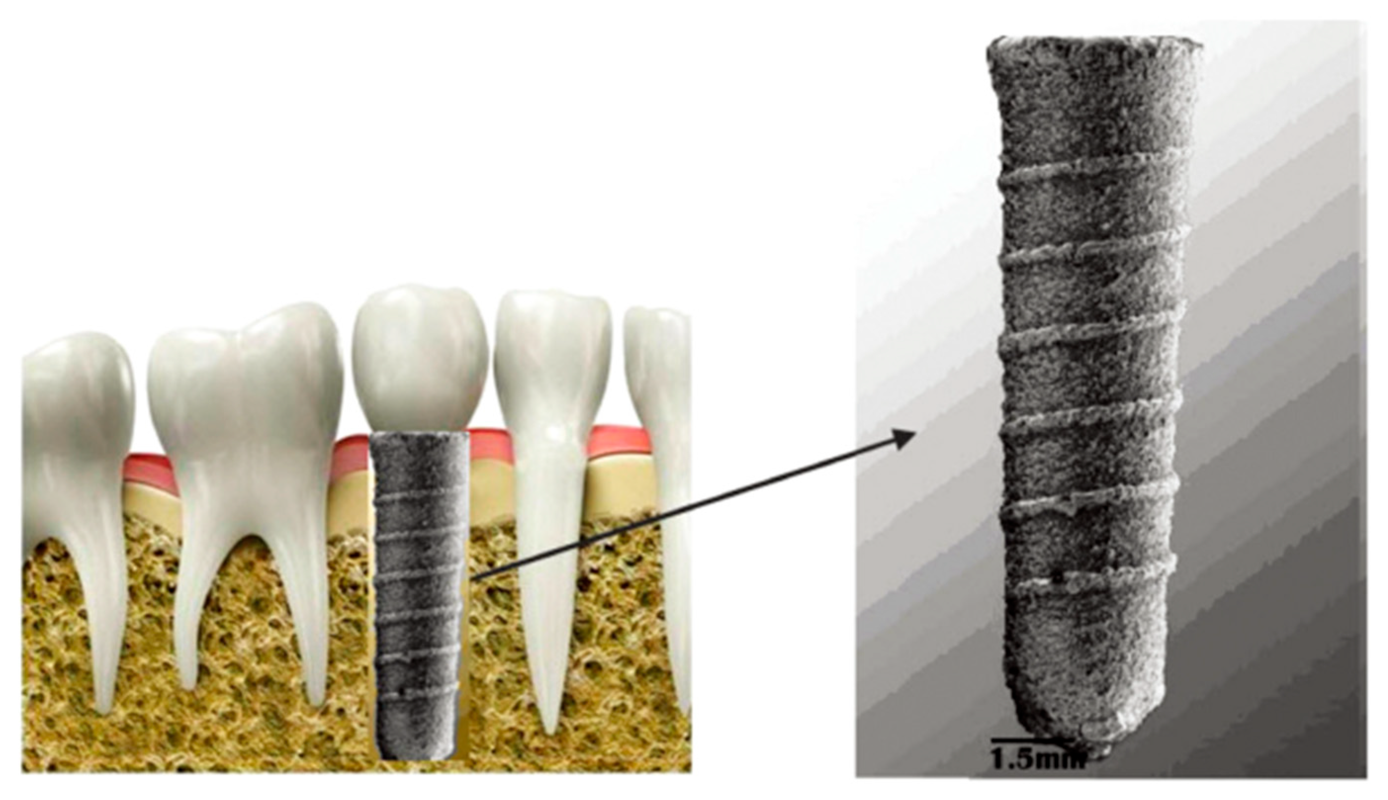
Figure 1. Porous implant Ti6Al4V [25].
Sci 07 00109 g001
Figure 2. Samples of the porous implant structure of (a) a titanium porous implant; and (b) a tantalum-coated implant [30].
Sci 07 00109 g002
Figure 5. Diagram of the SLA system (provided by 3Dceram) and examples of 3D-printed dental devices produced using the CeraFab System S65 Medical. Dental implants (top left), zirconia bridge (3Y-TZP, top right), recolored molar crown (center), uncolored molar crown (bottom right), and zirconia implant with abutment (bottom center) (Image courtesy of Lithoz GmbH, Vienna, Austria) [43].
Sci 07 00109 g005
Figure 6. Polymer dental implant [70].
Sci 07 00109 g006
Figure 7. Schematic representation of the SLM process (selective laser melting) [86].
Sci 07 00109 g007
Figure 8. Schematic representation of the EBM process (electron beam melting) [85].
Sci 07 00109 g008
Figure 9. Metal 3D-printing powders provided by Ametek: (a) Ti6Al4V powders produced by plasma spheroidization and (b) 316L powders produced by gas atomization [86].
Sci 07 00109 g009
Figure 10. Schematic representation of DMLS process (selective laser sintering) [92].
Sci 07 00109 g010
Figure 11. Exposure strategies and process parameters of the DMLS system [94].
Sci 07 00109 g011
Figure 12. Schematic representation of the SLA instrument (company 3Dceram) [100].
Sci 07 00109 g012
Figure 13. Schematic representation of SLS method (selective laser sintering) [109].
Sci 07 00109 g013
Figure 14. Graphical representation of the analysis of the number of publications from 2014 to 2023 by category in the Web of Science database.
Sci 07 00109 g014
Figure 15. Graphical representation of the analysis of the number of publications from 2014 to 2023 according to the type of published document in the Web of Science database.
Sci 07 00109 g015
Figure 16. Graphical representation of the analysis of the number of publications from 2014 to 2023 in the Web of Science database.
Sci 07 00109 g016
Figure 17. Graphical representation of the analysis of the number of publications from 2014 to 2023 in the Scopus database.
Sci 07 00109 g017
Figure 18. Graphical representation of the analysis of the number of publications from 2014 to 2023 by category in the Scopus database.
Sci 07 00109 g018
Figure 19. Graphical representation of the analysis of the number of publications from 2014 to 2023 according to the type of published document in the Scopus database.
Sci 07 00109 g019
Table 1. Evaluation of the materials’ properties.
MaterialTensile Strength (MPa)Fatigue ResistanceElastic Modulus (GPa)
Porous titanium550–900High10–30
Trabecular tantalum200–300Extremely high3–5
Zirconia900–1200Medium200–210
Polymers50–100Low1–5
Composite100–300Medium to high5–20
Table 2. Comparison of selected technologies.
TechnologyEnergy TypeApplicable MaterialsDisplacement AccuracySurface QualityLimitations
SLMLaser (infrared)Metals (titanium, aluminum, stainless steel, alloys)High (20–50 µm)Medium to highHigher residual stress, need for support
EBMElectron beam (vacuum)Metals (especially titanium alloys)Medium to high (50–100 µm)Coarse, requires adjustmentRequires vacuum, rougher surface
DMLSLaser (infrared)Metals (stainless steel, cobalt–chromium, titanium)High (20–50 µm)Medium to highSimilar to SLM, there may be higher exposure density
SLAUV–lightPolymers, ceramic suspensionsExtremely high (10–50 µm)Very smoothSensitive to resin properties, limited materials
SLSLaser (CO2)Polymers, compositesMedium (80–150 µm)Slightly mediumLow strength without sintering, limited detail
Disclaimer/Publisher’s Note: The statements, opinions and data contained in all publications are solely those of the individual author(s) and contributor(s) and not of MDPI and/or the editor(s). MDPI and/or the editor(s) disclaim responsibility for any injury to people or property resulting from any ideas, methods, instructions or products referred to in the content.