Article

Cryofibrinogenemia in PRECOVID-19 and COVID-19 Periods: Single University Study in Northern Spain

by
María del Amparo Sánchez López
1,†,
Carmen Lasa-Teja
2,†,
Mónica Renuncio-García
3,
Cristina Abraira-Meriel
1,
Saray Simón-Coloret
1,
Inmaculada Bertomeu-Genis
1,
Adrián Martín-Gutiérrez
2,
Carmen Secada-Gómez
2,
Carmen González-Vela
4,
Juan Irure-Ventura
3,
Marcos López-Hoyos
3,
Marcos A. González-López
1,‡ and
Ricardo Blanco
2,*,‡
1
Division of Dermatology, Hospital Universitario Marqués de Valdecilla, IDIVAL, ES-39008 Santander, Spain
2
Division of Rheumatology, Hospital Universitario Marqués de Valdecilla, IDIVAL, ES-39008 Santander, Spain
3
Division of Immunology, Hospital Universitario Marqués de Valdecilla, IDIVAL, ES-39008 Santander, Spain
4
Division of Pathology, Hospital Universitario Marqués de Valdecilla, IDIVAL, ES-39008 Santander, Spain
*
Author to whom correspondence should be addressed.
These authors contributed equally to this work.
These authors also contributed equally to this work.
Submission received: 18 November 2024 / Revised: 20 April 2025 / Accepted: 30 May 2025 / Published: 6 June 2025
(This article belongs to the Section Biology Research and Life Sciences)

Abstract

:
Cryofibrinogenemia (CF) may be secondary to COVID-19. To establish this relationship, in PRECOVID-19 and COVID-19 periods we assess: (a) frequency and clinical features in patients with CF; (b) study of CF syndrome. We study all cryofibrinogen tests performed in a single university hospital in Northern Spain, comparing two periods: PRECOVID-19 (July 2017–February 2020) and COVID-19 (March 2020–October 2022). CF syndrome was established with two positive cryofibrinogen tests plus compatible cutaneous manifestations and/or thrombotic events (TE). CF was found in 129/279 patients. In the COVID-19 period, they had more positive tests (50.2% vs. 28%; p = 0.0047), younger age (33 vs. 55 years, p = 0.054) and fewer cardiovascular (CV) risk factors (39.1% vs. 78.6%, p = 0.005). Cutaneous manifestations were the most frequent in both periods (81.4%), particularly purpuric macules (29.5%). Skin ulcers showed statistically significant differences, being more frequent in the PRECOVID-19 era (35.7% vs. 7.8%, p = 0.008). Thrombotic CV events were also observed (13.2%), particularly venous thromboembolisms (12.2%). Severe complications were more frequent in the PRECOVID-19 era, although this difference did not reach statistical significance (35.7% vs. 19.1%; p = 0.169). CF was secondary in 68/129 cases, mainly to SARS-CoV-2 (n = 45). CF syndrome was found in 27.9% of patients. After one year, most patients were clinically stable or in remission. Mild dermatological lesions were the most frequent manifestations, and most patients recovered.

1. Introduction

Cryofibrinogen is a cryoprotein that precipitates when plasma is cooled at 4 °C and redissolves at 37 °C, unlike cryoglobulins that precipitate in serum. Cryofibrinogenemia (CF) refers to the presence of cryofibrinogen in plasma. It was described for the first time by Korst and Kratochvil in 1955 [1]. Structurally, these precipitates are composed primarily of fibrinogen, fibrin, fibronectin, complement components, and occasionally immunoglobulins. CF may be asymptomatic or may lead to the development of a syndrome caused by the obstruction of blood vessels by this cryoprotein [2]. The pathogenesis of CF syndrome is not well understood, the diagnostic criteria are poorly defined, and the treatment and outcome are not established.
Multiple mechanisms are thought to be involved in the pathogenesis of CF, including structural alterations in fibrinogen that facilitate its precipitations at low temperatures and the formation of abnormal protein complex; inappropriate activation of platelets and the coagulation system, microvascular occlusion in cold-exposed areas leading in tissue damage; and direct endothelial injury [3,4,5,6].
The more common clinical manifestations of CF are dermatological such as ulcers, livedo reticularis, Raynaud´s phenomenon, urticarial reactions, maculopapular or petechial rashes and perniosis-like lesions. Systemic symptoms such as fever, myalgias, and arthralgias have also been described. Thrombotic events are somewhat less frequent, but tend to have more severe clinical consequences [3,4,5].
CF syndrome is broadly divided into 2 categories, primary or secondary to autoimmune diseases, neoplasms or infections.
Since the onset of the coronavirus disease-19 (COVID-19) pandemic, different dermatological manifestations associated with Severe Acute Respiratory Syndrome Coronavirus 2 (SARS-CoV-2) have been reported [7,8,9]. In this regard, Gómez-Fernández et al. [10] described the clinical, pathological, and immunological characteristics of 54 patients who presented perniosis-like lesions between April and May 2020. This study highlighted the high prevalence of CF, reaching up to two-thirds of the patients. However, there is uncertainty about the coincidence of such cases or the true association. Moreover, the impact of the COVID-19 pandemic on the incidence in CF and CF syndrome is unclear.
Taking all these considerations into account, in a sample of patients studied in two periods (PRECOVID-19 and COVID-19) from a single tertiary university hospital, the aim of our study was to assess in both periods (a) the frequency and clinical features of patients with CF, (b) the study of CF syndrome, and (c) the outcome of CF.

2. Materials and Methods

2.1. Study Design

We conducted an observational study in a single tertiary university hospital in northern Spain that included all cases of cryofibrinogen tests performed in two time periods: PRECOVID-19 (July 2017–February 2020) and COVID-19 (March 2020–October 2022). Cryofibrinogen testing was requested as part of the rheumatologic diagnostic approach in patients with or without clinical features suggestive of CF. All patients who had presented with at least one positive cryofibrinogen test were studied.

2.2. Data Collection

Epidemiological data (age, sex, date of positive cryofibrinogen test) and personal history of cardiovascular risk factors (CVRF) (current or former smoker, obesity, diabetes mellitus, high blood pressure, dyslipidemia) were obtained from all participants. Skin manifestations were collected: distal ulcers, Raynaud’s phenomenon, purpuric macules, perniosis, acrocyanosis, livedo and cold urticaria. In addition, the following clinical variables were collected: gastrointestinal symptoms (diarrhea, vomiting, digestive bleeding, heartburn), rheumatological symptoms (myalgia, arthralgia, arthritis, stiffness), respiratory symptoms (respiratory distress, cough, mucositis) and neurological symptoms (paresthesia, headache). Other collected data included: family history of familial CF syndrome, autoimmune or prothrombotic diseases (systemic lupus erythematosus (SLE), antiphospholipid syndrome (APS), vasculitis, tumors), histopathological findings in cutaneous biopsies, and concomitant SARS-CoV-2 infectious diseases (confirmed by polymerase chain reaction (PCR) and/or serological testing).
Data was extracted from medical records according to a specifically designed protocol, reviewed for confirmation, and stored in a computerized file. To minimize input errors, all data were double checked.

2.3. Clinical Definitions

The diagnosis of CF syndrome was established according to the modified diagnostic criteria previously proposed by Michaud et al. [3] and Grada et al. [6], such as: (a) presence of cryofibrinogen in plasm (at least two positive tests separated by a minimum of one month), (b) presence of compatible skin manifestations, including purpuric macules, perniosis-like lesions, Raynaud´s phenomenon, acrocyanosis, microlivedo, urticaria, and recurrent painful skin ulceration with no other etiological explanation, and/or (c) presence of any TE (arterial or venous). Additional criteria supporting the diagnosis of CF syndrome include cold sensitivity and typical biopsy findings. To diagnose primary or essential CF syndrome, it is necessary to exclude secondary causes of CF, such as autoimmune diseases, malignancies, infections or vaso-occlusive diseases. Otherwise, if any of these conditions are present, the diagnosis of secondary CF syndrome would be made. Asymptomatic CF patients were defined as those with a positive cryofibrinogen test who did not exhibit any clinical manifestations. Patients with incomplete CF syndrome were defined as symptomatic individuals who had only one positive cryofibrinogen test (Figure 1).
Severe clinical manifestations of CF were defined by the presence of symptomatic thrombosis such as venous thromboembolism (VTE), acute myocardial infarction (AMI), and distal ulceration.

2.4. Laboratory Tests

Cryofibrinogen is a cryoprotein that precipitates when plasma is cooled at 4 °C and redissolves at 37 °C [1]. Cryofibrinogen testing was performed by collecting plasma from whole blood samples into sodium-heparin tubes. Plasma was kept at 4 °C at least 72 h for precipitation. The cryoprecipitate was obtained by centrifugation and washing with 0.15 M sodium chloride at 4 °C. The cryoprecipitate was then resuspended in 0.3 M sodium chloride and incubated at 37 °C for one hour before immunodiffusion [11]. In the immunodiffusion assay, the sample is added on agarose plates containing anti-fibrinogen antibodies. A positive reaction for cryofibrinogen is evidenced by the appearance of a precipitin line at the interface between the sample and the antibody well, indicating the formation of antigen-antibody complexes.

2.5. Statistical Analysis

Statistical data analysis was carried out using the SPSS 22.0 statistical software package. Quantitative variables were expressed as median and interquartile range and were compared using the Mann-Whitney U test. Qualitative variables were expressed as percentages and compared using the Chi-Square and Fisher tests. Statistical significance was set at p < 0.05.

3. Results

3.1. CF in Both Periods

3.1.1. Frequency, Epidemiology and CVRF

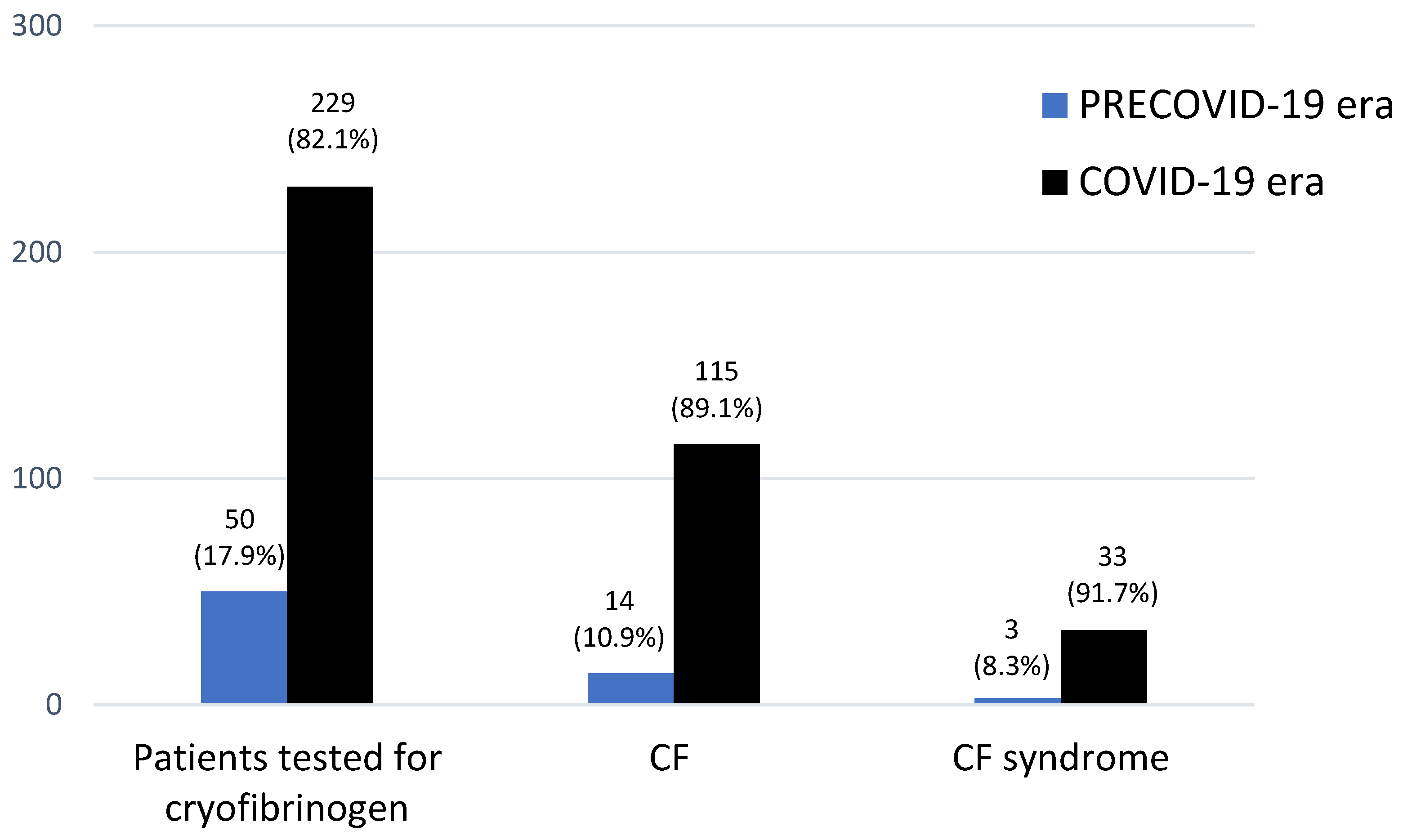
A total of 377 cryofibrinogen tests were performed in 279 patients in the PRECOVID-19 (n = 50) and COVID-19 (n = 229) periods. CF was found in 129 (46.2%) patients, being more frequent in the COVID-19 period (115/229; 50.2% vs. 14/50; 28%; p = 0.0047) (Figure 2). Patients who meet criteria for CF syndrome were not significantly more frequent in the COVID-19 period (33/229; 14.4% vs. 3/50; 6%; p = 0.079) (Figure 2).
More than half (n = 72; 55.8%), of the patients with a positive test underwent at least a second cryofibrinogen analysis, with both determinations being positive in 33 (27.9%) patients. Patients with CF in the COVID-19 era were younger and had lower CVRF (Table 1).

3.1.2. Clinical Features and Severity

Skin manifestations, described in Table 1, were the most frequent clinical manifestations in both groups (81.4%), especially purpuric macules (29.5%) followed by Raynaud´s phenomenon (23.3%). Distal skin ulcers were the only manifestations that showed statistically significant differences, being more frequent in the PRECOVID-19 era (35.7% vs. 7.8%, p = 0.008). The feet and/or hands were the most frequent localization in both groups (91.5%). Lower extremities and/or trunk were more frequent in the PRECOVID-19 group (28.6% vs. 6.1%; p = 0.019). The most reported skin symptoms in the overall groups were pain (29.5%) and itching (24%). Furthermore, 54.3% of patients had a history of sensitivity to cold.
Other clinical manifestations, at the time of the positive cryofibrinogen tests, were nonspecific and generally mild symptoms, such as respiratory (20.2%), rheumatological (14.7%), neurological (4.7%) and digestive symptoms (4.7%).
As mentioned above, the proportion of CF was lower in the PRECOVID-19 era. However, during this period, severe complications (n = 27) were more common (35.7% vs. 19.1%; p = 0.169). In both groups, these complications were associated with thrombotic CV events (n = 17), particularly VTE (n = 16) and AMI (n = 1), as well as cutaneous ulcerations (n = 14).

3.1.3. Histopathological Features

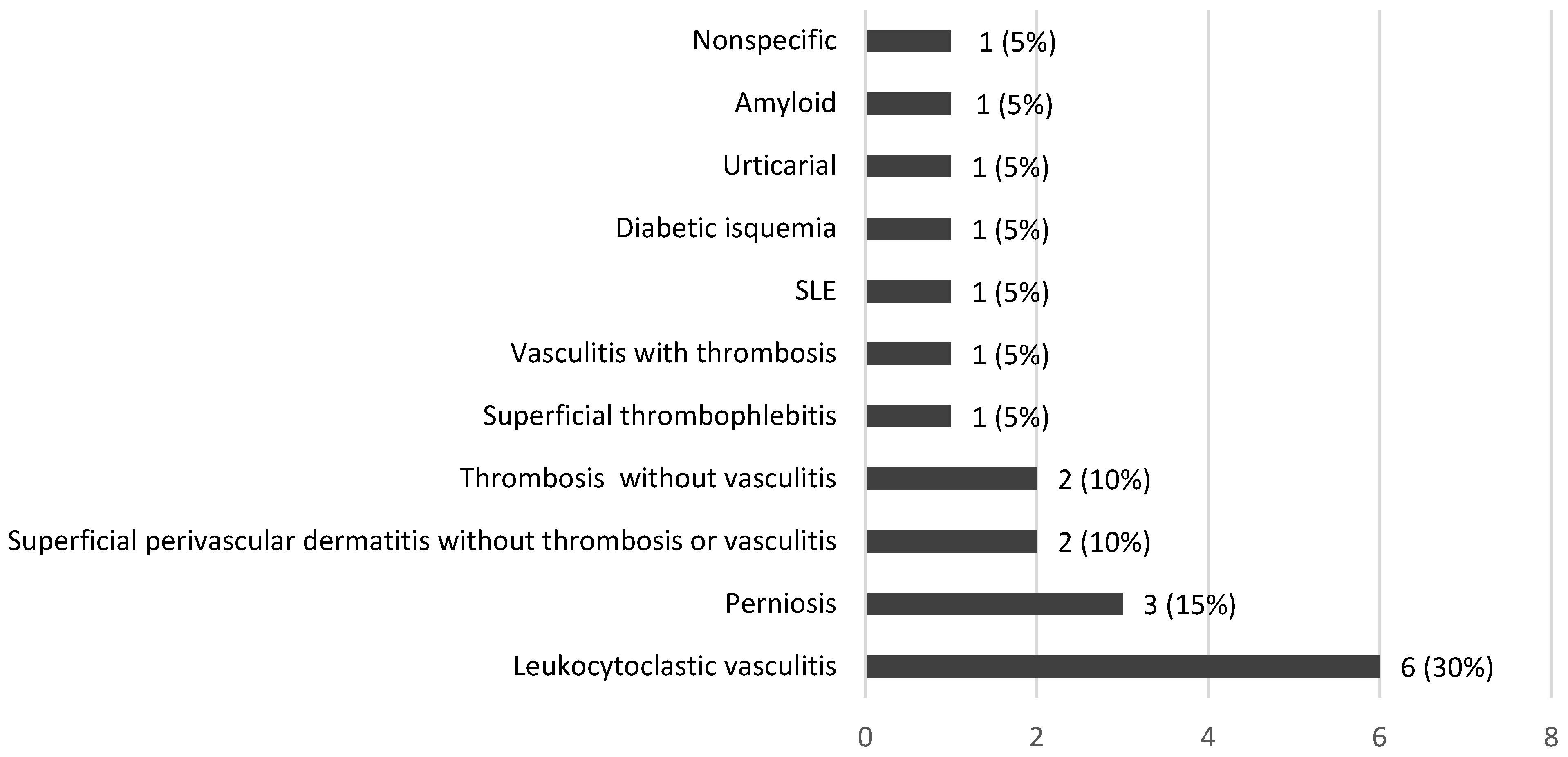
Skin biopsy was performed more frequently in the PRECOVID-19 era (35.7% vs. 13%; p = 0.043) (Table 1). The most common histopathological pattern was leukocytoclastic vasculitis (30%) (Figure 3). Leukocytoclastic vasculitis (40% vs. 26.7%; p = 0.613) and thrombosis (40% vs. 6.6%; p = 0.14) were not significantly more frequent during the PRECOVID-19 period.

3.1.4. Underlying Diseases

An underlying disease was observed in 52.7% cases, as detailed in Table 2. The proportion was similar in both periods. The most common underlying diseases were SARS-CoV-2 infection. SARS-CoV-2 pneumonia was identified in 7 patients.
In 9/13 patients with connective tissue diseases (all of them from the COVID-19 period), the diagnosis was established after the positive cryofibrinogen test. The diagnoses were: undifferentiated connective tissue disease (n = 5), serologic APS (n = 3), and APS. In the remaining 4 patients the diagnosis of connective tissue disease had previously been established; SLE, and APS, in the PRECOVID-19 period, and serological APS, and undifferentiated connective tissue disease in the COVID-19 period.
An underlying neoplasia was present in 7 patients in the PRECOVID-19 (n = 1), and in COVID-19 (n = 6) periods. Malignancies had been diagnosed at least 5 years before the positive cryofibrinogen test. The oncological diagnoses were cancer of the mouth, breast, colon, bladder and prostate carcinoma, acute myeloid leukaemia, essential thrombocytosis, and myelofibrosis; only this last patient was from the PRECOVID-19 era. Solid tumors were in remission. In contrast, the hematological neoplasms were active.
An underlying vasculitis or pseudovasculitis syndrome was observed in 5 patients. This was more frequent in the PRECOVID-19 period (21.4% vs. 1.7%; p = 0.009). Two of them had a pre-existing diagnosis, both in the PRECOVID-19 era: Henoch-Schoenlein Purpura (HSP), and Buerger disease. The other 3 patients were diagnosed after a positive cryofibrinogen test; Buerger disease in the PRECOVID-19 period and HSP (n = 2) during the COVID-19.
Veno-occlusive disease was observed in only 2 patients: central retinal vein thrombosis and Leriche’s syndrome.

3.2. CF Syndrome in Both Periods

3.2.1. Frequency and Epidemiology

The diagnosis of CF syndrome according to the proposed diagnostic criteria was found in 36 patients (27.9%), 3 in the PRECOVID-19 and 33 in the COVID-19 period. CF syndrome was somewhat higher in the COVID-19 period among patients with CF (33/115; 28.7% vs. 3/14; 21.4%; p = 0.756) (Figure 2).
The main general data of CF syndrome are summarized in Table 3. Patients with CF syndrome in the COVID-19 era were younger. Primary CF syndrome was non-statistically more frequent in the PRECOVID-19 era (67% vs. 39.4%; p = 0.559), while secondary CF was more common in the COVID-19 era (33% vs. 60.6%; p = 0.559). An underlying disease was observed in 20 (55.6%) cases.
CF syndrome was secondary to SARS-CoV-2 infection in 16 (48.5%) patients during COVID-19 era, with 80% (16/20 patients) of secondary CF syndrome cases during this period. In all these cases, SARS-CoV-2 infection was confirmed by microbiological tests. Other secondary causes included Buerger syndrome and essential thrombocythemia, diagnosed before the positive cryofibrinogen test and, undifferentiated connective tissue disease (n = 2) and APS diagnosed at the time of positive cryofibrinogen tests.

3.2.2. Clinical and Histopathological Features and Severity

In both periods, the most frequent clinical manifestations were cutaneous (Table 3). The most frequent location was the feet. Other clinical manifestations were non-specific and mild symptoms such as respiratory (19.4%), rheumatological (8.3%), neurological (2.8%).
Skin biopsies were performed on 2 patients, both in the COVID-19 period, revealing thrombosis without vasculitis and a perniosis pattern.
As mentioned, CF syndrome was found in 36 patients. In the remaining 93/129 (72.1%) patients with CF, the diagnosis of CF syndrome could not be established. These cases were either completely asymptomatic (14/129; 10.8%) or symptomatic but did not meet the criteria for CF syndrome (79/129; 61.2%). The proportion of asymptomatic patients was not statistically higher in the COVID-19 era compared to the PRECOVID-19 era (11.3% vs. 7.1%; p = 1), nor was the proportion of patients with ‘incomplete’ symptoms (71.4% vs. 60%; p = 0.116).
Severe CF syndrome was identified in 5 (13.9%) patients, all of them were diagnosed during the COVID-19 period: deep vein thrombosis (DVT) (n = 3), AMI with distal ulceration and isolated distal ulceration (Table 3).
Severe clinical manifestations were observed in 15/93 (16.1%) patients who did not meet the criteria for CF syndrome (3/11, 27.3%, in the PRECOVID-19 era vs. 12/82, 14.6%, in the COVID-19 era, p = 0.376). The severity in patients diagnosed with CF syndrome did not differ significantly compared to those with CF but without a diagnosis of CF syndrome (11% vs. 16.1%, p = 0.586).

3.2.3. Outcome

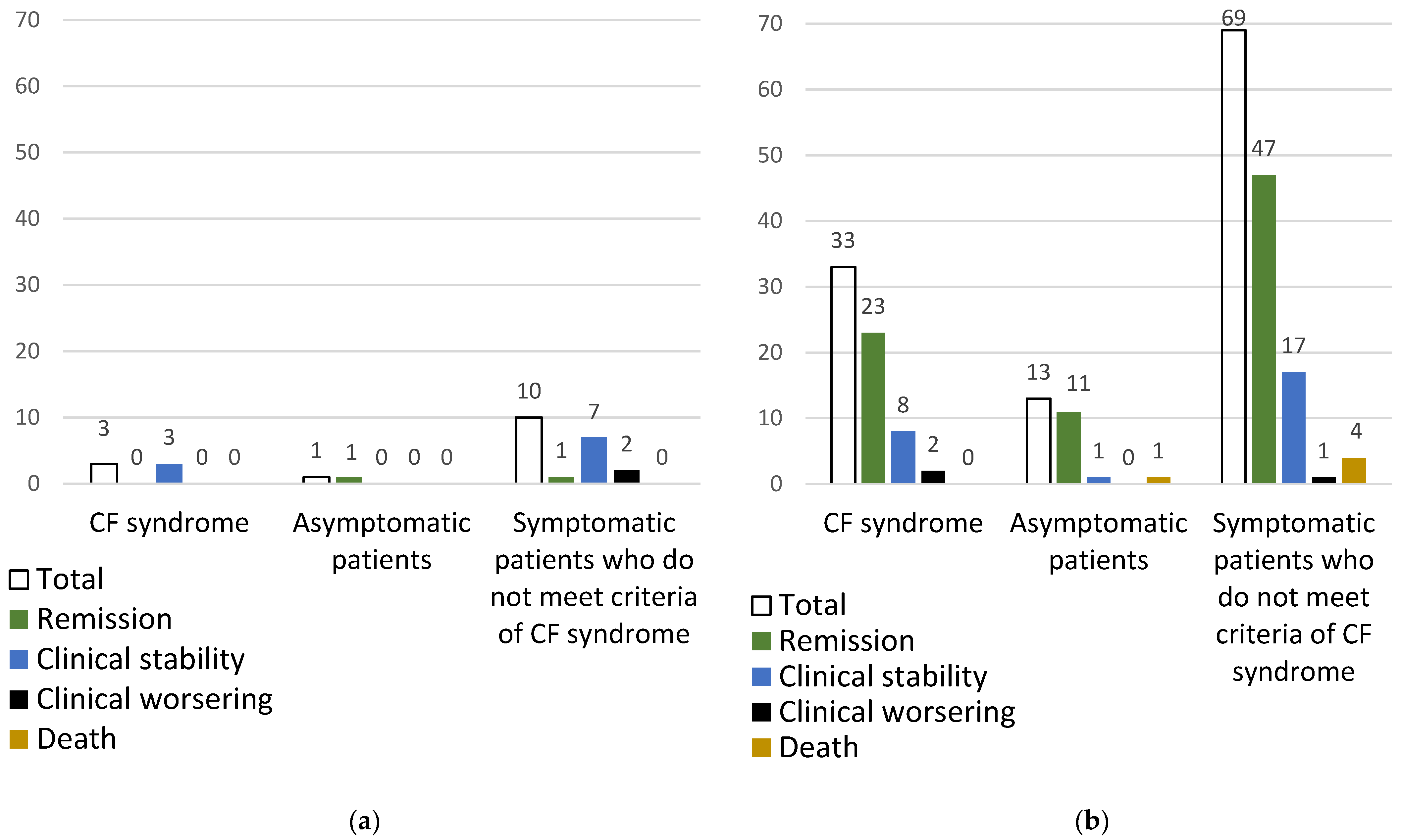
The outcome during one year of follow-up in patients with (a) CF syndrome, (b) asymptomatic and (c) symptomatic but not meeting criteria of CF syndrome are summarized in Figure 4a (PRECOVID-19) and in Figure 4b (COVID-19).
In the PRECOVID-19 period (Figure 4a), the patients with CF syndrome (n = 3) and the asymptomatic patient remained clinically stable. Of the remaining 10 symptomatic patients who did not meet the criteria for CF syndrome outcome was as follows: clinical remission (n = 1), clinical stability (n = 7) and clinical worsening (n = 2). Of these two later patients, one presented DVT and pretibial cutaneous ulcers after 11 months, and the other experienced an ischemic stroke after 11 months.
In the COVID-19 group (Figure 4b), of 33 patients with CF syndrome the clinical outcome was as follows: remission (n = 23), stability (n = 8) and worsening (n = 2). These last 2 patients presented an exacerbation of Raynaud’s phenomenon. In the group of asymptomatic patients (n = 13), they presented clinical remission (n = 11), clinical stability (n = 1) and mortality due to heart failure (n = 1). Of 69 patients who were symptomatic but did not meet the criteria for CF syndrome, the outcome was as follows: remission (n = 47), clinical stability (n = 17), worsening of Raynaud’s phenomenon and perniosis (n = 1) and death (n = 4). Mortality was due to heart failure (n = 3) and amyotrophic lateral sclerosis (n = 1).

4. Discussion

We present a series of patients with CF in two periods: PRECOVID-19 and COVID-19. The purpose of this study was to assess in both periods (a) the frequency and clinical features of patients with CF, (b) the study of CF syndrome, and (c) the outcome of CF.
Patients with CF in our COVID-19 series were more often male and younger, in contrast to our PRECOVID-19 series and other studies [3,12,13,14,15] conducted before to COVID-19 pandemic. The difference in mean age may be due to SARS-CoV-2 infection in younger patients, which may trigger CF [10,16]. Furthermore, we identified a statistically significant difference in CVRFs between the studied groups, probably due to the difference in mean age. Smith et al. [17] reported a diabetes prevalence of 13%, a figure comparable to that obtained in our sample.
Most of the published studies on CF were performed in the PRECOVID-19 era, reporting disparate prevalence results [2,6,18]. CF was identified in a range of 12–51% of patients screened for cryopathy [3]. Thus, H. Blain et al. [12] detected 16.8% of patients with CF while Saadoun et al. [13] reported a rate of 22.1%.
CF syndrome can be responsible for severe vasculopathies [3,6,19,20]. The most supported hypothesis is that an increase in the levels of plasmin inhibitors (α1-antitrypsin and α2-macroglobulin) leads to a decrease fibrinolysis and the accumulation of cryofibrinogen with consequent thrombosis of small vessels [5,12,17]. Also, cryofibrinogen can interact with circulating immunoglobulins or immune complex produced during infections, malignancies, or immune-mediated inflammatory diseases [2,21,22].
The proportion of essential and secondary CF reported in the literature [12,14,15] ranges from 30–57%, consistent with the findings in our cohort. There is a great disparity in the percentage of rheumatological diseases in patients with CF. For instance, concerning connective tissue diseases, different studies report 14–70%, and 0–30% for vasculitis [3,13]. Regarding the pattern observed in oncological pathology, we reported a prevalence lower than previous studies (8–40%) [3,13].
Regarding skin manifestations in patient with CF, our cohort showed a percentage comparable to that reported by other authors [12,13,14,15,22]. Purpuric macules were the most frequent skin manifestation observed in patients with CF and patients with CF syndrome. Similar results are reported in different studies (25–78%) [3,5,12,13,14,23]. In contrast, Soyfoo et al. [15] documented purpuric macules in only 4.7% of patients with CF. Unlike other causes of purpura, patients with CF are typically asymptomatic or mildly ill and the purpura is localized [24]. Raynaud’s phenomenon was observed in our PRECOVID-19 CF patients at a similar percentage to the 53.7% reported by Soyfoo et al. [15], but higher than the studies mentioned above, which range from 16.6–25% [3,5,12,13,14,23]. About skin ulcers, the results obtained during in the PRECOVID-19 era were comparable to those reported by Soyfoo et al. [15] (17–56%), whose study was conducted prior to the COVID-19 pandemic. Skin ulcers were significantly more frequent during the PRECOVID-19 era in our cohort, may be influenced by the older age and higher prevalence of CVRF observed in patients from this period, both of which were also statistically significant, as it is well documented that the presence of ulcers, especially in the lower extremities, is more common in elderly individuals and those with diabetes and arterial hypertension. Saadoun et al. [13], Blain et al. [12] and Soyfoo et al. [15] reported cold sensitivity in 40%, 43% and 57% of patients with CF respectively, findings that are consistent with our study. Urticarial lesions were rare in our series, occurring at lower rates than those reported by Saadoun et al. (11%) [13], Soyfoo et al. (3%) [15] or Michaud et al. (8.3%) [3]. We recorded livedo in line with previous studies (3–25%) [3,5,12,13,14,23]. Other skin symptoms, such as chilblains, perniosis-like lesions, and “blue finger”, have also been described [24,25].
In addition to cutaneous manifestations, CF can present with systemic symptoms and TEs [2]. H. Blain et al. [12] reported that 13% of patients with CF experienced a TE, a proportion similar to that described by Stanley B. Smith et al. (14%) [17] and in our cohort, but lower than the 43% reported by Saadoun et al. [13]. Notably, in patients with renal disease, Terrier et al. [26] observed an statistical increase in the frequency of severe TEs from 0–36% in patients with CF.
CF is also described in pediatric patients, often manifesting as a transient benign process related to infection. It is typically characterized by an acute onset of pain, erythema, and blistering of the fingers and toes [24,27], triggered by exposure to cold [28] or occurring as an essential feature [29].
Since the onset of the COVID-19 pandemic, many dermatological manifestations in association with SARS-CoV-2 infection have been reported [7,8,9]. Recent studies have confirmed the presence of patients with chilblains probably related to COVID-19 [16]. In this regard, Gómez-Fernández et al. [10] described the characteristics of 54 patients who presented perniosis-like lesions between April and May 2020. This study highlighted the high prevalence of CF in the patients studied, reaching figures of up to 66.7% of cases. The authors suggested that CF could be involved in the etiopathogenesis of the perniosis-like lesions and coagulopathies and thrombi observed in patients infected with SARS-CoV-2. Perry et al. [30] described secondary CF-induced livedo reticularis associated with COVID-19 and recrudescence, likely due to a viral infection other than SARS-CoV-2. Both studies, along with the present one, provide evidence of similar cutaneous findings between COVID-19 and CF, supporting the possibility that cases of SARS-CoV-2 induced CF are being missed. In the present study, we report that during the COVID-19 pandemic, there was an increase in CF and CF syndrome, although the manifestations were less severe.
Classically, the histopathological features of CF consist of vascular occlusion secondary to the presence of cryofibrinogen within small vessels [3,5,23,31]. In our study, this finding was also observed, which may explain the cutaneous manifestations seen in our patients, such as skin ulcers or purpuric macules secondary to vasculitis. However, we consider that we do not have enough histological samples to draw conclusions about the predominant histological pattern and whether there are differences between the two periods. Other specific findings include an occlusive eosinophilic deposit within the lumen and walls of blood vessels, with a variable inflammatory mononuclear perivascular infiltrate [32]. Leukocytoclastic vasculitis, necrosis of the dermis and epidermis, and deposits of immunoglobulins and complement are non-specific findings in lesions associated with CF [33]. Skin biopsies were significantly more frequent during the PRECOVID-19 period, a difference that is likely related to the general reduction in procedures during the COVID-19 era, aimed at minimizing the risk of viral transmission.
Cryoglobulins and cryofibrinogen may coexist in the same patient. Thus, in the largest series, Saadoun et al. [13] reported an association in up to 88.5%. Other authors support that the frequency of the development of a CF may exceed that of the cryoglobulinemia (CG) [34]. In our experience, none of the patients with CF had CG. Since they act through different mechanisms, the simultaneous detection of cryofibrinogen and cryoglobulins is essential to differentiate between immune-mediated vasculitis with CG, and more mechanical TEs with CF [20].
To our knowledge, only two authors have defined diagnostic criteria for CF, but they are not globally established. Therefore, the diagnostic criteria for the disease are poorly defined [3,6]. In the present study, we propose new diagnostic criteria modified from Grada et al. [6] and Michaud et al. [3] based on our daily clinical experience and management of patients with CF. We advocate the need to differentiate between an isolated positive cryofibrinogen test result and recurrent positive tests of cryofibrinogen accompanied by skin symptoms or TEs. This distinction is crucial as it delineates a group that has developed a potentially severe CF syndrome, who may benefit from treatment. When we obtain a positive cryofibrinogen test and suspect a CF syndrome (either with skin manifestations or TE), we propose frequent plasma cryofibrinogen tests (at least twice) and comprehensive follow-up; the outcome in most patients will be to clinical stability or remission. However, if we obtain a positive cryofibrinogen test but the patient remains asymptomatic, we recommend reviewing this test, and if the patient remains asymptomatic, no further follow-up is necessary.
The main limitations of this study include its retrospective design based on medical records, which might have distorted some data. Additionally, the unequal sample sizes between the two periods could have introduced bias in the interpretation of results. Finally, our results represent a limited sample from a region with a moderate population size and therefore may not be representative of our entire country.

5. Conclusions

Skin manifestations were the most frequent clinical manifestations in both periods, especially purpuric macules. PRECOVID-19 patients were older and more frequently presented skin ulcers that also exhibited severe histological features. CF was secondary in around half of cases, especially in relation to SARS-CoV-2 infection. The frequency of CF syndrome was somewhat higher in the COVID-19 period; however, the manifestations were less severe. In both periods, the outcome in most patients was clinical stability or remission. Patients with CF syndrome may benefit from treatment and follow-up, unlike asymptomatic patients or those who do not meet the criteria.

Author Contributions

Conceptualization: M.d.A.S.L., C.L.-T., M.R.-G., C.A.-M., S.S.-C., I.B.-G., A.M.-G., C.S.-G., C.G.-V., J.I.-V., M.L.-H., R.B. and M.A.G.-L. Methodology: M.d.A.S.L., C.L.-T., M.R.-G., R.B. and M.A.G.-L. Software: M.d.A.S.L., C.L.-T. and M.R.-G. Validation: M.d.A.S.L., C.L.-T., M.R.-G. and C.G.-V. Formal analysis: M.d.A.S.L., C.L.-T. and M.R.-G. Investigation: M.d.A.S.L., C.L.-T., M.R.-G., C.G.-V., M.L.-H., R.B. and M.A.G.-L. Resources: R.B. Data curation: M.d.A.S.L., C.L.-T., M.R.-G., C.A.-M., S.S.-C., I.B.-G., A.M.-G., C.S.-G., C.G.-V., J.I.-V. and M.L.-H. Writing—original draft preparation: M.d.A.S.L. and C.L.-T. Writing—review and editing: M.d.A.S.L., C.L.-T., M.R.-G., C.G.-V., J.I.-V., R.B. and M.A.G.-L. Visualization: M.d.A.S.L., C.L.-T. and R.B. Supervision: M.d.A.S.L., C.L.-T., M.L.-H., R.B. and M.A.G.-L. Project administration: M.d.A.S.L. and C.L.-T. Funding acquisition: R.B. All authors have read and agreed to the published version of the manuscript.

Funding

This research received no external funding.

Institutional Review Board Statement

The study was conducted in accordance with the Declaration of Helsinki and approved by the Ethics Committee of Hospital Universitario Marqués de Valdecilla (code: 2024.124, on 6 September 2024).

Informed Consent Statement

Not applicable.

Data Availability Statement

The original contributions presented in this study are included in the article. Further inquiries can be directed to the corresponding author.

Acknowledgments

We thank all the members of the different Medical Services and patients included in this project.

Conflicts of Interest

Dr. Ricardo Blanco received grants/research support from AbbVie, MSD, and Roche, and had consultation fees/participation in a company-sponsored speaker’s bureau from AbbVie, Pfizer, Roche, Bristol-Myers, Lilly, Galapagos, Novartis, Janssen, GSK, and MSD. The remaining authors have declared no conflicts of interest.

References

  1. Korst, D.R.; Kratochvil, C.H. Cryofibrinogen in a case of lung neoplasm associated with thrombophlebitis migrans. Blood 1955, 10, 945–953. [Google Scholar] [CrossRef] [PubMed]
  2. Moiseev, S.; Luqmani, R.; Novikov, P.; Shevtsova, T. Cryofibrinogenaemia-A neglected disease. Rheumatology 2017, 56, 1445–1451. [Google Scholar] [CrossRef]
  3. Michaud, M.; Pourrat, J. Cryofibrinogenemia. J. Clin. Rheumatol. 2013, 19, 142–148. [Google Scholar] [CrossRef]
  4. Santiago, M.B.; Melo, B.S. Cryofibrinogenemia: What Rheumatologists Should Know. Curr. Rheumatol. Rev. 2022, 18, 186–194. [Google Scholar] [CrossRef]
  5. Amdo, T.D.; Welker, J.A. An approach to the diagnosis and treatment of cryofibrinogenemia. Am. J. Med. 2004, 116, 332–337. [Google Scholar] [CrossRef] [PubMed]
  6. Grada, A.; Falanga, V. Cryofibrinogenemia-Induced Cutaneous Ulcers: A Review and Diagnostic Criteria. Am. J. Clin. Dermatol. 2017, 18, 97–104. [Google Scholar] [CrossRef] [PubMed]
  7. Martora, F.; Villani, A.; Fabbrocini, G.; Battista, T. COVID-19 and cutaneous manifestations: A review of the published literature. J. Cosmet. Dermatol. 2023, 22, 4–10. [Google Scholar] [CrossRef]
  8. Genovese, G.; Moltrasio, C.; Berti, E.; Marzano, A.V. Skin Manifestations Associated with COVID-19: Current Knowledge and Future Perspectives. Dermatology 2021, 237, 1–12. [Google Scholar] [CrossRef]
  9. Nakashima, C.; Kato, M.; Otsuka, A. Cutaneous manifestations of COVID-19 and COVID-19 vaccination. J. Dermatol. 2023, 50, 280–289. [Google Scholar] [CrossRef]
  10. Gómez-Fernández, C.; López-Sundh, A.E.; González-Vela, C.; Ocejo-Vinyals, J.G.; Mayor-Ibarguren, A.; Salas-Venero, C.A.; Gutiérrez-Larrañaga, M.; Tejerina-Puente, A.; Fariñas, M.C.; Cabero-Pérez, M.J.; et al. High prevalence of cryofibrinogenemia in patients with chilblains during the COVID-19 outbreak. Int. J. Dermatol. 2020, 59, 1475–1484. [Google Scholar] [CrossRef]
  11. Polyclonal Rabbit Fibrinogen No de catálogo A0080. Fibrinogen PRA 2010, 2, 44–45.
  12. Blain, H.; Cacoub, P.; Musset, L.; Costedoat-Chalumeau, N.; Silberstein, C.; Chosidow, O.; Godeau, P.; Frances, C.; Piette, J.C. Cryofibrinogenaemia: A study of 49 patients. Clin. Exp. Immunol. 2000, 120, 253–260. [Google Scholar] [CrossRef] [PubMed]
  13. Saadoun, D.; Elalamy, I.; Ghillani-Dalbin, P.; Sene, D.; Delluc, A.; Cacoub, P. Cryofibrinogenemia: New insights into clinical and pathogenic features. Am. J. Med. 2009, 122, 1128–1135. [Google Scholar] [CrossRef]
  14. Belizna, C.C.; Tron, F.; Joly, P.; Godin, M.; Hamidou, M.; Lévesque, H. Outcome of essential cryofibrinogenaemia in a series of 61 patients. Rheumatology 2008, 47, 205–207. [Google Scholar] [CrossRef]
  15. Soyfoo, M.S.; Goubella, A.; Cogan, E.; Wautrecht, J.C.; Ocmant, A.; Stordeur, P. Clinical significance of cryofibrinogenemia: Possible pathophysiological link with Raynaud’s phenomenon. J. Rheumatol. 2012, 39, 119–124. [Google Scholar] [CrossRef]
  16. Andina, D.; Noguera-Morel, L.; Bascuas-Arribas, M.; Gaitero-Tristán, J.; Alonso-Cadenas, J.A.; Escalada-Pellitero, S.; Hernández-Martín, Á.; de la Torre-Espi, M.; Colmenero, I.; Torrelo, A. Chilblains in children in the setting of COVID-19 pandemic. Pediatr. Dermatol. 2020, 37, 406–411. [Google Scholar] [CrossRef]
  17. Smith, S.B.; Arkin, C. Cryofibrinogenemia: Incidence, clinical correlations, and a review of the literature. Am. J. Clin. Pathol. 1972, 58, 524–530. [Google Scholar] [CrossRef]
  18. McKee, P.A.; Kalbfleisch, J.M. Incidence and significance of cryofibrinogenemia. J. Lab. Clin. Med. 1963, 61, 203–210. [Google Scholar]
  19. Jager, B.V. Cryofibrinogenemia. N. Engl. J. Med. 1962, 266, 579–583. [Google Scholar] [CrossRef]
  20. Sarda-Kolopp, M.N.; Miossec, P. Cryofibrinogen-Characteristics and Association with Cryoglobulin: A Retrospective Study out of a Series of 1, 712 Samples over 7 Years. Thromb Haemost. 2022, 123, 669–678. [Google Scholar] [CrossRef]
  21. Martin, S. Cryofibrinogenemia, monoclonal gammopathy, and purpura. Report of a case and review of the literature. Arch. Dermatol. 1979, 115, 208–211. [Google Scholar] [CrossRef] [PubMed]
  22. Matsuda, M.; Saida, T.; Hasegawa, R. Cryofibrinogen in the plasma of patients with skin ulcerative lesions on the legs: A complex of fibrinogen and cold insoluble globulin. Thromb. Res. 1976, 9, 541–552. [Google Scholar] [CrossRef]
  23. Belizna, C.; Loufrani, L.; Subra, J.F.; Godin, M.; Jolly, P.; Vitecocq, O.; Faller, B.; Ghali, A.; Tron, F.; Hamidou, M.; et al. A 5-year prospective follow-up study in essential cryofibrinogenemia patients. Autoimmun. Rev. 2011, 10, 559–562. [Google Scholar] [CrossRef] [PubMed]
  24. Ireland, T.A.; Werner, D.A.; Rietschel, R.L.; Patterson, J.H.; Spraker, M.K. Cutaneous lesions in cryofibrinogenemia. J. Pediatr. 1984, 105, 67–69. [Google Scholar] [CrossRef]
  25. Hassan, F.; Khoury, A.; Awad, J.; Jeries, H.; Naffaa, M.E. A very rare cause of blue finger: A case-based review. J. Scleroderma Relat. Disord. 2023, 8, NP1–NP5. [Google Scholar] [CrossRef]
  26. Terrier, B.; Izzedine, H.; Musset, L.; Ghillani, P.; Deray, G.; Saadoun, D.; Cacoub, P. Prevalence and clinical significance of cryofibrinogenaemia in patients with renal disorders. Nephrol. Dial. Transplant. 2011, 26, 3577–3581. [Google Scholar] [CrossRef] [PubMed]
  27. Robinson, M.G.; Troiano, G.; Cohen, H.; Foadi, M. Acute transient cryofibrinogenemia in infants. J. Pediatr. 1966, 69, 35–39. [Google Scholar] [CrossRef]
  28. William RGriswold, M.D.; Gilbert Simon, M.D.; Rawle MMcintosh, M.D. Anaphylactoid Purpura, Cold Exposure, and Cryofibrinogenemia. Ann. Intern. Med. 1973, 78, 611. [Google Scholar]
  29. Rachmilewitz, E.A. Essential cryofibrinogenemia. Clinical, pathological and immunological studies. Isr. J. Med. Sci. 1970, 6, 32–43. [Google Scholar]
  30. Perry, W.M.; Salame, N.; Swerlick, R.A.; Cheeley, J.T. The clot thickens with COVID-19 and cryofibrinogenemia: A thought-provoking association. JAAD Case Rep. 2022, 24, 24–28. [Google Scholar] [CrossRef]
  31. Ball, G.V.; Goldman, L.N. Chronic ulcerative colitis, skin necrosis, and cryofibrinogenemia. Ann. Intern. Med. 1976, 85, 464–466. [Google Scholar] [CrossRef] [PubMed]
  32. Nash, J.W.; Ross, P., Jr.; Crowson, N.A.; Taylor, J.; Morales, J.E.; Yunger, T.M.; Magro, C. The histopathologic spectrum of cryofibrinogenemia in four anatomic sites: Skin, lung, muscle, and kidney. Am. J. Clin. Pathol. 2003, 119, 114–122. [Google Scholar] [CrossRef]
  33. Zouboulis, C.C.; Gollnick, H.; Weber, S.; Peter, H.H.; Orfanos, C.E. Intravascular coagulation necrosis of the skin associated with cryofibrinogenemia, diabetes mellitus, and cardiolipin autoantibodies. J. Am. Acad. Dermatol. 1991, 25, 882–888. [Google Scholar] [CrossRef] [PubMed]
  34. Kalbfleisch, J.M.; Bird, R.M. Cryofibrinogenemia. N. Engl. J. Med. 1960, 3, 881–886. [Google Scholar] [CrossRef] [PubMed]
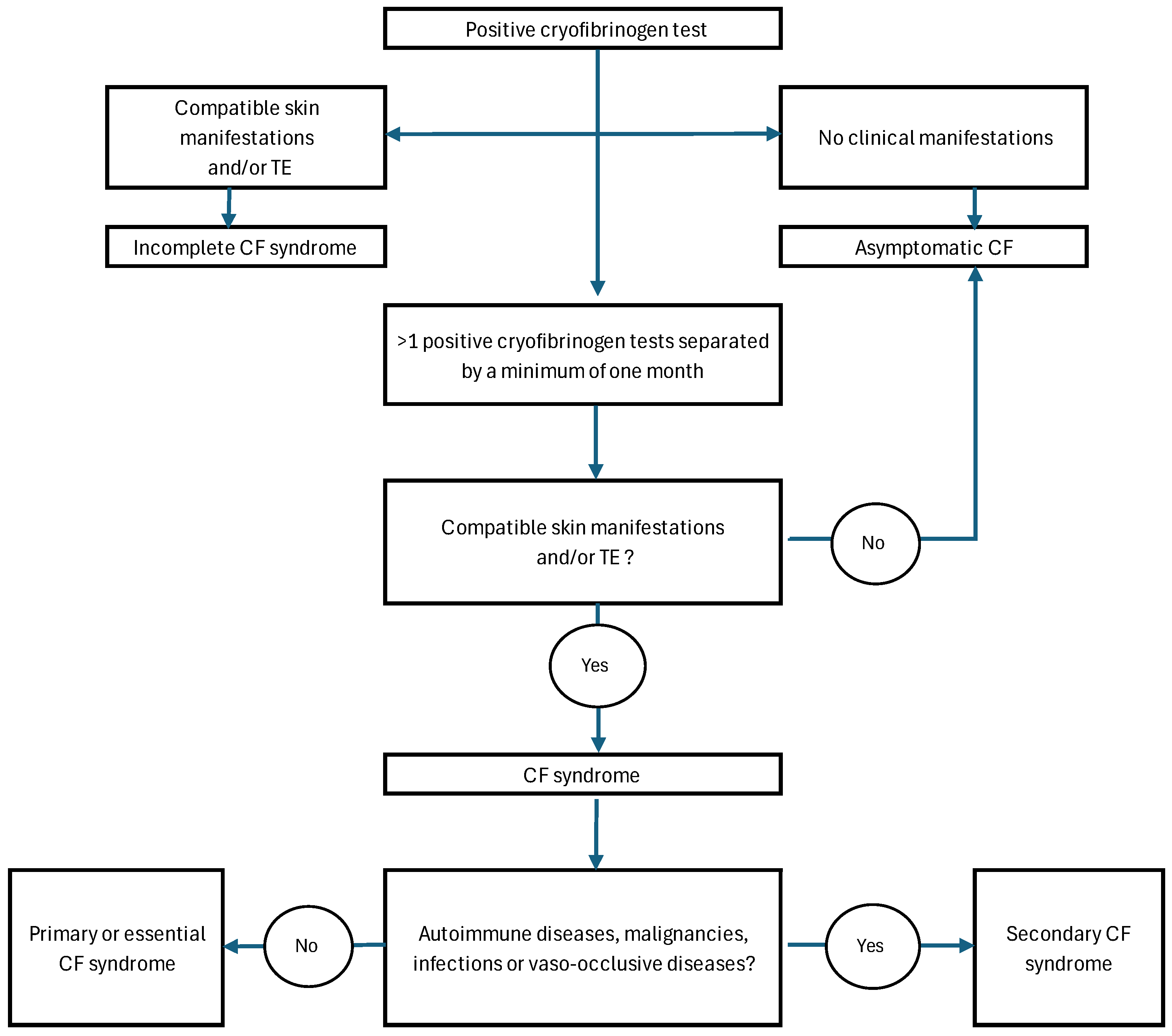
Figure 1. Diagnostic pathway of CF syndrome. CF: Cryofibrinogenemia; TE: Thrombotic Events.
Sci 07 00081 g001
Figure 2. Comparison between PRECOVID-19 and COVID-19 groups of: patients tested for cryofibrinogen, CF and CF syndrome. CF: cryofibrinogenemia.
Sci 07 00081 g002
Figure 3. Histopathological patterns observed in skin biopsies performed. SLE: Systemic lupus erythematosus.
Sci 07 00081 g003
Figure 4. Outcome after one year of follow-up in: (a) PRECOVID-19 period and (b) COVID-19 period. CF: Cryofibrinogenemia.
Sci 07 00081 g004
Table 1. Epidemiological and clinical features of patients with positive cryofibrinogen in PRECOVID-19 and COVID-19 periods.
Overall (n = 129)PRECOVID-19 (n = 14)COVID-19 (n = 115)p
Sex and age
  Female/Male, n (%)61/68 (47.3/52.7)7/7 (50/50) 54/61 (47/53)0.829
  Age, years, median [IQR] 37 [15–67]55 [36–75]33 [15–67]0.054
Cardiovascular Risk Factors, n (%) 56 (43.4)11 (78.6)45 (39.1)0.005
  Hypercholesterolemia34 (26.4)7 (50)27 (23.5)0.051
  High blood pressure33 (25.6)6 (42.9)27 (23.5)0.19
  Obesity23 (17.8)3 (21.4)20 (17.4)0.715
  Hypertriglyceridemia19 (14.7)3 (21.4)16 (13.9)0.433
  Smoker (past or present)16 (12.4)4 (28.6)12 (10.4)0.074
  Diabetes Mellitus15 (11.6)1 (7.1)14 (12.2)1
  Alcohol consumption13 (10.1)2 (14.3)11 (9.7)0.633
  Other toxics2 (1.6)0 (0)2 (1.7)1
Thrombotic Cardiovascular events, n (%)17 (13.2)2 (14.3)15 (13)1
  VTE (DVT, PE), n (%)16 (12.4)2 (14.3)14 (12.1)0.685
  AMI, n (%)1 (0.8)0 (0)1 (0.9)1
Patients with cutaneous lesions, n (%)105 (81.4)13 (92.9)92 (80)0.465
Type of skin lesions
  Purpuric macules, n (%)38 (29.5)3 (21.4)35 (30.4)0.757
  Raynaud, n (%)30 (23.3)6 (42.9)24 (20.9)0.091
  Perniosis, n (%)25 (19.4)0 (0)25 (21.7)0.07
  Acrocyanosis, n (%)19 (14.7)1 (7.1)18 (15.7)0.692
  Distal ulceration, n (%)14 (10.9)5 (35.7)9 (7.8)0.008
  Livedo, n (%)5 (3.9)2 (14.3)3 (2.6)0.091
  Cold urticaria, n (%)1 (0.8)0 (0)1 (0.9)1
Location of skin lesions
  Feet, n (%)38 (29.5)3 (21.4)35 (30.4)0.757
  Hands, n (%)31 (24)4 (28.6)27 (23.5)0.742
  Hands and feets, n (%)25 (19.4)2 (14.3)23 (20)1
  Other (lower extremities and trunk), n (%)11 (8.5)4 (28.6)7 (6.1)0.019
Cold sensitivity
  Yes, n (%)70 (54.3)7 (50)63 (54.8)0.734
  No, n (%)40 (31)7 (50)33 (28.7)0.129
  Not provided, n (%)19 (14.7)0 (0)19 (16.5)0.222
Skin biopsy, n (%)20 (15.5)5 (35.7)15 (13)0.043
AMI: Acute myocardial infarction, DVT: Deep Vein Thrombosis, PE: Pulmonary Embolism, VTE: Venous Thromboembolism.
Table 2. Underlying diseases in patients with positive cryofibrinogen tests in the PRECOVID-19 and COVID-19 era.
Overall (n = 129)PRECOVID-19 (n = 14)COVID-19 (n = 115)p
Underlying diseases, n (%)68 (52.7)7 (50)61 (53)1
SARS-CoV-2 infection, n (%)45 (34.9)0 (0)45 (39.1)0.002
Connective tissue disease, n (%)13 (10.1)2 (14.3)11 (9.6)0.633
  SLE1 (0.8)1 (7.1)0 (0)0.109
  APS2 (1.6)1 (7.1)1 (0.9)0.206
  Serological APS4 (3.1)0 (0)4 (3.5)1
  Undifferentiated2 (1.6)0 (0)2 (1.7)1
  Under investigation4 (3.1)0 (0)4 (3.5)1
Malignancies, n (%)7 (5.4)1 (7.1)6 (5.2)1
Vasculitis, n (%)5 (3.9)3 (21.4)2 (1.7)0.009
  Buerger, n (%)2 (1.6)2 (14.3)0 (0)0.011
  Henoch-Schoenlein Purpura, n (%)3 (1.6)1 (7.1)2 (1.7)0.294
Vaso-occlusive diseases, n (%)2 (1.6)1 (7.1)1 (0.9)0.206
  Leriche´s syndrome, n (%)1 (0.8)0 (0)1 (0.9)1
  CRVT, n (%)1 (0.8)1 (7.1)0 (0)0.109
APS: Antiphospholipid syndrome, CRVT: Central Retinal Vein thrombosis, SLE: Systemic lupus erythematosus.
Table 3. Epidemiological and clinical features of patients diagnosed with CF syndrome in the PRECOVID-19 and the COVID-19 era.
Overall (n = 36)PRECOVID-19 (n = 3)COVID-19 (n = 33)p
Sex and age
  Female/Male, n (%)18/18 (50/50)2/1 (67/33)16/17 (48.5/51.5)1
  Age, years, median [IQR] 28.5 [30]51 [40–56]17 [13–41]0.045
Diagnosis of CF syndrome
  Essential, n (%)15 (41.7)2 (66.7)13 (39.4)0.559
  Secondary, n (%)21 (58.3)1 (33.3)20 (60.6)0.559
Underlying diseases, n (%)20 (55.6)1 (33.3)19 (57.6)0.574
SARS-CoV-2 infection, n (%)16 (44.4)0 (0)16 (48.5)0.238
Connective tissue disease, n (%)3 (8.3)0 (0)3 (9)1
  APS, n (%)1 (2.7)0 (0)1 (3)1
  Others, n (%)2 (5.5)0 (0)2 (6)1
Vasculitis, n (%)1 (2.7)1 (33.3)0 (0)0.083
  Buerger, n (%)1 (2.7)1 (33.3)0 (0)0.083
Malignancies, n (%)1 (2.7)0 (0)1 (3)1
Thrombotic Cardiovascular events, n (%)4 (11.1)0 (0)4 (12.1)1
  DVT3 (8.3)0 (0)3 (9)1
  AMI1 (2.7)0 (0)1 (3)1
Patients with cutaneous lesions, n (%)35 (97.2)3 (100)32 (97)1
Type of skin lesions
  Purpuric macules, n (%)16 (44.4)1 (33.3)15 (45.5)1
  Raynaud, n (%)9 (25)2 (66.7)7 (21.2)0.148
  Perniosis, n (%)7 (19.4)0 (0)7 (21.2)1
  Acrocyanosis, n (%)7 (19.4)0 (0)6 (18.2)1
  Distal ulceration, n (%)2 (5.5)0 (0)2 (6)1
  Livedo, n (%)2 (5.5)0 (0)2 (6)1
Location of skin lesions
  Feet, n (%)18 (51.4)0 (0)18 (54.5)0.229
  Hands, n (%)9 (25.7)3 (100)6 (18.2)0.012
  Hands and feets, n (%)8 (22.9)0 (0)8 (24.2)1
AMI: Acute myocardial infarction, APS: Antiphospholipid syndrome, DVT: Deep Vein Thrombosis.
Disclaimer/Publisher’s Note: The statements, opinions and data contained in all publications are solely those of the individual author(s) and contributor(s) and not of MDPI and/or the editor(s). MDPI and/or the editor(s) disclaim responsibility for any injury to people or property resulting from any ideas, methods, instructions or products referred to in the content.