Article

Dapagliflozin and Silymarin Ameliorate Cisplatin-Induced Nephrotoxicity via Nrf2/HO-1 Upregulation: A Preclinical Mechanistic Study

by
Shakta Mani Satyam
1,*,
Laxminarayana Kurady Bairy
1,
Abdul Rehman
2,
Anuradha Asokan Nair
3,
Mohamed Farook
3,
Nirmal Nachiketh Binu
3,
Sofiya Khan
3,
Mohamed Yehya
3 and
Mohammed Moin Khan
3
1
Faculty of Pharmacology, RAK College of Medical Sciences, RAK Medical and Health Sciences University, Ras Al Khaimah P.O. Box 11172, United Arab Emirates
2
Faculty of Pathology, RAK College of Medical Sciences, RAK Medical and Health Sciences University, Ras Al Khaimah P.O. Box 11172, United Arab Emirates
3
RAK College of Medical Sciences, RAK Medical and Health Sciences University, Ras Al Khaimah P.O. Box 11172, United Arab Emirates
*
Author to whom correspondence should be addressed.
Submission received: 22 December 2024 / Revised: 16 April 2025 / Accepted: 6 May 2025 / Published: 8 May 2025

Abstract

This study evaluated the nephroprotective potential of dapagliflozin and silymarin, alone and in combination, against cisplatin-induced kidney damage in female Wistar rats. Cisplatin was administered at 3 mg/kg weekly to all groups except the normal controls, with treatments of silymarin (50 mg/kg/day), dapagliflozin (0.9 mg/kg/day), or both for 45 days. Dapagliflozin significantly reduced uric acid, the urea-to-creatinine ratio, and serum urea levels compared to nephrotoxic controls, while combination therapy showed further improvements. Both agents decreased inflammatory markers like TNF-alpha, IL-6, and IL-1 beta, with enhanced effects in combination. Oxidative stress markers, including nitrite and 4-HNE, were lowered, and antioxidant enzyme activities (catalase, SOD, and GSH-Px) were increased by dapagliflozin and silymarin, with the combined treatment yielding the most substantial improvements. Molecular analysis revealed elevated Nrf2 and HO-1 levels, which are critical for oxidative stress mitigation, particularly with combination therapy. Histologically, combination therapy preserved renal structure, closely resembling normal controls, while dapagliflozin and silymarin alone showed moderate inflammation and structural alterations. These findings highlight the effect of dapagliflozin and silymarin, especially in combination, to mitigate cisplatin-induced nephrotoxicity by reducing oxidative stress, inflammation, and apoptosis via modulation of the Nrf2/HO-1 pathway.

1. Introduction

Cancer continues to be one of the major global health challenges, with its incidence projected to increase over the next two decades [1]. Even if improvements in medical knowledge and technology have made it easier to detect and cure cancer, these developments are frequently accompanied by serious side effects that have a detrimental effect on patients’ health and quality of life [2]. Although the increase in cancer survival rates is praiseworthy, there is still reason for concern over the mounting load of comorbidities and side effects associated with treatment.
Cisplatin, a highly effective anticancer agent, is an integral part of chemotherapy regimens used to treat head and neck, lung, testicular, and ovarian cancers [3,4,5]. However, its clinical utility is often limited by nephrotoxic side effects, which can lead to dose reductions and compromise the overall effectiveness of cancer treatment [6]. The mechanisms behind cisplatin-induced kidney injury are complex and involve inflammation, damage to the proximal convoluted tubules, oxidative stress, and vascular damage in the nephrons [7,8,9].
Over the years, various nephroprotective strategies have been explored to mitigate cisplatin-induced renal damage. Clinical approaches such as intensive hydration regimens and forced diuresis with mannitol and magnesium supplementation have been shown to reduce renal toxicity, albeit with varying degrees of success [10,11]. The introduction of amifostine, an FDA-approved cytoprotective agent, offered an additional strategy to counteract cisplatin-induced nephrotoxicity, particularly in patients with advanced ovarian cancer undergoing repeated cisplatin administration. Despite its protective potential, amifostine’s clinical use is limited by significant side effects, including hypotension, nausea, vomiting, and severe hypersensitivity reactions, such as Stevens–Johnson syndrome (SJS) and toxic epidermal necrolysis (TEN), along with its high cost and requirement for intravenous administration [12,13]. These challenges highlight the urgent need for alternative, more tolerable nephroprotective interventions.
In the search for effective nephroprotective agents, silymarin has demonstrated promise in reducing cisplatin-induced kidney damage with minimal adverse effects [14,15,16]. Recognized primarily for its hepatoprotective properties, silymarin has also emerged as a potential nephroprotective agent due to its antioxidant, anti-apoptotic, and anti-inflammatory properties [17,18,19,20,21,22,23]. However, the extent of its renoprotective benefits in the context of cisplatin toxicity remains an area of ongoing investigation.
Recently, there has been growing interest in repurposing existing drugs to mitigate cisplatin-induced nephrotoxicity while enhancing the therapeutic window of chemotherapy. One such promising candidate is dapagliflozin, a sodium-glucose cotransporter 2 (SGLT2) inhibitor primarily used for managing type 2 diabetes mellitus. Emerging research suggests that dapagliflozin offers renoprotective benefits beyond glycemic control [24,25,26]. It has been shown to improve cardio-metabolic risk factors associated with ischemic heart disease [27] and exert protective effects in models of chronic kidney disease and diabetic nephropathy by reducing albuminuria, improving renal histology, and decreasing renal fibrosis markers [28,29]. Additionally, its free radical scavenging properties and role in calcium regulation position it as a compelling candidate for mitigating cisplatin-induced nephrotoxicity [30,31].
Given these potential benefits, this study aimed to explore the nephroprotective potential of dapagliflozin in combating cisplatin-induced nephrotoxicity and to investigate the mechanisms involved in its protective effects. By building on existing nephroprotective strategies and addressing the limitations of current interventions, this study seeks to contribute to the ongoing efforts to improve the safety and efficacy of cisplatin-based chemotherapy regimens.

2. Materials and Methods

2.1. Drugs and Reagents

Cisplatin (#232120) and silymarin (#S0292) were procured from Sigma-Aldrich, Bengaluru, India, while dapagliflozin was sourced from AstraZeneca, Bengaluru, India. Colorimetric assay kits for urea (#AV8960), uric acid (#AV8865), and creatinine (#AV8450) were purchased from Alliance Global, Dubai, UAE. Blood glucose strips were obtained from Life Pharmacy, Ras Al Khaimah, UAE. Kits for the colorimetric assays of GSH-Px (#E-BC-K096-S), CAT (#E-BC-K031-S), SOD (E-BC-K019-M), and nitrites (#E-BC-K070-S), along with rat ELISA kits for 4-HNE (#E-EL-0128), IL-1β (#E-EL-R0012), IL-6 (#E-EL-R0015), TNF-α (#E-EL-R2856), Nrf2 (#E-EL-R1052), and HO-1 (#E-EL-R0488), were obtained from Elabscience, Houston, USA.

2.2. Animals

At the Central Animal Research Facility (CARF) of RAK Medical and Health Sciences University (RAKMHSU), United Arab Emirates, thirty adult, female Wistar rats measuring 150–200 g and 8–10 weeks old were bred. The rats had unfettered access to water and regular rat chow while being housed in typical settings, which included a 12 h light/dark cycle, a temperature of 22–24 °C, and a relative humidity of 40–60%. The rats underwent a one-week acclimation period in the experimental holding room following mating, after which they were randomly assigned to experimental groups. The RAKMHSU Research and Ethics Committee granted ethical permission for the study (RAKMHSU-REC-114-2022/23-UG-M).

2.3. Justification for Cisplatin, Silymarin, and Dapagliflozin Dosage Selection

The cisplatin dose was standardized at 3 mg/kg, a dosage verified in prior studies to induce nephrotoxicity in rats [32]. The nephroprotective dose of silymarin, 50 mg/kg/day, was selected based on previous animal studies demonstrating its protective effects against kidney injury [19,22]. Earlier research has also shown that this dose of silymarin offers significant hepatoprotective benefits [33]. The dapagliflozin dose (0.9 mg/kg/day) was determined using the body surface area (BSA) conversion method proposed by Paget and Barnes [34,35]. This dosage corresponds to the FDA-approved human therapeutic dose of 10 mg/day (FDA paper). The calculation was performed using the following formula: Dose per kg for a rat = Dose for a 70 kg human × 0.018/0.2
In this equation, 0.018 represents the surface area ratio of a 0.2 kg rat relative to a 70 kg human.

2.4. Preparation and Administration of Drugs

Cisplatin was dissolved in normal saline (1 mL/kg) and administered intraperitoneally once per week, as normal saline is widely used as a vehicle for cisplatin due to its stability and compatibility with physiological conditions, ensuring optimal drug solubility and bioavailability [36]. Dapagliflozin and silymarin were both administered orally once a day after being dissolved in 2% gum acacia. Gum acacia is commonly used as a suspending agent in pharmacological studies because it enhances the dispersion of hydrophobic compounds, improves gastrointestinal absorption, and minimizes potential irritation, thereby ensuring consistent drug delivery and bioavailability [35,37,38].

2.5. Experimental Design

Following baseline body weight measurements, 30 female Wistar rats were divided into five groups (n = 6/group). Group I (normal control) received normal saline and 2% gum acacia. Group II (toxic control) was treated with cisplatin and 2% gum acacia. Group III was treated with cisplatin and silymarin, while Group IV received cisplatin and dapagliflozin. Group V received cisplatin along with both silymarin and dapagliflozin. Cisplatin was administered weekly in the appropriate groups, and silymarin and dapagliflozin were given daily. Body weights were recorded weekly throughout the study. Fasting blood glucose levels were assessed at the start and end of the experiment.

2.6. Collection of Blood and Serum Preparation

Blood samples were drawn from the retro-orbital plexus, and the serum was separated by centrifugation at 1609.92× g (3000 rpm) for 20 min at 4 °C. The serum samples were then stored at −80 °C for subsequent biochemical analysis.

2.7. Kidney Collection and Gross Examination

At the end of the study, animals were euthanized under anesthesia, and an incision was made to access the abdominal cavity. The kidneys were harvested for gross morphological analysis. The structure of the renal lobes, renal papillae, cortical and medullary thickness, and any signs of cortical necrosis or cysts were examined. Each rat’s kidney was kept in 10% formalin for histopathological analysis, while the other kidney was used for biochemical analysis.

2.8. Preparation of Kidney Homogenate

Kidney tissue homogenates (10%) were prepared in potassium phosphate buffer using a tissue homogenizer. Following centrifugation of the homogenates, the supernatants were kept for upcoming biochemical investigations at −80 °C.

2.9. Biochemical Estimations in Serum and Kidney Homogenate

Serum levels of urea, uric acid, and creatinine were measured using standard colorimetric assays with an autoanalyzer. Nitrites were measured by colorimetric assays to assess nitric oxide production, while superoxide dismutase (SOD), glutathione peroxidase (GSH-Px), and catalase (CAT) were measured to evaluate antioxidant defenses. A competitive ELISA was used to quantify serum 4-Hydroxy-2-nonenal (4-HNE), a marker of lipid peroxidation. Levels of Nrf2, heme oxygenase-1 (HO-1), interleukin-1 beta (IL-1β), interleukin-6 (IL-6), and tumor necrosis factor-alpha (TNF-α) were measured using sandwich ELISA. Each assay was performed according to the manufacturer’s instructions, and the standard curves were plotted using serial dilutions of known concentrations of the target analytes. The coefficient of determination (R2) values were consistently above 0.98, confirming the validity of the assay. Additionally, sample concentrations were derived from the standard curves using appropriate curve-fitting methods to maintain precision and reproducibility.

2.10. Histopathological Examination of the Kidney

Kidney tissue samples were fixed in 10% phosphate-buffered formalin, dehydrated, and embedded in paraffin. Sections of 6-micron thickness were cut using a microtome, stained with hematoxylin and eosin (H and E), and mounted on glass slides. The slides were examined under a light microscope (Olympus BX53) at 200× magnification, and photographs were taken for analysis.

2.11. Statistical Analysis

Data were expressed as mean ± standard deviation. SPSS (version 29) was used for statistical analysis, and a post hoc Tukey test was performed after a one-way ANOVA. The threshold for statistical significance was p < 0.05.

3. Results

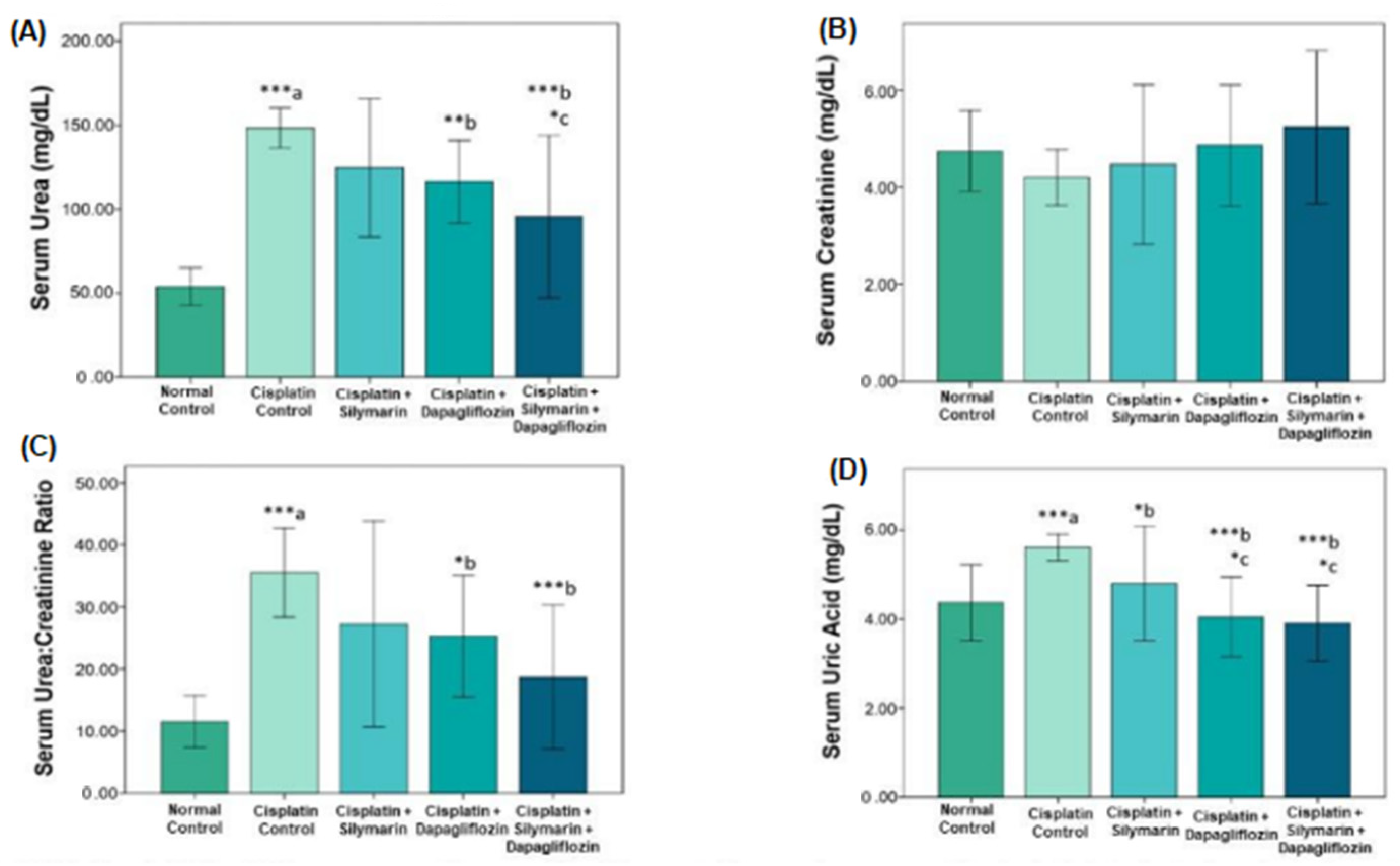
3.1. Impact on Tests of Renal Function

The kidney function tests revealed that the nephrotoxic control group exposed to cisplatin had significantly higher levels of serum urea (148.30 ± 5.95; Figure 1A), urea-to-creatinine ratio (35.50 ± 3.57; Figure 1B,C), and uric acid (5.61 ± 0.14; Figure 1D) than the normal control group (urea: 53.87 ± 5.57; urea-to-creatinine ratio: 11.50 ± 2.08; and uric acid: 4.36 ± 0.42). On the other hand, nephrotoxic rats treated with dapagliflozin showed a significant decrease in uric acid (4.04 ± 0.44; p < 0.001; Figure 1D), urea-to-creatinine ratio (25.28 ± 4.89; p = 0.022; Figure 1C), and serum urea (116.23 ± 12.34; p = 0.012; Figure 1A) when compared to the nephrotoxic control group. Uric acid levels significantly decreased after silymarin treatment (4.79 ± 0.64; p = 0.031; Figure 1D), but neither urea nor the urea-to-creatinine ratio changed significantly. Intriguingly, when compared to the nephrotoxic control group, the combination of dapagliflozin and silymarin demonstrated a notable improvement in kidney parameters, with notable decreases in urea (95.50 ± 24.17; p = 0.012; Figure 1A), urea-to-creatinine ratio (18.73 ± 5.81; p < 0.001; Figure 1C), and uric acid (3.90 ± 0.42; p < 0.001; Figure 1D). Additionally, the combined treatment group demonstrated a significant decrease in urea (95.50 ± 24.17; p = 0.027; Figure 1A) and uric acid (3.90 ± 0.42; p = 0.016; Figure 1D) when compared to the silymarin-only group (urea: 124.62 ± 20.55; uric acid: 4.79 ± 0.64; Figure 1A,D).

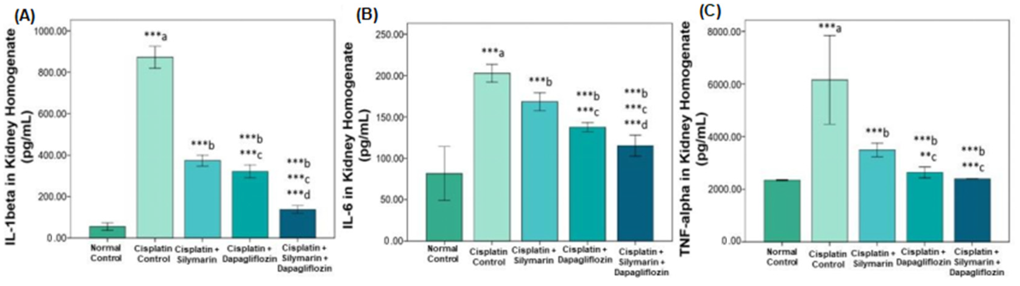
3.2. Impact on Inflammatory Cytokines

There was a significant increase (p < 0.001) in IL-1 beta (872.76 ± 26.29; Figure 2A), IL-6 (202.86 ± 5.37; Figure 2B), and TNF-alpha (6158.53 ± 843.26; Figure 2C) in the nephrotoxic control rats exposed to cisplatin compared to the normal control group (IL-1 beta: 54.87 ± 8.93; IL-6: 81.58 ± 16.23; TNF-alpha: 2345.74 ± 9.44). Treatment with dapagliflozin significantly reduced IL-1 beta (322.37 ± 15.42; p < 0.001; Figure 2A), IL-6 (137.60 ± 2.78; p < 0.001; Figure 2B), and TNF-alpha (2638.21 ± 105.39; p < 0.001; Figure 2C) compared to the nephrotoxic control group. Silymarin reduced IL-6 levels significantly (168.41 ± 5.36; p < 0.001; Figure 2B) but had no notable impact on IL-1 beta or TNF-alpha. Combination therapy with dapagliflozin and silymarin significantly reduced IL-1 beta (137.38 ± 9.32; p < 0.001; Figure 2A), IL-6 (115.28 ± 6.37; p < 0.001; Figure 2B), and TNF-alpha (2395.98 ± 4.89; p < 0.001; Figure 2C), compared to both the nephrotoxic control group and the silymarin-only group.

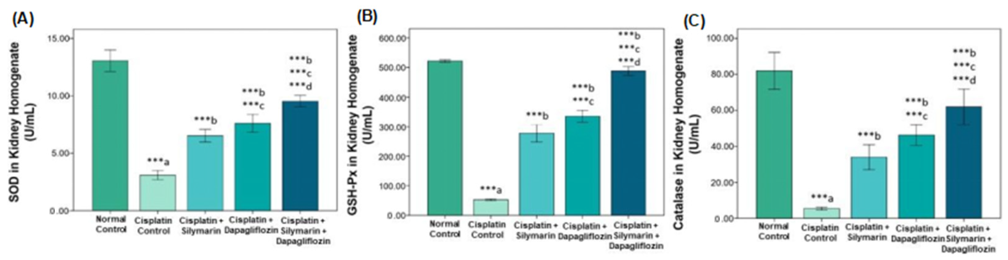
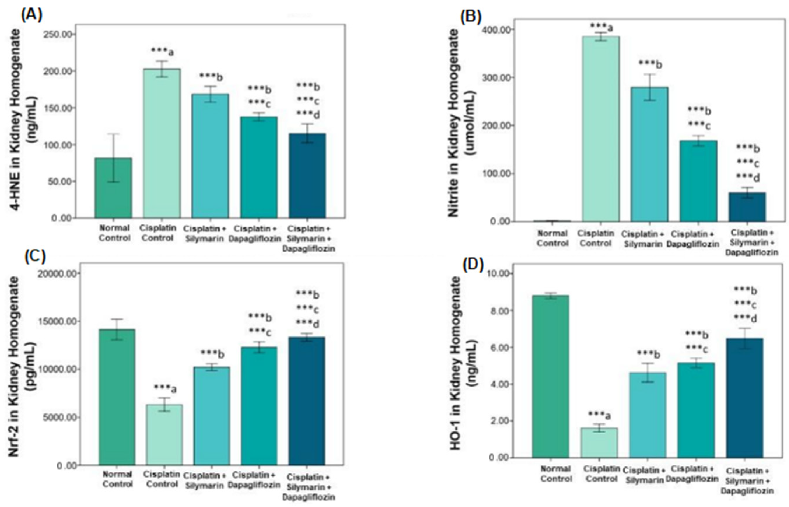
3.3. Effect on Oxidative Stress Biomarkers

The nephrotoxic control group exposed to cisplatin showed a significant reduction in SOD (3.09 ± 0.20; Figure 3A), GSH-Px (52.37 ± 1.24; Figure 3B), and CAT (5.43 ± 0.37; Figure 3C), alongside a marked increase in 4-HNE (202.86 ± 5.37; p < 0.001; Figure 4A) and nitrites (384.86 ± 4.25; p < 0.001; Figure 4B), compared to the normal control group. Treatment with dapagliflozin significantly lowered 4-HNE (137.60 ± 2.78; p < 0.001; Figure 4A) and nitrites (168.41 ± 5.36; p < 0.001; Figure 4B) and elevated SOD (7.60 ± 0.38; p < 0.001; Figure 3A), GSH-Px (335.23 ± 9.94; p < 0.001; Figure 3B), and CAT (46.27 ± 2.88; p < 0.001; Figure 3C) in comparison with the nephrotoxic control group. Silymarin significantly decreased nitrite levels (279.39 ± 13.55; p < 0.001; Figure 4B) and increased SOD (6.52 ± 0.27; p < 0.001; Figure 3A) and CAT (33.99 ± 3.48; p < 0.001; Figure 3C). The combination of dapagliflozin and silymarin led to further improvements, with notable increases in SOD (9.52 ± 0.25; p < 0.001; Figure 3A), GSH-Px (488.51 ± 7.32; p < 0.001; Figure 3B), and CAT (62.01 ± 4.90; p < 0.001; Figure 3C) and reductions in 4-HNE (115.28 ± 6.37; p < 0.001; Figure 4A) and nitrites (60.06 ± 5.33; p < 0.001; Figure 4B) compared to both the nephrotoxic control group and the silymarin-only group.

3.4. Nrf2/HO-1 Signaling Pathway Modification

Significant variations were seen in the Nrf2/HO-1 signaling pathway modification in response to various treatments. The levels of Nrf2 (6322.93 ± 350.59; Figure 4C) and HO-1 (1.61 ± 0.10; Figure 4D) were significantly lower (p < 0.001) in nephrotoxic rats exposed to cisplatin than in the normal control group (Nrf2: 14126.02 ± 531.65, Figure 4C; HO-1: 8.79 ± 0.07, Figure 4D). However, administering dapagliflozin alone to nephrotoxic rats significantly increased (p < 0.001) Nrf2 (12288.40 ± 278.56; Figure 4C) and HO-1 (5.14 ± 0.12; Figure 4D) levels, outperforming both the nephrotoxic control and the silymarin-only groups (Nrf2: 10216.13 ± 175.82; HO-1: 4.62 ± 0.25). Silymarin alone resulted in a significant increase in Nrf2 levels (p < 0.001), though not in HO-1 levels, compared to the nephrotoxic control group. Remarkably, the combined treatment of silymarin and dapagliflozin caused a substantial rise in both Nrf2 (13322.51 ± 202.73; p < 0.001; Figure 4C) and HO-1 (6.47 ± 0.27; p < 0.001; Figure 4D), surpassing the effects seen in the nephrotoxic control and silymarin-only groups.

3.5. Impact on Fasting Blood Glucose Levels and Body Weight

In contrast to the normal control group (92.83 ± 7.62), the cisplatin-exposed nephrotoxic group (110.33 ± 6.47; Figure 5A) showed a substantial (p < 0.05) decrease in fasting blood glucose levels. Nevertheless, during the course of the investigation, no significant variations (p > 0.05) in fasting blood glucose levels were observed across the other experimental groups. Regarding body weight (Figure 5B), no significant changes were detected at baseline across the groups. Nevertheless, a significant reduction in body weight (p < 0.05) was noticed in nephrotoxic rats treated with either dapagliflozin or the combination therapy (dapagliflozin and silymarin) compared to the normal control group over the treatment duration. From week 5 onwards, the cisplatin-treated rats experienced a marked weight reduction (p < 0.001) compared to the healthy controls, but no significant differences in body weight were observed between the dapagliflozin-only or combination-treated rats and the cisplatin-treated control (Figure 5B).

3.6. Effect on Kidney Histology

The normal control group exhibited typical renal cellular architecture. In contrast, the nephrotoxic control group treated with cisplatin displayed significant damage, including epithelial cell denudation, brush border loss, vascular congestion, cystic dilation, tubular inflammation, and interstitial inflammation. Rats treated with silymarin alone exhibited moderate cystic dilation, interstitial inflammation, and mild tubular inflammation. However, nephrotoxic rats treated with dapagliflozin showed only moderate interstitial inflammation. Notably, rats receiving both dapagliflozin and silymarin exhibited kidney histology that was nearly indistinguishable from normal control animals (Figure 6).

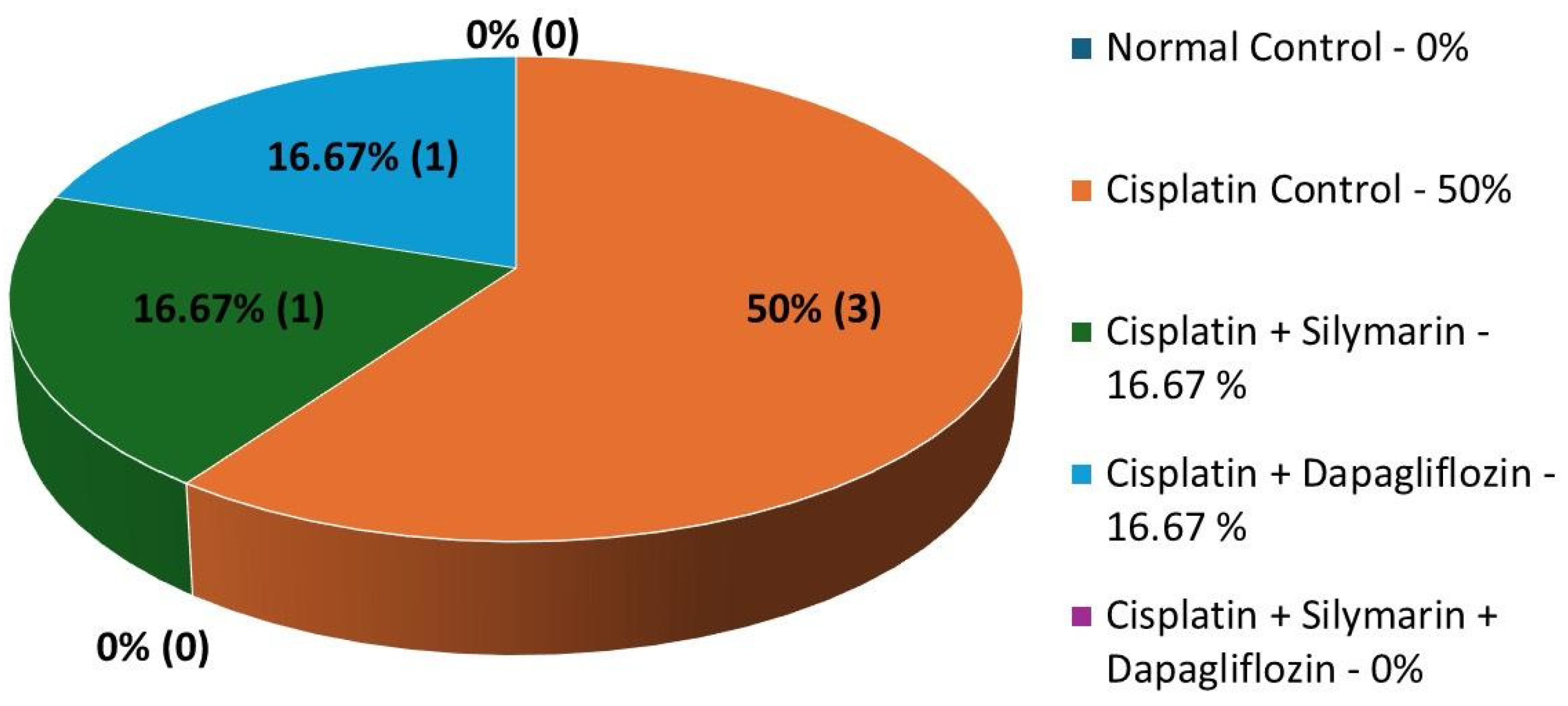
3.7. Impact on Mortality Rates

The cisplatin-treated nephrotoxic control group experienced a mortality rate of 50%, with three out of six rats succumbing to treatment. In contrast, the silymarin-only and dapagliflozin-only groups had a reduced mortality rate of 16.67%, with just one out of six rats dying in each group. Notably, no deaths occurred in the normal control group or in the combination-treated group (dapagliflozin and silymarin), where all rats survived. Due to the high mortality rate in the cisplatin-treated control group (Group II), attributed to cisplatin-induced nephrotoxicity, the Animal Ethics Committee, RAKMHSU approved the sanction of additional animals from the Central Animal Research Facility to ensure an adequate sample size for the study (Figure 7).

4. Discussion

This study employed an inclusive approach, integrating biochemical, histological, and molecular analyses to elucidate the protective mechanisms of dapagliflozin against cisplatin-induced nephrotoxicity. Cisplatin exerts its nephrotoxic effects through multiple molecular pathways, primarily by triggering oxidative stress, inflammation, mitochondrial dysfunction, and apoptosis within renal tubular cells [7,8,9]. A key driver of these detrimental effects is the excessive generation of reactive oxygen species (ROS), which disrupt cellular homeostasis and contribute to renal injury. The oxidative damage is further compounded by the suppression of cytoprotective pathways, particularly the nuclear erythroid 2-related factor 2 (Nrf2)/heme oxygenase-1 (HO-1) axis, which plays a crucial role in antioxidant defense [39,40,41,42].
The elevation of ROS levels leads to lipid peroxidation, a hallmark of oxidative stress, resulting in the accumulation of 4-hydroxy-2-nonenal (4-HNE), a cytotoxic byproduct that exacerbates renal cell damage [43,44,45]. Under physiological conditions, Nrf2 serves as a master regulator of cellular defense mechanisms by activating the transcription of antioxidant enzymes such as catalase, superoxide dismutase, and glutathione peroxidase, all of which counteract oxidative damage induced by cisplatin. HO-1, a downstream effector of Nrf2, further enhances renal protection by mitigating oxidative stress and improving renal function markers, including blood urea nitrogen (BUN) and serum creatinine levels.
Beyond oxidative stress, inflammation plays a pivotal role in cisplatin-induced nephrotoxicity. Pro-inflammatory cytokines, particularly interleukin-1β (IL-1β), are significantly upregulated following cisplatin administration, perpetuating an inflammatory cascade that exacerbates kidney damage. Elevated IL-1β levels in renal tissues have been directly linked to worsened nephrotoxicity. Interestingly, interleukin-6 (IL-6) appears to exert a contrasting effect, offering a protective role by suppressing pro-inflammatory cytokine expression and attenuating oxidative stress markers.
The degree of kidney damage is demonstrated by the significant increase in serum urea, urea-to-creatinine ratio, and uric acid in the cisplatin-induced nephrotoxic control group. Dapagliflozin treatment, either alone or combined with silymarin, significantly lowered these parameters, indicating a protective effect on kidney function. These outcomes agree with previous research demonstrating the ability of dapagliflozin to improve renal dysfunction across various animal models of kidney disease [28,29,46]. Heerspink et al.’s randomized controlled trial reported significant reductions in urinary albumin-to-creatinine ratio (UACR) and serum creatinine in diabetic patients treated with dapagliflozin, compared to a placebo [47]. In our study, while silymarin effectively reduced uric acid levels, its impact on urea and the urea-to-creatinine ratio was less pronounced. This aligns with findings from studies by Karimi et al. and Sonnenbichler et al., which reported similar effects of silymarin (the active ingredient in milk thistle extract) in animal models of renal disease [19,23]. Interestingly, the combination therapy with dapagliflozin and silymarin demonstrated significant improvements in kidney function, suggesting a synergistic effect. Histopathological assessments further confirmed a protective action against structural damage induced by cisplatin. Dapagliflozin, either alone or combined with silymarin, restored normal renal architecture, supporting its hypothesized potential to reverse kidney damage. These histological findings corroborate the observed biochemical improvements, reinforcing dapagliflozin’s renoprotective effects [48]. Dapagliflozin has been reported to attenuate glomerular hypertrophy and tubulointerstitial fibrosis in chronic kidney disease models, thus preserving renal structure [49]. The restoration of normal kidney architecture in our study suggests that the combination of dapagliflozin and silymarin not only alleviates the symptoms of nephrotoxicity but also promotes structural recovery.
Cisplatin-induced nephrotoxicity is characterized by increased pro-inflammatory cytokines. Dapagliflozin significantly decreased IL-1 beta, IL-6, and TNF-alpha in this trial, which is in line with research showing its anti-inflammatory qualities [50]. Silymarin also showed anti-inflammatory potential, particularly by reducing IL-6 levels. When used in combination, dapagliflozin and silymarin exhibited a pronounced anti-inflammatory effect, further indicating synergism. Reductions in pro-inflammatory cytokines such as TNF-α and IL-6 have been observed in dapagliflozin-treated diabetic mice [51]. Additionally, the reduction in nitrite levels observed with dapagliflozin treatment suggests decreased nitrosative stress [52]. Dapagliflozin further upregulated antioxidant enzymes such as catalase, superoxide dismutase, and glutathione peroxidase in our study, indicating a multifaceted mechanism by which it mitigates oxidative stress and inflammation in the kidneys [53].
Oxidative stress plays a crucial role in cisplatin-induced renal injury. In this study, dapagliflozin significantly reduced 4-HNE and nitrite levels while boosting antioxidant defenses (SOD, CAT, and GSH-Px), corroborating studies that suggest its antioxidative capacity [54]. Silymarin also displayed antioxidant activity, especially in lowering nitrite levels. An even greater benefit was obtained when dapagliflozin and silymarin were combined, highlighting their complementary roles in preventing oxidative stress. One important transcription factor, Nrf2, controls the production of detoxifying and antioxidant enzymes in response to redox shifts. Nrf2 separates from Keap1 under oxidative stress and moves into the nucleus, where it attaches itself to the antioxidant response element (ARE) in the promoter regions of phase II detoxifying enzymes like NQO1 and antioxidant genes like SOD, GSH-Px, and CAT [55]. Many cytoprotective genes are upregulated to preserve cellular homeostasis, and the Nrf2/HO-1 signaling pathway is essential for coordinating the antioxidant response. It has been demonstrated that Nrf2 activation raises the production of SOD isoforms, which are critical for scavenging superoxide radicals [56,57]. Although there is limited direct evidence connecting Nrf2 to catalase regulation, several studies indicate that Nrf2 stimulation increases catalase activity by influencing the cellular redox state [58,59]. Nrf2 activation also boosts the transcription of GSH-Px, which catalyzes a reduction in hydrogen peroxide, shielding cells from oxidative injury [60,61]. The Nrf2/HO-1 pathway indirectly reduces nitrite levels by modulating inflammatory and oxidative stress responses [62]. Nrf2 activation has also been shown to inhibit nitric oxide (NO) synthesis and nitrosative damage induced by oxidative stress while reducing the formation of 4-HNE and preventing lipid peroxidation [63]. Through its regulation of HO-1, Nrf2 can mitigate 4-HNE-induced oxidative damage [64]. In our study, cisplatin exposure led to a significant reduction in Nrf2 and HO-1 levels, reflecting compromised antioxidant defenses. Treatment with dapagliflozin dramatically restored these levels, indicating its role in boosting the Nrf2/HO-1 pathway. The observed antioxidative benefits, such as decreased nitrites and 4-HNE and increased enzymatic antioxidants like SOD, GSH-Px, and CAT, were probably caused by the combination-treated group’s high activation of this pathway. As evidenced by improvements in kidney function, inflammation, oxidative stress, and structural integrity, dapagliflozin, either by itself or in conjunction with silymarin, may help reverse the downregulation of the Nrf2/HO-1 pathway brought on by cisplatin-induced nephrotoxicity.
Our findings underscore the therapeutic potential of targeting the Nrf2/HO-1 pathway in the management of conditions characterized by inflammation and oxidative stress. We have demonstrated that dapagliflozin treatment upregulated Nrf2/HO-1 expression in a carbon tetrachloride-induced hepatotoxicity in rats, further supporting the involvement of this pathway in dapagliflozin-mediated renoprotection [37]. There is growing evidence that dapagliflozin modulates important signaling pathways, namely, Nrf2/HO-1, to provide its nephroprotective effects.
Previously, we have highlighted the transformative potential of drug repurposing by unveiling precise mechanistic insights to combat oxidative stress-associated diseases [33,37,38,65,66,67,68,69]. Dapagliflozin, a well-known SGLT2 inhibitor primarily used for its anti-diabetic effects, demonstrated significant nephroprotective properties in this study. Dapagliflozin’s kidney-protective benefits were not mediated by its glucose-lowering activity, as evidenced by the lack of significant variations in fasting blood glucose levels between the different groups. Additionally, blood glucose levels were monitored at both the beginning and end of the experiment to ensure consistency and avoid any discrepancies in the data. Several other studies have also demonstrated the nephroprotective effects of dapagliflozin in euglycemic rats, further supporting our assertion that its kidney-protective effects are independent of glucose reduction [70,71].
SGLT2 inhibitors, such as dapagliflozin, have been shown to provide nephroprotective effects that are independent of glucose lowering through mechanisms such as reducing intraglomerular pressure, improving tubular oxygenation, and alleviating renal inflammation and fibrosis [72]. Clinical studies have demonstrated that these drugs slow the decline in glomerular filtration rate (GFR) and reduce microalbuminuria in both diabetic and non-diabetic populations. Their benefits extend beyond glycemic control, as evidenced by improvements in renal function in non-diabetic individuals [73]. Furthermore, similar nephroprotective effects have been observed with other antidiabetic drugs, supporting the notion that kidney protection can occur through pathways unrelated to glucose control [74]. Therefore, the observed kidney protection in our study can likely be attributed to mechanisms independent of glucose lowering, as evidenced by the absence of significant changes in fasting blood glucose levels across the experimental groups.
Regarding the changes in body weight, no significant differences were detected at baseline across the experimental groups. However, significant weight reduction was observed in the nephrotoxic rats treated with dapagliflozin or the combination therapy (dapagliflozin and silymarin) compared to the normal control group during the treatment period. This observation may be explained by the metabolic effects of dapagliflozin. Dapagliflozin, a sodium-glucose co-transporter 2 (SGLT2) inhibitor, is known to enhance urinary glucose excretion by inhibiting glucose reabsorption in the proximal renal tubules. This increased glucose loss through urine results in a negative energy balance, which can lead to weight loss, a common clinical finding in patients with type 2 diabetes receiving dapagliflozin [75,76].
From week 5 onwards, cisplatin-treated rats experienced a significant reduction in body weight compared to healthy controls, as expected due to the nephrotoxic effects of cisplatin. However, no significant differences in body weight were observed between the dapagliflozin-only or combination-treated rats and the cisplatin-treated control group. This suggests that while dapagliflozin and the combination therapy contributed to weight loss, the nephrotoxic effects of cisplatin may have overshadowed or balanced out the metabolic changes induced by dapagliflozin.
Both the dapagliflozin- and combination-treated groups exhibited weight reduction, indicating a potential impact on metabolism and energy balance, which warrants further investigation. It is also noteworthy that dapagliflozin has been reported to influence fat mass reduction, potentially through mechanisms involving hormonal regulation and altered energy metabolism. Although the precise mechanisms remain unclear, studies have suggested that dapagliflozin may contribute to fat loss, possibly due to its effects on appetite regulation and increased fat oxidation, which are factors that can lead to a reduction in body weight [77,78,79]. Therefore, the weight loss observed in the dapagliflozin-treated and combination-treated groups could be attributed to a combination of increased energy expenditure, enhanced glucose excretion, and possible reductions in fat mass, all of which warrant further exploration in the context of nephrotoxicity and metabolic regulation.
The mortality rates observed in this study provide critical insights into the severe toxicity associated with cisplatin administration and the potential protective effects of silymarin and dapagliflozin. The 50% mortality rate recorded in the cisplatin-treated control group highlights the substantial nephrotoxicity and systemic toxicity induced by this chemotherapeutic agent. Cisplatin is well-documented to cause acute kidney injury (AKI), leading to renal failure and subsequent mortality in animal models due to oxidative stress, inflammatory responses, and apoptosis of renal tubular cells [40,80].
However, the mortality rate was significantly reduced to 16.67% in both the silymarin and dapagliflozin treatment groups when administered separately. Silymarin, a potent flavonoid with antioxidant and hepatoprotective properties, has been reported to mitigate oxidative stress and inflammation, two key contributors to cisplatin-induced renal toxicity. Similarly, dapagliflozin, an SGLT2 inhibitor primarily used in diabetes management, has shown renoprotective effects through its ability to reduce oxidative stress, modulate inflammation, and improve mitochondrial function [81]. The observed reduction in mortality in these groups indicates the potential of both compounds to confer substantial protection against cisplatin-induced toxicity.
Strikingly, the combination of dapagliflozin and silymarin resulted in complete survival, with no recorded deaths in this treatment group. This suggests a possible synergistic interaction between these two agents in counteracting cisplatin-induced toxicity. The underlying mechanisms of this synergy may involve an amplified antioxidant response, improved mitochondrial function, and enhanced anti-inflammatory effects, ultimately leading to superior protection against nephrotoxicity and systemic toxicity. While individual administration of silymarin and dapagliflozin demonstrated notable protective effects, their combination appears to exert a more pronounced protective impact, possibly through complementary or overlapping mechanisms that mitigate cisplatin’s deleterious effects more effectively.
The study’s comprehensive strategy, which combines biochemical, histological, and molecular investigations to properly analyze kidney function, inflammation, oxidative stress, and the underlying molecular pathways, is one of its many merits. Dapagliflozin, a well-known anti-diabetic drug, was repurposed without sacrificing safety at a dose of 0.9 mg/kg/day, which is equal to the therapeutic human dose of 10 mg/day. With an emphasis on the Nrf2/HO-1 signaling pathway’s regulation, which boosts both antioxidant and anti-inflammatory responses, the study also investigated the mechanisms behind the nephroprotective benefits of silymarin and dapagliflozin.
However, the study also has limitations. The small sample size and the use of a cisplatin-induced nephrotoxicity model without the presence of cancer may limit the study’s ability to accurately represent human physiology and pathophysiology, potentially restricting the generalizability of the findings to clinical settings. Although the results demonstrate the nephroprotective potential of dapagliflozin and its combination with silymarin against kidney damage caused by cisplatin, resolving these limitations could increase the findings’ validity and generalizability.

5. Conclusions

This study has demonstrated the significant nephroprotective benefits of dapagliflozin in a model of cisplatin-induced nephrotoxicity in Wistar rats, both when used alone and in combination with silymarin. Across a number of metrics, the combination treatment demonstrated synergistic effects that outperformed the separate effects of silymarin or dapagliflozin. The study revealed significant improvements in markers of oxidative stress, inflammation, renal function, and cellular structure, demonstrating the potential of this combined therapy to serve as a comprehensive treatment for cisplatin-induced renal damage.
The observed reductions in oxidative stress, inflammatory markers, and improvements in kidney function and tissue morphology suggest that this therapeutic combination could effectively treat several facets of renal injury. However, while the findings are promising, further research, including clinical trials, is essential to validate the safety and effectiveness of this combination in human patients. Patients receiving cisplatin-based chemotherapy may benefit from better clinical outcomes if the translational potential of dapagliflozin and silymarin for treating cisplatin-induced nephrotoxicity is investigated.

Author Contributions

Conceptualization, S.M.S.; methodology, S.M.S., L.K.B., A.R., A.A.N., M.F., N.N.B., S.K., M.Y., and M.M.K.; software, S.M.S., L.K.B., A.R., A.A.N., M.F., N.N.B., S.K., M.Y., and M.M.K.; validation, S.M.S., L.K.B., A.R., A.A.N., M.F., N.N.B., S.K., M.Y., and M.M.K.; formal analysis, S.M.S., L.K.B., A.R., A.A.N., M.F., N.N.B., S.K., M.Y., and M.M.K.; investigation, S.M.S., L.K.B., A.R., A.A.N., M.F., N.N.B., S.K., M.Y., and M.M.K.; resources, S.M.S., L.K.B., A.R., A.A.N., M.F., N.N.B., S.K., M.Y., and M.M.K.; data curation, S.M.S., L.K.B., A.R., A.A.N., M.F., N.N.B., S.K., M.Y., and M.M.K.; writing—original draft preparation, S.M.S.; writing—review and editing, S.M.S., L.K.B., A.R., A.A.N., M.F., N.N.B., S.K., M.Y., and M.M.K.; visualization, S.M.S., L.K.B., A.R., A.A.N., M.F., N.N.B., S.K., M.Y., and M.M.K.; supervision, S.M.S., L.K.B., and A.R.; project administration, S.M.S.; funding acquisition, S.M.S., L.K.B., and A.R. All authors have read and agreed to the published version of the manuscript.

Funding

This research received no external funding.

Institutional Review Board Statement

The animal study protocol was approved by the Ras Al Khaimah Medical and Health Sciences University Research and Ethics Committee (RAKMHSU-REC-114-2022/23-UG-M), UAE.

Informed Consent Statement

Not applicable.

Data Availability Statement

The data generated and analyzed in this study are included in this article.

Acknowledgments

The authors express their sincere appreciation to RAK Medical and Health Sciences University (RAKMHSU), UAE, for their unwavering support and resources that enabled the successful completion of this research.

Conflicts of Interest

The authors declare no conflicts of interest.

References

  1. Kocarnik, J.M.; Compton, K.; Dean, F.E.; Fu, W.; Gaw, B.L.; Harvey, J.D.; Henrikson, H.J.; Lu, D.; Pennini, A.; Xu, R.; et al. Cancer incidence, mortality, years of life lost, years lived with disability, and disability-adjusted life years for 29 cancer groups from 2010 to 2019: A systematic analysis for the global burden of disease study 2019. JAMA Oncol. 2022, 8, 420–444. [Google Scholar] [PubMed]
  2. Miller, R.P.; Tadagavadi, R.K.; Ramesh, G.; Reeves, W.B. Mechanisms of cisplatin nephrotoxicity. Toxins 2010, 2, 2490–2518. [Google Scholar] [CrossRef]
  3. Dasari, S.; Tchounwou, P.B. Cisplatin in cancer therapy: Molecular mechanisms of action. Eur. J. Pharmacol. 2014, 740, 364–378. [Google Scholar] [CrossRef]
  4. Ozkok, A.; Edelstein, C.L. Pathophysiology of cisplatin-induced acute kidney injury. BioMed Res. Int. 2014, 2014, 967826. [Google Scholar] [CrossRef] [PubMed]
  5. Pabla, N.; Dong, Z. Cisplatin nephrotoxicity: Mechanisms and renoprotective strategies. Kidney Int. 2008, 73, 994–1007. [Google Scholar] [CrossRef]
  6. Volarevic, V.; Djokovic, B.; Jankovic, M.G.; Harrell, C.R.; Fellabaum, C.; Djonov, V.; Arsenijevic, N. Molecular mechanisms of cisplatin-induced nephrotoxicity: A balance on the knife edge between renoprotection and tumor toxicity. J. Biomed. Sci. 2019, 26, 25. [Google Scholar] [CrossRef]
  7. Fuertes, M.A.; Castilla, J.; Alonso, C.; Pérez, J.M. Cisplatin biochemical mechanism of action: From cytotoxicity to induction of cell death through interconnections between apoptotic and necrotic pathways. Curr. Med. Chem. 2003, 10, 257–266. [Google Scholar] [CrossRef] [PubMed]
  8. Launay-Vacher, V.; Rey, J.-B.; Isnard-Bagnis, C.; Deray, G.; Daouphars, M. Prevention of cisplatin nephrotoxicity: State of the art and recommendations from the European Society of Clinical Pharmacy Special Interest Group on Cancer Care. Cancer Chemother. Pharmacol. 2008, 61, 903–909. [Google Scholar] [CrossRef]
  9. Wang, J.; Pabla, N.; Wang, C.-Y.; Wang, W.; Schoenlein, P.V.; Dong, Z. Caspase-mediated cleavage of ATM during cisplatin-induced tubular cell apoptosis: Inactivation of its kinase activity toward p53. Am. J. Physiol.-Ren. Physiol. 2006, 291, F1300–F1307. [Google Scholar] [CrossRef]
  10. Crona, D.J.; Faso, A.; Nishijima, T.F.; McGraw, K.A.; Galsky, M.D.; Milowsky, M.I. A systematic review of strategies to prevent cisplatin-induced nephrotoxicity. Oncologist 2017, 22, 609–619. [Google Scholar] [CrossRef]
  11. Sikking, C.; Niggebrugge-Mentink, K.L.; van der Sman, A.S.E.; Smit, R.H.P.; Bouman-Wammes, E.W.; Beex-Oosterhuis, M.M.; van Kesteren, C. Hydration methods for cisplatin containing chemotherapy: A systematic review. Oncologist 2024, 29, e173–e186. [Google Scholar] [CrossRef] [PubMed]
  12. Gu, J.; Zhu, S.; Li, X.; Wu, H.; Li, Y.; Hua, F. Effect of amifostine in head and neck cancer patients treated with radiotherapy: A systematic review and meta-analysis based on randomized controlled trials. PLoS ONE 2014, 9, e95968. [Google Scholar] [CrossRef] [PubMed]
  13. Ji, L.; Cui, P.; Zhou, S.; Qiu, L.; Huang, H.; Wang, C.; Wang, J. Advances of amifostine in radiation protection: Administration and delivery. Mol. Pharm. 2023, 20, 5383–5395. [Google Scholar] [CrossRef]
  14. Ibrahim, M.E.-T.; El Bana, E.; El-Kerdasy, H.I. Role of bone marrow derived mesenchymal stem cells and the protective effect of silymarin in cisplatin-induced acute renal failure in rats. Am. J. Med. Sci. 2018, 355, 76–83. [Google Scholar] [CrossRef]
  15. Iskander, A.; Yan, L.-J. Cisplatin-induced kidney toxicity: Potential roles of major NAD+-dependent enzymes and plant-derived natural products. Biomolecules 2022, 12, 1078. [Google Scholar] [CrossRef]
  16. Karimi, G.; Vahabzadeh, M.; Lari, P.; Rashedinia, M.; Moshiri, M. “Silymarin”, a promising pharmacological agent for treatment of diseases. Iran. J. Basic Med. Sci. 2011, 14, 308. [Google Scholar]
  17. Abenavoli, L.; Capasso, R.; Milic, N.; Capasso, F. Milk thistle in liver diseases: Past, present, future. Phytother. Res. 2010, 24, 1423–1432. [Google Scholar] [CrossRef] [PubMed]
  18. Akbari-Kordkheyli, V.; Abbaszadeh-Goudarzi, K.; Nejati-Laskokalayeh, M.; Zarpou, S.; Khonakdar-Tarsi, A. The protective effects of silymarin on ischemia-reperfusion injuries: A mechanistic review. Iran. J. Basic Med. Sci. 2019, 22, 968. [Google Scholar]
  19. Karimi, G.; Ramezani, M.; Tahoonian, Z. Cisplatin nephrotoxicity and protection by milk thistle extract in rats. Evid. -Based Complement. Altern. Med. 2005, 2, 383–386. [Google Scholar] [CrossRef]
  20. Mansour, H.H.; Hafez, H.F.; Fahmy, N.M. Silymarin modulates cisplatin-induced oxidative stress and hepatotoxicity in rats. BMB Rep. 2006, 39, 656–661. [Google Scholar] [CrossRef]
  21. Nicholson, M.K.; Asswad, R.G.; Wilding, J.P. Dapagliflozin for the treatment of type 2 diabetes mellitus—An update. Expert Opin. Pharmacother. 2021, 22, 2303–2310. [Google Scholar] [CrossRef]
  22. Shahbazi, F.; Dashti-Khavidaki, S.; Khalili, H.; Lessan-Pezeshki, M. Potential renoprotective effects of silymarin against nephrotoxic drugs: A review of literature. J. Pharm. Pharm. Sci. 2012, 15, 112–123. [Google Scholar] [CrossRef]
  23. Sonnenbichler, J.; Scalera, F.; Sonnenbichler, I.; Weyhenmeyer, R. Stimulatory effects of silibinin and silicristin from the milk thistle Silybum marianum on kidney cells. J. Pharmacol. Exp. Ther. 1999, 290, 1375–1383. [Google Scholar] [CrossRef] [PubMed]
  24. Bae, J.H.; Park, E.-G.; Kim, S.; Kim, S.G.; Hahn, S.; Kim, N.H. Effects of sodium-glucose cotransporter 2 inhibitors on renal outcomes in patients with type 2 diabetes: A systematic review and meta-analysis of randomized controlled trials. Sci. Rep. 2019, 9, 13009. [Google Scholar] [CrossRef] [PubMed]
  25. Chino, Y.; Samukawa, Y.; Sakai, S.; Nakai, Y.; Yamaguchi, J.; Nakanishi, T.; Tamai, I. SGLT2 inhibitor lowers serum uric acid through alteration of uric acid transport activity in renal tubule by increased glycosuria. Biopharm. Drug Dispos. 2014, 35, 391–404. [Google Scholar] [CrossRef]
  26. van Bommel, E.J.; Muskiet, M.H.; van Baar, M.J.; Tonneijck, L.; Smits, M.M.; Emanuel, A.L.; Bozovic, A.; Danser, A.J.; Geurts, F.; Hoorn, E.J.; et al. The renal hemodynamic effects of the SGLT2 inhibitor dapagliflozin are caused by post-glomerular vasodilatation rather than pre-glomerular vasoconstriction in metformin-treated patients with type 2 diabetes in the randomized, double-blind RED trial. Kidney Int. 2020, 97, 202–212. [Google Scholar] [CrossRef]
  27. Tanna, M.S.; Goldberg, L.R. The pleiotropic cardiovascular effects of sodium-glucose cotransporter-2 inhibitors. Curr. Opin. Cardiol. 2021, 36, 764–768. [Google Scholar] [CrossRef]
  28. Oraby, M.A.; El-Yamany, M.F.; Safar, M.M.; Assaf, N.; Ghoneim, H.A. Dapagliflozin attenuates early markers of diabetic nephropathy in fructose-streptozotocin-induced diabetes in rats. Biomed. Pharmacother. 2019, 109, 910–920. [Google Scholar] [CrossRef]
  29. Tang, L.; Wu, Y.; Tian, M.; Sjöström, C.D.; Johansson, U.; Peng, X.-R.; Smith, D.M.; Huang, Y. Dapagliflozin slows the progression of the renal and liver fibrosis associated with type 2 diabetes. Am. J. Physiol.-Endocrinol. Metab. 2017, 313, E563–E576. [Google Scholar] [CrossRef]
  30. Garvey, W.T.; Van Gaal, L.; Leiter, L.A.; Vijapurkar, U.; List, J.; Cuddihy, R.; Ren, J.; Davies, M.J. Effects of canagliflozin versus glimepiride on adipokines and inflammatory biomarkers in type 2 diabetes. Metabolism 2018, 85, 32–37. [Google Scholar] [CrossRef]
  31. Zaibi, N.; Li, P.; Xu, S.-Z. Protective effects of dapagliflozin against oxidative stress-induced cell injury in human proximal tubular cells. PLoS ONE 2021, 16, e0247234. [Google Scholar] [CrossRef] [PubMed]
  32. Perše, M.; Večerić-Haler, Ž. Cisplatin-induced rodent model of kidney injury: Characteristics and challenges. BioMed Res. Int. 2018, 2018, 1462802. [Google Scholar] [CrossRef]
  33. Satyam, S.M.; Bairy, L.K.; Ern, O.T.; Yen, Y.G.; Kanasin, A.; Muthaiah, T.; Ratnam, U.S.; Yadav, K. Influence of combination of docosahexaenoic acid supplement and a polyherbal formulation (Liv. 52) on carbon tetrachloride-induced hepatic injury: A preclinical study. J. Datta Meghe Inst. Med. Sci. Univ. 2020, 15, 114–117. [Google Scholar] [CrossRef]
  34. Paget, G.; Barnes, J. Toxicity tests. In Evaluation of Drug Activities; Elsevier: Amsterdam, The Netherlands, 1964; pp. 135–166. [Google Scholar]
  35. Satyam, S.M.; Bairy, L.K.; Shetty, P.; Sainath, P.; Bharati, S.; Ahmed, A.Z.; Singh, V.K.; Ashwal, A.J. Metformin and dapagliflozin attenuate doxorubicin-induced acute cardiotoxicity in Wistar rats: An electrocardiographic, biochemical, and histopathological approach. Cardiovasc. Toxicol. 2023, 23, 107–119. [Google Scholar] [CrossRef]
  36. Kim, Y.; Lee, H.-M. Acidic solvent improves cisplatin action in in-vitro. Biochem. Biophys. Res. Commun. 2024, 712, 149936. [Google Scholar] [CrossRef]
  37. Satyam, S.M.; Bairy, L.K.; Rehman, A.; Attia, M.; Ahmed, L.; Emad, K.; Jaafer, Y.; Bahaaeldin, A. Unlocking Synergistic Hepatoprotection: Dapagliflozin and Silymarin Combination Therapy Modulates Nuclear Erythroid 2-Related Factor 2/Heme Oxygenase-1 Pathway in Carbon Tetrachloride-Induced Hepatotoxicity in Wistar Rats. Biology 2024, 13, 473. [Google Scholar] [CrossRef]
  38. Satyam, S.M.; Bairy, L.K.; Rehman, A.; Farook, M.; Khan, S.; Nair, A.A.; Binu, N.N.; Yehya, M.; Khan, M.M. Dapagliflozin: A Promising Strategy to Combat Cisplatin-Induced Hepatotoxicity in Wistar Rats. Biology 2024, 13, 672. [Google Scholar] [CrossRef] [PubMed]
  39. Anees, L.M.; Abdel-Hamid, G.R.; Elkady, A.A. A nano based approach to alleviate cisplatin induced nephrotoxicity. Int. J. Immunopathol. Pharmacol. 2021, 35, 20587384211066441. [Google Scholar] [CrossRef]
  40. Fang, C.-Y.; Lou, D.-Y.; Zhou, L.-Q.; Wang, J.-C.; Yang, B.; He, Q.-J.; Wang, J.-J.; Weng, Q.-J. Natural products: Potential treatments for cisplatin-induced nephrotoxicity. Acta Pharmacol. Sin. 2021, 42, 1951–1969. [Google Scholar] [CrossRef] [PubMed]
  41. Li, S.; He, X.; Ruan, L.; Ye, T.; Wen, Y.; Song, Z.; Hu, S.; Chen, Y.; Peng, B.; Li, S. Protective effect of mannitol on cisplatin-induced nephrotoxicity: A systematic review and meta-analysis. Front. Oncol. 2021, 11, 804685. [Google Scholar] [CrossRef]
  42. Mercantepe, F.; Mercantepe, T.; Topcu, A.; Yılmaz, A.; Tumkaya, L. Protective effects of amifostine, curcumin, and melatonin against cisplatin-induced acute kidney injury. Naunyn-Schmiedeberg’s Arch. Pharmacol. 2018, 391, 915–931. [Google Scholar] [CrossRef]
  43. Dugbartey, G.J.; Bouma, H.R.; Lobb, I.; Sener, A. Hydrogen sulfide: A novel nephroprotectant against cisplatin-induced renal toxicity. Nitric Oxide 2016, 57, 15–20. [Google Scholar] [CrossRef] [PubMed]
  44. Malik, S.; Suchal, K.; Gamad, N.; Dinda, A.K.; Arya, D.S.; Bhatia, J. Telmisartan ameliorates cisplatin-induced nephrotoxicity by inhibiting MAPK mediated inflammation and apoptosis. Eur. J. Pharmacol. 2015, 748, 54–60. [Google Scholar] [CrossRef] [PubMed]
  45. Oh, G.-S.; Kim, H.-J.; Shen, A.; Lee, S.-B.; Yang, S.-H.; Shim, H.; Cho, E.-Y.; Kwon, K.-B.; Kwak, T.H.; So, H.-S. New therapeutic concept of NAD redox balance for cisplatin nephrotoxicity. BioMed Res. Int. 2016, 2016, 4048390. [Google Scholar] [CrossRef]
  46. Vallon, V. The mechanisms and therapeutic potential of SGLT2 inhibitors in diabetes mellitus. Annu. Rev. Med. 2015, 66, 255–270. [Google Scholar] [CrossRef]
  47. Heerspink, H.J.L.; Perco, P.; Mulder, S.; Leierer, J.; Hansen, M.K.; Heinzel, A.; Mayer, G. Canagliflozin reduces inflammation and fibrosis biomarkers: A potential mechanism of action for beneficial effects of SGLT2 inhibitors in diabetic kidney disease. Diabetologia 2019, 62, 1154–1166. [Google Scholar] [CrossRef]
  48. Chi, P.-J.; Lee, C.-J.; Hsieh, Y.-J.; Lu, C.-W.; Hsu, B.-G. Dapagliflozin ameliorates lipopolysaccharide related acute kidney injury in mice with streptozotocin-induced diabetes mellitus. Int. J. Med. Sci. 2022, 19, 729–739. [Google Scholar] [CrossRef]
  49. Afsar, B.; Afsar, R.E. Sodium–glucose cotransporter inhibitors and kidney fibrosis: Review of the current evidence and related mechanisms. Pharmacol. Rep. 2023, 75, 44–68. [Google Scholar] [CrossRef]
  50. Abdul-Ghani, M.; Del Prato, S.; Chilton, R.; DeFronzo, R.A. SGLT2 inhibitors and cardiovascular risk: Lessons learned from the EMPA-REG OUTCOME study. Diabetes Care 2016, 39, 717–725. [Google Scholar] [CrossRef]
  51. Cai, A.; Shen, J.; Yang, X.; Shao, X.; Gu, L.; Mou, S.; Che, X. Dapagliflozin alleviates renal inflammation and protects against diabetic kidney diseases, both dependent and independent of blood glucose levels. Front. Immunol. 2023, 14, 1205834. [Google Scholar] [CrossRef]
  52. Tai, S.; Zhou, Y.; Fu, L.; Ding, H.; Zhou, Y.; Yin, Z.; Yang, R.; Liu, Z.; Zhou, S. Dapagliflozin impedes endothelial cell senescence by activating the SIRT1 signaling pathway in type 2 diabetes. Heliyon 2023, 9, e19152. [Google Scholar] [CrossRef]
  53. Lee, O.Y.A.; Wong, A.N.N.; Ho, C.Y.; Tse, K.W.; Chan, A.Z.; Leung, G.P.-H.; Kwan, Y.W.; Yeung, M.H.Y. Potentials of natural antioxidants in reducing inflammation and oxidative stress in chronic kidney disease. Antioxidants 2024, 13, 751. [Google Scholar] [CrossRef] [PubMed]
  54. Arakawa, K.; Ishihara, T.; Oku, A.; Nawano, M.; Ueta, K.; Kitamura, K.; Matsumoto, M.; Saito, A. Improved diabetic syndrome in C57BL/KsJ-db/db mice by oral administration of the Na+-glucose cotransporter inhibitor T-1095. Br. J. Pharmacol. 2001, 132, 578–586. [Google Scholar] [CrossRef] [PubMed]
  55. Yu, X.; Cui, L.; Zhang, Z.; Zhao, Q.; Li, S. α-Linolenic acid attenuates doxorubicin-induced cardiotoxicity in rats through suppression of oxidative stress and apoptosis. Acta Biochim. Biophys. Sin. 2013, 45, 817–826. [Google Scholar] [CrossRef]
  56. Jung, K.-A.; Kwak, M.-K. The Nrf2 system as a potential target for the development of indirect antioxidants. Molecules 2010, 15, 7266–7291. [Google Scholar] [CrossRef]
  57. Rosa, A.C.; Corsi, D.; Cavi, N.; Bruni, N.; Dosio, F. Superoxide dismutase administration: A review of proposed human uses. Molecules 2021, 26, 1844. [Google Scholar] [CrossRef]
  58. Dinić, S.; Grdović, N.; Uskoković, A.; ĐORĐEVIĆ, M.; Mihailović, M.; Jovanović, J.A.; Poznanović, G.; Vidaković, M. CXCL12 protects pancreatic β-cells from oxidative stress by a Nrf2-induced increase in catalase expression and activity. Proc. Jpn. Acad. Ser. B 2016, 92, 436–454. [Google Scholar] [CrossRef] [PubMed]
  59. Lin, X.; Yang, H.; Zhou, L.; Guo, Z. Nrf2-dependent induction of NQO1 in mouse aortic endothelial cells overexpressing catalase. Free Radic. Biol. Med. 2011, 51, 97–106. [Google Scholar] [CrossRef]
  60. Harvey, C.J.; Thimmulappa, R.K.; Singh, A.; Blake, D.J.; Ling, G.; Wakabayashi, N.; Fujii, J.; Myers, A.; Biswal, S. Nrf2-regulated glutathione recycling independent of biosynthesis is critical for cell survival during oxidative stress. Free Radic. Biol. Med. 2009, 46, 443–453. [Google Scholar] [CrossRef]
  61. He, F.; Ru, X.; Wen, T. NRF2, a transcription factor for stress response and beyond. Int. J. Mol. Sci. 2020, 21, 4777. [Google Scholar] [CrossRef]
  62. Luo, J.-F.; Shen, X.Y.; Lio, C.K.; Dai, Y.; Cheng, C.S.; Liu, J.X.; Yao, Y.D.; Yu, Y.; Xie, Y.; Luo, P.; et al. Activation of Nrf2/HO-1 pathway by nardochinoid C inhibits inflammation and oxidative stress in lipopolysaccharide-stimulated macrophages. Front. Pharmacol. 2018, 9, 911. [Google Scholar] [CrossRef]
  63. Ayala, A.; Muñoz, M.F.; Argüelles, S. Lipid peroxidation: Production, metabolism, and signaling mechanisms of malondialdehyde and 4-hydroxy-2-nonenal. Oxidative Med. Cell. Longev. 2014, 2014, 360438. [Google Scholar] [CrossRef]
  64. Li, Y.; Zhao, T.; Li, J.; Xia, M.; Li, Y.; Wang, X.; Liu, C.; Zheng, T.; Chen, R.; Kan, D.; et al. Oxidative stress and 4-hydroxy-2-nonenal (4-HNE): Implications in the pathogenesis and treatment of aging-related diseases. J. Immunol. Res. 2022, 2022, 2233906. [Google Scholar] [CrossRef]
  65. Ahmed, A.Z.; Shetty, P.; Satyam, S.M.; D’souza, M.R.; Herle, A.M.; Singh, V.K. Methyl gallate mitigates doxorubicin-induced peripheral cytopenias: A preclinical experimental study. Res. J. Pharm. Technol. 2021, 14, 4529–4534. [Google Scholar] [CrossRef]
  66. Satyam, S.M.; Bairy, L.K. Neuronutraceuticals combating Neuroinflammaging: Molecular insights and translational challenges—A systematic review. Nutrients 2022, 14, 3029. [Google Scholar] [CrossRef] [PubMed]
  67. Satyam, S.M.; Bairy, L.K.; Pirasanthan, R. Influence of grape seed extract and zinc containing multivitamin-mineral nutritional food supplement on lipid profile in normal and diet-induced hypercholesterolemic rats. J. Clin. Diagn. Res. JCDR 2014, 8, HC12. [Google Scholar] [CrossRef]
  68. Satyam, S.M.; Bairy, L.K.; Pirasanthan, R.; Vaihnav, R.L. Grape seed extract and zinc containing nutritional food supplement decreases the oxidative stress induced by carbon tetrachloride in rats. Int. J. Pharm. Pharm. Sci. 2013, 5, 626–631. [Google Scholar]
  69. Satyam, S.M.; Kurady, L.B.; Prakash, J.; Syed, M.; Kumar, N.; Patil, J. Influence of ageless liquid with or without piperine on gentamicin-induced nephrotoxicity in wistar rats. Res. J. Pharm. Biol. Chem. Sci. 2016, 7, 1963–1976. [Google Scholar]
  70. Alanazi, W.A.; Alharbi, T.; Bin Anzan, K.M.; Alyahiya, M.K.; El-Nagar, D.M.; Almutairi, M.M.; Alhamami, H.N.; Albogami, A.M.; Mohany, M. The Role of Dapagliflozin in the Modulation of Hypothermia and Renal Injury Caused by Septic Shock in Euglycemic and Hyperglycemic Rat Models. Curr. Mol. Pharmacol. 2024, 17, e18761429329635. [Google Scholar] [CrossRef]
  71. Pirklbauer, M.; Sallaberger, S.; Staudinger, P.; Corazza, U.; Leierer, J.; Mayer, G.; Schramek, H. Empagliflozin inhibits IL-1β-mediated inflammatory response in human proximal tubular cells. Int. J. Mol. Sci. 2021, 22, 5089. [Google Scholar] [CrossRef]
  72. Song, J.; Li, X.; Ni, J. A Role for Sodium-Glucose Cotransporter 2 Inhibitors in the Treatment of Chronic Kidney Disease: A Mini Review. Kidney Blood Press. Res. 2023, 48, 599–610. [Google Scholar] [CrossRef]
  73. Speedtsberg, E.S.; Tepel, M. Narrative review investigating the nephroprotective mechanisms of sodium glucose cotransporter type 2 inhibitors in diabetic and nondiabetic patients with chronic kidney disease. Front. Endocrinol. 2023, 14, 1281107. [Google Scholar] [CrossRef]
  74. Chalmoukou, K.; Polyzos, D.; Manta, E.; Tatakis, F.; Konstantinidis, D.; Thomopoulos, C.; Costas, T. Renal outcomes associated with glucose-lowering agents: Systematic review and meta-analysis of randomized outcome trials. Eur. J. Intern. Med. 2022, 97, 78–85. [Google Scholar] [CrossRef]
  75. Ferrannini, E.; Ramos, S.J.; Salsali, A.; Tang, W.; List, J.F. Dapagliflozin monotherapy in type 2 diabetic patients with inadequate glycemic control by diet and exercise: A randomized, double-blind, placebo-controlled, phase 3 trial. Diabetes Care 2010, 33, 2217–2224. [Google Scholar] [CrossRef] [PubMed]
  76. Rosenstock, J.; Vico, M.; Wei, L.; Salsali, A.; List, J.F. Effects of dapagliflozin, an SGLT2 inhibitor, on HbA1c, body weight, and hypoglycemia risk in patients with type 2 diabetes inadequately controlled on pioglitazone monotherapy. Diabetes Care 2012, 35, 1473–1478. [Google Scholar] [CrossRef] [PubMed]
  77. Bolinder, J.; Ljunggren, Ö.; Kullberg, J.; Johansson, L.; Wilding, J.; Langkilde, A.M.; Sugg, J.; Parikh, S. Effects of dapagliflozin on body weight, total fat mass, and regional adipose tissue distribution in patients with type 2 diabetes mellitus with inadequate glycemic control on metformin. J. Clin. Endocrinol. Metab. 2012, 97, 1020–1031. [Google Scholar] [CrossRef]
  78. Devenny, J.J.; Godonis, H.E.; Harvey, S.J.; Rooney, S.; Cullen, M.J.; Pelleymounter, M.A. Weight loss induced by chronic dapagliflozin treatment is attenuated by compensatory hyperphagia in diet-induced obese (DIO) rats. Obesity 2012, 20, 1645–1652. [Google Scholar] [CrossRef]
  79. Pereira, M.J.; Lundkvist, P.; Kamble, P.G.; Lau, J.; Martins, J.G.; Sjöström, C.D.; Schnecke, V.; Walentinsson, A.; Johnsson, E.; Eriksson, J.W. A randomized controlled trial of dapagliflozin plus once-weekly exenatide versus placebo in individuals with obesity and without diabetes: Metabolic effects and markers associated with bodyweight loss. Diabetes Ther. 2018, 9, 1511–1532. [Google Scholar] [CrossRef]
  80. Perše, M. Cisplatin mouse models: Treatment, toxicity and translatability. Biomedicines 2021, 9, 1406. [Google Scholar] [CrossRef]
  81. Wheeler, D.C.; Stefánsson, B.V.; Jongs, N.; Chertow, G.M.; Greene, T.; Hou, F.F.; McMurray, J.J.V.; Correa-Rotter, R.; Rossing, P.; Toto, R.D.; et al. Effects of dapagliflozin on major adverse kidney and cardiovascular events in patients with diabetic and non-diabetic chronic kidney disease: A prespecified analysis from the DAPA-CKD trial. Lancet Diabetes Endocrinol. 2021, 9, 22–31. [Google Scholar] [CrossRef]
Figure 1. Kidney Function Tests in Cisplatin-Induced Nephrotoxicity in Rats. (A) Serum Urea levels (mg/dL) in different experimental groups. (B) Serum Creatinine levels (mg/dL) in different experimental groups. (C) Urea: Creatinine Ratio in different experimental groups. (D) Serum Uric Acid levels (mg/dL) in different experimental groups. *** p < 0.001, ** p < 0.01, * p < 0.05; a—compared to the normal healthy control group, b—compared to cisplatin-intoxicated nephrotoxic control group, c—compared to silymarin-treated nephrotoxic group. n = 6/group. All values on the y-axis depict the mean. Error bars +/- 2SD.
Sci 07 00059 g001
Figure 2. Inflammatory Cytokine Levels in Kidney Homogenate of Cisplatin-Induced Nephrotoxic Rats. (A) IL-1β: Interleukin-1 beta levels (pg/mL) in different experimental groups. (B) IL-6: Interleukin-6 levels (pg/mL) in different experimental groups. (C) TNF-α: Tumor necrosis factor-alpha levels (pg/mL) in different experimental groups. *** p < 0.001, ** p < 0.01; a—compared to the normal healthy control group, b—compared to cisplatin-intoxicated nephrotoxic control group, c—compared to silymarin-treated nephrotoxic group, d—compared to dapagliflozin-treated nephrotoxic group. n = 6/group. All values depict the mean. Error bars +/- 2SD.
Sci 07 00059 g002
Figure 3. Oxidative Stress Biomarkers in Kidney Homogenate of Cisplatin-Induced Nephrotoxic Rats. (A) SOD: Superoxide dismutase activity (U/mL) in different experimental groups. (B) GSH-Px: Glutathione peroxidase activity (U/mL) in different experimental groups. (C) Catalase: Catalase activity (U/mL) in different experimental groups. *** p < 0.001; a—compared to the normal healthy control group, b—compared to cisplatin-intoxicated nephrotoxic control group, c—compared to silymarin-treated nephrotoxic group, d—compared to dapagliflozin-treated nephrotoxic group. n = 6/group. All values on the y-axis depict the mean. Error bars +/- 2SD.
Sci 07 00059 g003
Figure 4. 4-HNE, Nitrite, Nrf2, and HO-1 Levels in Kidney Homogenate of Cisplatin-Induced Nephrotoxic Rats. (A) 4-HNE: 4-Hydroxynonenal levels (ng/mL) in different experimental groups. (B) Nitrite: Nitrite levels (µmol/mL) in different experimental groups. (C) Nrf2: Nuclear factor erythroid 2-related factor 2 levels (pg/mL) in different treatment groups. (D) HO-1: Heme oxygenase-1 levels (ng/mL) in different experimental groups. *** p < 0.001; a—compared to the normal healthy control group, b—compared to cisplatin-intoxicated nephrotoxic control group, c—compared to silymarin-treated nephrotoxic group, d—compared to dapagliflozin-treated nephrotoxic group. n = 6/group. All values on the y-axis depict the mean. Error bars +/- 2SD.
Sci 07 00059 g004
Figure 5. Effect on Baseline Fasting Blood Glucose and Body Weight in Cisplatin-Induced Nephrotoxic Rats. (A) Fasting Blood Glucose: Baseline and final fasting blood glucose levels (mg/dL) in different experimental groups. (B) Body Weight: Changes in body weight (g) over time (in weeks) for different experimental groups. * p < 0.05; a—compared to normal control. n = 6/group. All values on the y-axis depict the mean. Error bars +/- 2SD.
Sci 07 00059 g005
Figure 6. Qualitative histopathological examination of kidney (stained with H and E, observed under 200×). a. Denudation of epithelial cells, b. loss of brush border, c. vascular congestion, d. cystic dilatation, e. tubular inflammation, f. interstitial inflammation.
Sci 07 00059 g006
Figure 7. Effect of cisplatin on mortality percentage. Mortality rates in rats across different treatment groups are shown. The percentages shown represent individual group-specific mortality rates rather than overall mortality across all groups.
Sci 07 00059 g007
Disclaimer/Publisher’s Note: The statements, opinions and data contained in all publications are solely those of the individual author(s) and contributor(s) and not of MDPI and/or the editor(s). MDPI and/or the editor(s) disclaim responsibility for any injury to people or property resulting from any ideas, methods, instructions or products referred to in the content.

