Review

Advances in Pulsed Liquid-Based Nanoparticles: From Synthesis Mechanism to Application and Machine Learning Integration

1
Department of Physics & Astronomy, Western Kentucky University, Bowling Green, KY 42101, USA
2
Gatton Academy of Mathematics and Science, Western Kentucky University, Bowling Green, KY 42101, USA
3
Department of Biology, Western Kentucky University, Bowling Green, KY 42101, USA
*
Author to whom correspondence should be addressed.
Quantum Beam Sci. 2025, 9(4), 32; https://doi.org/10.3390/qubs9040032
Submission received: 1 July 2025 / Revised: 23 September 2025 / Accepted: 24 October 2025 / Published: 5 November 2025
(This article belongs to the Special Issue Quantum Beam Science: Feature Papers 2025)

Abstract

Pulsed liquid-based nanoparticle synthesis has emerged as a versatile and environmentally friendly approach for producing a wide range of nanomaterials with tunable properties. Unlike conventional chemical methods, pulsed techniques—such as pulsed laser ablation in liquids (PLAL), electrical discharge, and other energy-pulsing methods—enable the synthesis of high-purity nanoparticles without the need for toxic precursors or stabilizing agents. This review provides a comprehensive overview of the fundamental mechanisms driving nanoparticle formation under pulsed conditions, including plasma–liquid interactions, cavitation, and shockwave dynamics. We discuss the influence of key synthesis parameters, explore different pulsed energy sources, and highlight the resulting effects on nanoparticle size, shape, and composition. The review also surveys a broad spectrum of material systems and outlines advanced characterization techniques for analyzing synthesized nanostructures. Furthermore, we examine current and emerging applications in biomedicine, catalysis, sensing, energy, and environmental remediation. Finally, we address critical challenges such as scalability, reproducibility, and mechanistic complexity, and propose future directions for advancing the field through hybrid synthesis strategies, real-time diagnostics, and machine learning integration. By bridging mechanistic insights with practical applications, this review aims to guide researchers toward more controlled, sustainable, and innovative nanoparticle synthesis approaches.

1. Introduction

Nanoparticles (NPs), defined as materials with dimensions typically ranging from 1 to 100 nanometers, possess unique optical, electronic, thermal, and catalytic properties that distinguish them from their bulk counterparts. These characteristics originate from the high surface-to-volume ratio and the manifestation of quantum size effects at the nanoscale, enabling applications across diverse fields such as medicine, catalysis, sensing, optoelectronics, and energy storage [1,2,3,4]. Consequently, the development of reliable, reproducible, and scalable techniques for synthesizing high-purity and application-specific nanoparticles remains a central objective in nanoscience.
Nanoparticle synthesis techniques are classified into bottom-up and top-down strategies. Bottom-up methods involve assembling nanoparticles from atoms or molecules and include approaches such as chemical reduction, sol–gel processing, hydrothermal treatment, and microemulsion synthesis [5,6]. These techniques are valued for their fine control over particle size and shape. However, they often require stabilizers, surfactants, or reducing agents, which may introduce impurities or necessitate post-synthesis purification [7]. In contrast, top-down methods such as milling, lithography, and laser ablation rely on breaking down bulk materials into nanoscale units [8]. Among them, laser-based techniques, especially Pulsed Laser Ablation in Liquid (PLAL), have attracted significant attention as cleaner alternatives to chemical routes.
PLAL is a physical, surfactant-free synthesis method where intense pulsed laser irradiation is applied to a solid target submerged in a liquid medium. Upon laser impact, rapid heating leads to plasma formation, followed by cavitation bubble dynamics and ultrafast cooling, which results in the nucleation and growth of nanoparticles [9,10,11]. Unlike wet-chemical approaches, PLAL does not require precursors or stabilizers, offering high-purity, ligand-free nanoparticles directly dispersed in solutions. This feature is particularly advantageous for biomedical, sensing, and catalytic applications, where residual ligands can inhibit performance or cause toxicity [12,13,14].
The process begins when a pulsed laser beam, commonly from nanosecond, picosecond, or femtosecond sources, strikes the target material inside a liquid such as water, ethanol, or acetone (Figure 1). The absorbed energy creates a plasma plume containing atoms, ions, and electrons, which expand rapidly and transfer momentum and energy to the surrounding fluid. This interaction generates shockwaves and a cavitation bubble, within which nanoparticles nucleate as the plasma cools on microsecond timescales [15,16,17]. After the bubble collapses, the synthesized nanoparticles remain suspended in the liquid, forming a stable colloidal dispersion. Compared to gas-phase laser ablation or vacuum deposition methods, PLAL offers several benefits: synthesis under ambient conditions, scalability through liquid flow reactors, and compatibility with a wide range of solvents and target materials [18,19].
Laser parameters (wavelength, fluence, pulse duration, and repetition rate), as well as the choice of solvent and target material, provide substantial control over particle characteristics such as size, morphology, crystallinity, and surface chemistry [20,21,22,23,24]. These effects are discussed in detail in Section 3.2.1.
The choice of liquid medium in PLAL plays a crucial role not only in determining nanoparticle stability and surface functionality but also in mediating laser–plasma interactions. Aqueous media such as deionized water are commonly used for producing oxide-free metallic NPs, while organic solvents may be preferred for minimizing oxidation or tuning surface chemistry [25,26]. Furthermore, the use of reactive solvents or additives can facilitate the formation of alloy, core–shell, or doped nanoparticles, expanding the functional versatility of PLAL-derived nanomaterials [27]. The choice of solvent further influences nanoparticle stability and surface chemistry, and its effects are described in detail in Section 3.2.3.
The increasing interest in green synthesis methods has further highlighted PLAL as an environmentally friendly alternative. It generates minimal waste, uses no toxic chemicals, and enables synthesis under mild conditions. This aligns with broader trends in sustainable nanomanufacturing, particularly for sensitive applications in biology and environmental systems [28,29].
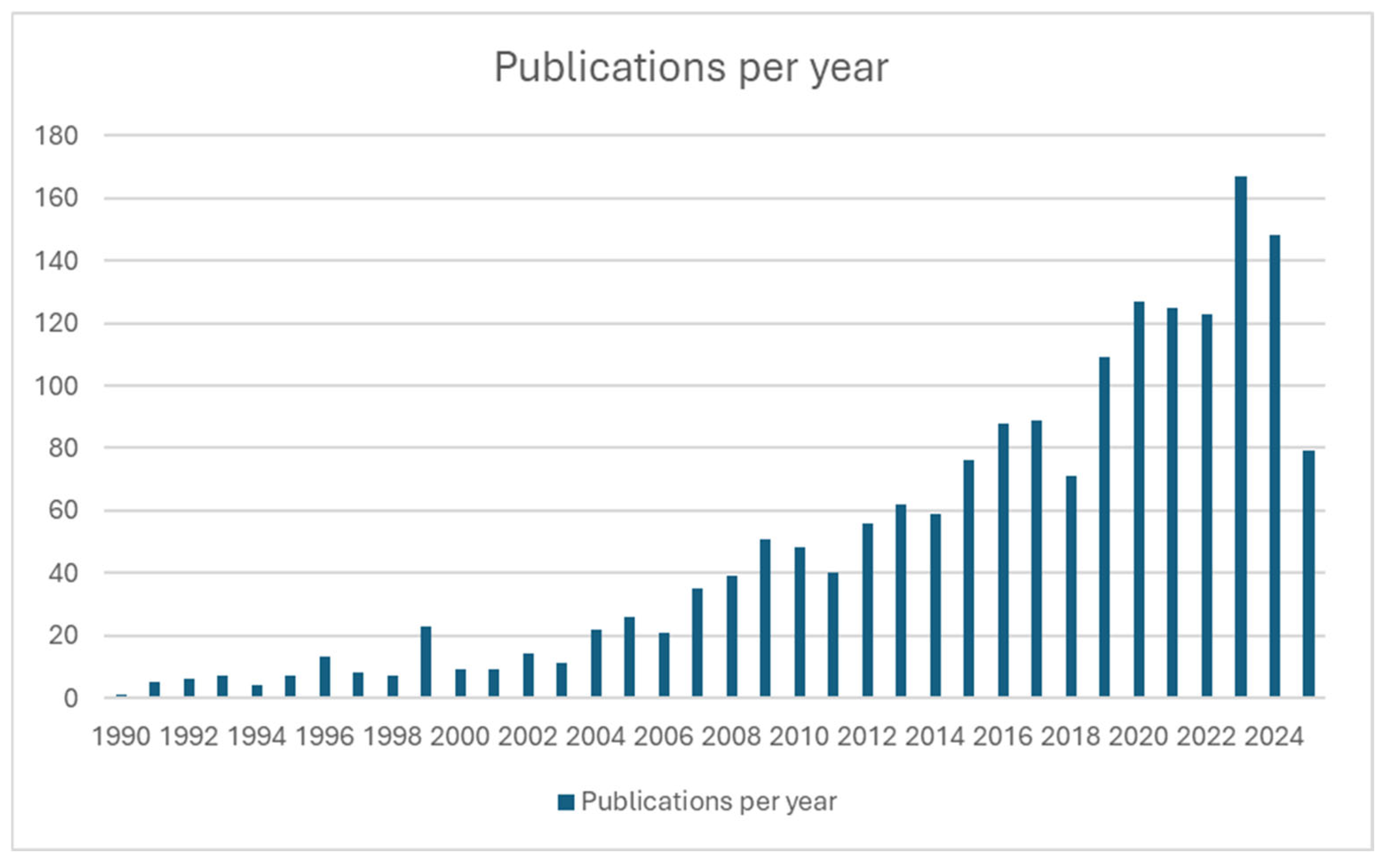
As a result of the above-mentioned properties, PLAL has gained widespread attention across interdisciplinary domains, including biomedicine, catalysis, environmental remediation, and energy storage. Its versatility in producing ligand-free, stable, and high-purity nanoparticles has positioned it as a preferred method for applications requiring minimal surface contamination. Additionally, advancements in laser technologies, diagnostics, and in situ characterization tools have contributed to more precise control and deeper mechanistic understanding of the PLAL process. These developments have further fueled research efforts, as reflected in the sharp increase in annual publications, as shown in Figure 2. The growing volume of literature not only reflects expanding academic interest but also signals PLAL’s potential transition from a niche research technique to a scalable solution for industrial nanomaterials production. The disciplinary distribution of these publications, illustrated in Figure 3, highlights the multidisciplinary appeal of PLAL, with significant contributions from applied physics, materials science, physical chemistry, and nanotechnology. This wide-ranging interest underscores the technique’s adaptability across diverse domains, from fundamental science to application-driven research.
Despite these advantages, PLAL still faces several challenges. First, its productivity is generally lower compared to chemical synthesis methods. The process is often batch-based, limiting throughput unless continuous-flow reactors are implemented [30]. Second, PLAL exhibits sensitivity to minor changes in experimental conditions, such as target surface roughness, beam stability, or liquid purity, which may lead to inconsistency in nanoparticle properties across syntheses [31]. Third, a comprehensive mechanistic understanding of NP formation remains incomplete. While the general sequence of plasma generation, bubble dynamics, and nucleation is well established, quantitative models linking these stages to final particle attributes are still under development [32].
To address these limitations, recent studies have introduced machine learning and real-time diagnostic tools to monitor and optimize synthesis conditions. Time-resolved spectroscopy, high-speed imaging, and in situ small-angle scattering methods have revealed valuable insights into nanoparticle formation mechanisms, bubble behavior, and solvent dynamics [33,34,35]. These tools are increasingly integrated with automated synthesis platforms for achieving more consistent and scalable results.
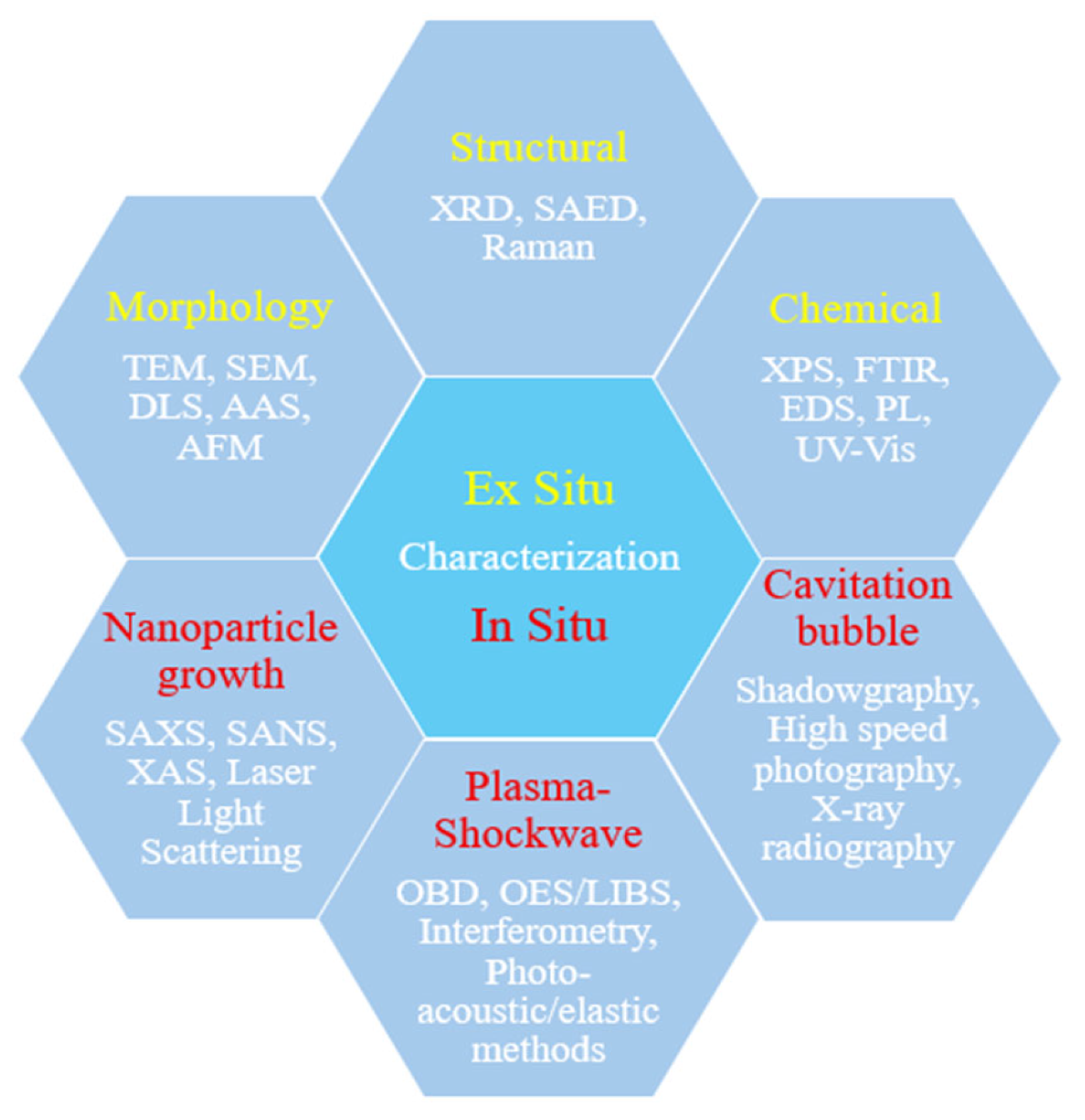
This review aims to provide a comprehensive overview of recent advances in pulsed liquid-based nanoparticle synthesis, with particular emphasis on PLAL, as illustrated in Figure 4. Section 2 highlights key applications of pulsed-synthesized nanoparticles in fields such as biomedicine, catalysis, sensing, and electronics. Section 3 introduces the fundamental principles of pulsed liquid-based synthesis, covering energy sources, solvent roles, pulse parameters, and the underlying mechanisms of PLAL. Section 4 discusses defect engineering via pulsed laser ablation with types of defects and mechanisms. Section 5 outlines representative material systems and compositions, including metals, oxides, alloys, core–shell structures, and emerging materials. Section 6 explores other pulsed energy techniques such as arc discharge and microplasma, outlining their mechanisms and comparative performance. Section 7 presents key characterization methods used to analyze nanoparticle morphology, structure, chemistry, and real-time dynamics. Section 8 and Section 9 address existing challenges, limitations, and offer future perspectives on scaling and optimizing pulsed nanoparticle synthesis methods. Finally, the conclusion summarizes key insights and reiterates the importance of advancing both mechanistic understanding and technical scalability to fully realize the potential of pulsed liquid-based nanomaterial production. By synthesizing current knowledge, this review aims to serve as a reference for researchers working toward controllable and sustainable nanoparticle production.

2. Applications

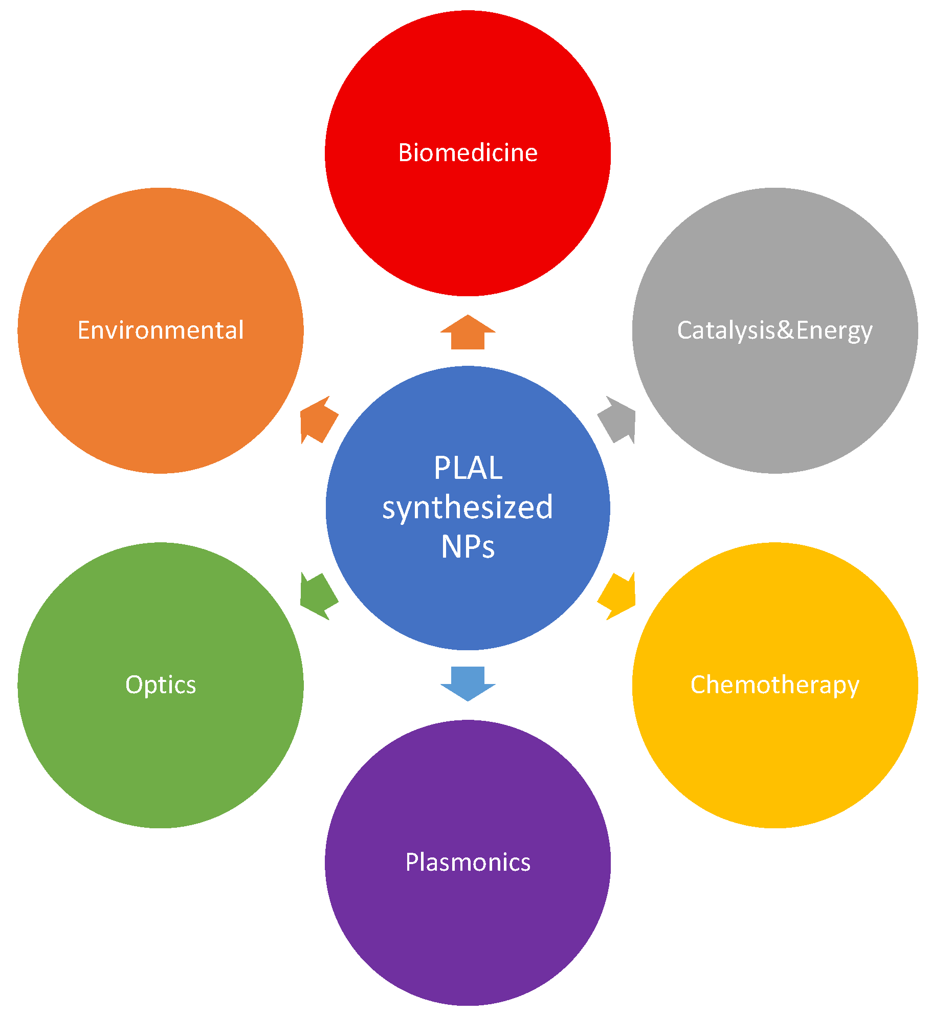
The pulsed laser ablation technique (PLAL) allows for the synthesis of nanostructures with tunable characteristics. In addition, the various materials that can be used in many different areas of applications, Figure 5. Below, we outline the major applications of PLAL-synthesized nanostructures currently being researched.

2.1. Biomedical

The nano-size of the particles enables various biomedical applications, such as drug delivery, photodynamic therapies, and bioimaging. Nanomedicines have been investigated as delivery agents by encapsulating drugs and delivering them to target tissues with a controlled release [36]. Metallic-based nanoparticles are being investigated for these applications due to their photostability, tunable optical properties, and unique interactions with biological molecules [37]. A summary of nanomaterials produced via pulsed laser ablation in liquid (PLAL) and their biomedical applications is presented in Table 1. Nanoparticle physiochemical properties affect how they can be utilized in drug delivery. Cellular internalization and localization are size-dependent, with internalization undergoing different mechanisms. Particles that are <200 nm in size undergo a clathrin-mediated pathway, while particles > 200 nm are internalized by caveolae-mediated endocytosis [38]. Caveolae-mediated endocytosis is kinetically slower than the clathrin-mediated pathway, which allows the size of the particle to affect circulation time. In addition, due to the high surface-area-to-volume ratio of nanoparticles, many active sites become accessible, allowing for surface chemistry modifications and functionalization with various drugs [39]. Al-Kinani et al. [40] designed a Fe@Au core–shell with encapsulated chitosan to improve the solubility and stability of curcumin, an anti-cancer drug, and attached folate, which acted as a targeting molecule to induce apoptosis in breast cancer cells. Mineral nanoparticles cause the overproduction of reactive oxygen species (ROS) in bacterial cells by damaging membranes, DNA, ribosomes, and proteins [41]. In addition to metal-based nanoparticles, lipid-based nanoparticles are being implemented in drug delivery systems that require the transportation of drugs through nonpolar hydrophobic layers, such as mucus. Yu et al. [42] are using the lipid-polymer nanoparticle to anchor a neonatal-Fc-Receptor Targeted peptide, allowing the structure to cross the mucus barrier and modify the receptor to prolong the retention of corticosteroids for treating asthma. Lipid polymer nanoparticles also exhibit low biotoxicity, which may make them advantageous for wound healing.
Nanomaterials can increase the production of ROS by the photosensitizer through surface plasmon resonance, which increases light absorption. An advantage of PDT over traditional drug delivery is that PDT has a lower chance of becoming obsolete due to antibiotic resistance because of the oxidative nature of ROS [43]. Metallic nanoparticles have been shown to improve the ROS production of photosensitizers, such as gold [44], silver [45], and platinum [46]. However, magnetic nanoparticles, such as metal oxides, can be influenced by an outside magnetic field, allowing them to be localized to the target site in a higher concentration [47]. This magnetic targeting relies on gradient magnetic fields, which may be difficult to achieve in deep tissue or organs with limited accessibility, since the magnetic gradient decreases with distance, the practical effective targeting range is often limited by the strength of the external field that can be safely applied [48].
Due to their unique optical properties arising from quantum effects and surface plasmon resonance, nanomaterials enable advanced imaging of living systems. Among these, metal-oxide nanoparticles are actively being investigated as contrast agents for magnetic resonance imaging (MRI). Xu et al. [49] demonstrated that gadolinium-doped iron oxide nanoparticles enhance the contrast of T1-weighted MRI images by facilitating deeper tissue penetration and improved magnetic coupling.
Table 1. Biomedical applications of PLAL.
Material
(Nanomaterial)
Laser TypeWavelengthPulseFluence/EnergyRep. RatePulse TimeMediumSize RangeApplicationReference
Fe3O4 nanoparticlesNd:YAG1064 & 532 nm10 ns16–32 J/cm21 Hz100 pulses/5 minDI water24 ± 6 nmDrug delivery, MRI, hyperthermia[50]
TeO2 nanoparticlesNd:YAG1064 nm100 ns~284 J/cm21 kHz5 minDI water~70 nm; aggl. ≤ 800 nmAntibacterial, anticancer, cytocompatible[51]
Gold nanoparticles (review)Various193–1064 nmfs/ns10–1000 J/cm21–10 Hz5–30 minWater/saline/THF/ethanol<5–100 nmMolecular imaging, drug delivery[52]
Gold NPs (bioconjugates, review)Nd:YAG, fs Ti:Sapph, excimer193–1064 nmfs/ns1–100 J/cm2Hz–kHzmin–hWater/buffers10–80 nmSERS biosensing, immunoassays, imaging[53]
Y-PSZ dental ceramic (microchanneling)DPSS Nd:YAG1064 nm120 ns5–100 kHzsecondsAir/water filmμm-scale channelsDental implants/tools[54]
AuNPs; Au/CNT nanocompositesQ-sw Nd:YAG1064/355 nm10 ns50/140 mJ/pulse10 Hz15–30 minWater; CNT dispersionAu 3–20 nmAnticancer (HCT-116, HeLa)[55]
Ferrite/ZnFe2O4 compositesQ-sw Nd:YAG532 nm9 ns300 mJ10 Hz40 minWater55–88 nm (PLAL)Drug delivery, hyperthermia, MRI[56]
FexOy@Au core–satelliteYb:KGW fs1025 nm420 fs50–100 µJ5–10 kHz20 + 15 min1 mM NaClCore 10–50 nm; Au 7.5 nmPhoto/magnetothermal therapy; MRI[57]
Pd nanoparticlesNd:YVO4 (diode-pumped)532 & 1064 nmns0.26/0.36 mJ20 kHzWater, methanol~6–15 nmAntimicrobial; cytocompatible (L929)[58]
SnO2 nanoparticlesQ-sw Nd:YAG1064 nm10 ns400–800 mJ/pulse1 Hz300 pulsesDDW17.6–25.8 nmAntibacterial; anticancer (A549)[59]
Au nanowires (HC-PCF biosensor)Nd:YAG (SHG/fund.)532 & 1064 nmns2 J (500 pulses)secondsEthanol → PCF≤40 nm dia; ≤3 µm lengthColon biosensor (SPR)[60]
Magnetic NPs (review)Nd:YAG, Ti:Sapph, etc.193–1064 nmfs/ps/ns0.1–10 J/cm2Hz–kHzmin–hWater/organic5–100 nmMRI, hyperthermia, delivery[61]
Elemental boron NPs (PEGylated)Yb:KGW fs1025–1030 nm270–480 fs350 µJ/pulse8 kHz7 hWater~37–40 nmBNCT, photoacoustic, photothermal[62]
ZnO@NiO core–shell m, Nd:YAG + plasma jet1064 nmns + plasma800 mJ/pulse7 Hz10 + 10 minDI water20–100 nmAntibacterial; cytotoxicity[63]
SeO2 nanoparticlesQ-sw Nd:YAG1064 nm~100 ns400 mJ/pulse8 Hz200 pulsesDI waterXRD ~39 nm; AFM ~150 nmAntibacterial; anticancer; aging effect[64]
Au/ZnO nanocompositesNd:YAG (Au PLAL) + UV1064 & 355 nm10 ns70 mJ (Au); 140 mJ (UV)10 Hz30 + 30 minWater—(TEM uniform Au)Anticancer (HCT116, HeLa), biocompatibility[65]
AuNPs (solvent effect, anticancer)Q-sw Nd:YAG532 & 1064 nm10 ns6 Hz300 pulses (~50 s)DDDW/NaOH/DMEMTEM 5–25 nmAnticancer (Hepa 1–6), IC50 35–74 µg/mL[66]
Au, Mg, Zn NPs (dental antibac.)Nd:YAG (SISMA OEM Plus)1064 nm≈35 ns0.3 mJ; 8488 J/cm220 kHz15 & 30 minDDW + SDS (0.025 M)Au 5–7.5 nm; Mg 1.9–3.8; Zn 120 → 19.5Antibacterial vs. oral pathogens; biocompatible[67]
Se NPs (amorph → trigonal)Nd:YAG1064 nm~100 ns0.25–0.75 J/cm2~3 kHz~5 min + autoclaveDI water70 ± 29 → 320 ± 28 nmBroad-spectrum antibacterial[68]
CQDs/GQDs (review)Nd:YAG; Ti:Sapph355/532/800/1064 nmfs → nsVaries10 Hz–1 kHzmin–hWater; organics; aminesCQDs 1–10 nm; GQDs < 100 nmBioimaging, PDT/PTT, delivery[69]
Pd–Cu bimetal nanoparticlesNd:YAG532 nm10 ns500 mJ/pulse4 HzPd 2–5 min; Cu 1–4 minDI waterFESEM 31–51 nm; XRD 3–16 nmAntibacterial; antioxidant; no hemolysis[70]
Metal-oxide core–shell NPs (mini-review)Nd:YAG (typ.)532/1064 nm (typ.)ns (fs/ps covered)Liquids (water common)Core 9–70 nm; shell 2–60 nmAntibacterial, anticancer, imaging, delivery[41]

2.2. Catalysis and Energy

Nanomaterials’ high surface area-to-volume ratio exposes active sites, enhancing catalysis and electrical conductivity, which makes them promising for applications in fuel cells and energy storage [49]. Hydrogen fuel cells are being actively researched as a clean and sustainable energy source; however, their efficiency is limited by the slow kinetics of the oxygen reduction reaction, which can be significantly improved using metallic nanoparticles, particularly those from the platinum group, due to their superior catalytic activity. In addition, alloys and core–shell structures can be used to limit the cost and improve the efficiency of the Pt catalyst [71]. PLAL can impact catalytic performance by introducing surface defects, vacancies, and under-coordinated atoms that act as additional active sites. These features enhance adsorption and activation of reactants [72]. Representative materials synthesized via PLAL for catalytic and energy-related applications are summarized in Table 2.
Materials are becoming desired in photocatalysis and supercapacitor energy storage due to their large specific capacitance and greater stability. Pandith et al. [73] showed that surface-modified CuO nanoparticles could be utilized as electrode materials to make pseudocapacitive reduction faster and as a photocatalyst in the degradation of AR88 dye.
Conducting polymer nanocomposites offer high electrical conductivity, flexibility, and tunable electric properties, making them ideal for energy storage and harvesting. When used in combination with other nanomaterials like metal oxides or carbon nanotubes, these composites improve specific capacitance, stability, and charge transport [74]. This makes them strong candidates as materials for supercapacitors, batteries, and flexible energy devices. Patil et al. [75] highlighted that incorporating metal oxide nanoparticles into conducting polymer matrices significantly enhances the electrochemical performance by increasing the active surface area and facilitating faster electron transfer, leading to improved energy storage capacity and cycling durability.
Table 2. Catalysis and energy applications of PLAL.
Material
(Nanomaterial)
Laser TypeWavelengthPulseFluence/EnergyRep. RatePulse TimeMediumSize RangeApplicationReference
TiO2, α-Fe2O3, TiO2–Fe2O3Q-sw Nd:YAG1064 nm8 ns3.5–17.7 J/cm26 Hz15 minWater12–32 nmPhotocatalysis, PEC[76]
Au/Ag core–shell seriesNd:YAG1064 nm6 ns60–90 mJ/pulse10 Hz10–15 minDI water35–50 nmDye degradation catalysis[77]
Ag–MWCNT compositesNd:YAG1064 nm7 ns~100 mJ/pulse10 Hz15 minMWCNT aqueousAg ~14 nm4-NP/MO/MB degradation[78]
Au/Ag (alloy, core–shell)Nd:YAG1064 nm7 ns60 mJ/pulse10 Hz7 minWater/HAuCl4Ag 20 nm; comp. 34 nm4-NP catalytic reduction[79]
Photocatalysis for wastewater (review)Various248–1064 nmfs/ns0.2–8 J/cm2; 40–350 mJ10 Hz–kHzmin–hWater/EtOH/mixed5–100 nmPollutant
degradation
[80]
Laser ablation in air (perspective)ns/fs (various)355–1064 nmns–fsVariableHz–kHzminAirOER/HER.
Li-ion
[81]
Cu nanoparticles (recycled vs. industrial)Nd:YAG1064 nm10 ns13 J/cm210 Hz10 minMethanol~2 & 30–100 nmHER/OER on Ni foam[82]
CuO@ZnO core–shellQ-sw Nd:YAG1064 nm10 ns500–900 mJ1 Hz5 min (200 pulses)Water19–70 nmMB photocatalysis[83]
Fe3O4 NPs (cat. + antibac.)Nd:YAG532 & 1064 nm10 ns22–26 J/cm21 Hz5 minWater30–65 nm (SEM)MB degradation[84]
Hydrogen gen./storage/detection (review)Nd:YAG, fiber, Ti:Sapph, excimer193–1064 nmfs/ps/ns0.1–100 J/cm2Hz–kHzmin–hWater/solvents/gas2–100 nmHER/PEC; sensing[85]
PLAL nanocatalysts (review)Various193–1064 nmfs/ps/ns0.1–100 J/cm2Hz–kHzmin–hVarious2–100 nmHER/ORR/CO2R/degradation[86]
Laser-synthesized NPs (broad review)Various193–1064 nmfs/ps/nsHz–MHzmin–cont.Water/organic2–200 nmCatalysis, energy, photonics[87]
FeNi alloy NPs (modeling)ps laser (simulated)800 nm10 ps600–3000 J/m2 (absorbed)ns–µs (model)Water (CG-MD)4–12 nmDefect-rich → catalysis[88]
Rh–Ni@graphitic-carbon (HER)Q-sw Nd:YAG1064 nm7 ns100 mJ (ablation); 90 mJ (dec.)10 Hz30 + 10 minAcetonitrile → aqueous saltsGC shell 1.7–12.9 nm; Rh few-nmHER: η10 ≈ 46 mV; Tafel 36 mV/dec[89]
Liquid pulsed laser propulsion (review)Nd:YAG, CO2, etc.532/1064/10.6 µmns/ps1–20 J/cm2Hz–kHzpulses → cont.Propellants (liq.)Micro-thrusters/propulsion[90]

2.3. Sensors and Environmental Remediation

Nanomaterials offer chemical sensitivity and surface specificity, which make them suitable for sensing trace elements and environmental pollutants. Metal oxides can be used to detect various toxic gas molecules in a sensor [91]. In addition, metal oxides can be improved by doping the materials with a second nanocatalyst. Feng et al. [92] demonstrated that doping a ZnO nanoplate with Pd nanoparticles creates a material that was used to develop a sensor that had a high specificity and selectivity for the detection of chlorobenzene by an oxidation reaction. Biosensing is an important area of research because it allows for the detection of certain biomolecules for diagnosis. Graphene quantum dots (GQDs) possess inherent photoluminescence, which makes them highly valuable in biosensing. Rasheed et al. [93] reviewed how functionalization of GQDs with various surface signaling molecules enables detection of diverse analytes, including Cu2+, Fe3+, Pb2+, Hg2+, enzymes, and antigens, through photoluminescence quenching mechanisms. A selection of nanomaterials synthesized via PLAL for sensing and environmental remediation applications is summarized in Table 3.
The wide band gap of organic-based nanomaterials and the delocalization of electrons, improving photostability of the material, make them outstanding candidates for photocatalysis of environmental pollutants. Bai et al. [94] demonstrated this potential by developing terpyridine-based metallo-cuboctahedron nanomaterials that achieved up to 95% degradation of persistent organic pollutants like ibuprofen under visible light. Metal oxides also exhibit photocatalytic activity, such as CuO, but there are still debates on the potential toxicity of the metal ions.
Table 3. Sensors and Environmental Remediation applications of PLAL.
Material (Nanomaterial)Laser TypeWavelengthPulseFluence/EnergyRep. RatePulse TimeMediumSize RangeApplicationReference
Polymer–Fe3O4 tabletsNd:YAG (2ω)532 nm6 ns9 J/cm210 HzAir (solid tablet)400 nm–4 µmOil–water separation[95]
Au–ZnO core–shell nanostructuresNd:YAG1064 nmns2000 mJ; 200 pulsesEthanol<50 nmOptical & biological sensors[96]
LIG on polyimideUV Nd:YVO4355 nmns0.11–0.6 J/cm2scanningAir (PI substrate)Porous, layeredHumidity/
ion sensors
[97]
Ag nanoparticles (gas sensor)Nd:YAG1064 nm10 ns~102 J/cm210 Hz20–80 minAgNO3 + TSC (aq)5–31 nmNH3/Ethanol gas sensing[98]
Au nanoparticles (colorimetry)Nd:YAG532 nm9 ns25–75 mJ10 Hz10–30 minMilli-Q waterTEM ~26 ± 2 nmLSPR colorimetric sensing[99]
Scale-up perspective (green)Nd:YAG, Ti:Sapph, fiber193–1064 nmfs/ps/nsHz–MHzmin–hWater/organics/flowBroadSensors & remediation[10]
ASS & synaptic devices (review)Nd:YAG, Ti:Sapph, fiber355–1064 nmfs/ps/nsHz–kHzmin–hWater/EtOH2–100 nmArtificial sensory systems; neuromorphic[100]
MoS2/WS2–Ag nanocompositesQ-sw Nd:YAG (Litron LPY 674G-10)8 ns26.3 J/cm210 Hz20 + 15 minDI waterAg 15–17 nm; few-layer TMDsDopamine/AA sensing; NLO limiting[101]
B,N co-doped GO (BNG1/BNG2)Nd:YAG (Quanta-Ray Pro 230-10)1064 nm10 ns625 mJ/pulse10 Hz60 minEthanol + NH3 + H3BO350–60 nmGas sensors (ethanol/acetone/NH3)[102]
rGO, Co3O4, rGO/Co3O4 composite filmsQ-sw Nd:YAG1064 nm10 ns140 mJ; 300–1000 pulses4 Hz75–250 sDI water (sequential)23–51 nm (FE-SEM)H2S/NO2 gas sensing[103]
PLAL for sensing & photonics (review)fs/ps/ns lasers355–1064 nmfs/ps/ns0.08–1000 J/cm2 (varies)Hz–kHz–MHzmin–hWater, acetone, ethanol, chloroform, SDS/PVP3–200 nm; periodic 95–350 nmSERS sensing (explosives, dyes, pesticides); NLO devices[104]

2.4. Electronics and Photonics

The tunability acquired by the PLAL technique makes synthesized materials advantageous in electronic devices to fine-tune the device to promote stability and longevity while also improving efficiency. Metallic nanoparticles offer high conductivity for conductive films. Pajor-wierzy et al. [105] displayed that Ni-Ag core–shell structures could be deposited onto films, while displaying a lower sintering temperature than bulk Ni and high conductivity. Then, in 2023, Pajor-wierzy et al. showed that the conductivity of Ni-Ag core–shell structures was thickness-dependent, which allows for greater tuning for electronic devices. A summary of representative nanomaterials synthesized via PLAL and their applications in electronics and photonics is presented in Table 4. Ceramic nanoparticles have applications in communication photonics due to their dielectric properties and microwave absorption. Didde & Dubey [106] demonstrated that sol–gel synthesized calcium-doped ZnAl2O4 ceramic nanoparticles exhibit a high dielectric constant (~13), low dielectric loss (0.024), and strong frequency-dependent conductivity, making them suitable for use in microstrip patch antennas for WLAN applications.

2.5. Case Studies Linking Synthesis Parameters, Defect Profiles, and Application Performance

Several studies have begun to quantitatively link pulsed-liquid synthesis conditions with resulting defect profiles and application-relevant performance, moving beyond qualitative descriptions. For instance, Ding et al. (2025) demonstrated how tuning the laser fluence and pulse parameters in PLAL of Ti3C2 MXene can control surface defect densities—specifically, oxygen-rich terminations and vacancy concentrations—resulting in enhanced sensing capabilities through improved charge transfer and conductivity in chemical sensing applications [121]. Similarly, Subhan et al. (2022) [19] systematically investigated how variations in laser fluence and liquid environment influence nanoparticle morphology and defect generation, establishing correlations between process settings, structural disorder, and consequent catalytic or optical properties of PMCs. Together, these works provide clear, measurable links between synthesis parameters (e.g., laser fluence, liquid chemistry), the nature and density of defects, and performance outcomes—an important step toward rational, defect-engineered pulsed liquid synthesis [19].

3. Fundamentals of Pulsed Liquid-Based Synthesis

3.1. Definitions and Key Terminology

Pulsed liquid-based nanoparticle synthesis (PLNS) refers to a group of top-down physical techniques that generate nanomaterials by delivering short bursts of energy to a solid or dispersed phase within a liquid medium. Other related approaches, including laser fragmentation, electrical discharge, and ultrasound-assisted synthesis, rely on similar principles of energy delivery followed by rapid physical and chemical transformations in the confined liquid environment [12], which will be mentioned later.

3.2. Key Parameters

3.2.1. Laser Wavelength and Pulse Duration

Laser wavelength and pulse duration are critical parameters in pulsed laser ablation in liquids (PLAL), governing energy absorption efficiency, plasma generation, cavitation dynamics, and ultimately the size, distribution, and crystallinity of the synthesized nanoparticles (NPs). Tuning these parameters enables precise control over NP formation pathways and product quality.
Shorter wavelengths, particularly in the ultraviolet (UV) range (e.g., 355 or 266 nm), are more strongly absorbed by most metals and semiconductors, which enhances material removal efficiency and favors the formation of smaller, more monodisperse nanoparticles. General trends reported for silver, copper, gold, and palladium confirm this effect: nanoparticles synthesized at 355 or 532 nm are consistently smaller than those formed at 1064 nm, due to stronger absorption and higher plasma density at shorter wavelengths [1,122,123].
Pulse duration determines whether ablation proceeds via thermal or non-thermal mechanisms. Femtosecond to low-picosecond pulses enable non-thermal ablation—such as Coulomb explosion and bond-breaking—by depositing energy faster than heat diffusion timescales. This results in highly supersaturated vapor phases and rapid quenching, which support the formation of small, defect-rich, and monodisperse NPs. For example, gold ablated with 10 ps pulses in water yielded highly uniform colloids with reduced aggregation compared to nanosecond pulses, which promoted droplet ejection and bimodal distributions due to melt-driven mechanisms [1].
Pulse duration determines whether ablation proceeds via thermal or non-thermal mechanisms. Femtosecond to picosecond pulses enable non-thermal ablation by depositing energy faster than heat can diffuse, producing highly supersaturated vapor phases and defect-rich, monodisperse nanoparticles. In contrast, nanosecond pulses promote melt-driven processes, leading to larger, often agglomerated particles due to extended heat-affected zones [1,124]. These general trends are summarized in Table 5.
These trends apply across different material systems. In silicon ablation using femtosecond pulses in ethanol, bimodal distributions emerged from competing ejection and condensation mechanisms, modifiable by pulse duration and focusing conditions [125]. For gold and silver, nanosecond pulses typically yield larger, more polydisperse particles (>50 nm), whereas picosecond pulses achieve narrower size distributions with suppressed aggregation [1,126]. Femtosecond and picosecond pulses are more efficient for ablation with minimal thermal damage, but also more prone to phenomena like optical breakdown and self-focusing at high fluence [127,128].
Summary of Observed Effects:
Table 5. Summary of the effect of wavelength and pulse duration on nanoparticle (NP) formation during PLAL.
ParameterEffect on NP FormationExample MaterialsReferences
Short wavelength (266–532 nm)Higher ablation efficiency, smaller NP size, narrow distributionCu, Ag, Pd, Au[1,122,123]
Long wavelength (1064 nm)Lower absorption, larger particles, higher ablation thresholdAu, Pd, Cu[1,122]
Femtosecond/picosecond pulsesNon-thermal ablation, high supersaturation, monodisperse NPsAu, Si, Ag[1,124,125,126]
Nanosecond pulsesThermal melting, ejection, bimodal distributions, larger aggregatesAu, Ag, Pd[1,126]
In conclusion, the combined tuning of laser wavelength and pulse duration offers powerful levers for engineering nanoparticle characteristics in PLAL. Shorter wavelengths and ultrashort pulses are generally preferred for achieving high-quality, narrowly dispersed, and chemically clean nanostructures—desirable for applications in catalysis, photonics, and biomedicine.

3.2.2. Laser Fluence and Repetition Rate

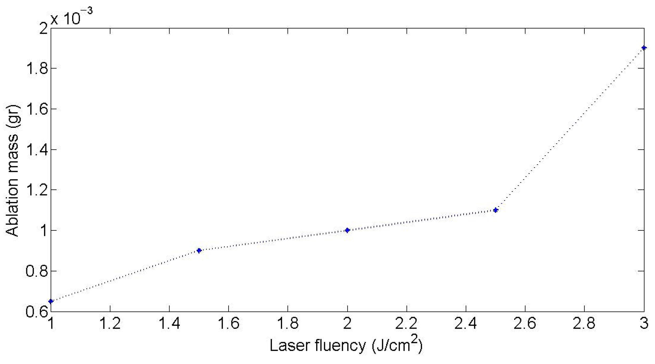
Laser fluence and repetition rate are pivotal in tuning nanoparticle (NP) size, yield, and colloidal stability in PLAL (Figure 6). Fluence values in the range of 1–10 J/cm2 are generally considered optimal for most material systems, providing a balance between effective ablation and minimal plasma shielding. Fluences below this range often result in insufficient material removal, while those above it can induce nonlinear optical effects, cavitation-driven scattering, and plasma plume shielding that reduce overall efficiency and increase particle polydispersity [1,122,129]. For example, in Figure 6, the ablation mass increases with fluence from ~1 to 3 J/cm2, with a sharper rise beyond 2.5 J/cm2, illustrating the threshold-like behavior of PLAL. At low fluence, ablation is inefficient, while higher fluence enhances efficiency but may also increase plasma shielding and nanoparticle polydispersity.
Fluence-dependent studies during silver ablation in water have shown that increasing fluence from approximately 2.1 to 3.2 kJ/cm2 leads to a decrease in average particle size from ~19 nm to ~7 nm, attributed to enhanced fragmentation and vaporization dynamics [122]. However, when fluence exceeds a critical value, plasma shielding impedes further energy deposition at the target interface, thereby reducing productivity and altering size distribution [129]. Fluence typically shows a logarithmic relationship with productivity. At low laser fluence levels, the energy delivered is insufficient to surpass the ablation threshold of the material, resulting in negligible material removal [131]. At excessively high fluence levels, the ablation process can reach a saturation point where further increases do not enhance material removal proportionally. This is due to plasma shielding, where the dense plasma absorbs incoming laser energy. Excessive fluence can also lead to thermal effects such as increased surface roughness or recast layers. In femtosecond laser ablation of silicon-on-insulator substrates, beyond a certain fluence, the ablation depth per pulse saturated and morphology deteriorated due to melting and resolidification [132]. Similarly, in the laser processing of RB-SiC ceramics, increasing laser energy beyond an optimal point led to the accumulation of ablation products on the surface, affecting microgroove quality [133].
Repetition rate also has a pronounced impact on ablation consistency and NP formation. At low repetition rates (e.g., 10–40 Hz), sufficient time exists between pulses for cavitation bubbles and thermal effects to dissipate, ensuring steady and reproducible ablation with uniform NP generation [1]. In contrast, at high repetition rates (>500 Hz), successive pulses may overlap with residual cavitation bubbles or uncooled regions, leading to beam scattering, irregular energy coupling, and larger or more aggregated nanoparticles.
A study involving picosecond laser ablation of Zn at 400 kHz demonstrated significant heat accumulation, resulting in the formation of rim structures and increased thermal damage to the target. These thermal effects promoted the generation of larger particles and reduced colloidal uniformity [134]. Conversely, when high repetition rate systems are combined with beam scanning or flow-cell geometries, they can yield high productivity while mitigating adverse thermal buildup. For instance, a ps-laser system operating at 5000 Hz with optimized beam movement achieved ~350 µg/min NP production while maintaining narrow size distributions [135].
Furthermore, increasing both pulse energy and repetition rate was shown to reduce the size and improve the uniformity of TiO2 nanoparticles, producing particles around 46 nm. In a study by Singh et al., TiO2 nanoparticles were synthesized using pulsed laser ablation in water with a laser operating at 35 mJ/pulse energy, 10 ns pulse width, and 10 Hz repetition rate. The resulting colloidal solution contained highly stable TiO2 nanoparticles, and characterization techniques such as UV-Visible absorption, photoluminescence, X-ray diffraction, SEM, and FTIR spectroscopy were used to analyze the nanomaterials. The study highlighted that variations in laser parameters significantly influenced nanoparticle size and uniformity [136].
Thus, fluence and repetition rate must be co-optimized for each material–liquid system to balance ablation efficiency with colloidal quality. Inappropriate values in either parameter may lead to undesirable outcomes such as low NP yields, excessive agglomeration, or inefficient energy use.

3.2.3. Role of Solvents and Surfactants
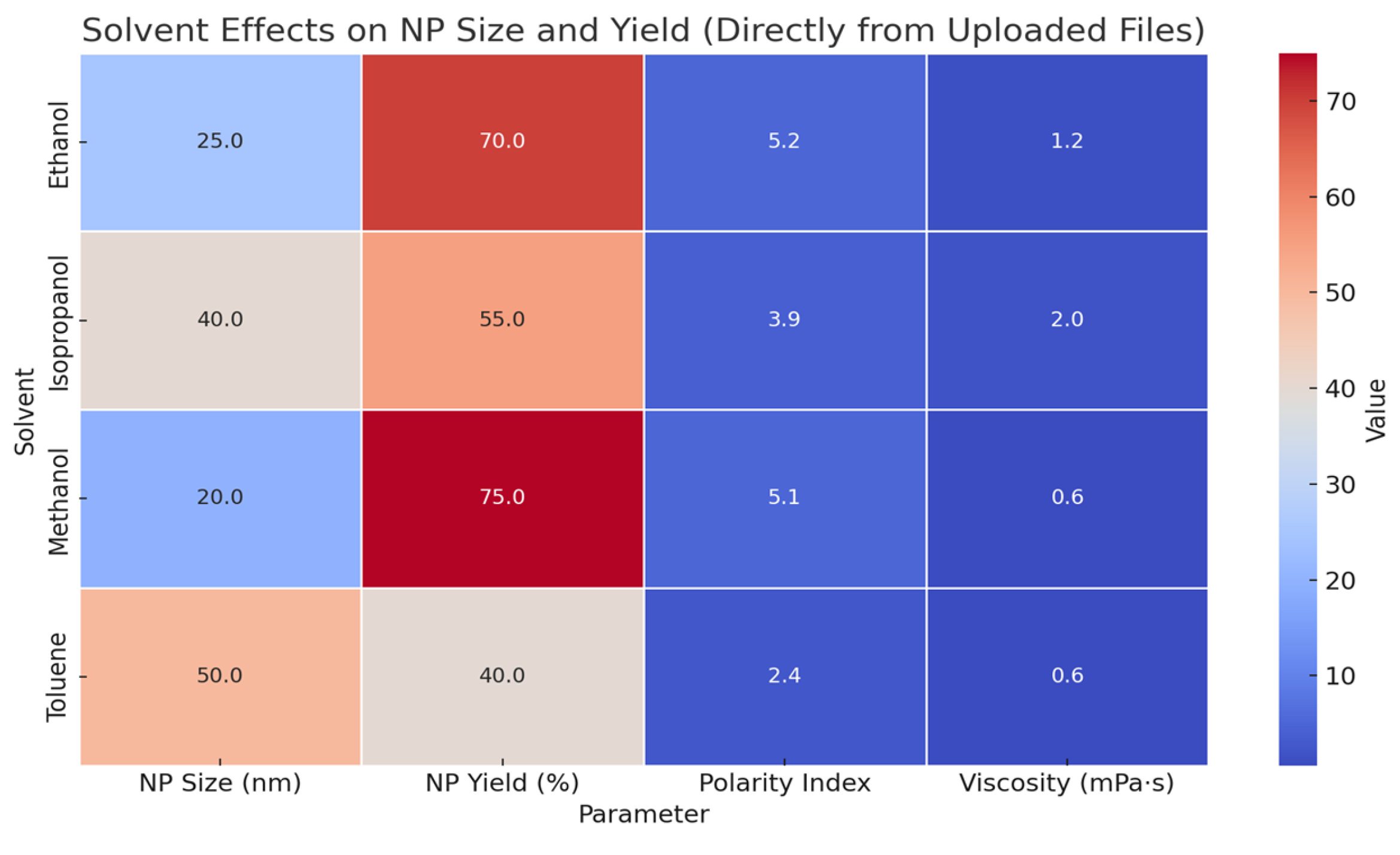
The solvent serves not only as a medium but as an active participant in the synthesis process. As shown in Figure 7, solvent properties such as polarity, viscosity, dielectric constant, boiling point, and thermal conductivity affect energy dissipation, plasma cooling, cavitation dynamics, and nanoparticle stabilization. Among the solvents studied, methanol resulted in the smallest average NP size (20 nm) and the highest yield (75%), which is attributed to its high polarity and low viscosity. In contrast, toluene produced the largest particles (50 nm) and the lowest yield (40%) due to its low polarity index and relatively high viscosity. Ethanol and isopropanol exhibited intermediate behavior, with ethanol favoring yield (70%) and isopropanol yielding moderately sized particles (40 nm). This data highlights the significant role solvent properties play in controlling NP characteristics during PLAL [124,137,138].
In addition, polar solvents promote faster heat transfer and may influence particle morphology by facilitating specific growth pathways. Forte et al. showed that the solvent medium directly affects the nonlinear behavior in the production of carbon chains [139]. In the synthesis of tungsten oxide nanocrystals, varying the solvent from ethanol to an ethanol/water mixture to pure water resulted in distinct morphologies—polymerized spherical structures, rod-shaped particles, and sheet-like formations, respectively [140]. These morphological changes are attributed to the differing polarities of the solvents used.
Viscosity critically impacts both the diffusion rates of reactants and bubble collapse dynamics. Higher viscosity solvents can slow down particle growth, leading to smaller particle sizes and more uniform distributions. In the synthesis of CuO nanoparticles, using ethanol (lower viscosity) resulted in smaller, more uniform spherical particles compared to those synthesized in propanol (higher viscosity), which produced a mix of needle-shaped and spherical particles [141].
In PLAL, viscosity differences also explain observed changes in gold nanoparticle sizes: gold NPs generated in ethanol with 0.1 M PVP K-15 had a mean size of 3.4 ± 0.8 nm, whereas in isopropanol (IPA), the size increased to 4.4 ± 1.1 nm, reflecting the higher viscosity of IPA (2.92 mPa·s vs. 1.81 mPa·s for ethanol) [142].
In highly viscous PVP solutions, the productivity of Au NPs decreased from 236 μg/h to 76 μg/h as viscosity rose from 0.94 to 6.39 mPa·s due to prolonged gas bubble shielding and reduced laser-target coupling [143].
The dielectric constant of a solvent influences the solvation of ions and the stabilization of charged species during nanoparticle formation [144]. Solvents with higher dielectric constants can better stabilize charged intermediates, affecting the nucleation rate and particle size. In the case of CdSe quantum dots, solvents with varying dielectric constants influenced the swelling of oleate-capped nanoparticles, affecting their dispersion and stability [145].
The boiling point and thermal conductivity of a solvent determine the thermal environment during synthesis. Solvents with lower boiling points can evaporate quickly, leading to rapid cooling and affecting the crystallinity and size of nanoparticles. In solvothermal synthesis, the choice of solvent with an appropriate boiling point is crucial for controlling the reaction temperature and pressure, thereby influencing the morphology and size of the resulting nanoparticles [146].
Figure 7. Influence of different solvents on nanoparticle (NP) size, yield, polarity index, and viscosity during pulsed laser ablation in liquid (PLAL) [111,128,143].
Qubs 09 00032 g007
Solvents with low thermal conductivity (e.g., CHCl3: 0.12 W/m·K) retained heat longer, resulting in smaller particle sizes (~3.5 ± 1.0 nm for Au), while solvents like ethanol offered better productivity through faster heat dissipation [142].
In contrast, water often promotes surface oxidation and larger particles due to its higher reactivity and cavitation energy. These effects are exacerbated in the absence of surfactants or stabilizers, leading to broader size distributions and aggregation [122,136].

3.2.4. Surfactants and Chemical Additives

Surfactants, such as CTAB (cetyltrimethylammonium bromide) or SDS (sodium dodecyl sulfate), are often introduced to control nanoparticle size and prevent agglomeration. They adsorb onto the nanoparticle surface, reducing surface energy and guiding anisotropic growth by selectively binding to specific crystal facets. SDS has been shown to induce zeta potentials exceeding −30 mV, an indicator of strong electrostatic stabilization, while excessive surfactant can trigger salting-out or pyrolysis effects that degrade colloidal quality [136].
In green chemistry contexts, efforts are underway to minimize or replace surfactants with biocompatible stabilizers or solvent-controlled self-assembly strategies. The addition of PVP during gold ablation was shown to reduce NP sizes from 4.2 nm to ~3.5 nm as PVP concentration increased from 10−4 to 10−3 mol/L, illustrating enhanced steric stabilization and suppressed agglomeration [143].

3.2.5. Target Composition and In Situ Doping

The target material’s composition—whether elemental, alloyed, or multilayer—directly determines the nanoparticle’s phase, size, and elemental distribution. For example, PLAL from complex alloys such as CoCrFeNiMn generates multielemental high-entropy alloy NPs with broadened size distributions, while elemental targets tend to produce more uniform, phase-pure particles [123].
Doping during PLAL is another avenue for functionality enhancement. In situ Eu3+ doping was achieved by introducing EuCl3 into aqueous PLAL systems, yielding YVO4:Eu3+ nanoparticles with an atomic Eu:Gd ratio of 0.83% and strong luminescence verified via LIBS [147]. Such doped nanoparticles hold promise for optical, bioimaging, and sensing applications.

3.3. Size Control, Crystallinity, and Surface Chemistry

3.3.1. Plasma and Bubble Dynamics

The evolution of plasma and cavitation bubbles directly influences the size, structure, and distribution of nanoparticles (NPs) synthesized via pulsed laser ablation in liquids (PLAL). During the early stages of ablation, plasma temperatures reach 4000–7000 K, and particle densities can approach ~1020 cm−3 [148,149]. Such high densities ensure extremely short mean free paths and strong inter-particle interactions, which support rapid ionization, charged cluster formation, and enhanced colloidal stability by suppressing uncontrolled aggregation.
These conditions initiate cavitation bubble formation, as plasma expansion rapidly vaporizes the surrounding liquid. The resulting cavitation bubble expands and subsequently collapses violently on nanosecond-to-microsecond timescales (Figure 8). This collapse is not merely a mechanical rebound; it generates localized temperatures exceeding 6000 K and pressures surpassing 106 Pa, creating the extreme conditions necessary for nucleation and condensation of atomic and ionic species into nanoparticles [1,126,137]. These general plasma and bubble behaviors and their impact on nanoparticle formation are summarized in Table 6.
In water-based laser ablation (Figure 8), the process begins with the arrival of a femtosecond laser pulse at the target surface (Domain 1, −20 to −0.5 ps). The high-intensity pulse immediately excites electrons through nonlinear absorption mechanisms, initiating plasma formation. Within the first picosecond (Domain 2), a dense, highly confined plasma is generated, composed of vaporized material, free electrons, and ions. Unlike in air, this plasma is strongly compressed by the surrounding liquid, preventing rapid expansion and resulting in extremely high localized pressures and temperatures, often exceeding several thousand Kelvin [126].
From 1 to 20 ps (Domain 3), the plasma begins to expand slightly, but confinement by the liquid causes a significant pressure build-up, leading to delayed and anisotropic plasma expansion—a phenomenon described as “plasma dilution” [1,139]. As the process proceeds into the 20–200 ps range (Domain 4), the plasma continues interacting with the surrounding liquid. The ablation plume is now fully confined, and this leads to the generation of a strong shockwave that propagates into the liquid. The high-pressure environment supports rapid thermalization and sets the stage for nanoparticle nucleation [126].
Between 200 ps and 1 ns (Domain 5), the plasma cools and condenses, enabling the formation of primary nanoparticles via vapor-phase condensation within the confined volume. These particles are typically smaller and more monodisperse than those produced in air due to the efficient confinement and rapid quenching [1,126]. Following this, from 1 ns to several microseconds (Domain 6), the expansion of vapor and hot gases causes the formation of a cavitation bubble. This bubble grows to its maximum radius within ~5–10 µs before collapsing violently around 10–30 µs, often emitting secondary shockwaves and occasionally fragmenting or reshaping existing nanoparticles [126,139].
Finally, in Domain 7, the system stabilizes, resulting in a colloidal suspension of nanoparticles dispersed in the liquid. The final state includes both the primary nanoparticles formed during plasma condensation and secondary particles or fragments generated during bubble collapse. Microbubbles may also persist temporarily, and the absence of surfactants or stabilizers ensures clean particle surfaces suitable for applications in catalysis, sensing, and biomedicine [1,139].

3.3.2. Rayleigh–Plesset and Gilmore Models

To quantitatively describe cavitation bubble dynamics in PLAL, two primary hydrodynamic models are commonly employed: the Rayleigh–Plesset equation and the Gilmore equation. Each describes different regimes of bubble behavior, with Rayleigh–Plesset suited for moderate-energy conditions in incompressible media, and Gilmore used when compressibility and shockwave effects dominate [1,148].
The Rayleigh–Plesset equation is a fundamental model that describes the radial dynamics of a single spherical bubble in an incompressible and viscous fluid. It is derived from the Navier–Stokes equations under the assumptions of spherical symmetry and a uniform far-field pressure. The governing form is:
p R t R t ¨ + 3 2 R ˙ t 2 = P B t P t 2 σ R t 4 μ R ˙ t R t
where
  • ρ = density of the liquid surrounding the liquid;
  • R (t) = Bubble radius at a time t;
  • R ˙ t   =   d R d t : Rate of change of radius—how fast the bubble expands or contracts;
  • R ¨ t = d 2 R d t 2 : Acceleration of the bubble wall;
  • P B t = Pressure inside the bubble;
  • P t = Pressure inside the liquid far from the bubble (ambient pressure);
  • μ = dynamic viscosity of the liquid;
  • σ = surface tension at the liquid–gas interface.
This equation is particularly effective in modeling expansion and collapse cycles of cavitation bubbles under nanosecond-pulse PLAL or moderate fluence regimes, where compressibility of the liquid can be neglected [137,148]. It enables estimation of:
  • Maximum bubble radius;
  • Collapse time;
  • Interfacial pressure;
  • Velocity profiles in the surrounding fluid.
In PLAL systems, the Rayleigh–Plesset model helps interpret how the initial vaporization of the target and liquid forms a transient cavitation bubble, and how the violent collapse of this bubble drives the condensation of metal vapor into nanoparticles [1,126]. The model is instrumental for:
  • Understanding early-stage bubble growth;
  • Analyzing laser–liquid interaction timescales;
  • Optimizing flow cell design to improve ablation efficiency and NP yield [124,148].
Although it does not capture shockwave formation or compressibility effects, which are better described by the Gilmore model, the Rayleigh–Plesset equation remains widely used in PLAL for predicting size distributions and identifying optimal synthesis conditions [4,98].
While the Rayleigh–Plesset equation provides valuable insight under moderate conditions, it becomes inadequate when bubble wall velocities approach the speed of sound in the liquid—a common occurrence in high-fluence or ultrashort-pulse PLAL. In such regimes, liquid compressibility and shockwave generation must be accounted for. This is where the Gilmore equation becomes essential.
The Gilmore equation extends Rayleigh–Plesset theory by incorporating fluid compressibility, allowing it to describe high-speed, large-amplitude oscillations of a cavitation bubble and the shockwaves emitted during collapse and rebound. The generalized form is:
1 R ˙ c ( t )   R R ¨ + 3 2   1 R ˙ 3 c ( t ) R ˙ 2 = 1 + R ˙ c ( t )   p B p t ρ t +   H B H
where
  • R (t) = bubble radius as a function of time;
  • R ˙ = d R d t = wall velocity;
  • R ¨ = d 2 R d 2 t = acceleration;
  • c(t) = local speed of sound in the liquid;
  • p B t = pressure inside the bubble;
  • p t   = pressure in the liquid at the bubble;
  • Ρ = density of surrounding liquid;
  • H B = enthalpy at the bubble wall;
  • H = enthalpy in the bulk fluid.
This equation captures nonlinear acoustic effects, such as:
  • Shockwave formation at collapse;
  • Energy dissipation during rebounds;
  • Variation in pressure transmission depending on compressibility gradients.
  • In the context of PLAL, the Gilmore model is particularly valuable for simulating:
  • Ultrashort laser pulse interactions where the plasma-induced shockwave propagates through the liquid at supersonic velocities;
  • Asymmetric bubble collapses near surfaces or boundaries, which can create jetting and influence NP morphology;
  • Post-collapse pressure rebounds, which may lead to secondary NP formation or fragmentation [126,148].
For example, Spellauge et al. [126] used time-resolved simulations of ultrashort pulse ablation in liquids and showed that bubble collapse following a femtosecond laser pulse generated pressure spikes of several hundred MPa within tens of nanoseconds—a regime that aligns well with Gilmore-based modeling. These insights support experimental observations of monodisperse Au and Si nanoparticles formed under fs and ps ablation conditions due to highly confined plasma and bubble dynamics [1].
Thus, the Gilmore equation serves as a predictive tool for designing PLAL systems involving high-intensity lasers, enabling researchers to link laser parameters directly to thermodynamic conditions that govern nanoparticle size, crystallinity, and yield [148].

3.3.3. Collapse-Driven Nucleation and NP Formation

The final stage of cavitation collapse is characterized by a sharp, localized compression of the gas and vapor inside the bubble. This leads to adiabatic heating, increasing the core temperature and pressure. These conditions favor the supersaturation of metal species, enabling homogeneous condensation of vaporized atoms and ions into nanoclusters. Such mechanisms dominate in the primary NP formation mode of PLAL [1,143].
Violent collapses also generate secondary effects such as liquid jets, shockwaves, and plasma re-excitation, all of which contribute to particle fragmentation, reshaping, or further nucleation depending on pulse timing and energy [126].

3.3.4. Computational Insights

Computational models integrating hydrodynamics, thermodynamics, and kinetic nucleation theory have been crucial for decoding these processes. Molecular dynamics and continuum simulations confirm that bubble collapse initiates a sharp thermal gradient and density jump, both critical for determining final NP size and crystallinity [124,143,148]. Such models also enable predictive tuning of particle characteristics by simulating variations in fluence, solvent properties, or laser pulse structure.
Table 6. Summary of key plasma and bubble dynamics parameters affecting nanoparticle (NP) synthesis during PLAL.
ParameterTypical RangeEffect on NP PropertiesRepresentative Values & Sources
Plasma Temperature4000–7000 KHigher temperatures enhance nucleation; optimal for small, spherical NPs. Lower temperatures favor larger, irregular particles.At 6000 K, spherical Al NPs formed with narrow size distribution.
Plasma Electron Density~1025–1027 m−3High density favors electrostatic charging of NPs → colloidal stability, repulsion—based dispersion.Electron charging occurs at ps scale when density ≈ 1026 m−3.
Plasma Lifetime~10–1000 ns (fs–ns pulses)Long-lived plasma leads to coalescence; short-lived plasma leads to finer NPs.Cooling rate ~10 K/ns observed within first 200 ns of LAL.
Bubble Maximum Radius2–4 mm (depends on solvent & pulse energy)Larger bubble volume supports more particles; collapse pressure affects secondary NP formation.Ag NPs released during 2–4 mm radius bubbles in water.
Bubble Lifetime200–600 μsLonger lifetime → larger, more crystalline particles; short lifetime → metastable phases.Ni NPs: hcp in ACN (shorter collapse), fcc in methanol (longer).
Collapse PressureUp to several hundred MPaSudden rise in temperature and pressure at collapse nucleates dense and crystalline particles via supersaturation.The Rayleigh–Plesset & Gilmore models simulate collapse at high P, T (≥1000 K, ≥100 MPa).
Shockwave Velocity1500–2700 m/sHigh velocity enhances compression of vapor phase, initiating particle nucleation and fragmentation.2600 m/s before 200 ns (Tsuji et al.), 1700 m/s after 500 μm.
Number of Laser Pulses per Bubble1–5 pulses (depends on repetition rate & bubble)Multiple pulses inside bubble lifetime can cause reshaping, fragmentation, or secondary NP formation.Enhanced NP ejection observed between bubble collapse and rebound phase.
NP Size (primary particles)10–30 nmSmall, monodisperse particles formed inside cavitation bubble center or early plasma stages.Primary Ag NPs 10–20 nm under 4000–6000 K plasma.
NP Size (secondary particles)>40–100 nmFormed later by agglomeration, molten droplet coalescence, or during bubble rebound.Secondary particles ~60–100 nm after bubble collapse.

3.3.5. Post-Synthesis Modifications

In pulsed laser ablation in liquids (PLAL), the primary nanoparticles (NPs) often exhibit wide size distributions, metastable phases, and surface defects due to the ultrafast and nonequilibrium synthesis environment. To enhance the functionality, uniformity, and stability of colloidal products, various post-synthesis laser-based strategies have been developed. Below are some of the post-modification methods.
Laser Fragmentation in Liquid (LFL)
Laser Fragmentation in Liquid (LFL) is one of the most widely used techniques to reduce NP size, narrow the distribution, and tailor optical or catalytic properties. LFL involves the re-irradiation of a colloidal dispersion using nanosecond or picosecond laser pulses, which leads to fragmentation through photothermal evaporation, Coulomb explosion, or plasma-mediated ablation, depending on the laser fluence and pulse duration [1,124,126].
Experiments have shown that Au NPs originally ~40–50 nm in diameter can be fragmented down to ~3–5 nm using LFL under optimized pulse energy and repetition rates [124,126,135]. Spellauge et al. demonstrated that by tuning the flow rate and pulse repetition in a passage reactor setup, fragmentation efficiency and NP monodispersity could be tightly controlled [126]. This is especially advantageous in catalytic applications, where reducing particle size and increasing surface defect density enhances catalytic performance due to increased surface-to-volume ratios and accessible active sites [135,138].
Additionally, LFL has been used on metal oxide systems, such as ZnO and CeO2, to introduce oxygen vacancies and modulate bandgap properties, improving photocatalytic or sensing behavior without requiring chemical etching or doping [124,138].
Laser Melting in Liquid (LML)
Laser Melting in Liquid (LML) provides a complementary pathway for annealing and recrystallizing nanoparticles. Unlike LFL, the laser fluence in LML is kept below the ablation threshold to induce localized surface melting, allowing recrystallization that improves crystallinity, phase purity, and optical response, while maintaining colloidal size and shape [3,11]. This process is particularly relevant for applications where low defect density and high uniformity are essential, such as in photonics or biomedical imaging.
Ligand-Assisted PLAL
Ligand-assisted PLAL enhances colloidal stability and introduces chemical functionality by adding surface-active molecules (e.g., citrate, CTAB, PEG, amino acids) during laser ablation. These ligands immediately adsorb to freshly nucleated NP surfaces, offering steric and electrostatic stabilization, suppressing agglomeration, and providing tunable surface chemistry for downstream applications [122,138].
For instance, De Anda Villa et al. demonstrated that gold NPs synthesized in the presence of ethanol exhibit reduced oxidation and improved long-term stability compared to those made in water [102]. Moreover, ligand-assisted PLAL has been employed to produce biocompatible NPs with enhanced dispersibility in physiological media, enabling applications in drug delivery, imaging, and biosensing [122]. A comparative overview of the major laser- and discharge-based nanoparticle synthesis techniques in liquids is summarized in Table 7.
Real-Time Feedback and Optical Diagnostics
Modern PLAL systems increasingly utilize in situ monitoring tools such as dynamic light scattering (DLS), UV–vis spectroscopy, and plasma emission spectroscopy to provide real-time feedback on NP size, concentration, and formation dynamics [1,135,138]. These diagnostic techniques allow for closed-loop control of laser parameters (fluence, focus, repetition rate) and synthesis environment, thereby enabling reproducible and scalable NP production.
Time-Resolved Spectroscopy
Time-resolved absorption and emission spectroscopy is vital for probing short-lived intermediate species, plasma cooling dynamics, and solvent breakdown pathways during NP formation. These tools have provided insights into how plasma–liquid interactions, nucleation kinetics, and bubble collapse events shape the size, composition and crystallinity of final NPs [1,126,148]. Such spectroscopic evidence is critical for rational design and mechanistic modeling of PLAL systems.

3.3.6. Plasma Diagnostics:

Advanced plasma diagnostics play a pivotal role in understanding and optimizing PLAL, particularly in linking plasma characteristics to nanoparticle (NP) formation dynamics. Time-resolved optical emission spectroscopy (OES), intensified charge-coupled device (ICCD) imaging, and laser-induced breakdown spectroscopy (LIBS) are among the most informative techniques employed in this context.
ICCD imaging enables visualization of the spatiotemporal evolution of laser-induced plasma plumes. For example, during nanosecond PLAL on aluminum in water, ICCD snapshots captured at delay times of 40 ns with 20 ns gate width revealed a plasma core with steep temperature gradients, where the central region displayed lower temperatures compared to peripheral zones. These variations directly affect the nucleation and growth behavior of NPs—plasma interiors tend to form well-crystallized, spherical particles, while peripheries yield more irregular morphologies due to non-equilibrium condensation [1,124].
Time-resolved OES provides quantitative insights into plasma temperature and electron density. Temperatures exceeding 10,000 K and electron densities between 1019 and 1020 cm−3 have been measured within 100 ns of laser pulse interaction, demonstrating conditions suitable for both ionization and high-energy plasma reactions. The appearance of specific species such as AlO and Al lines at 394/396 nm has been used to trace oxidation kinetics in real-time [1].
Plasma uniformity is another critical parameter influencing NP homogeneity. In a study using time-resolved emission imaging during PLAL, highly resolved spatial and spectral data revealed that the center of the plasma plume contributes disproportionately to particle formation, yielding narrow size distributions, while edge regions correlated with broader distributions and potential agglomeration [150].
The plasma–liquid interface is also highly reactive, often facilitating early-stage chemical reactions within nanoseconds. For example, De Giacomo et al. showed that within 2 μs after laser impact, plasma–liquid interactions lead to the formation of oxidized and doped species inside cavitation bubbles, influenced by the availability of oxygen and solvated ions [1]. Moreover, double-pulse PLAL has been reported to extend plasma lifetimes and enhance emission intensity, providing more controlled reaction windows for nanoparticle formation [1].
In practical terms, plasma diagnostics inform the optimization of laser fluence, pulse width, and repetition rate to tailor nanoparticle properties. For instance, a delay time of 300 ns has been identified as optimal for capturing maximum emission intensity from key plasma species, guiding synchronization for LIBSS and other time-gated detection methods [125,148].

4. Defect Engineering via Pulsed Liquid Ablation

Defect engineering involves the design and optimization of crystallographic defects (point, planar-line, and interfacial-strain defects) by intentional generation to improve a nanomaterial’s electronic, catalytic, optical, and mechanical properties [86,151,152]. Because the defects are active sites and channels for transport, catalysis-electrocatalysis, sensing, and photonics performance often correlate with the type, density, and distribution of defects instead of composition. PLAL enables controlled defect generation via ultrafast plasma chemistry, cavitation-shock, and immediate quenching, with control achieved through parameters such as pulse width-fluence-repetition, double-pulse delay, wavelength-spot, liquid chemistry and dissolved gases [121,124,151].
The defect densities greatly affect the electronic, optical, and catalytic properties of materials [86]. Thus, precise control over both bulk and surface defect densities in nanoparticles is critical for optimizing their catalytic performance. For instance, analysis of the bandgaps and Urbach energies reveals that the enhanced photothermal conversion efficiencies (PTCEs) of the laser-synthesized nanoparticles arise primarily from defect states and structural disorder, which enable sub-bandgap photon absorption and promote non-radiative recombination of photoexcited carriers [152]. Lau et al. developed a mechanistic model illustrating how laser processing parameters control the generation of both bulk and surface defects in ZnO and TiO2 nanoparticles, demonstrating how tuning these defect densities impacts their photoelectrochemical performance [153]. Similarly, laser ablation in a liquid medium enables the fabrication of FeO nanoparticles, revealing how variations in synthesis conditions influence size-dependent optical properties and, by extension, defect structures [154]. Furthermore, it was reported that the short-pulsed laser ablation in liquid can be used to synthesize blue luminescent silicon nanocrystals, highlighting the role of defect states and surface passivation in tuning optical emission [155]. Gadolinium silicide (Gd-Si) nanoparticles synthesized by laser ablation exhibit superparamagnetic behavior due to their small size, where structural defects significantly influence their magnetic moments and grain structure [156]. Room temperature ferromagnetism (RTF) can be induced in nanoparticles of nonmagnetic oxides, such as ZnO, TiO2, CeO2, and Al2O3, when synthesized using liquid-phase pulsed laser ablation (PLAL). This phenomenon is attributed to defects like oxygen and vacancies created on the nanoparticle surface and in the volume during PLAL, leading to exchange interactions between localized electron spin moments [157].
Building on this, Honda et al. (2016) demonstrated that millisecond-pulsed laser ablation in liquid allows for the synthesis of ZnO nanorods and spheres with tunable size, shape, surface chemistry, and defect profiles, clearly showing how laser parameters directly influence defect formation and morphology [158]. Oxygen defects were also found to induce strongly coupled Pt/Metal Oxides/rGO nanocomposites for methanol oxidation reaction in a laser-synthesized nonstoichiometric metal oxides (TiO2–x or SnO2–x) [159]. The pulsed laser fragmentation in liquid (PLFL) was used to produce sub-5 nm cobalt oxide nanoparticles with abundant oxygen vacancies and Co2+ defects, achieving enhanced electrocatalytic performance toward the oxygen evolution reaction, as evidenced by lower overpotential and Tafel slope compared to pristine and nanocast cobalt oxides [160].
Defective molybdenum sulfide quantum dots (MS-QDs) are highly active electrocatalysts for the hydrogen evolution reaction (HER) because defects increase the number of active sites, improve conductivity, and create a large surface area, making the overall electrocatalytic process more efficient compared to bulk MoS2 [161]. These defects, which can include vacancies and oxidized structures formed during synthesis, are crucial for boosting the performance of molybdenum disulfide (MoS2) for hydrogen production. Zhang et al. (2019) demonstrated the synthesis of carbon-encapsulated core–shell nanostructures including metal, metal carbide, and metal/metal-oxide particles via laser ablation in organic solvents, where the metal’s carbon solubility and catalytic activity drive carbon shell crystallinity and defect density [162].
Interface and defect engineering of titanium dioxide (TiO2) supported palladium (Pd) or platinum (Pt) catalysts involves creating controlled defects, such as oxygen vacancies (OVs) or Ti3+ species, and modifying the metal-support interface to enhance the electrocatalytic nitrogen reduction reaction (NRR). This engineering improves the catalyst’s ability to activate N2, balance hydrogen evolution reaction (HER) with NRR, and boost the NRR’s activity and selectivity for ammonia production under ambient conditions [163]. More recently, by varying the laser fluence, Lasemi et al. (2024) achieved tunable defect densities in CuZn alloy nanoparticles (including nanotwinned and faulted structures), enabling systematic control over size, composition, and reactivity in model ethylene hydrogenation catalysts [72].
Taken together, complementary pulsed-liquid studies demonstrate vacancy and surface-state control across materials classes: PLFL yields oxygen-vacancy/Co2+-rich CoOx with superior OER activity, ms-PLAL tunes ZnO morphology and surface states, ns-PLPP inserts specific cation vacancies in Co3O4 with single-pulse resolution, and PLAL in organics forms defect-rich carbon shells around active cores [124,158,160].
Collectively, these examples provide a systematic playbook—adjust laser fluence/duration, liquid chemistry, and post-processing dose, then iterate with TEM/XPS/Raman/EPR-guided feedback to deliberately engineer the type, density, and distribution of catalytically relevant defects in laser-synthesized nanoparticles.

4.1. Types of Defects

Crystalline defects are categorized by their dimensionality. Point defects involve individual atoms and include vacancies, interstitials, and substitutional impurities, affecting properties like diffusion and conductivity. Line defects, or dislocations, such as edge and screw dislocations, enable plastic deformation and influence mechanical strength. Planar defects, including grain boundaries, stacking faults, and twin boundaries, disrupt the lattice over a plane and impact toughness and deformation behavior. Interface strain defects occur at boundaries between different crystal regions, where misfit strain is accommodated by elastic distortion or dislocation formation.
A range of advanced techniques is used to characterize defects in crystalline materials. X-ray diffraction (XRD) provides insights into lattice strain, grain size, and dislocation density, while high-resolution XRD and transmission electron microscopy (TEM) reveal planar defects and atomic-scale structures. Scanning electron microscopy (SEM) and atomic force microscopy (AFM) assess surface morphology and microstructural features, and electron backscatter diffraction (EBSD) maps grain orientations. Techniques like positron annihilation spectroscopy (PAS), EPR, NMR, and optical spectroscopy probe vacancy defects and electronic states. Defect engineering plays a vital role in applications from semiconductor doping and quantum sensing to alloy strengthening and energy storage. Understanding and controlling defects is crucial for optimizing performance in structural materials, electronic devices, and energy systems, with characterization methods integral to quality control and failure analysis across industries.

4.2. Mechanism

Defect engineering in pulsed-liquid laser synthesis arises from the interplay of confined plasma formation and cavitation bubble dynamics, which together act as a transient nanoreactor. The ultra-fast, high-temperature plasma generated at the target–liquid interface is confined by the surrounding fluid, producing steep quenching rates and pressure gradients that stabilize metastable structures such as stacking faults, twins, and high dislocation densities—features rarely accessible through near-equilibrium wet syntheses [164,165]. Inside the cavitation bubble, nanoparticle nucleation, growth, and repeated re-irradiation occur on microsecond timescales, while bubble collapse imparts shock waves that further restructure the particles, enabling controlled introduction of vacancies and interfacial strain [166,167]. For example, femtosecond pulsed laser ablation in liquid (PLAL) of CuZn alloys systematically increased stacking-fault and nanotwin densities as laser fluence rose, directly modulating catalytic activity in ethylene hydrogenation [72]. Similarly, pulsed laser fragmentation in liquid (PLFL) produced CoOx nanoparticles rich in oxygen vacancies and Co2+ sites, enhancing oxygen evolution reaction performance [160], while nanosecond post-processing in a liquid jet selectively introduced cobalt vacancies into mesostructured Co3O4 without altering particle morphology [160]. Beyond oxides and alloys, ablation in organic solvents yields hybrid carbon-encapsulated nanostructures where defect-rich graphitic shells form under bubble-confined carbonization, tuning both conductivity and interfacial strain [162]. Collectively, these studies illustrate how laser-driven plasma and cavitation dynamics uniquely enable defect control in metals, oxides, and nanocomposites, offering pathways unavailable to conventional solution-phase syntheses.
To facilitate interdisciplinary understanding, we include schematic and tabular summaries that map synthesis–defect–property–application relationships across pulsed liquid methods. A conceptual diagram illustrates how laser parameters (pulse width, fluence, repetition rate, liquid chemistry) influence plasma and cavitation dynamics, leading to specific defect types such as oxygen vacancies, dislocations, or surface terminations. These defect states are then linked to functional properties (e.g., bandgap narrowing, catalytic site density, magnetic ordering), which ultimately determine application performance in catalysis, sensing, energy storage, or photonics. In parallel, a comparative table clarifies methodological distinctions among pulsed laser ablation in liquid (PLAL), pulsed laser fragmentation in liquid (PLFL), pulsed laser melting in liquid (PLML), and pulsed laser post-processing (PLPP), highlighting the characteristic defect formation pathways and representative applications. These relationships are summarized in Table 8.
Together, these visuals serve as concise reference points, distilling the most practical take-home messages for readers from diverse disciplines.

5. Material Systems and Compositions

Pulsed liquid-based nanoparticle synthesis methods have enabled the fabrication of a broad spectrum of materials with tunable physical and chemical properties. The choice of material system plays a critical role in determining the application potential, stability, and functionality of the synthesized nanoparticles. Below, we outline the major material classes synthesized via pulsed liquid-based techniques along with their characteristic features.
The diversity of materials that can be produced via PLAL is also expanding. Nanoparticles of metals (Au, Ag, Cu), oxides (TiO2, ZnO), semiconductors (Si, Ge), chalcogenides (CdS, PbTe), and even nitrides and carbides have been synthesized successfully [168,169]. Advanced structures like quantum dots, hollow spheres, nanosheets, and dendritic particles have also been reported. The laser–liquid interface offers a dynamic reaction zone that facilitates the formation of such unconventional structures, often difficult to achieve through other techniques [8,116,169].

5.1. Metals

Metallic nanoparticles are of great interest due to their ability to produce a wide band of surface plasmon resonance (SPR), which results from an interaction between electromagnetic waves and electrons in the conduction band, generating a very strong electric field in the proximity of their surfaces [170]. Noble metallic nanoparticles (Ag, Au, Pd, and Pt) are also characterized by tunable optical and photoelectric properties, high corrosion and oxidation stability, and exhibit a very low biotoxicity. Ag nanoparticles are known for their potent antimicrobial properties, which have shown promise as disinfectant agents [171]. Au nanoparticles exhibit strong catalytic properties and stability that have been used in drug delivery systems and bioimaging [172]. Pd nanoparticles exhibit a unique affinity for hydrogen gas and alkynes [173], which has been shown to be significant in many organic reactions [174]. Showing applications in pharmaceuticals as drug synthesis catalysts [175]. Pt nanoparticles are regarded as the most efficient electrocatalyst for hydrogen evolution reactions used in hydrogen fuel cells due to their high stability and reactivity towards hydrogen [176]. Copper nanoparticles are preferable to the other metallic particles in electrical properties because of the lower cost compared and the high electrical conductivity [177]. Even as a bulk material, copper is widely used in electronics because of its conductivity; however, as nanoparticles, it exhibits greater conductivity due to the higher surface-area-to-volume ratio. However, there are inherent limitations associated with these nanoparticles, such as the cytotoxic effects of oxidized silver on mammalian cells, the high susceptibility of copper to oxidation, and the significant cost of platinum. These challenges are the focus of ongoing research, with potential solutions including the controlled release of nanoparticles and the use of composite materials such as metal alloys or core–shell structures to enhance biocompatibility, stability, and economic feasibility.

5.2. Metal Oxides & Chalcogenides

The properties of metallic nanoparticles change significantly upon oxidation due to the formation of an oxide layer, which alters their reactivity, electronic structure, and surface chemistry. Most metallic nanoparticles are subject to rapid oxidation, especially at the nanoscale, where their high surface area-to-volume ratio accelerates the process [178]. Notably, oxidized metallic nanoparticles have been shown to exhibit ferromagnetic and antiferromagnetic properties that are not typically observed in their unoxidized metallic nanoparticle counterparts. In a study done by Wang et al. [179], it was found that oxidized iron nanoparticles exhibited greater magnetic effects than chemically reduced iron nanoparticles. Due to changes in surface chemistry, metal oxide nanoparticles efficiently react with nanostructures to improve certain properties, such as conductivity and magnetism. For example, Puscasu et al. [180] demonstrated that iron oxide nanoparticles could be deposited onto a silicon matrix to alter magnetic properties for the use as magnetic vectors for targeted biomolecules. Similarly, Yasmin et al. [181] showed that metal oxide/graphene nanosheets significantly improved the thermal conductivity of conventional thermal fluids. One of the major drawbacks of metal oxides is the cytotoxicity [182]. Many different methods have arisen to mitigate this drawback, which include alloys, core–shell structures, and dispersion.
Metal oxide nanoparticles demonstrate a wide range of functional properties, making them crucial in biomedical applications, such as drug delivery and antimicrobials, and in electronics. However, their largely ionic nature and wide band gaps contrast with the more tunable, semiconducting characteristics found in chalcogenide nanomaterials, which are increasingly attracting attention in energy and optoelectronic technologies. Chalcogenide nanoparticles offer a solution to electronics that require a fine-tuning of conductivity and even switching between conductive states. The conductivity band gaps of copper chalcogenides vary with the sizes and types of chalcogenide atoms, which makes them very advantageous in electrical applications [183]. Chalcogenides are often combined with other metallic nanoparticles (Ag, Au, Pt, and Cu), which allows the properties to be a combination of the two elements. For example, copper nanoparticles exhibit high thermal conductivity and high electrical conductivity. However, when paired with a chalcogenide, such as selenides or sulfides, they exhibit a low thermal conductivity paired with a tunable conductivity [184]. Chalcogenide nanoparticles still lack a conventional synthesis method [185].

5.3. Emerging Materials

Despite the versatility of traditional nanoparticles, there remain challenges related to stability, biocompatibility, and property optimization. To overcome these limitations, researchers have begun investigating emerging material systems like MXenes and quantum dots, which bring forth distinct structural features and surface chemistry.
MXenes are 2D structures composed of a transition metal, carbide or nitride, and various surface terminations. MXenes hold value because of their unique properties of high conductivity and functionalized surfaces that make them hydrophilic and ready to bond to surfaces, while also efficiently absorbing electromagnetic waves [186]. These traits specifically allow for the creation of conductive fabrics and gels for wearable electronics: the surface terminations can be manipulated to react with the fibers in the fabric or gel, and the high conductivity allows for the efficient transmission of current through the material [187]. While MXenes have not yet been directly synthesized using PLAL, they represent a possible direction for the technique. However, MXenes have been used alongside PLAL to produce a unique core–shell structure of TiO2–TiC with a Pt-decorated surface for hydrogen evolution kinetics [188], and in the assistance for the ablation of cells, particularly for the future development of tumor therapy [189].
Quantum dots are semiconductor zero-dimensional particles that exhibit unique fluorescent properties due to quantum confinement, which allows them to have unique possibilities in medical imaging and electronic sensors [190]. Quantum dots are governed by quantum mechanics because they are smaller than their excitation Bohr radius [191]. The quantum confinement causes a well of delocalized electrons confined to size-specific levels. The confined energy level of the electrons causes the emission of very size-specific wavelengths due to the distinct excitation levels of the electrons. The quantum size effect allows for tunability of the optical properties because, as the size of the nanoparticle decreases, the bandgap increases, causing the emission wavelengths to decrease [192]. This allows for the possibility of multicolor bioimaging for treatment. Quantum dots have been synthesized by PLAL and represent a unique area for optoelectronics and bioimaging. However, quantum dots have been shown to have significant biotoxicity [193]. The molecular mechanism of toxicity is still being discovered and is thought to negatively impact major organelles and alter cellular and membrane proteins [194].

5.4. Representative Case Studies

5.4.1. Au–Ag Alloy and Core–Shell Nanoparticles

Pulsed laser ablation in liquids (PLAL) has enabled the synthesis of Au–Ag bimetallic nanoparticles (BNPs) in both core–shell and alloy forms with tunable optical and structural properties. In one study using a 1064 nm laser and chloroauric acid in aqueous solution, TEM analysis revealed core–shell structures where the Ag core was surrounded by a 5–15 nm Au shell, with total particle diameters ranging from 10 to 40 nm depending on the precursor concentration and laser energy [195].
The localized surface plasmon resonance (LSPR) of these BNPs was tunable across a wide spectral range. For instance, the LSPR peak shifted from 521 nm (pure Au) and 392 nm (pure Ag) to intermediate values between 400–500 nm for Au–Ag core–shell and alloyed particles, depending on the Ag/Au ratio [138,195]. With higher Ag content (e.g., Ag75Au25), the LSPR peak blue-shifted toward 398 nm, while for Au-rich alloys (e.g., Ag25Au75), the peak red-shifted toward 515 nm, enabling fine spectral tuning for biosensing and photothermal imaging [138].
The choice of ethanol as a solvent plays a dual role: it reduces oxidation by providing a mildly reducing environment and contributes to colloidal stability through organic adsorption, thus preventing agglomeration [1]. Ethanol and other alcohols can form stabilizing enolate or alcoholate layers around nanoparticles during PLAL, supporting long-term dispersion and improved zeta potential behavior [1].

5.4.2. Comparison with Chemical Methods

Chemically synthesized Au–Ag NPs often suffer from organic contamination due to the use of ligands or reductants. These agents induce bathochromic (red) shifts in the LSPR and can complicate applications requiring clean surfaces, such as catalysis or in vivo biomedical use. By contrast, laser-synthesized nanoparticles in ethanol or water achieve monodispersity without the need for stabilizers, providing cleaner surfaces and sharper LSPR bands due to the absence of chemical residues [1].
Additionally, the alloy composition can be finely tuned by adjusting the laser fluence and irradiation time. For example, Nguyen et al. reported that increasing laser power density from 0.8 to 1.6 W/cm2 during a 30-min PLAL session shifted the LSPR peak from 516 nm to 492 nm, suggesting successful alloying and reduced particle size [195].

5.4.3. Silver Nanoparticles

The synthesis of silver nanoparticles (Ag NPs) via pulsed laser ablation in liquids (PLAL) reveals pronounced differences in oxidation state, size distribution, and colloidal stability depending on the solvent medium—particularly between ethanol and deionized (DI) water. Numerous studies have shown that ethanol significantly suppresses the surface oxidation of Ag NPs, offering a cleaner, more stable colloid compared to water-based synthesis.

5.4.4. Oxidation Behavior of Ag NPs

X-ray photoelectron spectroscopy (XPS) and UV–Vis spectroscopy demonstrate that Ag NPs generated in DI water suffer from greater surface oxidation. In one study, the surface of Ag NPs in water showed an increase in Ag2O presence over time due to oxidative aging. This oxidation manifested as a red shift and broadening in the plasmon resonance band over several weeks, with mean particle sizes increasing from ~12 nm to over 25 nm due to coalescence and oxide growth [1,196].
By contrast, when Ag targets were ablated in ethanol, the plasmon band of the resulting colloid remained stable (centered near 400–410 nm), indicating low oxidation and particle integrity. This stabilization is attributed to ethanol’s lower oxygen content and its mild reducing properties, which hinder Ag+ formation and subsequent Ag2O growth [122,196].

5.4.5. Zeta Potential and Stability of Ag NPs

Ag NPs generated in ethanol typically show zeta potentials in the range of −30 to −45 mV, compared to values near −10 to −20 mV in water, indicating superior colloidal stability. Zeta potential values above ±30 mV are considered indicative of highly stable colloids, due to strong electrostatic repulsion preventing aggregation [1,129].

5.4.6. Size and Dispersion Characteristics of Ag NPs

Dynamic light scattering (DLS) and transmission electron microscopy (TEM) reveal that Ag NPs formed in ethanol are smaller and more monodisperse than their counterparts in water. In ethanol, mean hydrodynamic diameters of 10–15 nm were consistently reported, with narrow polydispersity indices. In contrast, water-based Ag NPs frequently exhibited bimodal size distributions with larger aggregates forming over time, especially in the absence of surfactants [122].

5.4.7. Functional Implications of Ag NPs

The improved purity and lower oxidation levels of Ag NPs synthesized in ethanol make them better suited for applications requiring clean metal surfaces, such as surface-enhanced Raman scattering (SERS), biosensing, and antimicrobial coatings. Ethanol-based Ag NPs demonstrate stronger antimicrobial activity due to higher availability of metallic Ag0 sites for ion release and enhanced interaction with microbial membranes [2,122]. The comparative effects of solvent media on the physicochemical properties of Ag NPs synthesized by PLAL are summarized in Table 9.
Comparative Summary:
Table 9. Comparison of ethanol and deionized water as solvents in PLAL-based silver nanoparticle (Ag NP) synthesis.
ParameterEthanolDeionized Water
OxidationMinimal Ag2O formation [122,196]Progressive Ag2O formation [196]
Mean Particle Size~12 nm [1]Up to 25+ nm over time [196]
Zeta Potential−30 to −45 mV [1,129]−10 to −20 mV [1]
Plasmon PeakStable at 400–410 nm [197]Red-shifted and broadened [196]
ApplicationsSERS, antimicrobial, sensing [2,197]Less ideal due to oxidation [196]
In conclusion, ethanol not only serves as a superior medium for generating clean and stable Ag nanoparticles via PLAL but also enhances their performance across diverse biomedical and sensing applications.

5.4.8. Silicon-Based Nanoparticles

Silicon nanoparticles (Si NPs) synthesized by pulsed laser ablation in liquids (PLAL) have emerged as a promising platform for optoelectronic and biomedical applications due to their tunable size, crystallinity, and photoluminescent behavior. In particular, femtosecond laser ablation of silicon in ethanol has proven effective for producing colloidal Si NPs with controlled properties, where both direct ejection and vapor condensation pathways contribute to particle formation [125].
Using a single-crystal Si wafer submerged in 95% ethanol and irradiated with femtosecond laser pulses (35–900 fs) at a fluence of 4 J/cm2, researchers observed bimodal nanoparticle distributions. Shorter pulses (<100 fs) yielded abundant sub-3 nm particles, while longer pulses (e.g., 900 fs) predominantly produced spherical particles in the 30–100 nm range. This behavior reflects a shift from explosive ejection and plasma nucleation mechanisms at high peak power (shorter pulses) to a more thermally dominated melt ejection regime at lower peak power (longer pulses) [125].
High-resolution transmission electron microscopy (HRTEM) revealed that 20–40 nm particles were polycrystalline, composed mainly of 5–10 nm Si crystallites with a diamond cubic structure. Some particles exhibited inclusions of cubic silicon carbide (SiC), attributed to ethanol decomposition during laser irradiation. Notably, the oxide shell on these particles was very thin, indicating minimal surface oxidation under ethanol-mediated PLAL [125].
Optical characterization via Raman spectroscopy revealed a downshift and asymmetric broadening of the first-order Si peak, consistent with phonon confinement in nanoscale crystallites. A unique Raman feature at ~150 cm−1 indicated disorder-activated transverse acoustical (DATA) scattering, further confirming the nanoscale disorder and finite-size effects [125].
Photoluminescence (PL) spectra collected under 488 nm excitation showed broad emission bands centered around 635 nm. These emissions were present in both deposited films and colloidal solutions and were notably absent in Si NPs prepared in aqueous media, highlighting the crucial role of ethanol in surface passivation and defect mediation. The PL behavior was attributed to both quantum confinement and disorder-induced states in the crystalline cores [125].
Moreover, electron energy loss spectroscopy (EELS) confirmed the presence of silicon plasmon peaks around 16 eV, with only minimal contributions from oxidized species. Even in particles as small as 2–3 nm, the oxidation level remained low, reinforcing ethanol’s protective role. Compared to Si NPs made in water, those synthesized in ethanol demonstrated superior colloidal stability, narrower size distribution, and enhanced luminescence efficiency [125].

5.4.9. Oxide and Doped Nanoparticles

The use of pulsed laser ablation in liquids (PLAL) for synthesizing oxide and doped oxide nanoparticles has proven effective for engineering multifunctional materials with tunable optical, structural, and electronic properties. A notable example is the synthesis of europium-doped gadolinium oxide (Gd2O3:Eu3+) nanoparticles via PLAL, using an undoped Gd2O3 target immersed in aqueous EuCl3 solutions. This method enables incorporation of dopant ions into the nanoparticle core, bypassing conventional solid-state diffusion limitations [147].
Core–Shell Morphologies and Phase Control: Iron and titanium oxide nanoparticles formed via PLAL in ethanol frequently adopt core–shell structures, where a metallic or oxide-rich core is encapsulated by a thin shell of oxidized species. For example, laser ablation of Fe targets in ethanol leads to Fe@FexOy core–shell nanoparticles, attributed to partial oxidation during the fast quenching of the plasma plume. This morphology is critical in applications such as photocatalysis and magnetism, as it allows independent tuning of core and shell functionalities [129].
Doping Strategy with EuCl3: Doping was achieved by ablating monoclinic Gd2O3 targets in EuCl3 aqueous solutions with molarities ranging from 10−5 to 10−3 mol/L. A laser system delivering 500 ps pulses at 1064 nm and ~1.5 mJ pulse energy was employed. XRD and SAED confirmed that the resulting particles retained the monoclinic phase, regardless of doping level, demonstrating structural stability during Eu incorporation. Luminescence spectroscopy further validated that Eu3+ ions were successfully doped into the Gd2O3 lattice rather than merely adsorbed on the surface. This was evidenced by sharp emission bands at 620–640 nm and 705–720 nm, characteristic of Eu3+ in monoclinic Gd2O3 crystalline sites [147].
Quantitative Doping Efficiency: LIBS analysis of the highest doped samples (ablated in 10−3 mol/L EuCl3) revealed an [Eu]/[Gd] atomic ratio of 0.83 ± 0.015%, while plasma diagnostics estimated 0.55 ± 0.37%, confirming successful doping into the particle core during the plasma cooling stage [147]. The linear dependence of luminescence intensity on EuCl3 concentration over two orders of magnitude highlights the controllability of the doping process.
Photoluminescence Performance: The Eu-doped Gd2O3 nanoparticles demonstrated red luminescence with long-lived emission lifetimes in the millisecond range and no evidence of concentration quenching, confirming that doping remained in the dilute regime suitable for bioimaging applications. Notably, control samples of undoped Gd2O3 later mixed with EuCl3 showed only weak emission, supporting that core doping—not surface adsorption—was responsible for the observed luminescence [147].
Biomedical and Photovoltaic Relevance: Due to the low toxicity and high contrast behavior of Gd3+ in MRI and the sharp emission of Eu3+, Gd2O3:Eu3+ nanoparticles serve as dual-mode imaging agents combining magnetic and optical modalities. Furthermore, the high bandgap and stability of TiO2 and Fe3O4 oxide particles, often doped or combined with luminescent centers, position these materials as strong candidates for use in dye-sensitized solar cells or as photocatalysts [1,129].

6. Electrical Discharge and Other Pulsed Energy Techniques

Electrical discharge-based methods constitute a versatile class of nanoparticle (NP) synthesis strategies that harness transient plasma formation through high-voltage pulses in liquid or gas–liquid environments. Unlike pulsed laser ablation in liquids (PLAL), where material removal is driven by localized photon absorption and subsequent thermal effects, these techniques rely on electrical breakdown and plasma-mediated erosion of electrodes. Variants of this approach—including spark discharge, arc discharge, and nanosecond microplasmas—exhibit distinct plasma dynamics, energy deposition modes, and material interactions. Collectively, they expand the design space of pulsed energy-driven nanomaterial synthesis, offering advantages in scalability, material diversity, and process cost.
Fundamentally, these methods differ from PLAL in their energy coupling mechanism. Spark and arc discharges initiate through dielectric breakdown and electron avalanche formation in a confined medium. This leads to the formation of high-temperature plasma zones where electrode material is vaporized, followed by rapid nucleation and condensation. The broader spatial extent and temporal persistence of these plasmas, relative to PLAL, often result in wider particle size distributions, as evidenced by several studies. For instance, Agati et al. reported the formation of Sn/Zn nanoparticles via nanosecond pulsed discharge in toluene, yielding a heterogeneous mixture of <10 nm alloyed particles, 12–20 nm core–shell structures, and even >100 nm oxidized Sn-rich domains [197]. Similarly, Chang et al. demonstrated that spark discharge synthesis of nano-tungsten colloids produced average particle sizes ranging from 216 to 252 nm, with high polydispersity (PDI up to 0.983) depending on the discharge timing, further highlighting the intrinsic variability of this technique [198].

6.1. Spark Discharge Nanoparticle Synthesis

Spark discharge nanoparticle generation involves the formation of transient microplasmas between submerged metal electrodes subjected to pulsed high voltage. Each discharge event erodes a small quantity of material from the electrode surface through Joule heating, producing vaporized species that condense into nanoparticles upon cooling in the dielectric liquid. The discharge occurs once the electric field across the electrode gap surpasses the breakdown threshold of the fluid medium [199,200].
The morphology and composition of the synthesized nanoparticles depend on several parameters, including the capacitance and inductance of the circuit, pulse repetition rate, interelectrode spacing, and the physical and chemical properties of the electrodes. A range of metallic nanoparticles, such as silver [199,201], gold [199], nickel [198], cobalt [198], and copper [201]—as well as alloys like Sn/Zn [197] and Cu/Zn [201]—have been successfully produced using this method. The absence of chemical precursors allows for the generation of clean-surface nanoparticles, which is particularly advantageous for catalytic and biomedical applications.
More recent developments in spark discharge synthesis involve the use of asymmetric or composite electrode configurations to achieve in situ alloying. Adjusting the relative surface areas, erosion rates, or polarity of each electrode enables compositional control over the resulting nanoparticles. Additionally, the incorporation of flow-through reactor designs has been shown to improve productivity by facilitating continuous operation and minimizing agglomeration through rapid convective removal of the particle-laden fluid.
From a thermophysical standpoint, the plasma formed during spark discharge exhibits quench rates on the order of 106 K/s, promoting the formation of nanoscale domains and, in some cases, amorphous or metastable phases. Nonetheless, operating at excessive pulse energies or frequencies can induce particle agglomeration via turbulent mixing or secondary discharges. As such, process optimization must balance productivity with control over particle size and phase purity.
Relative to PLAL, spark discharge systems offer significantly lower capital cost and are less sensitive to the optical properties of the working fluid. However, they typically yield broader particle size distributions and suffer from issues such as electrode wear and potential contamination from metal vapor. Strategies to mitigate these limitations include the use of chemically inert electrode materials (e.g., tungsten) or the integration of dielectric barrier layers.

6.2. Arc Discharge Techniques

Arc discharge operates at higher power levels than spark discharge and is characterized by a sustained current flow across the electrodes. Once ignited, the arc is maintained by a continuous high current, resulting in a long-lived, high-temperature plasma column that drives extensive erosion of the electrode surface [202]. This intense local heating leads to the vaporization of the electrode material, which subsequently condenses into nanoparticles upon mixing with the surrounding fluid.
Arc discharge is well-suited for the synthesis of refractory and carbon-based nanostructures. Historical implementations of this technique have focused on producing carbon nanotubes and fullerenes in gaseous or liquid environments. More recently, the method has been extended to the generation of metallic and alloy nanoparticles in both aqueous and organic solvents. The high plasma enthalpy supports the formation of metal carbides and oxides, while extended residence times allow for interdiffusion and shell formation in composite systems [202,203].
Despite its versatility, arc discharge often produces polydisperse particle populations due to the spatial and temporal variability of the arc column. Furthermore, oxidation of the particle surface can occur unless the system is operated under inert or reducing atmospheres. Post-processing steps such as centrifugation or chemical etching may therefore be required to purify and stabilize the product.
Compared to PLAL, arc discharge enables access to higher-temperature reaction pathways and exotic materials, but at the expense of size control and process complexity. In contrast to laser systems, which allow for localized energy deposition and micro-scale resolution, arc-based methods generally result in volumetric heating and lower spatial selectivity.

6.3. Nanosecond Pulsed Discharges and Microplasmas

Nanosecond pulsed discharges (NPDs) and microplasma systems operate in the regime of ultrafast, high-voltage electrical pulses applied across narrow electrode gaps. These discharges generate short-lived, non-equilibrium plasmas with high electron densities and electric fields, capable of initiating chemical reactions, decomposing precursors, and producing nanoparticles in both gas and liquid phases [197,204].
In NPDs, the transient nature of the plasma suppresses bulk heating and enhances the production of radicals, ions, and solvated electrons. The technique is especially suitable for synthesis in reactive or optically dense media, as it does not rely on laser focusing or beam propagation. Experimental setups often involve pin-to-pin or pin-to-plate electrode arrangements, and can be implemented in batch, microfluidic, or recirculating flow configurations. Common electrode materials include tungsten, platinum, and carbon due to their resilience under repetitive pulsing.
Recent studies have demonstrated that coupling NPD with hydrodynamic cavitation or reactive gas injection can improve NP synthesis efficiency. The implosive collapse of cavitation bubbles enhances local pressures and temperatures, thereby augmenting nucleation rates. The presence of injected oxygen or nitrogen further modifies plasma chemistry and surface functionalities of the resulting nanoparticles, enabling tailored compositions for specific applications [205].
Compared to PLAL, NPD-based systems offer operational simplicity, reduced optical alignment requirements, and compatibility with scaled-up or continuous production schemes [206]. Challenges include the need for precise electrical control, electrode degradation over time, and difficulties in achieving tight size distributions without additional downstream processing.

6.4. Ultrasonics

In ultrasound-assisted synthesis, high-frequency acoustic waves (>20 kHz) produce cavitation bubbles in the liquid, which collapse violently to generate localized hotspots with temperatures of several thousand Kelvin and high pressures. These extreme conditions can initiate chemical reactions, promote nanoparticle nucleation, and assist in the fragmentation and dispersion of pre-formed particles [1]. Shockwaves and microjets resulting from bubble collapse help break apart agglomerates, improving particle uniformity and colloidal stability. Studies have shown that increasing sonication time reduces nanoparticle size and enhances surface area, crystallinity, and optical bandgap properties. For example, SnO2 nanoparticles synthesized via ultrasonic irradiation showed a bandgap widening from 3.6 to 3.77 eV and significant improvements in porosity and gas sensitivity [2].

6.5. Mechanistic Distinctions and Reaction Zones

While all discharge-based nanoparticle (NP) synthesis methods rely on plasma-induced nucleation and rapid quenching, their mechanisms differ significantly in terms of temporal scales, spatial confinement, and energy distribution. These variations critically affect the structure and composition of the resulting nanomaterials.
Spark discharges, which typically persist for microseconds, produce highly localized hot spots and exhibit rapid electrical breakdown at the electrode interface. These conditions induce extreme temperatures and pressures that vaporize electrode materials almost instantaneously, leading to nucleation of metallic or alloy NPs within nanoseconds of plasma quenching [200,203].
Arc discharges, on the other hand, last for milliseconds and maintain a broader, elongated plasma region between electrodes. This allows for sustained energy deposition and more extensive plasma–electrode interactions. As a result, arc discharges can yield larger particles, layered structures, and even graphitized carbon morphologies [202,207].
Nanosecond pulsed discharges represent an ultrafast approach where energy is confined within extremely short bursts (≤100 ns), creating dense, transient plasmas with localized pressure waves. These conditions are conducive to forming metastable phases or doped carbon nanostructures due to the sharply confined reaction volume and extremely fast cooling rates [204,205].
These discharge methods give rise to three spatially defined reaction zones:
  • Hot Zone: Closest to the discharge plasma, this region reaches several thousand Kelvin, leading to the immediate vaporization and ionization of electrode material. Rapid cooling in this zone leads to the formation of small, spherical, and highly crystalline nanoparticles. For instance, Chang et al. reported the synthesis of ~7–8 nm TiO2 and CuO NPs in this region under spark discharge conditions [198].
  • Intermediate Zone: Extending outward, this area contains partially ionized vapor where recombination reactions, radical-induced alloying, and ion–molecule collisions dominate. Here, processes such as core–shell formation or alloying (e.g., Sn/Zn and Cu/Zn) are facilitated by sufficient thermal energy and reactive species density [197].
  • Cold Zone: Situated furthest from the plasma source, this region features low temperatures and energy densities. It promotes condensation-driven growth of larger particles, agglomeration, or surface oxidation. Material formed here often exhibits reduced crystallinity or porosity due to slower quenching dynamics.
Controlling the geometry of the electrode gap, pulse energy, and ambient medium (e.g., water, alcohols, or gas mixtures) enables precise modulation of NP characteristics such as size, crystallinity, and composition [197,201,207].

6.6. Comparative Assessment with PLAL

PLAL remains one of the most controllable methods for NP synthesis, enabling fine-tuning of particle size, crystallinity, and purity via laser parameters such as fluence, repetition rate, and wavelength. However, the technique requires expensive pulsed laser systems, sensitive optical alignment, and may be constrained by the absorption and scattering properties of the liquid medium.
Electrical discharge methods, in contrast, offer robustness, lower equipment costs, and compatibility with a wider range of solvent systems, including opaque or corrosive media [197,199]. Spark discharge setups are compact and straightforward, while arc systems can handle higher loads and produce exotic phases. Nanosecond discharges combine features of both, enabling transient, localized energy input without the need for photonic control.
The trade-off lies primarily in control and reproducibility. PLAL offers narrower size distributions and fewer impurities, whereas discharge methods provide greater throughput and material flexibility. For industrial applications where cost and scalability dominate, electrical discharges may present a more practical alternative.

6.7. Examples of Synthesized Materials

Numerous materials have been successfully synthesized using discharge techniques. Spark discharge enables the formation of metallic NPs (Au, Ag, Cu), oxides (NiO, ZnO), and metal alloy systems (e.g., Sn/Zn, Cu/Zn) [199,201]. Arc discharge is well-suited for synthesizing carbon nanostructures, TiC, and hybrid composites under inert or hydrocarbon-rich conditions [202,208]. NPDs have demonstrated the ability to produce reactive species-rich environments conducive to forming nitrides, core–shell particles, and surface-functionalized NPs in aqueous or mixed-phase systems [197,205]. A comparative overview of nanoparticle synthesis methods—including PLAL, spark discharge, arc discharge, and nanosecond/microplasma systems—is summarized in Table 10.
For example, nanosecond pulsed discharge in ammonium nitrate solution has yielded nitrogen-doped carbon NPs with electrochemical functionality [205]. Similarly, iron oxide NPs synthesized in saline environments have been applied as contrast agents in biomedical imaging. The tunability of discharge parameters—pulse width, gas composition, electrode polarity—allows these systems to be tailored for specific material systems and applications [200].
Together, these results highlight the versatility of electrical discharge methods as scalable, environmentally benign, and compositionally diverse alternatives to PLAL for nanoparticle production across scientific and industrial domains.
Comparison of PLAL vs. Electrical Discharge Methods:
Table 10. Comparative overview of nanoparticle synthesis methods including PLAL, spark discharge, arc discharge, and nanosecond/microplasma techniques. PLAL employs short laser pulses (fs–ns), enabling precise NP size control, superior surface cleanliness, and synthesis of diverse materials such as metals, oxides, and quantum dots.
Feature/ParameterPLALSpark DischargeArc DischargeNanosecond/
Microplasma
Energy SourceLaser pulses (ns–fs)High-voltage capacitor pulsesContinuous high-current arcHigh-voltage nanosecond pulses
Plasma Duration~10−9–10−12 s~10−6–10−3 s~10−3–1 s1–1000 ns
Material InputSolid targetMetal electrodesMetal or carbon electrodesElectrodes + liquid/gas
Control Over NP SizeHigh (via fluence, pulse width)Moderate (via pulse energy/freq)Low to moderateHigh (via pulse duration/energy)
Surface CleanlinessHigh (no stabilizers)HighModerate (risk of contamination)High
Product TypesMetals, oxides, QDsMetals, alloys, some oxidesCarbon NPs, metal carbides, alloysAlloys, nitrides, reactive clusters
ThroughputLow–moderateModerateHighLow–moderate
ScalabilityLimited by laser power/opticsHigh (simple circuits)Moderate (thermal management needed)Moderate
Setup CostHighLowModerateModerate

7. Characterization Techniques

Figure 9 presents a systematic overview of the characterization techniques used in the study of nanoparticles synthesized by Pulsed Laser Ablation in Liquids (PLAL). It divides these techniques into two main categories: ex situ and in situ.
Ex Situ characterization
Comprehensive characterization of nanoparticles (NPs) synthesized via pulsed liquid-based methods is essential for understanding their size, structure, composition, and functionality. Characterization not only validates the success of synthesis but also informs process optimization, especially for applications in catalysis, sensing, and biomedicine. Techniques typically fall into four major categories: morphological, structural, chemical, and in situ or dynamic analyses. Ex situ characterization involves analyzing dried or isolated samples after synthesis, offering detailed structural and compositional insights. In contrast, in situ techniques monitor processes in real-time during or immediately after synthesis, providing critical data on NP formation mechanisms and transient behaviors. This section outlines the most used techniques, discusses their advantages and limitations, and highlights their relevance to Pulsed Laser Ablation in Liquid (PLAL) and other pulsed synthesis routes.

7.1. Morphological Characterization

Transmission Electron Microscopy (TEM) is a primary tool for analyzing the size, shape, and internal structure of nanoparticles. TEM offers sub-nanometer resolution and enables direct imaging of the crystalline structure via lattice fringes. It is particularly useful for confirming monodispersity, examining core–shell morphologies, and detecting defects or grain boundaries in individual particles. Selected Area Electron Diffraction (SAED), often performed in tandem with TEM, reveals crystal structures and phase composition by producing diffraction patterns characteristic of specific crystal structures [209].
Scanning Electron Microscopy (SEM), although less powerful in resolution compared to TEM, provides surface morphology data over larger fields of view and is more suitable for bulk material characterization. SEM is frequently used in combination with Energy Dispersive X-ray Spectroscopy (EDS) for elemental mapping and qualitative composition analysis [210].
Dynamic Light Scattering (DLS) is widely employed for determining the hydrodynamic diameter of nanoparticles in colloidal suspension. It is based on the scattering of laser light by particles undergoing Brownian motion. DLS provides ensemble-averaged size data and is particularly useful for tracking colloidal stability and aggregation behavior over time. However, it tends to overestimate particle sizes in polydisperse or non-spherical samples and is less accurate for very small particles (<10 nm) [211].
Acoustic Attenuation Spectroscopy (AAS) is an emerging tool for analyzing particle size distribution (PSD) in concentrated colloidal systems. Unlike DLS, AAS does not require sample dilution, making it ideal for real-time process control in industrial or high-throughput environments. AAS operates by measuring the frequency-dependent attenuation of ultrasonic waves as they pass through the colloid, which correlates with particle size and volume fraction. Its ability to characterize suspensions under native conditions enhances its relevance to PLAL and other pulsed techniques, where post-synthesis modification is minimized [212].
Atomic Force Microscopy (AFM) is an essential technique for the precise characterization of nanoparticle morphology, size and surface topography. AFM achieves sub-nanometer vertical resolution, enabling accurate measurements of particle height and thickness, even for non-spherical and complex geometries. It operates effectively in various environments, including air and liquid, facilitating real-time and ex situ analyses. Due to tip geometry effects, AFM images require correction for lateral dilation, typically addressed by tip reconstruction methods. Furthermore, AFM allows evaluation of mechanical properties, such as elasticity and adhesion, and is often integrated with complementary techniques, like Kelvin Probe Force Microscopy (KPFM) or infrared nanospectroscopy, enhancing its capabilities in surface chemistry and nanometrology [213,214].

7.2. Structural Characterization

X-ray Diffraction (XRD) remains the gold standard for determining crystal structure and phase composition of nanomaterials. XRD patterns are matched against standard databases (e.g., PDF cards) to identify material phases. In PLAL, XRD is crucial for confirming whether synthesized nanoparticles are crystalline, amorphous, or mixed phase. Additionally, Scherrer analysis applied to the XRD peak broadening can be used to estimate crystallite size, particularly in the 5–50 nm range [215].
Selected Area Electron Diffraction (SAED), mentioned earlier in conjunction with TEM, offers localized structural information and can distinguish between polycrystalline and single-crystal domains. While XRD provides bulk-averaged data, SAED enables structural analysis at the single-particle level, making it especially valuable when dealing with heterostructures or core–shell particles [216].
Raman Spectroscopy is another structural tool, particularly suited for oxide nanoparticles or carbon-based materials synthesized via PLAL. It provides insights into vibrational modes and molecular bonding. Raman spectra can help identify oxidation states, crystalline disorder, and phase transitions in materials like TiO2, ZnO, or graphene derivatives [217]. In certain materials, shifts or broadening of Raman peaks can also reveal information about particle size or strain.

7.3. Chemical and Surface Characterization

X-ray Photoelectron Spectroscopy (XPS) offers detailed surface chemical information, including elemental composition, chemical bonding states, and oxidation levels. Since PLAL often yields ligand-free nanoparticles, XPS is useful for assessing the presence of surface oxides, hydroxyls, or other adsorbed species that form because of laser–liquid interactions. High-resolution spectra can deconvolute overlapping peaks and quantify the ratio of metallic to oxidized species [218].
Fourier-Transform Infrared Spectroscopy (FTIR) is widely used to probe molecular vibrations and functional groups, especially when nanoparticles are synthesized in organic or reactive liquids. FTIR can detect residual solvent molecules, surface-bound ligands, or reaction byproducts. Although less surface-specific than XPS, FTIR is a fast and non-destructive technique that complements other chemical characterization tools [219].
Energy-Dispersive X-ray Spectroscopy (EDS), often attached to SEM or TEM setups, provides semi-quantitative elemental analysis. It is especially helpful for identifying dopants, alloy compositions, or inhomogeneities in multicomponent systems. EDS mapping can spatially resolve elemental distributions, revealing whether target ablation in PLAL results in uniform or segregated compositions [210].
UV–Visible (UV–Vis) Absorption Spectroscopy is another essential method, especially for noble metal nanoparticles like Au or Ag, which exhibit localized surface plasmon resonance (LSPR). The position and shape of the absorption peak are sensitive to particle size, shape, and surrounding medium, allowing indirect monitoring of colloid properties. UV–Vis spectroscopy is also useful for real-time tracking of particle growth or degradation during synthesis or storage [220].
Photoluminescence (PL) is a powerful optical characterization technique used to probe the electronic structure, defect states, and surface chemistry of nanomaterials. Upon excitation by a light source, typically in the UV or visible range, materials emit photons through radiative recombination of electron–hole pairs. The resulting emission spectrum provides insight into bandgap energies, quantum confinement effects, and surface/interface defects. Under high excitation powers, PL transitions from plasmon-mediated emission to blackbody-like radiation due to nanoparticle heating, enabling its application in nanoscale thermometry. The spectral tunability, non-destructive nature, and sensitivity to structural and chemical environments make PL a vital technique for evaluating nanomaterial quality in fields ranging from optoelectronics to bioimaging [116,221,222].

In Situ and Real-Time Techniques

In situ diagnostics are increasingly vital for understanding the rapid dynamics of pulsed synthesis processes like PLAL. Real-time techniques can provide kinetic data on nucleation, growth, and agglomeration—information that is often lost in post-synthesis characterization.

7.4. Cavitation Bubble Characterization

Shadowgraphy is one of the most widely employed optical diagnostic techniques in PLAL for visualizing cavitation bubble dynamics and nanoparticle ejection processes. It operates on the principle that any absorbing or scattering object—such as a cavitation bubble—will cast a shadow when placed in the path of a parallel light beam. This shadow is recorded on a camera sensor, typically a CCD or intensified CCD (ICCD), with ICCDs offering nanosecond time resolution crucial for capturing transient phenomena like plasma plume formation and shockwave emission. In a standard setup, a continuous wave (CW) laser or flash lamp illuminates the laser-induced breakdown region, and the transmitted light is captured by a sensor synchronized with the pump laser pulse. While shadowgraphy provides excellent contrast and simplicity for observing cavitation bubble growth, oscillation, and collapse, it is inherently limited to capturing only the outer boundary of the bubble, without resolving internal structures. High-speed cameras can overcome temporal limitations by capturing sequential frames at various time delays, enabling detailed reconstruction of bubble behavior over microsecond timescales. Despite its limitations in spatial resolution and internal detail, shadowgraphy remains a robust and accessible technique for investigating the dynamics of bubble evolution and its critical role in nanoparticle generation during PLAL [30,223].
High-speed imaging of cavitation bubble formation and collapse, typically performed with nanosecond to microsecond resolution, offers visual confirmation of the ablation regime and bubble dynamics. This information is crucial for optimizing laser parameters to improve nanoparticle yield and uniformity [224].
X-ray Radiography (XR) is one of the diagnostic techniques analogous to shadowgraphy, but it utilizes X-rays instead of visible light for imaging. While vaporized ablation products are generally opaque to visible light, leading to significant scattering or extinction, X-rays, being more energetic and highly penetrative, can traverse dense regions with minimal attenuation. This makes XR particularly suitable for visualizing internal dynamics of the cavitation bubble during pulsed laser ablation in liquid (PLAL), such as nanoparticle formation and ejection processes. In a typical XR setup, as demonstrated by Ibrahimkutty et al., a white X-ray beam is directed through the cavitation region, and the transmitted signal is captured using a gated X-ray detector [225]. Denser regions within the bubble absorb more X-rays, appearing brighter, while areas with less absorption appear darker. Despite its imaging advantages, a potential concern arises regarding the interaction between energetic X-rays and nanoparticles. Absorption of X-rays might alter nanoparticle morphology, yet to date, there is no comprehensive study addressing these effects. Further investigation is needed to assess whether XR, although non-invasive in principle, might unintentionally influence the nanoparticles it aims to characterize [225,226,227].

7.5. Plasma-Shockwave Characterization

Optical Beam Deflection (OBD) is a straightforward yet powerful technique for probing the dynamic processes occurring during pulsed laser ablation in liquids (PLAL). It operates by detecting changes in the refractive index of the surrounding medium, which result from phenomena such as plasma formation, cavitation bubble dynamics, and shockwave propagation. As a continuous-wave (CW) laser beam passes near the ablation zone, refractive index gradients cause the beam to deflect, and these deflections are captured in real time by a photodiode and recorded using a digital storage oscilloscope (DSO). The OBD signal—typically containing peaks and dips—can be correlated with distinct events such as the generation of primary shockwaves, bubble expansion and collapse, and subsequent secondary shockwaves. The major advantages of OBD include its compact and cost-effective setup, ease of operation, and the ability to capture comprehensive dynamic information from a single laser pulse [30,228]. This makes OBD particularly valuable for high-resolution temporal studies of laser–matter interactions in PLAL environments.
Optical Emission Spectroscopy (OES), also known as Laser-Induced Breakdown Spectroscopy (LIBS), is a powerful technique for analyzing the plasma generated during pulsed laser ablation in liquids (PLAL). It enables the evaluation of critical plasma parameters, including electron density, temperature, flow velocity, and plume morphology, based on time-resolved emission spectra. In the work by Dell’Aglio et al., OES was employed using a spectrograph coupled to an ICCD camera synchronized with nanosecond Nd:YAG laser pulses. Plasma radiation from a silver target submerged in deionized water was collected through an optical system involving a mirror and a quartz lens and transmitted to the spectrometer via fiber optics. Emission spectra were recorded with defined delay and gate times (250 ns delay, 5 μs gate) to capture the transient evolution of plasma. Electron density was estimated from the Stark broadening of the Ag I emission line at 520.90 nm. This setup allowed a detailed comparison between single-pulse and double-pulse LAL, demonstrating the efficacy of OES in real-time characterization of laser-produced plasma in liquid environments [229]. However, conducting LIBS in a liquid environment poses challenges due to rapid plasma quenching by the surrounding fluid. To address this, techniques like multiple-pulse LIBS have been employed to reheat the plasma and extend its emission lifetime, allowing for more accurate spectroscopic analysis in PLAL systems [30].
Photo-acoustic method is a non-optical diagnostic technique widely used to investigate shockwave dynamics during pulsed laser ablation in liquids (PLAL). It relies on piezoelectric transducers to detect acoustic signals generated by plasma-induced shockwaves and stress waves within the target material. Typically, transducers are strategically placed—one in contact with the surrounding liquid and another on the target substrate—to capture both the primary shockwaves in the fluid and stress waves in the solid. The generated acoustic signals are time-resolved and recorded using a digital storage oscilloscope (DSO), offering microsecond-level resolution. The data enables researchers to analyze shockwave propagation, interaction with boundaries, and secondary shock phenomena related to cavitation bubble collapse. While the technique offers robust and direct measurement of mechanical wave behavior in PLAL systems, its effectiveness is bound by the temporal resolution of the transducers. Nonetheless, by adjusting sensor positions, parameters such as shockwave velocity and pressure can also be quantitatively determined [230].
Photo-elastic imaging method is a non-invasive diagnostic technique used to visualize transient stress distributions in transparent solids during pulsed laser ablation in liquid (PLAL). It exploits the birefringent properties induced in materials under mechanical stress: when polarized light passes through a stressed region of a transparent medium, it experiences phase retardation proportional to the local stress. This leads to the formation of characteristic fringe patterns that can be captured using a polariscope and a time-gated camera system. In PLAL studies, photo-elastic imaging has revealed multiple wavefronts, including primary shock waves in the liquid and stress waves like P-waves within the solid substrate. Notably, recent work by Nguyen et al. introduced the concept of a planar head wave and its solid counterpart—the planar stress wave—visualized using this technique. These waves, parallel to the target surface, travel at acoustic speeds and increase in prominence with cumulative laser shots. Such observations underscore the sensitivity of photo-elastic imaging to both conventional and previously undetected stress phenomena, making it a powerful tool for exploring dynamic mechanical processes at the solid–liquid interface during laser ablation [231].
Interferometry is a non-destructive, phase-contrast technique widely employed in PLAL studies to monitor real-time phenomena such as plasma formation and shockwave propagation. Unlike amplitude-based diagnostics, interferometry relies on detecting changes in optical path length via interference patterns, making it highly sensitive to variations in refractive index, electron density, and ambient mass density. In time-resolved configurations like the Mach–Zehnder interferometer, this technique enables precise tracking of both primary and reflected shockwaves, including those under confined geometries, as demonstrated by Choudhury et al. Analysis of fringe shifts allows extraction of two-dimensional spatial distributions of plasma and surrounding medium densities, offering valuable insight into laser–matter interaction dynamics and shockwave–plasma interplay [232].
Hybrid diagnostic systems in PLAL combine two or more complementary techniques—such as shadowgraphy, optical beam deflection (OBD), laser light scattering (LLS), and optical emission spectroscopy (OES) to simultaneously monitor different aspects of nanoparticle generation and plasma dynamics. These integrated setups are synchronized to capture events like cavitation, shockwaves, and plasma emission in real time, minimizing errors from shot-to-shot fluctuations and improving data correlation. For example, coupling LLS with shadowgraphy helps localize scattered light within the bubble, while OBD with shadowgraphy confirms shockwave propagation. Such hybrid systems enhance understanding of PLAL processes while maintaining a compact, non-interfering experimental design [233].

7.6. Nanoparticle Growth Characterization

Laser Light Scattering (LLS) serves as a powerful diagnostic method in pulsed laser ablation in liquid (PLAL), specifically for tracking the dynamics of nanoparticle growth within cavitation bubbles. The technique relies on the fact that nanoparticles larger than a few nanometers can scatter incident laser light, with the scattered signals providing valuable spatial and temporal data on particle nucleation and development. In contrast to traditional shadowgraphy, where the detection is aligned with the probe beam, LLS detects light scattered at right angles using CCD or ICCD cameras. Often combined with shadowgraphy in a unified setup, this approach enables simultaneous visualization of both scattering and shadow features, allowing for precise identification of nanoparticle growth regions. Time-resolved imaging at varying delays after the pump laser pulse reveals early scattering signals that indicate rapid particle formation—an effect attributed to the relatively low temperatures within the cavitation bubble. Additionally, the observation of scattered light near the bubble edges and in nearby satellite bubbles implies ongoing particle growth influenced by thermal gradients. This real-time capability makes LLS highly valuable for understanding nanoparticle formation mechanisms and fine-tuning PLAL conditions for controlled synthesis [30,234,235].
Time-Resolved Spectroscopy, including time-resolved UV–Vis or photoluminescence (PL), can capture transient states of particle formation or intermediate species. Such techniques are critical for building mechanistic models of laser-induced plasma evolution and bubble dynamics, which are central to PLAL processes [236].
Small-Angle X-ray Scattering (SAXS) and Small-Angle Neutron Scattering (SANS) have also been applied for in situ nanoparticle characterization in liquids. These techniques are non-invasive and can provide information about particle size distribution, shape, and agglomeration behavior without requiring drying or isolation [237,238].
X-ray Absorption Spectroscopy (XAS) is an advanced characterization technique widely employed for probing the local atomic and electronic structures of materials, particularly nanoparticles. XAS is element-specific, providing detailed insights into oxidation states, coordination chemistry, and local atomic arrangements around a specific absorbing atom. The method consists of two key regimes: X-ray Absorption Near Edge Structure (XANES) and Extended X-ray Absorption Fine Structure (EXAFS). XANES, also known as Near Edge X-ray Absorption Fine Structure (NEXAFS), measures the absorption coefficient near the absorption edge, providing information on the oxidation state, vacant electronic states, and site symmetry of the absorbing element. It is highly sensitive to subtle electronic structural changes, enabling the detection of chemical states even at very low concentrations. EXAFS probes oscillations in absorption occurring beyond the absorption edge, typically extending several hundred electron volts past it. It delivers quantitative data on interatomic distances, coordination numbers, types of neighboring atoms, and structural disorder (through Debye–Waller factors). EXAFS is invaluable for analyzing non-crystalline, amorphous, or nanostructured materials, as it doesn’t require long-range order [226,227,239].
Combining multiple characterization methods often provides a more comprehensive picture. For example, correlating TEM with XPS allows linking morphology with surface chemistry, while combining DLS with UV–Vis can assess colloidal stability over time. Multimodal approaches are especially valuable in PLAL, where laser–material interactions and solvent chemistry create a diverse range of particle characteristics. Recent advances in correlative microscopy, such as combining electron microscopy with atomic force microscopy (AFM) or confocal Raman imaging, are pushing the boundaries of nanoscale analysis. These techniques offer spatial and chemical data at sub-10 nm resolution, critical for understanding heterogeneity in core–shell or doped systems.

7.7. In Situ and Time-Resolved Characterization of Defects

Ultrafast ablation processes occurring on the picosecond timescale are of central significance, since they establish the transient thermodynamic and kinetic conditions that ultimately dictate nanoparticle nucleation and growth. Recent experimental studies have further advanced real-time tracking of defect dynamics during laser-based nanoparticle synthesis [240,241,242,243]. Time-resolved shadowgraphy is a powerful optical imaging technique used to visualize the expansion and collapse of the cavitation bubbles as well as the evolution of the size and crystallinity of the nanoparticles [233,244,245]. Cheng et al. demonstrated in situ liquid-phase 4D-STEM to directly visualize and quantify the formation and phase evolution of copper nanoparticles, capturing defect nucleation and morphological transitions during growth in a liquid environment [246]. Furthermore, Basagni et al. investigated metastable Au–Fe alloy nanoparticles produced via LAL, characterizing their dynamic structural transformations—activation of amorphous-to-crystalline transitions and compositional rearrangements—upon thermal aging in vacuum and atmospheric conditions, thereby revealing how non-equilibrium defect-rich architectures evolve in realistic environments [247]. Moreover, using ultrafast pump–probe microscopy, the complete spatiotemporal evolution of picosecond laser-induced ablation dynamics in gold immersed in air and water has been visualized. Transient reflectivity measurements demonstrated that the water confinement layer strongly modulates the ablation behavior across the full timescale investigated, spanning from picoseconds to microseconds [126]. These recent in situ techniques complement the ultrafast observations such as single-pulse XFEL CDI, time-resolved SAXS/WAXS, ultrafast electron diffraction, and optical pump–probe methods—by extending the temporal span from sub-picosecond lattice dynamics to longer-term defect stability and functionality relationships [240]. The femtosecond X-ray scattering with atomistic modeling was employed to track defect evolution in laser-ablated gold films, showing how transient density fluctuations and lattice disorder emerge in real time and govern ablation pathways and material functionality [248]. Altogether, these multi-timescale approaches decisively bridge real-time defect generation, post-synthesis evolution, and functional performance in laser-synthesized nanoparticles.
Robust characterization is essential for advancing pulsed liquid-based synthesis methods such as PLAL. A variety of tools—ranging from conventional electron microscopy to emerging techniques like acoustic attenuation spectroscopy—enable researchers to evaluate nanoparticle properties comprehensively. The growing interest in real-time and in situ diagnostics is expected to play a transformative role in understanding formation mechanisms and optimizing synthesis protocols. As machine learning and high-throughput experimentation continue to develop, they will likely be integrated with advanced characterization workflows to facilitate rapid screening and predictive modeling of nanoparticle properties.

7.8. Computational and Machine Learning Advances in Defect Engineering

The large-scale molecular dynamics simulations were used to unravel the microscopic mechanisms of femtosecond laser spallation and ablation in metal targets, exposing how stress confinement and phase explosion interplay at atomic scales—demonstrating that subsurface void formation and spallation-driven fragmentation critically determine the emergence of defect structures and their influence on material outcomes [249].
On the machine learning front, Plettenberg et al. (2023) developed a neural network interatomic potential explicitly dependent on electronic temperature, allowing large-scale MD simulations with near-first-principles accuracy under nonequilibrium, laser-excited conditions—pivotal for modeling defect-initiating processes under ultrafast excitation [250]. Complementing this, Nyabadza & Brabazon (2025) introduced a machine-learning recommender system trained on PLAL experimental data, using models like XGBoost and K-Nearest Neighbors to predict processing parameters for targeted nanoparticle size and concentration—indirectly steering defect content of the final products [251]. “Machine learning with an XGBoost classifier revealed a critical pulse energy (~0.258 mJ) in Si3N4 laser ablation and demonstrated that both pulse energy and interval time strongly determine ablation quality [252]. Use of deep learning models demonstrated that plasma imaging and acoustic signals can act as powerful surrogates for real-time diagnostics in pulsed laser ablation: convolutional neural networks accurately predicted prior pulse energy (R2 ≈ 0.98) solely from plasma images, while conditional GANs reconstructed the resulting surface morphology with high structural similarity, enabling visualization of ablated features even when direct observation is obscured [253]. Together, these advances demonstrate how physics-based simulations and machine learning are converging to provide predictive control over pulsed liquid-based synthesis, linking ultrafast defect formation mechanisms to process parameters and final material functionality. Such integration marks an important step toward rational defect engineering, enabling the design of nanoparticles with tailored structural and functional properties rather than relying solely on empirical optimization.

7.9. Machine Learning for Defect Detection and Process Optimization

Recent advances in machine learning (ML) have begun to transform defect engineering in pulsed liquid–based synthesis from largely empirical tuning to predictive and mechanistic approaches. For example, Plettenberg et al. (2023) developed a neural network interatomic potential explicitly dependent on electronic temperature, enabling large-scale molecular dynamics simulations with near-first-principles accuracy to predict defect initiation under ultrafast laser excitation [250]. Complementing this, Nyabadza and Brabazon (2025) introduced a recommender system trained on PLAL datasets that applied ensemble methods such as XGBoost and K-nearest neighbors to predict process parameters for targeted nanoparticle sizes and defect densities [250,251]. Guo et al. [254] developed an unsupervised one-class support vector machine for high-resolution scanning TEM images, enabling automated identification of point and line defects such as vacancies and twin boundaries in 2D materials without the need for labeled training sets [254].
In another approach, Groschner et al. designed a pipeline that combines a U-Net convolutional neural network for nanoparticle segmentation with a Random Forest classifier for defect classification, achieving an 86% accuracy in detecting stacking faults in TEM images with a Dice coefficient of 0.8 [255].
Strengthening the efficiency of ML-based microscopy, Sun et al. (2022) introduced NSNet, a lightweight deep-learning framework for segmentation of nanoparticles in SEM/TEM images, achieving 86.2% accuracy and processing up to 11 images per second on embedded hardware—demonstrating the feasibility of real-time morphology analysis in complex, high-throughput environments [256].
These advances highlight how ML not only accelerates the statistical evaluation of large microscopy datasets but also provides a scalable framework for linking synthesis parameters to emergent defect landscapes in nanoparticles produced by pulsed liquid methods.

8. Challenges and Limitations

Despite the advances and unique advantages of pulsed liquid-based nanoparticle synthesis (PLNS), the technique faces several critical challenges that limit its broader industrial and commercial implementation. These challenges include issues related to scalability, reproducibility, incomplete mechanistic understanding, safety, environmental impact, and cost barriers.

8.1. Scalability and Reproducibility

While PLNS offers unparalleled purity and surfactant-free nanoparticle production, scaling the process to meet industrial demands remains a significant hurdle. Most current systems are limited to batch-mode operations, producing only milligram-to-gram quantities per hour. In batch setups, fluctuations in laser fluence, target erosion profiles, cavitation bubble dynamics, and solvent degradation lead to run-to-run inconsistencies in particle size and yield. Reproducibility is further hindered by the sensitivity of nanoparticle properties to slight variations in laser wavelength, repetition rate, and solvent aging effects [124,129]. Although continuous-flow PLAL setups have been developed to improve scalability and stability [1,16,227], they often require complex engineering solutions and remain difficult to standardize across laboratories or scales beyond pilot production.

8.2. Incomplete Mechanistic Understanding

PLA involves highly dynamic and multiscale processes, including plasma formation, shockwave propagation, cavitation dynamics, and nanoparticle nucleation that unfold within femtoseconds to microseconds. Capturing these transient events in situ remains technically demanding. The underlying physicochemical mechanisms, such as the roles of ionization, surface passivation, and secondary laser–particle interactions, are still not fully understood, limiting predictive control of particle properties [148,149,257]. Advanced modeling tools (e.g., coupled hydrodynamic and molecular dynamics simulations) and ultrafast diagnostics (e.g., ICCD imaging, time-resolved spectroscopy) help clarify these processes, but a complete mechanistic framework remains elusive.

8.3. Environmental and Safety Considerations

Although PLNS is often promoted as a “green synthesis” method, its environmental impact is context-dependent. The use of organic solvents such as acetone, ethanol, and DMF introduces flammability, toxicity, and waste management concerns. In addition, the process generates aerosols and fine nanoparticles that can become airborne during liquid handling, posing inhalation risks if proper fume extraction is not implemented. Safety protocols are also necessary to handle high-power pulsed lasers and high-voltage discharge systems. Without robust engineering controls, the risks of ocular damage, thermal injury, or laser-induced plasma emissions are non-negligible [122,137].

8.4. Equipment and Cost Barriers

PLAL and related PLNS methods require precise instrumentation, including femtosecond or nanosecond lasers, beam delivery optics, flow cells, target motion stages, and often high-resolution diagnostics. These components demand substantial capital investment and technical maintenance, which creates a high barrier to entry for small labs or startups. Moreover, the limited availability of plug-and-play commercial PLAL systems means that many researchers must build custom setups, further complicating standardization and comparison across studies [10,124]. Without the development of user-friendly, modular platforms, the technique may remain confined to specialized academic or national lab environments.
In summary, while pulsed liquid-based synthesis holds great promise for environmentally conscious, high-purity nanomaterial production, its wider adoption will depend on resolving these foundational limitations. Continued efforts in mechanistic modeling, system standardization, solvent safety, and instrument accessibility are essential for transitioning the method from laboratory curiosity to industrial mainstay.

9. Future Perspectives

The field of pulsed liquid-based nanoparticle synthesis has matured significantly in recent years, offering high-purity, surfactant-free nanomaterials with tunable properties. However, several frontiers remain under active exploration. The integration of data-driven tools, hybrid synthesis methods, sustainable design, and industrial scalability will shape the next generation of advancements in this domain.

9.1. Integration with Artificial Intelligence (AI)/Machine Learning (ML) for Process Optimization

Pulsed liquid techniques, such as pulsed laser ablation in liquids (PLAL) and spark discharge synthesis, involve numerous interdependent parameters such as pulse duration, fluence, repetition rate, solvent properties, target material, and ambient conditions [166,171,223,225,257,258,259]. Those parameters collectively influence nanoparticle size, composition, and morphology. Traditional trial-and-error optimization is time-consuming, costly, and inefficient. The integration of artificial intelligence (AI) and machine learning (ML) offers a transformative pathway to accelerate process optimization and improve reproducibility.
By training ML models on historical synthesis data, researchers can predict outcome metrics such as nanoparticle size distributions, yield, or phase composition, and even identify optimal synthesis windows. Recent studies have demonstrated the ability of neural networks and decision-tree algorithms to correlate laser parameters with nanoparticle characteristics across diverse solvent systems [260,261,262]. For instance, it was shown that a two-step machine learning framework combining Bayesian optimization with a deep neural network was used to optimize the synthesis of silver nanoparticles. This approach effectively guides experiments toward desired optical properties with fewer iterations [260]. Similarly, Kusne et al. (2020) developed a closed-loop system based on Bayesian active learning that was capable of autonomously discovering new materials by navigating the parameter space with minimal experimental runs [263]. In another example, an active machine learning framework employing Bayesian optimization was applied to determine optimal process parameters in 3D printing, resulting in high geometric accuracy with fewer iterations [264]. These examples highlight how active learning and Bayesian optimization can efficiently direct experiments toward high-value parameter spaces with reduced trial-and-error cycles [263,264,265,266].
In the near future, closed-loop AI-controlled synthesis systems may autonomously tune pulse conditions in real-time to achieve desired nanoparticle outputs [267,268,269]. Recent developments in this area have shown that fully automated systems integrating robotics, machine learning, and in situ characterization can perform closed-loop optimization of nanoparticle synthesis. One such system successfully adjusted synthesis parameters in real-time to optimize nanoparticle size and optical properties with minimal human input, demonstrating the potential of autonomous process control [270]. Another case study highlighted how an AI-guided platform accelerated the optimization of catalytic nanoparticles by continuously updating model predictions based on experimental feedback [271]. A separate study developed a synthesis robot equipped with real-time spectroscopic feedback, which explored and refined nanostructure synthesis conditions using artificial intelligence [267]. In the context of pulsed laser deposition, researchers have demonstrated autonomous thin-film synthesis by combining in situ spectroscopy with machine learning algorithms, allowing the system to adapt laser parameters dynamically to improve material properties [272]. Additionally, a Bayesian state estimation framework has been used to enable real-time control in thin-film synthesis, paving the way for intelligent systems that can learn and adjust on the fly during laser-based fabrication [273]. These efforts collectively underscore the transformative potential of closed-loop AI systems in achieving reproducible, efficient, and high-precision nanoparticle synthesis.

9.2. Hybrid Techniques and Combinatorial Approaches

Combining pulsed liquid-based synthesis with other techniques—such as plasma treatment, chemical reduction, or photochemical activation—presents opportunities to enhance control over nucleation and growth processes. Hybrid methods enable the formation of complex architectures (e.g., alloyed core–shell structures, doped oxides) and functional coatings that are difficult to achieve through single-step pulsed methods alone [274,275].
For example, post-synthetic plasma processing of PLAL-derived nanoparticles can modify surface states without altering core structure, improving catalytic or sensing performance. Similarly, coupling PLAL with solvothermal or microwave-assisted steps has yielded improved crystallinity or phase selectivity in metal oxides and chalcogenides [276,277,278]. The combinatorial design of solvent systems, surfactants, and pulse sequences also enables access to metastable phases or anisotropic morphologies, expanding the design space for novel materials [279].

9.3. Towards Greener and Sustainable Synthesis

One of the major advantages of pulsed liquid techniques is the ability to synthesize nanoparticles in the absence of chemical reducing agents, surfactants, or hazardous precursors. Nonetheless, the choice of solvent, target material, and energy input still contributes to the environmental footprint. Future research must focus on enhancing the sustainability profile of these methods by:
  • Using renewable or benign solvents (e.g., water, ethanol, plant extracts),
  • Recovering and recycling ablation targets and solvents,
  • Improving energy efficiency via high-repetition, low-energy laser systems or pulsed power sources,
  • Developing life cycle assessments (LCA) for pulsed synthesis routes.
Additionally, scaling up nanoparticle production in environmentally controlled settings, without sacrificing monodispersity or purity, will be key to aligning with green chemistry principles.

9.4. Trends in Industrial Adoption

While pulsed liquid synthesis methods are well-established in academic laboratories, their industrial adoption has been limited due to challenges in scalability, throughput, and process consistency [10,19,280]. However, recent advancements in continuous-flow PLAL systems, multi-target ablation chambers, and automated sample handling have opened the door to small-scale commercial production of plasmonic nanoparticles and catalysts [281].
One of the key advantages of pulsed laser-assisted synthesis is its speed and efficiency. This technique enables the rapid generation of nanomaterials smaller than 100 nm with uniform size distribution. Depending on the laser parameters, bulk production can be completed in approximately one hour or less [12]. Notably, scale-up to synthesis rates of several grams per hour has already been demonstrated, highlighting the method’s feasibility for industrial-scale applications [21,22,282].
Industries focused on printed electronics, antimicrobial coatings, battery additives, and photonic devices are beginning to explore the benefits of high-purity, surfactant-free nanomaterials produced by pulsed methods. Moreover, increasing demand for custom, on-demand nanoparticle formulations—particularly in biomedicine and flexible electronics—aligns well with the modular and controllable nature of pulsed synthesis systems. Strategic collaborations between academia and industry, supported by government funding for advanced manufacturing, will likely accelerate the commercial translation of pulsed liquid-based nanoparticle technologies.

9.5. Safety, Environmental, and Regulatory Considerations

Safety, environmental, and regulatory considerations are increasingly relevant for defect-rich nanoparticles produced by pulsed liquid methods. Their high surface area and vacancy-driven reactivity can elevate risks of oxidative stress, ion release, and unpredictable environmental fate compared to bulk counterparts [283]. Laboratory studies and regulatory agencies highlight the need for strict handling protocols, waste treatment to prevent nanoparticle release into water systems, and thorough characterization of defect states (e.g., vacancies, oxidation levels) when assessing hazards [284,285]. In practice, compliance frameworks such as the U.S. EPA’s TSCA (which governs nanoscale substances via premanufacture and one-time reporting) [286] and the EU’s REACH nanoform guidelines (especially challenging for multicomponent systems) [287] require not only size and surface chemistry disclosure, but also stability and defect-related details during registration.

10. Conclusions

Pulsed liquid-based nanoparticle synthesis represents a convergence of laser physics, fluid dynamics, materials science, and sustainable chemistry. As outlined in this review, techniques such as Pulsed Laser Ablation in Liquid (PLAL), Laser Fragmentation in Liquid (LFL), Laser Melting in Liquid (LML), and Electric Discharge Machining (EDM) offer unique, non-chemical routes to engineer nanoparticles with tightly controlled physical and chemical characteristics. These methods eliminate the need for surfactants or reducing agents, thus producing ligand-free, high-purity colloidal dispersions that are especially attractive for applications in catalysis, biomedical imaging, energy storage, and optoelectronics.
Key findings from this review emphasize the importance of laser parameters such as pulse energy, duration, and repetition rate in determining the ablation regime and subsequent particle characteristics. Similarly, solvent selection plays a dual role as both a reaction medium and a thermodynamic boundary condition, influencing plasma quenching, cavitation bubble dynamics, and long-term colloidal stability. The integration of beam-shaping optics, advanced diagnostics, and real-time feedback systems is facilitating greater reproducibility and enabling closed-loop synthesis with minimal user intervention.
A particularly exciting trend is the synergy between pulsed synthesis and artificial intelligence/machine learning (AI/ML) frameworks. Data-driven approaches are already demonstrating predictive capabilities for tailoring particle size, yield, and morphology, while Bayesian optimization and active learning methods are helping to minimize experimental workload. Looking ahead, the development of autonomous AI-guided synthesis platforms—where real-time spectral feedback controls laser parameters dynamically—could enable fully self-optimized nanoparticle production systems.
At the industrial scale, the transition from batch-mode laboratory setups to continuous-flow and high-repetition-rate systems is unlocking new possibilities for commercial translation. Use cases in antimicrobial coatings, printed electronics, photothermal therapy agents, and battery materials illustrate the breadth of applications. Furthermore, the rising demand for on-demand, customizable nanoparticle formulations, particularly in biomedical and flexible electronics markets, positions pulsed liquid-based methods as ideal candidates for decentralized and modular manufacturing.
Despite these advances, challenges remain in scalability, long-term stability, and standardization of synthesized products. Future research must also address green engineering concerns by incorporating low-energy lasers, bio-based solvents, and safer target materials. Nevertheless, the interdisciplinary nature and inherent adaptability of pulsed liquid-based techniques ensure their continued relevance in addressing both fundamental and applied challenges in nanomaterials science.
In conclusion, the evolution of pulsed liquid-based synthesis reflects a broader shift toward cleaner, smarter, and more responsive nanomanufacturing strategies. By merging precise physical control with intelligent automation, these techniques are not only reshaping how we synthesize materials but also how we design systems for a more sustainable technological future.

Author Contributions

Methodology, A.O.E.; validation, A.O.E.; formal analysis, B.G. (Begench Gurbandurdyyev), B.A., B.G. (Brayden Gross), S.B.E. and A.O.E.; investigation, A.O.E.; data curation, A.O.E.; writing—original draft preparation, B.G. (Begench Gurbandurdyyev), B.A., B.G. (Brayden Gross), S.B.E. and A.O.E.; writing—review and editing, B.G. (Begench Gurbandurdyyev), B.A., B.G. (Brayden Gross), S.B.E. and A.O.E.; visualization, B.G. (Begench Gurbandurdyyev), B.A. and A.O.E.; supervision, A.O.E.; project administration, A.O.E.; funding acquisition, A.O.E. All authors have read and agreed to the published version of the manuscript.

Funding

This project is fully supported by Kentucky Biomedical Research Infrastructure Network and INBRE (KBRIN) 5P20GM 103436-23 and NSF MRI (Award Number 1920069), KY NSF EPSCoR RA (#3200002692-23-011) and NASA award number 80NSSC21M0362.

Data Availability Statement

Not applicable.

Acknowledgments

We thank John Andersland for his help and expertise on SEM measurements.

Conflicts of Interest

The authors declare no conflicts of interest.

References

  1. Zhang, D.; Gökce, B.; Barcikowski, S. Laser Synthesis and Processing of Colloids: Fundamentals and Applications. Chem. Rev. 2017, 117, 3990–4103. [Google Scholar] [CrossRef]
  2. Amendola, V.; Pilot, R.; Frasconi, M.; Maragò, O.M.; Iatì, M.A. Surface Plasmon Resonance in Gold Nanoparticles: A Review. J. Phys. Condens. Matter 2017, 29, 203002. [Google Scholar] [CrossRef]
  3. Trout, C.J.; Kumpf, P.; Sipps, K.; Griepenburg, J.C.; O’Malley, S.M. The Influence of Alkanethiols on the Production of Hydrophobic Gold Nanoparticles via Pulsed Laser Ablation in Liquids. Nanomanufacturing 2021, 1, 98–108. [Google Scholar] [CrossRef]
  4. Allamyradov, Y.; ben Yosef, J.; Kylychbekov, S.; Majidov, I.; Khuzhakulov, Z.; Er, A.Y.; Kitchens, C.; Banga, S.; Er, A.O. The Role of Efflux Pump Inhibitor in Enhancing Antimicrobial Efficiency of Ag NPs and MB as an Effective Photodynamic Therapy Agent. Photodiagnosis Photodyn. Ther. 2024, 47, 104212. [Google Scholar] [CrossRef] [PubMed]
  5. Sylvestre, J.-P.; Poulin, S.; Kabashin, A.V.; Sacher, E.; Meunier, M.; Luong, J.H.T. Surface Chemistry of Gold Nanoparticles Produced by Laser Ablation in Aqueous Media. J. Phys. Chem. B 2004, 108, 16864–16869. [Google Scholar] [CrossRef]
  6. Mafuné, F.; Kohno, J.; Takeda, Y.; Kondow, T.; Sawabe, H. Formation and Size Control of Silver Nanoparticles by Laser Ablation in Aqueous Solution. J. Phys. Chem. B 2000, 104, 9111–9117. [Google Scholar] [CrossRef]
  7. Zhang, D.; Li, Z.; Liang, C. Diverse Nanomaterials Synthesized by Laser Ablation of Pure Metals in Liquids. Sci. China Phys. Mech. Astron. 2022, 65, 274203. [Google Scholar] [CrossRef]
  8. Daniel, M.-C.; Astruc, D. Gold Nanoparticles:  Assembly, Supramolecular Chemistry, Quantum-Size-Related Properties, and Applications toward Biology, Catalysis, and Nanotechnology. Chem. Rev. 2004, 104, 293–346. [Google Scholar] [CrossRef]
  9. Reich, S.; Schönfeld, P.; Letzel, A.; Kohsakowski, S.; Olbinado, M.; Gökce, B.; Barcikowski, S.; Plech, A. Fluence Threshold Behaviour on Ablation and Bubble Formation in Pulsed Laser Ablation in Liquids. ChemPhysChem 2017, 18, 1084–1090. [Google Scholar] [CrossRef]
  10. Khairani, I.Y.; Mínguez-Vega, G.; Doñate-Buendía, C.; Gökce, B. Green Nanoparticle Synthesis at Scale: A Perspective on Overcoming the Limits of Pulsed Laser Ablation in Liquids for High-Throughput Production. Phys. Chem. Chem. Phys. 2023, 25, 19380–19408. [Google Scholar] [CrossRef]
  11. Reichenberger, S.; Marzun, G.; Muhler, M.; Barcikowski, S. Perspective of Surfactant-Free Colloidal Nanoparticles in Heterogeneous Catalysis. ChemCatChem 2019, 11, 4489–4518. [Google Scholar] [CrossRef]
  12. Theerthagiri, J.; Karuppasamy, K.; Lee, S.J.; Shwetharani, R.; Kim, H.-S.; Pasha, S.K.K.; Ashokkumar, M.; Choi, M.Y. Fundamentals and Comprehensive Insights on Pulsed Laser Synthesis of Advanced Materials for Diverse Photo- and Electrocatalytic Applications. Light Sci. Appl. 2022, 11, 250. [Google Scholar] [CrossRef]
  13. Elahi, N.; Rizwan, M. Progress and Prospects of Magnetic Iron Oxide Nanoparticles in Biomedical Applications: A Review. Artif. Organs 2021, 45, 1272–1299. [Google Scholar] [CrossRef]
  14. Allamyradov, Y.; ben Yosef, J.; Annamuradov, B.; Ateyeh, M.; Street, C.; Whipple, H.; Er, A.O. Photodynamic Therapy Review: Past, Present, Future, Opportunities and Challenges. Photochem 2024, 4, 434–461. [Google Scholar] [CrossRef]
  15. Dell’Aglio, M.; Gaudiuso, R.; De Pascale, O.; De Giacomo, A. Mechanisms and Processes of Pulsed Laser Ablation in Liquids during Nanoparticle Production. Appl. Surf. Sci. 2015, 348, 4–9. [Google Scholar] [CrossRef]
  16. Gatsa, O.; Tahir, S.; Flimelová, M.; Riahi, F.; Doñate-Buendia, C.; Gökce, B.; Bulgakov, A.V. Unveiling Fundamentals of Multi-Beam Pulsed Laser Ablation in Liquids toward Scaling up Nanoparticle Production. Nanomaterials 2024, 14, 365. [Google Scholar] [CrossRef]
  17. Luo, J.; Niu, Z. Jet and Shock Wave from Collapse of Two Cavitation Bubbles. Sci. Rep. 2019, 9, 1352. [Google Scholar] [CrossRef] [PubMed]
  18. Kim, M.; Osone, S.; Kim, T.; Higashi, H.; Seto, T. Synthesis of Nanoparticles by Laser Ablation: A Review. KONA Powder Part. J. 2017, 34, 80–90. [Google Scholar] [CrossRef]
  19. Subhan, A.; Mourad, A.-H.I.; Al-Douri, Y. Influence of Laser Process Parameters, Liquid Medium, and External Field on the Synthesis of Colloidal Metal Nanoparticles Using Pulsed Laser Ablation in Liquid: A Review. Nanomaterials 2022, 12, 2144. [Google Scholar] [CrossRef]
  20. Bulgakov, A.V.; Bulgakova, N.M. Recent Advances in Nanoparticle Generation in Liquids by Lasers: Revealing Formation Mechanisms and Tailoring Properties. Sci. China Phys. Mech. Astron. 2022, 65, 274207. [Google Scholar] [CrossRef]
  21. Alhajj, M.; Ghoshal, S.K. Sustainability, Safety, Biocompatibility and Benefits of Laser Ablated Gold, Silver and Copper Nanoparticles: A Comprehensive Review. J. Mol. Liq. 2024, 414, 126130. [Google Scholar] [CrossRef]
  22. Buzea, C.; Pacheco, I.I.; Robbie, K. Nanomaterials and Nanoparticles: Sources and Toxicity. Biointerphases 2007, 2, MR17–MR71. [Google Scholar] [CrossRef] [PubMed]
  23. Khuzhakulov, Z.; Kylychbekov, S.; Allamyradov, Y.; Majidov, I.; Ben Yosef, J.; Er, A.Y.; Kitchens, C.; Banga, S.; Badarudeen, S.; Er, A.O. Formation of Picosecond Laser-Induced Periodic Surface Structures on Steel for Knee Arthroplasty Prosthetics. Front. Met. Alloys 2023, 1, 1090104. [Google Scholar] [CrossRef]
  24. Majidov, I.; Allamyradov, Y.; Kylychbekov, S.; Khuzhakulov, Z.; Er, A.O. Phase Transition and Controlled Zirconia Implant Patterning Using Laser-Induced Shockwaves. Appl. Sci. 2025, 15, 362. [Google Scholar] [CrossRef]
  25. Sajid, M.; Płotka-Wasylka, J. Nanoparticles: Synthesis, Characteristics, and Applications in Analytical and Other Sciences. Microchem. J. 2020, 154, 104623. [Google Scholar] [CrossRef]
  26. Kim, K.-M.; Kim, T.-H.; Kim, H.-M.; Kim, H.-J.; Gwak, G.-H.; Paek, S.-M.; Oh, J.-M. Colloidal Behaviors of ZnO Nanoparticles in Various Aqueous Media. Toxicol. Environ. Health Sci. 2012, 4, 121–131. [Google Scholar] [CrossRef]
  27. Saimon, J.A.; Salim, E.T.; Amin, M.H.; Fakhri, M.A.; Azzahrani, A.S.; Ali, A.B.M.; Gopinath, S.C.B. Ag@WO3 Core–Shell Nanocomposite for Wide Range Photo Detection. Sci. Rep. 2024, 14, 28192. [Google Scholar] [CrossRef]
  28. Sahoo, A.; Dixit, T.; Kumari, A.; Gupta, S.; Kothandaraman, R.; Rajeev, P.P.; Rao, M.S.R.; Krishnan, S. Facile Control of Giant Green-Emission in Multifunctional ZnO Quantum Dots Produced in a Single-Step Process: Femtosecond Pulse Ablation. Nanoscale Adv. 2025, 7, 524–535. [Google Scholar] [CrossRef]
  29. Mavridi-Printezi, A.; Menichetti, A.; Mordini, D.; Amorati, R.; Montalti, M. Recent Applications of Melanin-like Nanoparticles as Antioxidant Agents. Antioxidants 2023, 12, 863. [Google Scholar] [CrossRef]
  30. Mehta, K.; Baruah, P.K. A Comprehensive Review and Outlook on the Experimental Techniques to Investigate the Complex Dynamics of Pulsed Laser Ablation in Liquid for Nanoparticle Synthesis. Rev. Sci. Instrum. 2022, 93, 091501. [Google Scholar] [CrossRef]
  31. Elsayed, K.A.; Imam, H.; Ahmed, M.A.; Ramadan, R. Effect of Focusing Conditions and Laser Parameters on the Fabrication of Gold Nanoparticles via Laser Ablation in Liquid. Opt. Laser Technol. 2013, 45, 495–502. [Google Scholar] [CrossRef]
  32. Blažeka, D.; Car, J.; Krstulović, N. Concentration Quantification of TiO2 Nanoparticles Synthesized by Laser Ablation of a Ti Target in Water. Materials 2022, 15, 3146. [Google Scholar] [CrossRef]
  33. Hwang, J.S.; Park, J.-E.; Kim, G.W.; Nam, H.; Yu, S.; Jeon, J.S.; Kim, S.; Lee, H.; Yang, M. Recycling Silver Nanoparticle Debris from Laser Ablation of Silver Nanowire in Liquid Media toward Minimum Material Waste. Sci. Rep. 2021, 11, 2262. [Google Scholar] [CrossRef] [PubMed]
  34. Yang, L.; Wang, H.; Leng, D.; Fang, S.; Yang, Y.; Du, Y. Machine Learning Applications in Nanomaterials: Recent Advances and Future Perspectives. Chem. Eng. J. 2024, 500, 156687. [Google Scholar] [CrossRef]
  35. De Bonis, A.; Sansone, M.; D’Alessio, L.; Galasso, A.; Santagata, A.; Teghil, R. Dynamics of Laser-Induced Bubble and Nanoparticles Generation during Ultra-Short Laser Ablation of Pd in Liquid. J. Phys. D Appl. Phys. 2013, 46, 445301. [Google Scholar] [CrossRef]
  36. Patra, J.K.; Das, G.; Fraceto, L.F.; Campos, E.V.R.; del Pilar Rodriguez-Torres, M.; Acosta-Torres, L.S.; Diaz-Torres, L.A.; Grillo, R.; Swamy, M.K.; Sharma, S.; et al. Nano Based Drug Delivery Systems: Recent Developments and Future Prospects. J. Nanobiotechnol. 2018, 16, 71. [Google Scholar] [CrossRef] [PubMed]
  37. Jiang, W.; Kim, B.Y.S.; Rutka, J.T.; Chan, W.C.W. Nanoparticle-Mediated Cellular Response Is Size-Dependent. Nat. Nanotech 2008, 3, 145–150. [Google Scholar] [CrossRef]
  38. Rejman, J.; Oberle, V.; Zuhorn, I.S.; Hoekstra, D. Size-Dependent Internalization of Particles via the Pathways of Clathrin- and Caveolae-Mediated Endocytosis. Biochem. J. 2004, 377, 159–169. [Google Scholar] [CrossRef]
  39. Whitesides, G.M.; Grzybowski, B. Self-Assembly at All Scales. Science 2002, 295, 2418–2421. [Google Scholar] [CrossRef]
  40. Al-Kinani, M.A.; Haider, A.J.; Al-Musawi, S. Design and Synthesis of Nanoencapsulation with a New Formulation of Fe@Au-CS-CU-FA NPs by Pulsed Laser Ablation in Liquid (PLAL) Method in Breast Cancer Therapy: In Vitro and In Vivo. Plasmonics 2021, 16, 1107–1117. [Google Scholar] [CrossRef]
  41. Talib, S.M.; Haider, A.J.; Al-Musawi, S.; Al-Joudi, F.S.; Ahmed, S.A. Laser-Fabricated Metal Oxide Core–Shell Nanoparticles for Biomedical Applications: A Mini Review. Plasmonics 2025, 20, 7509–7526. [Google Scholar] [CrossRef]
  42. Yu, Y.; Ni, M.; Zheng, Y.; Huang, Y. Airway Epithelial-Targeted Nanoparticle Reverses Asthma in Inhalation Therapy. J. Control. Release 2024, 367, 223–234. [Google Scholar] [CrossRef] [PubMed]
  43. Anas, A.; Sobhanan, J.; Sulfiya, K.M.; Jasmin, C.; Sreelakshmi, P.K.; Biju, V. Advances in Photodynamic Antimicrobial Chemotherapy. J. Photochem. Photobiol. C Photochem. Rev. 2021, 49, 100452. [Google Scholar] [CrossRef]
  44. Lavaee, F.; Motamedifar, M.; Rafiee, G. The Effect of Photodynamic Therapy by Gold Nanoparticles on Streptococcus Mutans and Biofilm Formation: An in Vitro Study. Lasers Med. Sci. 2022, 37, 1717–1725. [Google Scholar] [CrossRef] [PubMed]
  45. Abdelgwad, M.; Sabry, D.; Mohamed Abdelgawad, L.; Mohamed Elroby Ali, D. In Vitro Differential Sensitivity of Head and Neck Squamous Cell Carcinoma to Cisplatin, Silver Nanoparticles, and Photodynamic Therapy. Rep. Biochem. Mol. Biol. 2022, 11, 224–237. [Google Scholar] [CrossRef]
  46. Liu, X.-L.; Dong, X.; Yang, S.-C.; Lai, X.; Liu, H.-J.; Gao, Y.; Feng, H.-Y.; Zhu, M.-H.; Yuan, Y.; Lu, Q.; et al. Biomimetic Liposomal Nanoplatinum for Targeted Cancer Chemophototherapy. Adv. Sci. 2021, 8, 2003679. [Google Scholar] [CrossRef]
  47. Balhaddad, A.A.; Mokeem, L.; Alsahafi, R.; Weir, M.D.; Xu, H.H.K.; Melo, M.A.S. Magnetic-Based Photosensitizer to Improve the Efficiency of Antimicrobial Photodynamic Therapy against Mature Dental Caries-Related Biofilms. MedComm–Biomater. Appl. 2023, 2, e67. [Google Scholar] [CrossRef]
  48. Lee, J.-H.; Kim, J.; Cheon, J. Magnetic Nanoparticles for Multi-Imaging and Drug Delivery. Mol. Cells 2013, 35, 274–284. [Google Scholar] [CrossRef]
  49. Dessie, Y.; Tilahun, E.; Wondimu, T.H. Functionalized Carbon Electrocatalysts in Energy Conversion and Storage Applications: A Review. Heliyon 2024, 10, e39395. [Google Scholar] [CrossRef]
  50. Abdulwahid, F.S.; Haider, A.J.; Al-Musawi, S. Effect of Laser Parameter on Fe3O4 NPs Formation by Pulsed Laser Ablation in Liquid. AIP Conf. Proc. 2023, 2769, 020039. [Google Scholar] [CrossRef]
  51. Hesabizadeh, T.; Hicks, E.; Medina Cruz, D.; Bourdo, S.E.; Watanabe, F.; Bonney, M.; Nichols, J.; Webster, T.J.; Guisbiers, G. Synthesis of “Naked” TeO2 Nanoparticles for Biomedical Applications. ACS Omega 2022, 7, 23685–23694. [Google Scholar] [CrossRef]
  52. Mat Isa, S.Z.; Zainon, R.; Tamal, M. State of the Art in Gold Nanoparticle Synthesisation via Pulsed Laser Ablation in Liquid and Its Characterisation for Molecular Imaging: A Review. Materials 2022, 15, 875. [Google Scholar] [CrossRef] [PubMed]
  53. Altuwirqi, R.M. Graphene Nanostructures by Pulsed Laser Ablation in Liquids: A Review. Materials 2022, 15, 5925. [Google Scholar] [CrossRef] [PubMed]
  54. Rout, S.; Panigrahi, D.; Patel, S.K.; Dhupal, D. Microchanneling on Bio-Inert Dental Ceramic Using Dry Pulsed Laser Ablation and Liquid Supported Pulsed Laser Ablation Approach. Opt. Lasers Eng. 2021, 144, 106654. [Google Scholar] [CrossRef]
  55. Al Baroot, A.; Elsayed, K.A.; Khan, F.A.; Haladu, S.A.; Ercan, F.; Çevik, E.; Drmosh, Q.A.; Almessiere, M.A. Anticancer Activity of Au/CNT Nanocomposite Fabricated by Nanosecond Pulsed Laser Ablation Method on Colon and Cervical Cancer. Micromachines 2023, 14, 1455. [Google Scholar] [CrossRef]
  56. Almessiere, M.A.; Güner, S.; Slimani, Y.; Hassan, M.; Baykal, A.; Gondal, M.A.; Baig, U.; Trukhanov, S.V.; Trukhanov, A.V. Structural and Magnetic Properties of Co0.5Ni0.5Ga0.01Gd0.01Fe1.98O4/ZnFe2O4 Spinel Ferrite Nanocomposites: Comparative Study between Sol-Gel and Pulsed Laser Ablation in Liquid Approaches. Nanomaterials 2021, 11, 2461. [Google Scholar] [CrossRef]
  57. Popov, A.A.; Swiatkowska-Warkocka, Z.; Marszalek, M.; Tselikov, G.; Zelepukin, I.V.; Al-Kattan, A.; Deyev, S.M.; Klimentov, S.M.; Itina, T.E.; Kabashin, A.V. Laser-Ablative Synthesis of Ultrapure Magneto-Plasmonic Core-Satellite Nanocomposites for Biomedical Applications. Nanomaterials 2022, 12, 649. [Google Scholar] [CrossRef]
  58. Fernández-Arias, M.; Vilas, A.M.; Boutinguiza, M.; Rodríguez, D.; Arias-González, F.; Pou-Álvarez, P.; Riveiro, A.; Gil, J.; Pou, J. Palladium Nanoparticles Synthesized by Laser Ablation in Liquids for Antimicrobial Applications. Nanomaterials 2022, 12, 2621. [Google Scholar] [CrossRef]
  59. Hadi, A.J.; Nayef, U.M.; Jabir, M.S.; Mutlak, F.A.-H. Laser-Ablated Tin Dioxide Nanoparticle Synthesis for Enhanced Biomedical Applications. Plasmonics 2023, 18, 1667–1677. [Google Scholar] [CrossRef]
  60. Fakhri, M.A.; Salim, E.T.; Sulaiman, G.M.; Albukhaty, S.; Ali, H.S.; Salim, Z.T.; Gopinath, S.C.B.; Hashim, U.; Al-aqbi, Z.T. Gold Nanowires Based on Photonic Crystal Fiber by Laser Ablation in Liquid to Improve Colon Biosensor. Plasmonics 2023, 18, 2447–2463. [Google Scholar] [CrossRef]
  61. Semaltianos, N.G.; Karczewski, G. Laser Synthesis of Magnetic Nanoparticles in Liquids and Application in the Fabrication of Polymer–Nanoparticle Composites. ACS Appl. Nano Mater. 2021, 4, 6407–6440. [Google Scholar] [CrossRef]
  62. Pastukhov, A.I.; Belyaev, I.B.; Bulmahn, J.C.; Zelepukin, I.V.; Popov, A.A.; Zavestovskaya, I.N.; Klimentov, S.M.; Deyev, S.M.; Prasad, P.N.; Kabashin, A.V. Laser-Ablative Aqueous Synthesis and Characterization of Elemental Boron Nanoparticles for Biomedical Applications. Sci. Rep. 2022, 12, 9129. [Google Scholar] [CrossRef] [PubMed]
  63. Imran, H.J.; Hubeatir, K.A.; Aadim, K.A. A Novel Method for ZnO@NiO Core–Shell Nanoparticle Synthesis Using Pulse Laser Ablation in Liquid and Plasma Jet Techniques. Sci. Rep. 2023, 13, 5441. [Google Scholar] [CrossRef] [PubMed]
  64. Yaseen, M.; Ali, A.; Abd, A. Bio-Logical Effect on Fungi, Bacteria and Cancer Cells with a Study of the Aging of Selenium Dioxide Particles Produced by Pulsed Laser. J. Optoelectron. Biomed. Mater. 2025, 17, 87–97. [Google Scholar] [CrossRef]
  65. Alheshibri, M.; Elsayed, K.A.; Khan, F.A.; Haladu, S.A.; Ercan, F.; Çevik, E.; Drmosh, Q.A.; Kayed, T.S.; Almessiere, M.A. Tuning the Morphology of Au/ZnO Nanocomposite Using Pulsed Laser Ablation for Anticancer Applications. Arab. J. Sci. Eng. 2024, 49, 1063–1074. [Google Scholar] [CrossRef]
  66. Khamees, E.J.; Rashid, E.Y.; Al-Kufaishi, Z.H.J.; Mohammed, K.A.; Al-Taee, R.A.M.; Sharma, S.; Kalyani, T.; Sharma, M.; Kulshreshta, A.; Kumar, A.; et al. Nanosecond-Pulsed Laser Ablation Synthesis of Gold Nanoparticles in DDDW, NaOH, and DMEM Liquid Media: Unveiling of Microstructural Morphological, Chemical, Optical, and Structural Characterizations and Cytotoxic Evaluation of Enhanced Anticancer Efficacy. Gold. Bull. 2025, 58, 10. [Google Scholar] [CrossRef]
  67. Costa, D.C.; Fernandes, M.; Moura, C.; Miranda, G.; Silva, F.; Carvalho, Ó.; Madeira, S. Laser Ablation in Liquid-Assisted Synthesis of Three Types of Nanoparticles for Enhanced Antibacterial Applications. Int. J. Precis. Eng. Manuf. Green Technol. 2025, 12, 1699–1717. [Google Scholar] [CrossRef]
  68. Hesabizadeh, T. Selenium Nanoparticles Synthesized by Pulsed Laser Ablation in Liquids for Antimicrobial Applications. Ph.D. Thesis, University of Arkansas at Little Rock, Little Rock, AR, USA, 2025. [Google Scholar]
  69. Cortes, F.R.U.; Falomir, E.; Doñate-Buendía, C.; Mínguez-Vega, G. A Review on Pulsed Laser-Based Synthesis of Carbon and Graphene Quantum Dots in Liquids: From Fundamentals, Chemistry to Bio Applications and Beyond. J. Phys. Chem. C 2025, 129, 10378–10414. [Google Scholar] [CrossRef]
  70. Al-Helaly, M.T.; Hussain, S.A.; Shaheed, M.A. Investigation of Structural and Optical Properties of Palladium–Copper Nanoparticles Synthesized by Pulsed Laser Ablation Method for Biomedical Applications. J. Appl. Bioanal. 2025, 11, 521–531. [Google Scholar] [CrossRef]
  71. Wu, Z.-P.; Caracciolo, D.T.; Maswadeh, Y.; Wen, J.; Kong, Z.; Shan, S.; Vargas, J.A.; Yan, S.; Hopkins, E.; Park, K.; et al. Alloying–Realloying Enabled High Durability for Pt–Pd-3d-Transition Metal Nanoparticle Fuel Cell Catalysts. Nat. Commun. 2021, 12, 859. [Google Scholar] [CrossRef]
  72. Lasemi, N.; Wicht, T.; Bernardi, J.; Liedl, G.; Rupprechter, G. Defect-Rich CuZn Nanoparticles for Model Catalysis Produced by Femtosecond Laser Ablation. ACS Appl. Mater. Interfaces 2024, 16, 38163–38176. [Google Scholar] [CrossRef]
  73. Pandith, A.; Jayaprakash, G.K.; ALOthman, Z.A. Surface-Modified CuO Nanoparticles for Photocatalysis and Highly Efficient Energy Storage Devices. Environ. Sci. Pollut. Res. 2023, 30, 43320–43330. [Google Scholar] [CrossRef]
  74. Rehman, Z.U.; Raza, M.A.; Chishti, U.N.; Hussnain, A.; Maqsood, M.F.; Iqbal, M.Z.; Iqbal, M.J.; Latif, U. Role of Carbon Nanomaterials on Enhancing the Supercapacitive Performance of Manganese Oxide-Based Composite Electrodes. Arab. J. Sci. Eng. 2023, 48, 8371–8386. [Google Scholar] [CrossRef]
  75. Patil, P.H.; Kulkarni, V.V.; Jadhav, S.A. An Overview of Recent Advancements in Conducting Polymer–Metal Oxide Nanocomposites for Supercapacitor Application. J. Compos. Sci. 2022, 6, 363. [Google Scholar] [CrossRef]
  76. Alshammari, T.K.; Ghoshal, S.K.; Bakhtiar, H.; Salim, A.A.; Alias, S.S. Elucidating the Structural and Spectroscopic Attributes of Titania-Iron Oxide (TiO2-(α-Fe2O3)) Nanocomposites: Showcasing a Pulsed Laser Ablation in Liquid Approach. Mater. Chem. Phys. 2024, 318, 129235. [Google Scholar] [CrossRef]
  77. Siddiq, M.; Ur Rehman, Z.; Asim Rasheed, M.; Ul Hassan, S.M.; Qayyum, H.; Mehmood, S.; Qayyum, A. Synthesis of Bimetallic Core/Shell Nanoparticles via Pulse Laser Ablation and Their Catalytic Effectiveness in Dye Degradation. J. Laser Appl. 2024, 36, 032008. [Google Scholar] [CrossRef]
  78. Mostafa, A.M.; Mwafy, E.A.; Awwad, N.S.; Ibrahium, H.A. Synthesis of Multi-Walled Carbon Nanotubes Decorated with Silver Metallic Nanoparticles as a Catalytic Degradable Material via Pulsed Laser Ablation in Liquid Media. Colloids Surf. A Physicochem. Eng. Asp. 2021, 626, 126992. [Google Scholar] [CrossRef]
  79. Mostafa, A.M.; Mwafy, E.A.; Awwad, N.S.; Ibrahium, H.A. Catalytic Activity of Ag Nanoparticles and Au/Ag Nanocomposite Prepared by Pulsed Laser Ablation Technique against 4-Nitrophenol for Environmental Applications. J. Mater. Sci. Mater. Electron. 2021, 32, 11978–11988. [Google Scholar] [CrossRef]
  80. Li, Y.; Xiao, L.; Zheng, Z.; Yan, J.; Sun, L.; Huang, Z.; Li, X. A Review on Pulsed Laser Fabrication of Nanomaterials in Liquids for (Photo)Catalytic Degradation of Organic Pollutants in the Water System. Nanomaterials 2023, 13, 2628. [Google Scholar] [CrossRef]
  81. Lin, Z.; Shen, S.; Wang, Z.; Zhong, W. Laser Ablation in Air and Its Application in Catalytic Water Splitting and Li-Ion Battery. iScience 2021, 24, 102469. [Google Scholar] [CrossRef]
  82. Lo Pò, C.; Boscarino, S.; Scalese, S.; Boninelli, S.; Grimaldi, M.G.; Ruffino, F. Pulsed Laser Ablation of Recycled Copper in Methanol: A New Route toward Sustainable Plasmonic and Catalytic Nanostructures. Appl. Surf. Sci. Adv. 2025, 26, 100712. [Google Scholar] [CrossRef]
  83. Aziz, S.M.A.; Nayef, U.M.; Rasheed, M. Synthesis of CuO@ZnO Nanoparticle Core–Shell Formed via Laser Ablation in Liquid for Photocatalytic Applications. Plasmonics 2025, 20, 2595–2605. [Google Scholar] [CrossRef]
  84. Attallah, A.H.; Abdulwahid, F.S.; Ali, Y.A.; Haider, A.J. Enhanced Characteristics of Iron Oxide Nanoparticles for Efficient Pollutant Degradation via Pulsed Laser Ablation in Liquid. Plasmonics 2024, 19, 2581–2594. [Google Scholar] [CrossRef]
  85. Shabalina, A.V.; Svetlichnyi, V.A.; Kulinich, S.A. Green Laser Ablation-Based Synthesis of Functional Nanomaterials for Generation, Storage, and Detection of Hydrogen. Curr. Opin. Green Sustain. Chem. 2022, 33, 100566. [Google Scholar] [CrossRef]
  86. Forsythe, R.C.; Cox, C.P.; Wilsey, M.K.; Müller, A.M. Pulsed Laser in Liquids Made Nanomaterials for Catalysis. Chem. Rev. 2021, 121, 7568–7637. [Google Scholar] [CrossRef]
  87. Yogesh, G.K.; Shukla, S.; Sastikumar, D.; Koinkar, P. Progress in Pulsed Laser Ablation in Liquid (PLAL) Technique for the Synthesis of Carbon Nanomaterials: A Review. Appl. Phys. A 2021, 127, 810. [Google Scholar] [CrossRef]
  88. Chen, C.; Zhigilei, L.V. Atomistic Modeling of Pulsed Laser Ablation in Liquid: Spatially and Time-Resolved Maps of Transient Nonequilibrium States and Channels of Nanoparticle Formation. Appl. Phys. A 2023, 129, 288. [Google Scholar] [CrossRef]
  89. Oh, Y.; Vamsi Krishna, B.N.; Jung, H.J.; Toor, A.; Lee, S.J. Rhodium Nanoparticle-Supported Graphitic Carbon-Encapsulated Nickel Metal Core Electrocatalyst via Pulsed Laser Ablation for Hydrogen Evolution Reaction. ACS Appl. Mater. Interfaces 2025, 17, 44402–44410. [Google Scholar] [CrossRef]
  90. Li, S.; Du, B.; Cui, Q.; Ye, J.; Cui, H.; Gao, H.; Wang, Y.; Zheng, Y.; Han, J. A Review on Liquid Pulsed Laser Propulsion. Aerospace 2025, 12, 604. [Google Scholar] [CrossRef]
  91. Xue, S.; Cao, S.; Huang, Z.; Yang, D.; Zhang, G. Improving Gas-Sensing Performance Based on MOS Nanomaterials: A Review. Materials 2021, 14, 4263. [Google Scholar] [CrossRef]
  92. Feng, Z.; Gao, C.; Ma, X.; Zhan, J. Well-Dispersed Pd Nanoparticles on Porous ZnO Nanoplates via Surface Ion Exchange for Chlorobenzene-Selective Sensor. RSC Adv. 2019, 9, 42351–42359. [Google Scholar] [CrossRef] [PubMed]
  93. Rasheed, P.A.; Ankitha, M.; Pillai, V.K.; Alwarappan, S. Graphene Quantum Dots for Biosensing and Bioimaging. RSC Adv. 2024, 14, 16001–16023. [Google Scholar] [CrossRef] [PubMed]
  94. Bai, Q.; Huang, Y.; Chen, Z.; Pan, Y.; Zhang, X.; Long, Q.; Yang, Q.; Wu, T.; Xie, T.-Z.; Wang, M.; et al. Terpyridine-Based Metallo-Cuboctahedron Nanomaterials for Efficient Photocatalytic Degradation of Persistent Organic Pollutants. Nano Res. 2024, 17, 6833–6840. [Google Scholar] [CrossRef]
  95. Gera, T.; Kondász, B.; Smausz, T.; Kopniczky, J.; Hodovány, S.; Ajtai, T.; Szabó-Révész, P.; Ambrus, R.; Csóka, I.; Hopp, B. Pulsed Laser Ablation of Polymer-Based Magnetic Nanocomposites for Oil Spill Remediation. Clean. Mater. 2024, 11, 100235. [Google Scholar] [CrossRef]
  96. Hassan, N.; Fakhri, M.; Salim, E. Physical Properties of Pure Gold Nanoparticles and Gold Doped ZnO Nanoparticles Using Laser Ablation in Liquid For Sensor Applications. Eng. Technol. J. 2022, 40, 422–427. [Google Scholar] [CrossRef]
  97. Hong, S.; Kim, J.; Jung, S.; Lee, J.; Shin, B.S. Surface Morphological Growth Characteristics of Laser-Induced Graphene with UV Pulsed Laser and Sensor Applications. ACS Mater. Lett. 2023, 5, 1261–1270. [Google Scholar] [CrossRef]
  98. Kalai Priya, A.; Yogesh, G.K.; Subha, K.; Kalyanavalli, V.; Sastikumar, D. Synthesis of Silver Nano-Butterfly Park by Using Laser Ablation of Aqueous Salt for Gas Sensing Application. Appl. Phys. A 2021, 127, 292. [Google Scholar] [CrossRef]
  99. Lanza, G.; Betancourth, D.; Avila, A.; Riascos, H.; Perez-Taborda, J.A. Control of the Size Distribution of AuNPs for Colorimetric Sensing by Pulsed Laser Ablation in Liquids. Kuwait J. Sci. 2025, 52, 100294. [Google Scholar] [CrossRef]
  100. Choi, J.-G.; Baek, S.; Lee, J.; Park, S. Scalable Metal-Based Nanoparticle Synthesis via Laser Ablation in Liquids for Transformative Sensory and Synaptic Devices. Int. J. Extrem. Manuf. 2025, 7, 062001. [Google Scholar] [CrossRef]
  101. Nancy, P.; Arya Nair, J.S.; Thomas, S.; Sandhya, K.Y.; Kalarikkal, N. Laser Driven Exfoliation and in Situ Engineering of MoS2/WS2–Ag Nanocomposites for High-Performance Electrochemical Sensing and Photonic Applications. New J. Chem. 2025, 49, 15023–15037. [Google Scholar] [CrossRef]
  102. Puliyasseri, R.; Anbalagan, K.P.; Sastikumar, D. Single-Step Synthesis of N, B Co-Doped Graphene Oxide by Nanosecond Pulsed Laser Ablation in Liquid for Clad-Modified Fiber Optic Gas Sensing Application. Appl. Phys. A 2025, 131, 596. [Google Scholar] [CrossRef]
  103. Sabbar, G.; Mohammed, W.M.; Al-Nafiey, A. Temperature-Dependent Gas Sensing Properties of Cobalt Oxide, Reduced Graphene Oxide, and Composite Thin Films Synthesized by Pulsed Laser Ablation. Arab. J. Sci. Eng. 2025, 50, 1–15. [Google Scholar] [CrossRef]
  104. Byram, C.; Moram, S.S.B.; Banerjee, D.; Beeram, R.; Rathod, J.; Soma, V.R. Review of Ultrafast Laser Ablation for Sensing and Photonic Applications. J. Opt. 2023, 25, 043001. [Google Scholar] [CrossRef]
  105. Pajor-Świerzy, A.; Kozak, K.; Duraczyńska, D.; Wiertel-Pochopień, A.; Zawała, J.; Szczepanowicz, K. Silver Shell Thickness-Dependent Conductivity of Coatings Based on Ni@Ag Core@shell Nanoparticles. Nanotechnol. Sci. Appl. 2023, 16, 73–84. [Google Scholar] [CrossRef]
  106. Didde, S.; Dubey, R.S. Sol–Gel Derived Ceramic Nanoparticles as an Alternative Material for Microstrip Patch Antenna in WLAN Applications. Sci. Rep. 2024, 14, 13684. [Google Scholar] [CrossRef]
  107. Nyabadza, A.; McCarthy, É.; Vázquez, M.; Brabazon, D. Post-Fabrication Adjustment of Metalloid Mg–C-Graphene Nanoparticles via Pulsed Laser Ablation for Paper Electronics and Process Optimisation. Mater. Des. 2024, 240, 112869. [Google Scholar] [CrossRef]
  108. Nyabadza, A.; Vázquez, M.; Coyle, S.; Fitzpatrick, B.; Brabazon, D. Magnesium Nanoparticle Synthesis from Powders via Pulsed Laser Ablation in Liquid for Nanocolloid Production. Appl. Sci. 2021, 11, 10974. [Google Scholar] [CrossRef]
  109. Nyabadza, A.; Vázquez, M.; Fitzpatrick, B.; Brabazon, D. Effect of Liquid Medium and Laser Processing Parameters on the Fabrication of Carbon Nanoparticles via Pulsed Laser Ablation in Liquid towards Paper Electronics. Colloids Surf. A Physicochem. Eng. Asp. 2022, 636, 128151. [Google Scholar] [CrossRef]
  110. Zhang, D.; Li, X.; Fu, Y.; Yao, Q.; Li, Z.; Sugioka, K. Liquid Vortexes and Flows Induced by Femtosecond Laser Ablation in Liquid Governing Formation of Circular and Crisscross LIPSS. Opto-Electron. Adv. 2022, 5, 210066. [Google Scholar] [CrossRef]
  111. Marabotti, P.; Peggiani, S.; Vidale, A.; Spartaco Casari, C. Pulsed Laser Ablation in Liquid of Sp-Carbon Chains: Status and Recent Advances. Chin. Phys. B 2022, 31, 125202. [Google Scholar] [CrossRef]
  112. Parameswaran Sreekala, A.; Raveendran Nair, P.; Kundalam Kadavath, J.; Krishnan, B.; Avellaneda, D.A.; Anantharaman, M.R.; Shaji, S. Laser Processing in Liquids: Insights into Nanocolloid Generation and Thin Film Integration for Energy, Photonic, and Sensing Applications. Beilstein J. Nanotechnol. 2025, 16, 1428–1498. [Google Scholar] [CrossRef]
  113. Abdul Amir, H.A.A.; Fakhri, M.A.; Alwahib, A.A. Synthesized of GaN Nanostructure Using 1064 Nm Laser Wavelength by Pulsed Laser Ablation in Liquid. Eng. Technol. J. 2022, 40, 404–411. [Google Scholar] [CrossRef]
  114. Ismail, R.A.; Erten-Ela, S.; Ali, A.K.; Yavuz, C.; Hassoon, K.I. Pulsed Laser Ablation of Tin Oxide Nanoparticles in Liquid for Optoelectronic Devices. Silicon 2021, 13, 3229–3237. [Google Scholar] [CrossRef]
  115. Altowyan, A.S.; Mostafa, A.M.; Ahmed, H.A. Effect of Liquid Media and Laser Energy on the Preparation of Ag Nanoparticles and Their Nanocomposites with Au Nanoparticles via Laser Ablation for Optoelectronic Applications. Optik 2021, 241, 167217. [Google Scholar] [CrossRef]
  116. Ye, F.; Musselman, K.P. Synthesis of Low Dimensional Nanomaterials by Pulsed Laser Ablation in Liquid. APL Mater. 2024, 12, 050602. [Google Scholar] [CrossRef]
  117. Tsuta, M.; Nakamura, S.; Kato, A. Micronization of KSrPO4:Eu and KBaPO4:Eu Phosphor Particles for White Light-Emitting Diodes by Pulsed Laser Ablation in Liquid. Opt. Laser Technol. 2021, 135, 106725. [Google Scholar] [CrossRef]
  118. Sulaiman, E.M.; Mutlak, F.A.-H.; Nayef, U.M. High-Performance Photodetector of Au–MgO/PS Nanostructure Manufactured via Pulsed Laser Ablation Technique. Opt. Quantum Electron. 2022, 54, 744. [Google Scholar] [CrossRef]
  119. Rasheed, S.A.; Nayef, U.M.; Muayad, M.W. Synthesis of Al2O3 Nanoparticles via Laser Ablation for Photodetectors Application. Plasmonics 2025, 20, 6611–6621. [Google Scholar] [CrossRef]
  120. Ishikawa, Y.; Tsuji, T.; Sakaki, S.; Koshizaki, N. Pulsed Laser Melting in Liquid for Crystalline Spherical Submicrometer Particle Fabrication–Mechanism, Process Control, and Applications. Prog. Mater. Sci. 2023, 131, 101004. [Google Scholar] [CrossRef]
  121. Ding, J.; Yuan, Q.; Xin, B.; Li, J.; Liu, S.; Wang, Y.; Deng, H.; Chen, Q. Atomic-Scale Defect and Surface Chemistry Control via Pulsed Laser Ablation to Boost Ethanol Response of MXene Sensors. Surf. Interfaces 2025, 73, 107469. [Google Scholar] [CrossRef]
  122. Nyabadza, A.; Vazquez, M.; Brabazon, D. A Review of Bimetallic and Monometallic Nanoparticle Synthesis via Laser Ablation in Liquid. Crystals 2023, 13, 253. [Google Scholar] [CrossRef]
  123. Altakroury, A.R.; Gatsa, O.; Riahi, F.; Fu, Z.; Flimelová, M.; Samokhvalov, A.; Barcikowski, S.; Doñate-Buendía, C.; Bulgakov, A.V.; Gökce, B. Size Control of Nanoparticles Synthesized by Pulsed Laser Ablation in Liquids Using Donut-Shaped Beams. Beilstein J. Nanotechnol. 2025, 16, 407–417. [Google Scholar] [CrossRef]
  124. Fazio, E.; Gökce, B.; De Giacomo, A.; Meneghetti, M.; Compagnini, G.; Tommasini, M.; Waag, F.; Lucotti, A.; Zanchi, C.G.; Ossi, P.M.; et al. Nanoparticles Engineering by Pulsed Laser Ablation in Liquids: Concepts and Applications. Nanomaterials 2020, 10, 2317. [Google Scholar] [CrossRef]
  125. Kuzmin, P.G.; Shafeev, G.A.; Bukin, V.V.; Garnov, S.V.; Farcau, C.; Carles, R.; Warot-Fontrose, B.; Guieu, V.; Viau, G. Silicon Nanoparticles Produced by Femtosecond Laser Ablation in Ethanol: Size Control, Structural Characterization, and Optical Properties. J. Phys. Chem. C 2010, 114, 15266–15273. [Google Scholar] [CrossRef]
  126. Spellauge, M.; Doñate-Buendía, C.; Barcikowski, S.; Gökce, B.; Huber, H.P. Comparison of Ultrashort Pulse Ablation of Gold in Air and Water by Time-Resolved Experiments. Light Sci. Appl. 2022, 11, 68. [Google Scholar] [CrossRef]
  127. Chichkov, B.N.; Momma, C.; Nolte, S.; von Alvensleben, F.; Tünnermann, A. Femtosecond, Picosecond and Nanosecond Laser Ablation of Solids. Appl. Phys. A 1996, 63, 109–115. [Google Scholar] [CrossRef]
  128. Pou-Álvarez, P.; Riveiro, A.; Nóvoa, X.R.; Fernández-Arias, M.; del Val, J.; Comesaña, R.; Boutinguiza, M.; Lusquiños, F.; Pou, J. Nanosecond, Picosecond and Femtosecond Laser Surface Treatment of Magnesium Alloy: Role of Pulse Length. Surf. Coat. Technol. 2021, 427, 127802. [Google Scholar] [CrossRef]
  129. Amendola, V.; Amans, D.; Ishikawa, Y.; Koshizaki, N.; Scirè, S.; Compagnini, G.; Reichenberger, S.; Barcikowski, S. Room-Temperature Laser Synthesis in Liquid of Oxide, Metal-Oxide Core-Shells, and Doped Oxide Nanoparticles. Chem.–A Eur. J. 2020, 26, 9206–9242. [Google Scholar] [CrossRef] [PubMed]
  130. Mirghassemzadeh, N.; Ghamkhari, M.; Dorranian, D. Dependence of Laser Ablation Produced Gold Nanoparticles Characteristics on the Fluence of Laser Pulse. Soft Nanosci. Lett. 2013, 3, 121–125. [Google Scholar] [CrossRef]
  131. Das, A.; Ghosh, A.; Chattopadhyaya, S.; Ding, C.-F. A Review on Critical Challenges in Additive Manufacturing via Laser-Induced Forward Transfer. Opt. Laser Technol. 2024, 168, 109893. [Google Scholar] [CrossRef]
  132. Zhang, H.; van Oosten, D.; Krol, D.M.; Dijkhuis, J.I. Saturation Effects in Femtosecond Laser Ablation of Silicon-on-Insulator. Appl. Phys. Lett. 2011, 99, 231108. [Google Scholar] [CrossRef]
  133. Yang, F.; Kang, R.; Ma, H.; Ma, G.; Wu, D.; Dong, Z. Effect of Femtosecond Laser Processing Parameters on the Ablation Microgrooves of RB-SiC Composites. Materials 2023, 16, 2536. [Google Scholar] [CrossRef]
  134. Mustafa, H.; Matthews, D.T.A.; Römer, G.R.B.E. The Role of Pulse Repetition Rate on Picosecond Pulsed Laser Processing of Zn and Zn-Coated Steel. Opt. Laser Technol. 2020, 131, 106408. [Google Scholar] [CrossRef]
  135. Ryabchikov, Y.V.; Kana, A.; Mirza, I. Performance of Pico-Second Laser-Designed Silicon/Gold Composite Nanoparticles Affected by Precision of Focus Position. Crystals 2025, 15, 132. [Google Scholar] [CrossRef]
  136. Nyabadza, A.; McCarthy, É.; Makhesana, M.; Heidarinassab, S.; Plouze, A.; Vazquez, M.; Brabazon, D. A Review of Physical, Chemical and Biological Synthesis Methods of Bimetallic Nanoparticles and Applications in Sensing, Water Treatment, Biomedicine, Catalysis and Hydrogen Storage. Adv. Colloid Interface Sci. 2023, 321, 103010. [Google Scholar] [CrossRef]
  137. Schoppink, J.J.; Krizek, J.; Moser, C.; Fernandez Rivas, D. Cavitation Induced by Pulsed and Continuous-Wave Fiber Lasers in Confinement. Exp. Therm. Fluid Sci. 2023, 146, 110926. [Google Scholar] [CrossRef]
  138. Fazio, E.; Saija, R.; Santoro, M.; Abir, S.; Neri, F.; Tommasini, M.; Ossi, P.M. On the Optical Properties of Ag–Au Colloidal Alloys Pulsed Laser Ablated in Liquid: Experiments and Theory. J. Phys. Chem. C 2020, 124, 24930–24939. [Google Scholar] [CrossRef]
  139. Forte, G.; D’Urso, L.; Fazio, E.; Patanè, S.; Neri, F.; Puglisi, O.; Compagnini, G. The Effects of Liquid Environments on the Optical Properties of Linear Carbon Chains Prepared by Laser Ablation Generated Plasmas. Appl. Surf. Sci. 2013, 272, 76–81. [Google Scholar] [CrossRef]
  140. Zhang, K.; Zhu, P.; Zhang, R. The Effect of Different Solvents on the Morphology and Gas-Sensitive Properties of Tungsten Oxide Nanoparticles. Appl. Phys. A 2025, 131, 104. [Google Scholar] [CrossRef]
  141. Gholami, T.; Seifi, H.; Dawi, E.A.; Pirsaheb, M.; Seifi, S.; Aljeboree, A.M.; Hamoody, A.-H.M.; Altimari, U.S.; Ahmed Abass, M.; Salavati-Niasari, M. A Review on Investigating the Effect of Solvent on the Synthesis, Morphology, Shape and Size of Nanostructures. Mater. Sci. Eng. B 2024, 304, 117370. [Google Scholar] [CrossRef]
  142. Hettiarachchi, B.S.; Takaoka, Y.; Uetake, Y.; Yakiyama, Y.; Yoshikawa, H.Y.; Maruyama, M.; Sakurai, H. Mechanistic Study in Gold Nanoparticle Synthesis through Microchip Laser Ablation in Organic Solvents. Metals 2024, 14, 155. [Google Scholar] [CrossRef]
  143. Hettiarachchi, B.S.; Takaoka, Y.; Uetake, Y.; Yakiyama, Y.; Lim, H.H.; Taira, T.; Maruyama, M.; Mori, Y.; Yoshikawa, H.Y.; Sakurai, H. Uncovering Gold Nanoparticle Synthesis Using a Microchip Laser System through Pulsed Laser Ablation in Aqueous Solution. Ind. Chem. Mater. 2024, 2, 340–347. [Google Scholar] [CrossRef]
  144. Leekumjorn, S.; Gullapalli, S.; Wong, M.S. Understanding the Solvent Polarity Effects on Surfactant-Capped Nanoparticles. J. Phys. Chem. B 2012, 116, 13063–13070. [Google Scholar] [CrossRef] [PubMed]
  145. Singh, D.P.; Gupta, S.K.; Manohar, R.; Varia, M.C.; Kumar, S.; Kumar, A. Effect of Cadmium Selenide Quantum Dots on the Dielectric and Physical Parameters of Ferroelectric Liquid Crystal. J. Appl. Phys. 2014, 116, 034106. [Google Scholar] [CrossRef]
  146. Wu, Y.-C.; Tai, Y.-C. Effects of Alcohol Solvents on Anatase TiO2 Nanocrystals Prepared by Microwave-Assisted Solvothermal Method. J. Nanoparticle Res. 2013, 15, 1686. [Google Scholar] [CrossRef]
  147. Chemin, A.; Lam, J.; Laurens, G.; Trichard, F.; Motto-Ros, V.; Ledoux, G.; Jarý, V.; Laguta, V.; Nikl, M.; Dujardin, C.; et al. Doping Nanoparticles Using Pulsed Laser Ablation in a Liquid Containing the Doping Agent. Nanoscale Adv. 2019, 1, 3963–3972. [Google Scholar] [CrossRef]
  148. Kanitz, A.; Kalus, M.-R.; Gurevich, E.L.; Ostendorf, A.; Barcikowski, S.; Amans, D. Review on Experimental and Theoretical Investigations of the Early Stage, Femtoseconds to Microseconds Processes during Laser Ablation in Liquid-Phase for the Synthesis of Colloidal Nanoparticles. Plasma Sources Sci. Technol. 2019, 28, 103001. [Google Scholar] [CrossRef]
  149. Itina, T.E. On Nanoparticle Formation by Laser Ablation in Liquids. J. Phys. Chem. C 2011, 115, 5044–5048. [Google Scholar] [CrossRef]
  150. Motto-Ros, V.; Pelascini, F.; Dell’Aglio, M.; De Giacomo, A. Fate of Laser-Induced Plasma Material: Particle Formation and Re-Deposition Effects in Micro-LIBS Scanning Applications. Spectrochim. Acta Part B At. Spectrosc. 2025, 230, 107240. [Google Scholar] [CrossRef]
  151. Zhang, D.; Liu, J.; Li, P.; Tian, Z.; Liang, C. Recent Advances in Surfactant-Free, Surface-Charged, and Defect-Rich Catalysts Developed by Laser Ablation and Processing in Liquids. ChemNanoMat 2017, 3, 512–533. [Google Scholar] [CrossRef]
  152. Ye, F.; Ayub, A.; Karimi, R.; Wettig, S.; Sanderson, J.; Musselman, K.P. Defect-Rich MoSe2 2H/1T Hybrid Nanoparticles Prepared from Femtosecond Laser Ablation in Liquid and Their Enhanced Photothermal Conversion Efficiencies. Adv. Mater. 2023, 35, 2301129. [Google Scholar] [CrossRef] [PubMed]
  153. Lau, M.; Reichenberger, S.; Haxhiaj, I.; Barcikowski, S.; Müller, A.M. Mechanism of Laser-Induced Bulk and Surface Defect Generation in ZnO and TiO2 Nanoparticles: Effect on Photoelectrochemical Performance. ACS Appl. Energy Mater. 2018, 1, 5366–5385. [Google Scholar] [CrossRef]
  154. Liu, P.; Cai, W.; Zeng, H. Fabrication and Size-Dependent Optical Properties of FeO Nanoparticles Induced by Laser Ablation in a Liquid Medium. J. Phys. Chem. C 2008, 112, 3261–3266. [Google Scholar] [CrossRef]
  155. Švrček, V.; Kondo, M. Blue Luminescent Silicon Nanocrystals Prepared by Short Pulsed Laser Ablation in Liquid Media. Appl. Surf. Sci. 2009, 255, 9643–9646. [Google Scholar] [CrossRef]
  156. Tarasenka, N.; Butsen, A.; Pankov, V.; Tarasenko, N. Structural Defects and Magnetic Properties of Gadolinium Silicide Nanoparticles Synthesized by Laser Ablation Technique in Liquid. Phys. Status Solidi (b) 2013, 250, 809–814. [Google Scholar] [CrossRef]
  157. Singh, S.C.; Kotnala, R.K.; Gopal, R. Room Temperature Ferromagnetism in Liquid-Phase Pulsed Laser Ablation Synthesized Nanoparticles of Nonmagnetic Oxides. J. Appl. Phys. 2015, 118, 064305. [Google Scholar] [CrossRef]
  158. Honda, M.; Goto, T.; Owashi, T.; Rozhin, A.G.; Yamaguchi, S.; Ito, T.; Kulinich, S.A. ZnO Nanorods Prepared via Ablation of Zn with Millisecond Laser in Liquid Media. Phys. Chem. Chem. Phys. 2016, 18, 23628–23637. [Google Scholar] [CrossRef]
  159. Wu, S.; Liu, J.; Ye, Y.; Tian, Z.; Zhu, X.; Liang, C. Oxygen Defects Induce Strongly Coupled Pt/Metal Oxides/rGO Nanocomposites for Methanol Oxidation Reaction. ACS Appl. Energy Mater. 2019, 2, 5577–5583. [Google Scholar] [CrossRef]
  160. Yu, M.; Waag, F.; Chan, C.K.; Weidenthaler, C.; Barcikowski, S.; Tüysüz, H. Laser Fragmentation-Induced Defect-Rich Cobalt Oxide Nanoparticles for Electrochemical Oxygen Evolution Reaction. ChemSusChem 2020, 13, 520–528. [Google Scholar] [CrossRef]
  161. Ou, G.; Fan, P.; Ke, X.; Xu, Y.; Huang, K.; Wei, H.; Yu, W.; Zhang, H.; Zhong, M.; Wu, H.; et al. Defective Molybdenum Sulfide Quantum Dots as Highly Active Hydrogen Evolution Electrocatalysts. Nano Res. 2018, 11, 751–761. [Google Scholar] [CrossRef]
  162. Zhang, D.; Zhang, C.; Liu, J.; Chen, Q.; Zhu, X.; Liang, C. Carbon-Encapsulated Metal/Metal Carbide/Metal Oxide Core–Shell Nanostructures Generated by Laser Ablation of Metals in Organic Solvents. ACS Appl. Nano Mater. 2019, 2, 28–39. [Google Scholar] [CrossRef]
  163. Lv, J.; Tian, Z.; Dai, K.; Ye, Y.; Liang, C. Interface and Defect Engineer of Titanium Dioxide Supported Palladium or Platinum for Tuning the Activity and Selectivity of Electrocatalytic Nitrogen Reduction Reaction. J. Colloid Interface Sci. 2019, 553, 126–135. [Google Scholar] [CrossRef] [PubMed]
  164. Two Mechanisms of Nanoparticle Generation in Picosecond Laser Ablation in Liquids: The Origin of the Bimodal Size Distribution—Nanoscale (RSC Publishing). Available online: https://pubs.rsc.org/en/content/articlelanding/2018/nr/c7nr08614h (accessed on 7 September 2025).
  165. Ko, B.; Lu, W.; Sokolov, A.V.; Lee, H.W.H.; Scully, M.O.; Zhang, Z. Multi-Pulse Laser-Induced Bubble Formation and Nanoparticle Aggregation Using MoS2 Nanoparticles. Sci. Rep. 2020, 10, 15753. [Google Scholar] [CrossRef] [PubMed]
  166. Reich, S.; Schönfeld, P.; Wagener, P.; Letzel, A.; Ibrahimkutty, S.; Gökce, B.; Barcikowski, S.; Menzel, A.; dos Santos Rolo, T.; Plech, A. Pulsed Laser Ablation in Liquids: Impact of the Bubble Dynamics on Particle Formation. J. Colloid Interface Sci. 2017, 489, 106–113. [Google Scholar] [CrossRef]
  167. Riahi, F.; Bußmann, A.; Doñate-Buendia, C.; Adami, S.; Adams, N.A.; Barcikowski, S.; Gökce, B. Characterizing Bubble Interaction Effects in Synchronous-Double-Pulse Laser Ablation for Enhanced Nanoparticle Synthesis. Photon. Res. PRJ 2023, 11, 2054–2071. [Google Scholar] [CrossRef]
  168. Baig, U.; Khan, A.; Gondal, M.A.; Dastageer, M.A.; Akhtar, S. Single-Step Synthesis of Silicon Carbide Anchored Graphitic Carbon Nitride Nanocomposite Photo-Catalyst for Efficient Photoelectrochemical Water Splitting under Visible-Light Irradiation. Colloids Surf. A Physicochem. Eng. Asp. 2021, 611, 125886. [Google Scholar] [CrossRef]
  169. Miskin, C.K.; Deshmukh, S.D.; Vasiraju, V.; Bock, K.; Mittal, G.; Dubois-Camacho, A.; Vaddiraju, S.; Agrawal, R. Lead Chalcogenide Nanoparticles and Their Size-Controlled Self-Assemblies for Thermoelectric and Photovoltaic Applications. ACS Appl. Nano Mater. 2019, 2, 1242–1252. [Google Scholar] [CrossRef]
  170. Bonatti, L.; Gil, G.; Giovannini, T.; Corni, S.; Cappelli, C. Plasmonic Resonances of Metal Nanoparticles: Atomistic vs. Continuum Approaches. Front. Chem. 2020, 8, 340. [Google Scholar] [CrossRef]
  171. Hakimov, S.; Kylychbekov, S.; Harness, B.; Neupane, S.; Hurley, J.; Brooks, A.; Banga, S.; Er, A.O. Evaluation of Silver Nanoparticles Attached to Methylene Blue as an Antimicrobial Agent and Its Cytotoxicity. Photodiagnosis Photodyn. Ther. 2022, 39, 102904. [Google Scholar] [CrossRef]
  172. Burlec, A.F.; Corciova, A.; Boev, M.; Batir-Marin, D.; Mircea, C.; Cioanca, O.; Danila, G.; Danila, M.; Bucur, A.F.; Hancianu, M. Current Overview of Metal Nanoparticles’ Synthesis, Characterization, and Biomedical Applications, with a Focus on Silver and Gold Nanoparticles. Pharmaceuticals 2023, 16, 1410. [Google Scholar] [CrossRef]
  173. Choe, K.; Zheng, F.; Wang, H.; Yuan, Y.; Zhao, W.; Xue, G.; Qiu, X.; Ri, M.; Shi, X.; Wang, Y.; et al. Fast and Selective Semihydrogenation of Alkynes by Palladium Nanoparticles Sandwiched in Metal–Organic Frameworks. Angew. Chem. Int. Ed. 2020, 59, 3650–3657. [Google Scholar] [CrossRef]
  174. Mastalir, Á.; Molnár, Á. Palladium Nanoparticles Supported on Porous Silica Materials as Heterogeneous Catalysts of C−C Coupling and Cross-Coupling Reactions. ChemCatChem 2023, 15, e202300643. [Google Scholar] [CrossRef]
  175. Díaz-Vázquez, E.D.; Cuellar, M.A.; Heredia, M.D.; Barolo, S.M.; González-Bakker, A.; Padrón, J.M.; Budén, M.E.; Martín, S.E.; Uberman, P.M. Palladium Nanoparticles for the Synthesis of Phenanthridinones and Benzo[c]Chromenes via C–H Activation Reaction. RSC Adv. 2024, 14, 18703–18715. [Google Scholar] [CrossRef]
  176. Hansen, J.N.; Prats, H.; Toudahl, K.K.; Mørch Secher, N.; Chan, K.; Kibsgaard, J.; Chorkendorff, I. Is There Anything Better than Pt for HER? ACS Energy Lett. 2021, 6, 1175–1180. [Google Scholar] [CrossRef]
  177. Molahalli, V.; Sharma, A.; Bijapur, K.; Soman, G.; Shetty, A.; Sirichandana, B.; Patel, B.G.M.; Chattham, N.; Hegde, G. Nanomaterials. In Copper-Based Nanomaterials in Organic Transformations; ACS Symposium Series; American Chemical Society: Washington, DC, USA, 2024; Volume 1466, pp. 1–33. [Google Scholar]
  178. Koga, K.; Hirasawa, M. Anisotropic Growth of NiO Nanorods from Ni Nanoparticles by Rapid Thermal Oxidation. Nanotechnology 2013, 24, 375602. [Google Scholar] [CrossRef]
  179. Wang, P.; Shi, T.; Mehta, N.; Yang, S.; Wang, H.; Liu, D.; Zhu, Z. Changes in Magnetic Properties of Magnetite Nanoparticles Upon Microbial Iron Reduction. Geochem. Geophys. Geosyst. 2022, 23, e2021GC010212. [Google Scholar] [CrossRef]
  180. Puscasu, E.; Sacarescu, L.; Lupu, N.; Grigoras, M.; Oanca, G.; Balasoiu, M.; Creanga, D. Iron Oxide-Silica Nanocomposites Yielded by Chemical Route and Sol–Gel Method. J. Sol-Gel Sci. Technol. 2016, 79, 457–465. [Google Scholar] [CrossRef]
  181. Yasmin, H.; Giwa, S.O.; Noor, S.; Sharifpur, M. Thermal Conductivity Enhancement of Metal Oxide Nanofluids: A Critical Review. Nanomaterials 2023, 13, 597. [Google Scholar] [CrossRef] [PubMed]
  182. Sutunkova, M.P.; Klinova, S.V.; Ryabova, Y.V.; Tazhigulova, A.V.; Minigalieva, I.A.; Shabardina, L.V.; Solovyeva, S.N.; Bushueva, T.V.; Privalova, L.I. Comparative Evaluation of the Cytotoxic Effects of Metal Oxide and Metalloid Oxide Nanoparticles: An Experimental Study. Int. J. Mol. Sci. 2023, 24, 8383. [Google Scholar] [CrossRef]
  183. Mbewana-Ntshanka, N.G.; Moloto, M.J.; Mubiayi, P.K. Antimicrobial Activity of the Synthesized of Copper Chalcogenide Nanoparticles. J. Nanotechnol. 2021, 2021, 6675145. [Google Scholar] [CrossRef]
  184. Khongiang, L.; Deb, S.; Kalita, P.K. Influence of Capping Agents for Controlling Structural and Optical Properties of Copper Chalcogenide (CuS) Nanoparticles. J. Phys. Conf. Ser. 2024, 2919, 012008. [Google Scholar] [CrossRef]
  185. Ramirez, O.; Ramasamy, P.; Chan Choi, Y.; Lee, J.-S. Morphology Transformation of Chalcogenide Nanoparticles Triggered by Cation Exchange Reactions. Chem. Mater. 2019, 31, 268–276. [Google Scholar] [CrossRef]
  186. Gogotsi, Y.; Anasori, B. The Rise of MXenes. ACS Nano 2019, 13, 8491–8494. [Google Scholar] [CrossRef] [PubMed]
  187. Jin, Y.; Ma, Z.; Wu, M.; Wang, L. Preparation of MXene with High Conductivity and Its Application on Conductive Fabrics. Appl. Nanosci. 2022, 12, 2317–2329. [Google Scholar] [CrossRef]
  188. Park, C.E.; Jeong, G.H.; Theerthagiri, J.; Lee, H.; Choi, M.Y. Moving beyond Ti2C3Tx MXene to Pt-Decorated TiO2@TiC Core–Shell via Pulsed Laser in Reshaping Modification for Accelerating Hydrogen Evolution Kinetics. ACS Nano 2023, 17, 7539–7549. [Google Scholar] [CrossRef]
  189. Kyrylenko, S.; Gogotsi, O.; Baginskiy, I.; Balitskyi, V.; Zahorodna, V.; Husak, Y.; Yanko, I.; Pernakov, M.; Roshchupkin, A.; Lyndin, M.; et al. MXene-Assisted Ablation of Cells with a Pulsed Near-Infrared Laser. ACS Appl. Mater. Interfaces 2022, 14, 28683–28696. [Google Scholar] [CrossRef]
  190. Yang, H.-L.; Bai, L.-F.; Geng, Z.-R.; Chen, H.; Xu, L.-T.; Xie, Y.-C.; Wang, D.-J.; Gu, H.-W.; Wang, X.-M. Carbon Quantum Dots: Preparation, Optical Properties, and Biomedical Applications. Mater. Today Adv. 2023, 18, 100376. [Google Scholar] [CrossRef]
  191. Zhu, S.; Song, Y.; Zhao, X.; Shao, J.; Zhang, J.; Yang, B. The Photoluminescence Mechanism in Carbon Dots (Graphene Quantum Dots, Carbon Nanodots, and Polymer Dots): Current State and Future Perspective. Nano Res. 2015, 8, 355–381. [Google Scholar] [CrossRef]
  192. Wagner, A.M.; Knipe, J.M.; Orive, G.; Peppas, N.A. Quantum Dots in Biomedical Applications. Acta Biomater. 2019, 94, 44–63. [Google Scholar] [CrossRef]
  193. Le, N.; Zhang, M.; Kim, K. Quantum Dots and Their Interaction with Biological Systems. Int. J. Mol. Sci. 2022, 23, 10763. [Google Scholar] [CrossRef]
  194. Okafor, O.; Kim, K. Cytotoxicity of Quantum Dots in Receptor-Mediated Endocytic and Pinocytic Pathways in Yeast. Int. J. Mol. Sci. 2024, 25, 4714. [Google Scholar] [CrossRef]
  195. Alheshibri, M. Fabrication of Au–Ag Bimetallic Nanoparticles Using Pulsed Laser Ablation for Medical Applications: A Review. Nanomaterials 2023, 13, 2940. [Google Scholar] [CrossRef] [PubMed]
  196. De Anda Villa, M.; Gaudin, J.; Amans, D.; Boudjada, F.; Bozek, J.; Evaristo Grisenti, R.; Lamour, E.; Laurens, G.; Macé, S.; Nicolas, C.; et al. Assessing the Surface Oxidation State of Free-Standing Gold Nanoparticles Produced by Laser Ablation. Langmuir 2019, 35, 11859–11871. [Google Scholar] [CrossRef] [PubMed]
  197. Agati, M.; Hamdan, A.; Boninelli, S. Formation of Sn/Zn Alloy or Core-Shell Nanoparticles via Pulsed Nanosecond Discharges in Liquid Toluene. Mater. Chem. Phys. 2022, 292, 126858. [Google Scholar] [CrossRef]
  198. Chang, C.-Y.; Tseng, K.-H.; Chang, J.-T.; Chung, M.-Y.; Lin, Z.-Y. A Study of Nano-Tungsten Colloid Preparing by the Electrical Spark Discharge Method. Micromachines 2022, 13, 2009. [Google Scholar] [CrossRef]
  199. Kohut, A.; Kéri, A.; Horváth, V.; Kopniczky, J.; Ajtai, T.; Hopp, B.; Galbács, G.; Geretovszky, Z. Facile and Versatile Substrate Fabrication for Surface Enhanced Raman Spectroscopy Using Spark Discharge Generation of Au/Ag Nanoparticles. Appl. Surf. Sci. 2020, 531, 147268. [Google Scholar] [CrossRef]
  200. Titov, E.; Bodrikov, I.; Titov, D. Control of the Energy Impact of Electric Discharges in a Liquid Phase. Energies 2023, 16, 1683. [Google Scholar] [CrossRef]
  201. Casian-Plaza, F.A.; Janovszky, P.M.; Palásti, D.J.; Kohut, A.; Geretovszky, Z.; Kopniczky, J.; Schubert, F.; Živković, S.; Galbács, Z.; Galbács, G. Comparison of Three Nanoparticle Deposition Techniques Potentially Applicable to Elemental Mapping by Nanoparticle-Enhanced Laser-Induced Breakdown Spectroscopy. Appl. Surf. Sci. 2024, 657, 159844. [Google Scholar] [CrossRef]
  202. Asyunin, V.I.; Bushin, S.A.; Davydov, S.G.; Dolgov, A.N.; Pilyushenko, A.V.; Pshenichnyi, A.A.; Revazov, V.O.; Yakubov, R.K. The Effect of Pulsed Vacuum-Arc Discharge on the Surface of Elements of a Discharger. Tech. Phys. Lett. 2016, 42, 368–371. [Google Scholar] [CrossRef]
  203. Shao, T.; Wang, R.; Zhang, C.; Yan, P. Atmospheric-Pressure Pulsed Discharges and Plasmas: Mechanism, Characteristics and Applications. High Volt. 2018, 3, 14–20. [Google Scholar] [CrossRef]
  204. Dobrynin, D.; Rakhmanov, R.; Fridman, A. Nanosecond-Pulsed Spark Discharge Plasma in Liquid Nitrogen: Synthesis of Polynitrogen from NaN3. J. Phys. D Appl. Phys. 2019, 52, 455502. [Google Scholar] [CrossRef]
  205. Wu, Q.; Luo, H.; Wang, H.; Liu, Z.; Zhang, L.; Li, Y.; Zou, X.; Wang, X. Simultaneous Hydrodynamic Cavitation and Nanosecond Pulse Discharge Plasma Enhanced by Oxygen Injection. Ultrason. Sonochem. 2023, 99, 106552. [Google Scholar] [CrossRef] [PubMed]
  206. Miao, Y.; Yokochi, A.; Jovanovic, G.; Zhang, S.; von Jouanne, A. Application-Oriented Non-Thermal Plasma in Chemical Reaction Engineering: A Review. Green Energy Resour. 2023, 1, 100004. [Google Scholar] [CrossRef]
  207. Anwar, M.; Saraswati, T.E.; Anjarwati, L.; Moraru, D.; Udhiarto, A.; Adriyanto, F.; Maghfiroh, H.; Nuryadi, R. Probing Ionization Characteristics of Under-Water Plasma Arc Discharge Using Simultaneous Current and Voltage versus Time Measurement in Carbon Nanoparticle Synthesis. Micro Nano Eng. 2022, 14, 100099. [Google Scholar] [CrossRef]
  208. Hayashi, Y.; Diono, W.; Takada, N.; Kanda, H.; Goto, M. Glycine Oligomerization by Pulsed Discharge Plasma over Aqueous Solution under Atmospheric Pressure. ChemEngineering 2018, 2, 17. [Google Scholar] [CrossRef]
  209. Ramezani, S. NANOPARTICLES, from Theory to Application (Gunter Schmid). Available online: https://www.academia.edu/5517804/NANOPARTICLES_from_theory_to_application_gunter_schmid_ (accessed on 6 April 2025).
  210. Duma, Z.-S.; Sihvonen, T.; Havukainen, J.; Reinikainen, V.; Reinikainen, S.-P. Optimizing Energy Dispersive X-Ray Spectroscopy (EDS) Image Fusion to Scanning Electron Microscopy (SEM) Images. Micron 2022, 163, 103361. [Google Scholar] [CrossRef]
  211. Berne, B.J.; Pecora, R. Dynamic Light Scattering: With Applications to Chemistry, Biology, and Physics; Dover Publications: Garden City, NY, USA, 2000; ISBN 978-0-486-41155-2. [Google Scholar]
  212. DosRamos, J.G. Acoustic Attenuation Spectroscopy for Process Control of Dispersed Systems. IOP Conf. Ser. Mater. Sci. Eng. 2012, 42, 012023. [Google Scholar] [CrossRef]
  213. Bellotti, R.; Picotto, G.B.; Ribotta, L. AFM Measurements and Tip Characterization of Nanoparticles with Different Shapes. Nanomanuf. Metrol. 2022, 5, 127–138. [Google Scholar] [CrossRef]
  214. Majeed, M.S.; Hassan, S.M.; Fadhil, S.A. AgO Nanoparticles Synthesis by Different Nd:YAG Laser Pulse Energies. Lasers Manuf. Mater. Process. 2022, 9, 228–240. [Google Scholar] [CrossRef]
  215. Cullity, B.D. Elements of X-Ray Diffraction; Addison-Wesley Publishing Company: Boston, MA, USA, 1956; ISBN 978-0-201-01230-9. [Google Scholar]
  216. Sun, Y.; Xia, Y. Shape-Controlled Synthesis of Gold and Silver Nanoparticles. Science 2002, 298, 2176–2179. [Google Scholar] [CrossRef]
  217. Ferrari, A.C.; Robertson, J. Interpretation of Raman Spectra of Disordered and Amorphous Carbon. Phys. Rev. B 2000, 61, 14095–14107. [Google Scholar] [CrossRef]
  218. Beamson, G.; Briggs, D. High Resolution Monochromated X-Ray Photoelectron Spectroscopy of Organic Polymers: A Comparison between Solid State Data for Organic Polymers and Gas Phase Data for Small Molecules. Mol. Phys. 1992, 76, 919–936. [Google Scholar] [CrossRef]
  219. Griffiths, P.R. Fourier Transform Infrared Spectrometry. Science 1983, 222, 297–302. [Google Scholar] [CrossRef]
  220. Kreibig, U.; Vollmer, M. Optical Properties of Metal Clusters; Springer Science & Business Media: Berlin/Heidelberg, Germany, 2013. [Google Scholar]
  221. Camarda, P.; Messina, F.; Vaccaro, L.; Agnello, S.; Buscarino, G.; Schneider, R.; Popescu, R.; Gerthsen, D.; Lorenzi, R.; Gelardi, F.M.; et al. Luminescence Mechanisms of Defective ZnO Nanoparticles. Phys. Chem. Chem. Phys. 2016, 18, 16237–16244. [Google Scholar] [CrossRef] [PubMed]
  222. Fang, X.; Wang, J.; Qiu, M. Metallic Photoluminescence of Plasmonic Nanoparticles in Both Weak and Strong Excitation Regimes. Nanophotonics 2024, 13, 3355–3361. [Google Scholar] [CrossRef] [PubMed]
  223. Huang, H.; Lai, J.; Lu, J.; Li, Z. Pulsed Laser Ablation of Bulk Target and Particle Products in Liquid for Nanomaterial Fabrication. AIP Adv. 2019, 9, 015307. [Google Scholar] [CrossRef]
  224. Tsuji, T.; Thang, D.H.; Okazaki, Y.; Nakanishi, M.; Tsuboi, Y.; Tsuji, M. Preparation of Silver Nanoparticles by Laser Ablation in Polyvinylpyrrolidone Solutions. Appl. Surf. Sci. 2008, 254, 5224–5230. [Google Scholar] [CrossRef]
  225. Ibrahimkutty, S.; Wagener, P.; Rolo, T.D.S.; Karpov, D.; Menzel, A.; Baumbach, T.; Barcikowski, S.; Plech, A. A Hierarchical View on Material Formation during Pulsed-Laser Synthesis of Nanoparticles in Liquid. Sci. Rep. 2015, 5, 16313. [Google Scholar] [CrossRef]
  226. Reich, S.; Göttlicher, J.; Letzel, A.; Gökce, B.; Barcikowski, S.; dos Santos Rolo, T.; Baumbach, T.; Plech, A. X-Ray Spectroscopic and Stroboscopic Analysis of Pulsed-Laser Ablation of Zn and Its Oxidation. Appl. Phys. A 2017, 124, 71. [Google Scholar] [CrossRef]
  227. Reich, S.; Göttlicher, J.; Ziefuss, A.; Streubel, R.; Letzel, A.; Menzel, A.; Mathon, O.; Pascarelli, S.; Baumbach, T.; Zuber, M.; et al. In Situ Speciation and Spatial Mapping of Zn Products during Pulsed Laser Ablation in Liquids (PLAL) by Combined Synchrotron Methods. Nanoscale 2020, 12, 14011–14020. [Google Scholar] [CrossRef]
  228. Sánchez Aké, C.; Sobral, H.; Villagrán Muniz, M.; Escobar-Alarcón, L.; Camps, E. Characterization of Laser Ablation Plasmas by Laser Beam Deflection. Opt. Lasers Eng. 2003, 39, 581–588. [Google Scholar] [CrossRef]
  229. Dell’Aglio, M.; Gaudiuso, R.; ElRashedy, R.; De Pascale, O.; Palazzo, G.; De Giacomo, A. Collinear Double Pulse Laser Ablation in Water for the Production of Silver Nanoparticles. Phys. Chem. Chem. Phys. 2013, 15, 20868–20875. [Google Scholar] [CrossRef] [PubMed]
  230. Valverde-Alva, M.A.; García-Fernández, T.; Villagrán-Muniz, M.; Sánchez-Aké, C.; Castañeda-Guzmán, R.; Esparza-Alegría, E.; Sánchez-Valdés, C.F.; Llamazares, J.L.S.; Herrera, C.E.M. Synthesis of Silver Nanoparticles by Laser Ablation in Ethanol: A Pulsed Photoacoustic Study. Appl. Surf. Sci. 2015, 355, 341–349. [Google Scholar] [CrossRef]
  231. Matsukura, M.; Ito, Y. Time-Resolved Photoelasticity Imaging of Transient Stress Fields in Solids Induced by Intense Laser Pulses. J. Phys. Conf. Ser. 2007, 59, 749. [Google Scholar] [CrossRef]
  232. Choudhury, K.; Singh, R.K.; Narayan, S.; Srivastava, A.; Kumar, A. Time Resolved Interferometric Study of the Plasma Plume Induced Shock Wave in Confined Geometry: Two-Dimensional Mapping of the Ambient and Plasma Density. Phys. Plasmas 2016, 23, 042108. [Google Scholar] [CrossRef]
  233. Chen, J.; Li, X.; Gu, Y.; Wang, H.; Song, X.; Zeng, H. Probing Mesoscopic Process of Laser Ablation in Liquid by Integrated Method of Optical Beam Deflection and Time-Resolved Shadowgraphy. J. Colloid Interface Sci. 2017, 489, 38–46. [Google Scholar] [CrossRef]
  234. Soliman, W.; Takada, N.; Sasaki, K. Growth Processes of Nanoparticles in Liquid-Phase Laser Ablation Studied by Laser-Light Scattering. Appl. Phys. Express 2010, 3, 035201. [Google Scholar] [CrossRef]
  235. Xu, R. Light Scattering: A Review of Particle Characterization Applications. Particuology 2015, 18, 11–21. [Google Scholar] [CrossRef]
  236. Vogel, A.; Venugopalan, V. Mechanisms of Pulsed Laser Ablation of Biological Tissues. Chem. Rev. 2003, 103, 577–644. [Google Scholar] [CrossRef]
  237. Fankuchen, I. Small-Angle Scattering of X-Rays. André Guinier and Gérard Fournet. Translated by Christopher B. Walker. Wiley, New York; Chapman & Hall, London, 1955. Xi + 268 Pp. Illus. $7.50. Science 1956, 123, 591–592. [Google Scholar] [CrossRef]
  238. Cheng, L.; Ye, A.; Yang, Z.; Gilbert, E.P.; Knott, R.; de Campo, L.; Storer, B.; Hemar, Y.; Singh, H. Small-Angle X-Ray Scattering (SAXS) and Small-Angle Neutron Scattering (SANS) Study on the Structure of Sodium Caseinate in Dispersions and at the Oil-Water Interface: Effect of Calcium Ions. Food Struct. 2022, 32, 100276. [Google Scholar] [CrossRef]
  239. Mourdikoudis, S.; Pallares, R.M.; Thanh, N.T.K. Characterization Techniques for Nanoparticles: Comparison and Complementarity upon Studying Nanoparticle Properties. Nanoscale 2018, 10, 12871–12934. [Google Scholar] [CrossRef] [PubMed]
  240. Spellauge, M.; Redka, D.; Mo, M.; Song, C.; Huber, H.P.; Plech, A. Time-Resolved Probing of Laser-Induced Nanostructuring Processes in Liquids. Beilstein J. Nanotechnol. 2025, 16, 968–1002. [Google Scholar] [CrossRef] [PubMed]
  241. Chen, C.; Spellauge, M.; Redka, D.; Auer, R.; Doñate-Buendia, C.; Barcikowski, S.; Gökce, B.; Huber, H.P.; Zhigilei, L.V. Time-Resolved Probing and Modeling of Optical Signatures of Ultrashort-Pulse Laser Spallation and Phase Explosion in Iron-Nickel Targets. Phys. Rev. B 2025, 111, 174301. [Google Scholar] [CrossRef]
  242. Chen, C.; Zhigilei, L.V. Ultrashort Pulse Laser Ablation in Liquids: Probing the First Nanoseconds of Underwater Phase Explosion. Light Sci. Appl. 2022, 11, 111. [Google Scholar] [CrossRef]
  243. Ando, K.; Nakajima, T. Clear Observation of the Formation of Nanoparticles inside the Ablation Bubble through a Laser-Induced Flat Transparent Window by Laser Scattering. Nanoscale 2020, 12, 9640–9646. [Google Scholar] [CrossRef]
  244. Zhang, N.; Zhu, X.; Yang, J.; Wang, X.; Wang, M. Time-Resolved Shadowgraphs of Material Ejection in Intense Femtosecond Laser Ablation of Aluminum. Phys. Rev. Lett. 2007, 99, 167602. [Google Scholar] [CrossRef]
  245. Coviello, V.; Reffatto, C.; Fawaz, M.W.; Mahler, B.; Sollier, A.; Lukic, B.; Rack, A.; Amans, D.; Amendola, V. Time-Resolved Dynamics of Laser Ablation in Liquid with Gas-Evolving Additives: Toward Molding the Atomic Structure of Nonequilibrium Nanoalloys. Adv. Sci. 2025, 12, 2416035. [Google Scholar] [CrossRef]
  246. Cheng, N.; Sun, H.; Pivak, Y.; Liebscher, C.H. Direct Visualization and Quantitative Insights into the Formation and Phase Evolution of Cu Nanoparticles via In Situ Liquid Phase 4D-STEM. Adv. Sci. 2025, 12, 2500706. [Google Scholar] [CrossRef]
  247. Basagni, A.; Torresan, V.; Marzola, P.; van Raap, M.B.F.; Nodari, L.; Amendola, V. Structural Evolution under Physical and Chemical Stimuli of Metastable Au–Fe Nanoalloys Obtained by Laser Ablation in Liquid. Faraday Discuss. 2023, 242, 286–300. [Google Scholar] [CrossRef]
  248. Sun, Y.; Chen, C.; Albert, T.J.; Li, H.; Arefev, M.I.; Chen, Y.; Dunne, M.; Glownia, J.M.; Jerman, M.; Hoffmann, M.; et al. Dynamics of Nanoscale Phase Decomposition in Laser Ablation. Commun. Mater. 2025, 6, 69. [Google Scholar] [CrossRef]
  249. Wu, C.; Zhigilei, L.V. Microscopic Mechanisms of Laser Spallation and Ablation of Metal Targets from Large-Scale Molecular Dynamics Simulations. Appl. Phys. A 2014, 114, 11–32. [Google Scholar] [CrossRef]
  250. Plettenberg, P.; Bauerhenne, B.; Garcia, M.E. Neural Network Interatomic Potential for Laser-Excited Materials. Commun. Mater. 2023, 4, 63. [Google Scholar] [CrossRef]
  251. Nyabadza, A.; Brabazon, D. Machine Learning-Based Recommender System for Pulsed Laser Ablation in Liquid: Recommendation of Optimal Processing Parameters for Targeted Nanoparticle Size and Concentration Using Cosine Similarity and KNN Models. Crystals 2025, 15, 662. [Google Scholar] [CrossRef]
  252. Tsai, C.-C.; Yiu, T.-H. Investigation of Laser Ablation Quality Based on Data Science and Machine Learning XGBoost Classifier. Appl. Sci. 2024, 14, 326. [Google Scholar] [CrossRef]
  253. Kawaoto, R.; Namba, T.; Ohtsuki, Y.; Yan, F.; Nakajima, T. Predicting Ablation-Assisted Nanosecond Laser Fabrication of Glass Optical Diffusers by Machine Learning. Results Opt. 2025, 21, 100865. [Google Scholar] [CrossRef]
  254. Guo, Y.; Kalinin, S.V.; Cai, H.; Xiao, K.; Krylyuk, S.; Davydov, A.V.; Guo, Q.; Lupini, A.R. Defect Detection in Atomic-Resolution Images via Unsupervised Learning with Translational Invariance. Npj Comput. Mater. 2021, 7, 180. [Google Scholar] [CrossRef]
  255. Groschner, C.K.; Choi, C.; Scott, M.C. Machine Learning Pipeline for Segmentation and Defect Identification from High-Resolution Transmission Electron Microscopy Data. Microsc. Microanal. 2021, 27, 549–556. [Google Scholar] [CrossRef]
  256. Sun, Z.; Shi, J.; Wang, J.; Jiang, M.; Wang, Z.; Bai, X.; Wang, X. A Deep Learning-Based Framework for Automatic Analysis of the Nanoparticle Morphology in SEM/TEM Images. Nanoscale 2022, 14, 10761–10772. [Google Scholar] [CrossRef]
  257. Novotný, K.; Krempl, I.; Afonin, I.; Prokeš, L.; Vaňhara, P.; Havel, J. Laser-Induced Plasma Spectroscopy in Pulsed Laser Ablation Synthesis of Nanoparticles in Liquid. Talanta 2025, 290, 127767. [Google Scholar] [CrossRef]
  258. Belekov, E. Improved Antimicrobial Properties of Methylene Blue Attached to Silver Nanoparticles. Photodiagnosis Photodyn. Ther. 2020, 32, 102012. [Google Scholar] [CrossRef]
  259. Kholikov, K. Improved Singlet Oxygen Generation and Antimicrobial Activity of Sulphur-Doped Graphene Quantum Dots Coupled with Methylene Blue for Photodynamic Therapy Applications. Photodiagn. Photodyn. Ther. 2018, 24, 7–14. [Google Scholar] [CrossRef]
  260. Mekki-Berrada, F.; Ren, Z.; Huang, T.; Wong, W.K.; Zheng, F.; Xie, J.; Tian, I.P.S.; Jayavelu, S.; Mahfoud, Z.; Bash, D.; et al. Two-Step Machine Learning Enables Optimized Nanoparticle Synthesis. Npj Comput. Mater. 2021, 7, 55. [Google Scholar] [CrossRef]
  261. Ortiz-Perez, A.; van Tilborg, D.; van der Meel, R.; Grisoni, F.; Albertazzi, L. Machine Learning-Guided High Throughput Nanoparticle Design. Digit. Discov. 2024, 3, 1280–1291. [Google Scholar] [CrossRef]
  262. Lv, H.; Chen, X. Intelligent Control of Nanoparticle Synthesis through Machine Learning. Nanoscale 2022, 14, 6688–6708. [Google Scholar] [CrossRef] [PubMed]
  263. Kusne, A.G.; Yu, H.; Wu, C.; Zhang, H.; Hattrick-Simpers, J.; DeCost, B.; Sarker, S.; Oses, C.; Toher, C.; Curtarolo, S.; et al. On-the-Fly Closed-Loop Materials Discovery via Bayesian Active Learning. Nat. Commun. 2020, 11, 5966. [Google Scholar] [CrossRef]
  264. Johnson, J.E.; Jamil, I.R.; Pan, L.; Lin, G.; Xu, X. Bayesian Optimization with Gaussian-Process-Based Active Machine Learning for Improvement of Geometric Accuracy in Projection Multi-Photon 3D Printing. Light Sci. Appl. 2025, 14, 56. [Google Scholar] [CrossRef]
  265. Chitturi, S.R.; Ramdas, A.; Wu, Y.; Rohr, B.; Ermon, S.; Dionne, J.; da Jornada, F.H.; Dunne, M.; Tassone, C.; Neiswanger, W.; et al. Targeted Materials Discovery Using Bayesian Algorithm Execution. Npj Comput. Mater. 2024, 10, 156. [Google Scholar] [CrossRef]
  266. Di Fiore, F.; Nardelli, M.; Mainini, L. Active Learning and Bayesian Optimization: A Unified Perspective to Learn with a Goal. Arch. Comput. Methods Eng. 2024, 31, 2985–3013. [Google Scholar] [CrossRef]
  267. Jiang, Y.; Salley, D.; Sharma, A.; Keenan, G.; Mullin, M.; Cronin, L. An Artificial Intelligence Enabled Chemical Synthesis Robot for Exploration and Optimization of Nanomaterials. Sci. Adv. 2022, 8, eabo2626. [Google Scholar] [CrossRef]
  268. Harris, S.B.; Biswas, A.; Yun, S.J.; Roccapriore, K.M.; Rouleau, C.M.; Puretzky, A.A.; Vasudevan, R.K.; Geohegan, D.B.; Xiao, K. Autonomous Synthesis of Thin Film Materials with Pulsed Laser Deposition Enabled by In Situ Spectroscopy and Automation. Small Methods 2024, 8, 2301763. [Google Scholar] [CrossRef]
  269. Harris, S.B.; López Fajardo, R.Y.; Puretzky, A.A.; Xiao, K.; Bao, F.; Vasudevan, R.K. Online Bayesian State Estimation for Real-Time Monitoring of Growth Kinetics in Thin Film Synthesis. Nano Lett. 2025, 25, 2444–2451. [Google Scholar] [CrossRef]
  270. Park, J.; Kim, Y.M.; Hong, S.; Han, B.; Nam, K.T.; Jung, Y. Closed-Loop Optimization of Nanoparticle Synthesis Enabled by Robotics and Machine Learning. Matter 2023, 6, 677–690. [Google Scholar] [CrossRef]
  271. Citrine Informatics. AI-Guided Closed-Loop Synthesis of Nanoparticles for Catalysis. Case Study. 2021. Available online: https://citrine.io/Ai-Guided-Closed-Loop-Synthesis-of-Nanoparticles-for-Catalysis/ (accessed on 26 May 2025).
  272. Harris, S.B.; Biswas, A.; Yun, S.J.; Rouleau, C.M.; Puretzky, A.A.; Vasudevan, R.K.; Geohegan, D.B.; Xiao, K. Autonomous Synthesis of Thin Film Materials with Pulsed Laser Deposition Enabled by In Situ Spectroscopy and Automation. arXiv 2023, arXiv:2308.08700. [Google Scholar] [CrossRef] [PubMed]
  273. Harris, S.B.; Fajardo, R.; Puretzky, A.A.; Xiao, K.; Bao, F.; Vasudevan, R.K. Bayesian State Estimation Unlocks Real-Time Control in Thin Film Synthesis. arXiv 2024, arXiv:2410.23895. [Google Scholar] [CrossRef]
  274. Balachandran, A.; Sreenilayam, S.P.; Madanan, K.; Thomas, S.; Brabazon, D. Nanoparticle Production via Laser Ablation Synthesis in Solution Method and Printed Electronic Application—A Brief Review. Results Eng. 2022, 16, 100646. [Google Scholar] [CrossRef]
  275. Wu, R.; Du, Q.; Zhang, H.; Zhang, P.; Lei, X.; Zhang, F. A Comprehensive Review: The Approach for Fabrication of Core/Shell Au Nanocomposite and Modification, Properties, Applications of Au NPs. J. Saudi Chem. Soc. 2024, 28, 101824. [Google Scholar] [CrossRef]
  276. Nandihalli, N.; Gregory, D.H.; Mori, T. Energy-Saving Pathways for Thermoelectric Nanomaterial Synthesis: Hydrothermal/Solvothermal, Microwave-Assisted, Solution-Based, and Powder Processing. Adv. Sci. 2022, 9, 2106052. [Google Scholar] [CrossRef]
  277. Hamad, A.H. The Effect of Microwave Irradiation on the Laser-Generated Ag-TiO2 Compound Nanoparticles. ARO-Sci. J. KOYA Univ. 2025, 13, 111–116. [Google Scholar] [CrossRef]
  278. Zito, C.A.; Orlandi, M.O.; Volanti, D.P. Accelerated Microwave-Assisted Hydrothermal/Solvothermal Processing: Fundamentals, Morphologies, and Applications. J. Electroceram 2018, 40, 271–292. [Google Scholar] [CrossRef]
  279. Paul, N.; Sawate, A.; Sugano, S.; Katayama, T.; Furube, A.; Koinkar, P. Formation of WO3/MoS2/rGO Composite Prepared by Integration of Pulse Laser Ablation and Hydrothermal Method to Enhance Optical and Photocatalytic Activity. Surf. Rev. Lett. 2025, 32, 2540005. [Google Scholar] [CrossRef]
  280. Towards Scalable Large-Area Pulsed Laser Deposition. Available online: https://www.mdpi.com/1996-1944/14/17/4854 (accessed on 10 September 2025).
  281. Khairani, I.Y.; Spellauge, M.; Riahi, F.; Huber, H.P.; Gökce, B.; Doñate-Buendía, C. Parallel Diffractive Multi-Beam Pulsed-Laser Ablation in Liquids Toward Cost-Effective Gram Per Hour Nanoparticle Productivity. Adv. Photonics Res. 2024, 5, 2300290. [Google Scholar] [CrossRef]
  282. Barcikowski, S.; Menéndez-Manjón, A.; Chichkov, B.; Brikas, M.; Račiukaitis, G. Generation of Nanoparticle Colloids by Picosecond and Femtosecond Laser Ablations in Liquid Flow. Appl. Phys. Lett. 2007, 91, 083113. [Google Scholar] [CrossRef]
  283. Nel, A.E.; Mädler, L.; Velegol, D.; Xia, T.; Hoek, E.M.V.; Somasundaran, P.; Klaessig, F.; Castranova, V.; Thompson, M. Understanding Biophysicochemical Interactions at the Nano–Bio Interface. Nat. Mater. 2009, 8, 543–557. [Google Scholar] [CrossRef]
  284. Keller, A.A.; McFerran, S.; Lazareva, A.; Suh, S. Global Life Cycle Releases of Engineered Nanomaterials. J. Nanopart Res. 2013, 15, 1692. [Google Scholar] [CrossRef]
  285. Lowry, G.V.; Gregory, K.B.; Apte, S.C.; Lead, J.R. Transformations of Nanomaterials in the Environment. Environ. Sci. Technol. 2012, 46, 6893–6899. [Google Scholar] [CrossRef]
  286. Petersen, E.J.; Ceger, P.; Allen, D.G.; Coyle, J.; Derk, R.; Garcia-Reyero, N.; Gordon, J.; Kleinstreuer, N.C.; Matheson, J.; McShan, D.; et al. U.S. Federal Agency Interests and Key Considerations for New Approach Methodologies for Nanomaterials. ALTEX-Altern. Anim. Exp. 2022, 39, 183–206. [Google Scholar] [CrossRef]
  287. Hunt, N.; Kestens, V.; Rasmussen, K.; Badetti, E.; Soeteman-Hernández, L.G.; Oomen, A.G.; Peijnenburg, W.; Hristozov, D.; Rauscher, H. Regulatory Preparedness for Multicomponent Nanomaterials: Current State, Gaps and Challenges of REACH. NanoImpact 2025, 37, 100538. [Google Scholar] [CrossRef]
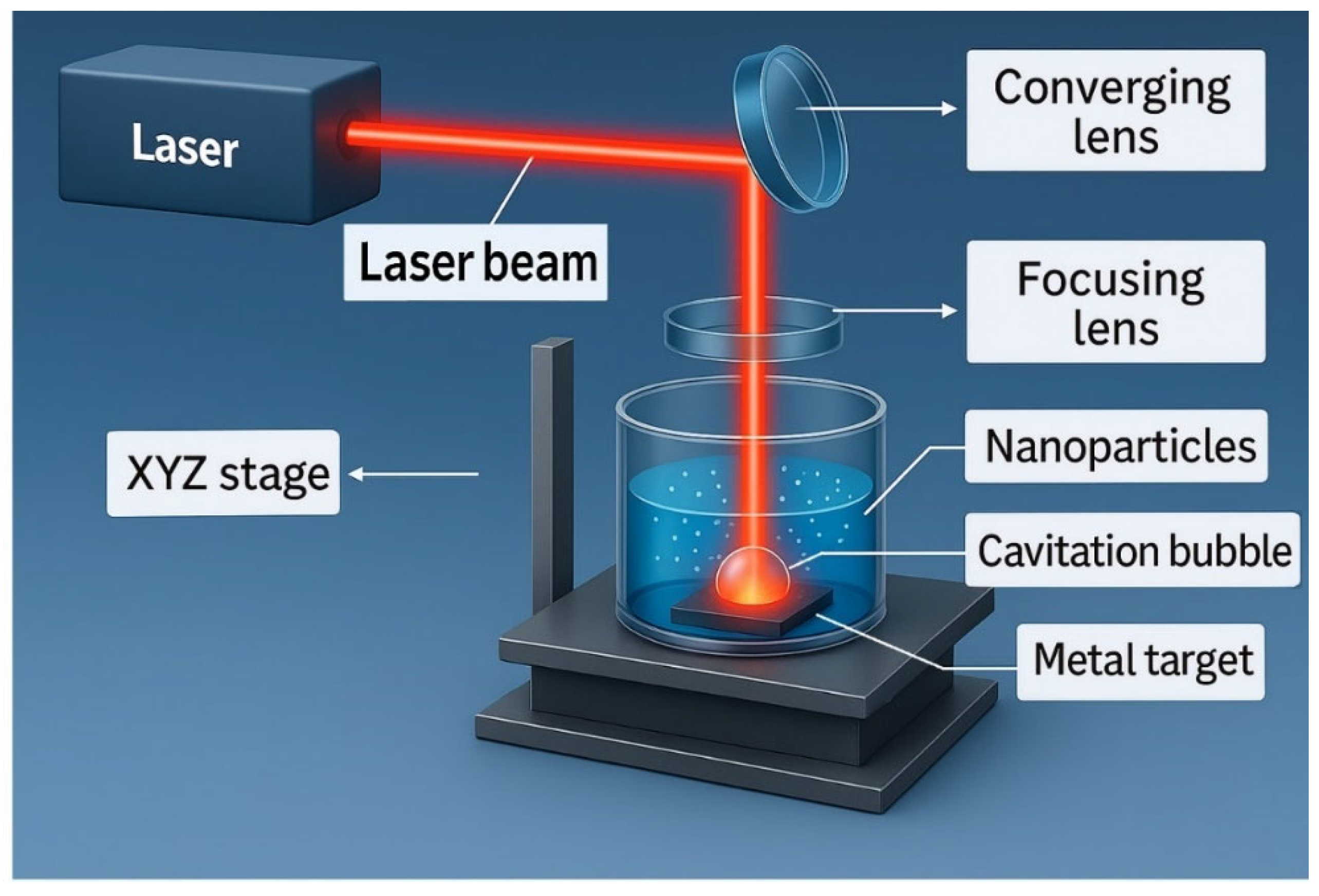
Figure 1. Schematic representation of a typical Pulsed Laser Ablation in Liquid (PLAL) setup used for nanoparticle synthesis.
Qubs 09 00032 g001
Figure 2. Annual number of publications related to pulsed laser ablation in liquid (PLAL) from 1990 to 2024. The data were obtained from the Web of Science database by searching for the phrase “pulsed laser ablation”.
Qubs 09 00032 g002
Figure 3. Distribution of publications related to pulsed laser ablation in liquid (PLAL) across various scientific disciplines, based on data retrieved from the Web of Science database. The search was conducted using the term “pulsed laser ablation” in the title or abstract fields.
Qubs 09 00032 g003
Figure 4. Schematic overview of the review.
Qubs 09 00032 g004
Figure 5. The major applications of PLAL.
Qubs 09 00032 g005
Figure 6. Ablation mass as a function of laser fluence during PLAL [130].
Qubs 09 00032 g006
Figure 8. The ultrafast time-resolved sequence of physical events occurring during pulsed laser ablation in liquid (PLAL), specifically in water, divided into seven domains across timescales from femtoseconds to microseconds [126].
Qubs 09 00032 g008
Figure 9. Characterization techniques of NP synthesis by PLAL.
Qubs 09 00032 g009
Table 4. Electronics and photonics applications of PLAL.
Material (Nanomaterial)Laser TypeWavelengthPulseFluence/EnergyRep. RatePulse TimeMediumSize RangeApplicationReference
Mg–C–Graphene nanoparticlesNd:YAG (WEDGE HF 1064)1064 nm0.2–0.9 ns1–2 J/cm210–20 kHz85 minIPA/IPA–HCl/IPA–NaOH60–300 nm → <90 nmPaper electronics, printed inks[107]
Magnesium (Mg) nanoparticles from powdersNd:YAG (WEDGE HF)1064 nm600 ps1.83–1.91 J/cm210 kHz2, 5, 25 minIPA53–239 nmFlexible electronics, conductive inks[108]
Carbon nanoparticles (CNPs)Nd:YAG (WEDGE HF)1064 nm600 ps1.63–1.91 J/cm210 kHz8 minWater/Ethanol/Mg-solution10–1389 nm (by medium)Paper/flexible electronics[109]
LIPSS on Si and W (fs structuring)Femtosecond (FCPA μJewel D-1000-UG3)1045 nm457 fs5.5–77 J/cm2100 kHzscanningWater~100–200 nm periodicityPhotonics: antireflection, colorization[110]
sp-carbon chains (polyynes/cumulenes)Nd:YAG, fs Ti:Sapph, excimer193–1064 nmfs/ns0.3–5 J/cm210 Hz–1 kHz5–180 minWater & organicsCnH2 (n = 6–30)NLO, optoelectronics, sensing[111]
Ag nanoparticles via donut beamsNd:YAG + DOE532 nm7 ns3.2 vs. 1.2 J/cm210 Hz15 minWater20–30 nm (narrower with donut)Plasmonics, SERS[112]
GaN nanostructures (on porous Si)Q-sw Nd:YAG1064 nmns1600 mJ/pulse4 Hz500 pulsesEthanol (drop-cast)Grain ~132 nmUV detectors, LEDs[113]
SnO2 nanoparticles (device)Q-sw Nd:YAG (SHG)532 nm7 ns~12 J/cm210 HzMethanol/3 mM NaCl25–65 nmn-SnO2/p-Si photodetectors[114]
PLAL for photonics/opto/quantum (review)Ti:Sapph, Nd:YAG, fiber, excimer193–1064 nmfs/ps/ns0.01–10 J/cm2Hz–MHzmin–hWater, ethanol, acetone2–200 nmPhotonics, quantum emitters, metamaterials[12]
Ag NPs; Au@Ag core–shell (NLO)Nd:YAG (fund.)1064 nm7 ns50–250 mJ/pulse10 Hz10 minWater/HAuCl49–40 nmOptical limiting/switching[115]
Low-dimensional nanomaterials (review)Nd:YAG, Ti:Sapph, excimer, fiber193–1064 nmfs/ps/ns0.01–10 J/cm2Hz–kHzmin–hWater/ethanol/DMF/PEG/IPAQDs 1–200 nm; 2D; 1DPL devices, UV PDs, bioimaging[116]
Phosphor micronization (KSrPO4:Eu, KBaPO4:Eu)Nd:YAG/Nd:YVO4 harmonics532/355/266 nm4–10 ns~1–8 J/cm2 (est.)10–20 Hz15–30 minWater~2.0 µm → ~1.0 µmW-LED phosphors[117]
Au:MgO on porous Si (photodetector)Nd:YAG1064 nmns600–1000 mJ/pulse10 Hz100 pulsesCTAB aq.; Au → Mg colloid—(size ↑ with energy)UV–Vis–NIR photodetection[118]
Al2O3 nanoparticles (device)Q-sw Nd:YAG532 nm10 ns400–1000 mJ/pulse1 HzDI water (3 mL)30–100 nm; XRD ~53 nmPhotodetectors on porous Si[119]
PLM crystalline spheres (review)Nd:YAG, Ti:Sapph, excimer193–1064 nmfs/ps/ns0.1–10 J/cm2Hz–kHzmin–hWater/ethanol/organics10–200 nm (uniform spheres)Optical materials, photonics (primary), catalysis, biomedicine[120]
Table 7. Summary of laser- and discharge-based nanoparticle synthesis methods in liquids, emphasizing energy sources, material states, mechanisms, particle types, and the influence of the liquid medium.
MethodLaser/Energy SourceMaterial StateMechanismParticle TypeLiquid Role
PLAL (Pulsed Laser Ablation in Liquid)Pulsed LaserSolid TargetAblation → Plasma → Bubble CollapseFreshly generated nanoparticlesMedium for plasma formation, cooling, and confinement
LFL (Laser Fragmentation in Liquid)Pulsed/Continuous LaserColloidal SuspensionLaser-induced photofragmentationSmaller or uniform nanoparticlesEnergy transfer and cooling
LML (Laser Melting in Liquid)Pulsed/Continuous LaserColloidal SuspensionParticle melting → ReshapingSpherical nanoparticlesShape control and thermal sink
EDM (Electrical Discharge Machining in Liquid)Electrical DischargeSolid ElectrodesMicro-explosions, vaporization, quenchingMetal or metal oxide nanoparticlesDielectric medium enables discharge and cooling
Table 8. Summary of synthesis–defect–property–application relationships across pulsed liquid–based nanoparticle synthesis methods.
TechniqueKey ParametersTypical Defects GeneratedProperties AffectedRepresentative Applications
PLAL (Pulsed Laser Ablation in Liquid)Pulse width, fluence, repetition rate, wavelength, liquid chemistryOxygen vacancies, surface terminations, lattice strain, dislocationsBandgap narrowing, enhanced catalytic site density, increased photothermal conversionPhotocatalysis (H2 evolution, CO2 reduction), photothermal therapy, sensors
PLFL (Pulsed Laser Fragmentation in Liquid)High fluence, short pulses, multipulse irradiationVacancies (O, S, N), high surface disorder, amorphous shellsOptical emission tuning, increased carrier recombination/trap density, conductivity modulationQuantum dots for LEDs, bioimaging, defect-rich catalysts for OER/HER
PLML (Pulsed Laser Melting in Liquid)Nanosecond–millisecond pulses, lower fluence, controlled heatingGrain boundaries, twin defects, recrystallization-induced strainParticle size/morphology uniformity, magnetic domain behaviorMagnetic nanomaterials, plasmonic tuning, shape-controlled catalysts
PLPP (Pulsed Laser Post-Processing in Liquid)Secondary irradiation of preformed colloids, wavelength-specific targetingControlled surface vacancies, interface modifications, cation exchangeSurface chemistry tailoring, charge transfer kineticsElectrocatalysis (methanol oxidation, N2 reduction), hybrid composites with tailored interfaces
Disclaimer/Publisher’s Note: The statements, opinions and data contained in all publications are solely those of the individual author(s) and contributor(s) and not of MDPI and/or the editor(s). MDPI and/or the editor(s) disclaim responsibility for any injury to people or property resulting from any ideas, methods, instructions or products referred to in the content.