Abstract
This work is focused on an accurate experimental determination of the thermal 33S(n,)30Si cross-section. This cross-section is a critical parameter for the potential use of 33S as a cooperative target in boron neutron capture therapy or to understand its role in the stellar nucleosynthesis of 36S. At present, there are large discrepancies in this experimental value; therefore, in this work we measured it relative to the 10B(n,)7Li standard cross-section at the high flux reactor of the Institut Laue-Langevin. The experimental setup was based on a double-sided silicon strip detector. Two 33S samples were used. One 10B sample was used as reference. Particular attention was taken to the characterization of the mass thickness of the samples before and after the experiment because of the high volatility of 33S. Such work was already published in a dedicated paper. A cross-check of the 10B sample was carried out with the neutron flux monitor at the n_TOF-CERN facility. The obtained cross-section of (280 ± 33) mb is significantly higher than the existing data.
1. Introduction
Neutron capture is responsible for the generation of most of the elements heavier than iron and specific neutron-rich nuclides below iron. In both, the astrophysical s- and r-processes, the capture of neutrons is usually followed by a radiative decay of the compound nucleus, incrementing the mass by one unit. Thus, neutron captures and emissions lead generally to a monotonous rise of mass and nuclear charge. Only for certain light nuclides is this monotony broken by the neutron-induced emission of protons or alpha particles populating lower-Z products. In particular, (n,) reactions may perturb the regular flow of the s- and r-processes to heavier masses and affect the abundance of specific nuclides [1]. Although astrophysical energies are higher than thermal energies, it has been shown that in neutron-induced charged particle emission reactions, the thermal value used for normalization of the cross-section has a direct impact on the nucleosynthesis calculations [2]. Therefore, a precise and accurate value of the thermal cross-section is required for many isotopes.
This is the case of the 33S(n,)30Si reaction which influences the nucleosynthesis of the neutron-rich isotope 36S. To study such influence, the data from two experimental 33S(n,)30Si cross-sections in the keV region were used [3,4]. Such experiments provided data only above 10 keV, far above the thermal values and without information on the possible 1/v region between thermal and 10 keV. Both sets of data were used in astrophysical models showing a significant overproduction of 36S; therefore, the origin of the 36S in the solar system is still an open question [5].
On the other hand, a few years ago, the 33S(n,)30Si reaction was proposed as a possible cooperative neutron target in boron neutron capture therapy (BNCT) [6]. BNCT is based on the absorption of 10B in the tumour tissue and the subsequent irradiation with low energy neutrons [7]. Such neutrons must have enough energy to penetrate the tissue and to reach the tumour area with thermal energy for maximizing the capture on 10B. Then, the emitted ions (Q = 2.79 MeV) irreversibly damage the cell in which 10B was absorbed. Neutrons with energies of a few keV are considered the best option [7,8]. The 33S(n,)30Si reaction has a high Q-value (3.49 MeV) and important resonances in the keV region. Thus, the presence of 33S in a tumour close to the skin could enhance the dose in BNCT treatments. This effect was studied with Monte Carlo simulations showing an improvement in the dose due to the presence of 33S being interesting for tumours close to the skin [9,10].
Both applications, astrophysics and BNCT, motivated two experiments at the n_TOF facility at CERN [11,12]. In Praena et al. [11] the obtained -strength of the 13.5-keV resonance, crucial for BNCT, was shown to be the double that measured by Wagemans et al. [4], in agreement with the transmission measurement of Coddens et al. [13]. The measured data cover the range from 10 to 300 keV. In Sabaté-Gilarte et al. [12] experimental data from thermal to 10 keV were measured for the first time showing a clear behaviour. However, neither of the previous works provided the thermal value of the cross-section. Therefore, in order to complete the experimental work, it is mandatory to conduct a dedicated measurement of the thermal cross-section in view of the discrepant data of such value.
The status of the 33S(n,)30Si cross-section thermal value can be found in EXFOR database [14], and it is summarized in Table 1.

Table 1.
Experimental values of the 33S(,)30Si cross-section available in the EXFOR database [14].
Seppi [15] obtained a value of 8 mb, which is far below the values of subsequent measurements. Münnich [16] obtained its value relative to the 6Li(n,t)4He cross-section using a radium–beryllium neutron source moderated with 5 cm of paraffin. Benisz et al. [17] provided two values at two different energies: thermal (25.3 meV) obtained with a polonium–beryllium source moderated with 7 cm of paraffin and a slightly higher energy (35 meV) measured with a monochromatic beam at the Ewa nuclear reactor. Asghar and Emsallem [18] conducted measurements at the H22 external thermal neutron beam at the Institut Laue-Langevin (ILL). Their experiment was mainly aimed at obtaining a partial cross-section for the 33S(n,) but also provided a value for the 33S(n,) cross-section. Later, a preliminary value of (115 ± 10) mb was reported by Geerts et al. [19] in a work dedicated to the characterization of 33S samples performed with a technique based on the evaporation of sulphur onto a formvar substrate. The sample was a formvar–sulphur–formvar sandwich. This kind of samples were used in the work of Wagemans et al. in which natural sulphur and enriched (59.15%) 33S samples were used [20]. The experiment was carried out at the neutron guide H22D at the high flux reactor of ILL. The setup was based on a surface barrier detector mounted outside of the neutron beam. The value provided by Wagemans et al. was (110 ± 10) mb [20].
The present work describes the measurement of the 33S(,)30S cross-section with cold neutrons at the Institut Laue-Langevin (ILL, Grenoble). It will be shown that a significantly higher thermal-equivalent value than all previous measurements has been found. In future works, our thermal value will be used to provide for the first time a comprehensive experimental 33S(n,)30Si cross-section ranging from thermal to 300 keV. Then, its role in the 36S nucleosynthesis and BNCT will be studied.
2. Experimental Setup
2.1. The PF1B Neutron Beam at ILL
The experiment was carried out at the cold neutron beam instrument PF1B at the High Flux Reactor of the Institut Laue-Langevin in Grenoble. The ballistic supermirror neutron guide H113 [21] delivers a cold neutron beam with an average energy of about 4 meV with a negligible background of epithermal and fast neutrons and -rays [22]. The thermal neutron equivalent capture flux in the centre of the beam spot was measured by gold foil activation being ≈8.5· with negligible variations along the experiment. The very thin samples provided insurance that 4 and 25.3 meV neutrons experience negligible attenuation in the sample, and the involved cross-sections are in a regime, with no resonances; thus, the value obtained at 4 meV scales to 25.3 meV.
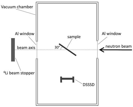
Figure 1 shows a sketch of the experimental. The vacuum chamber was made of stainless steel with entrance and exit windows made of aluminium of m thickness. The inside part of the chamber was carefully covered with borated rubber in order to absorb scattered neutrons. To suppress the background of the emitted -particles and 7Li nuclei, the borated rubber was clad in a double envelope of m aluminium foils. The pressure inside the chamber during the experiment was lower than mbar.

Figure 1.
Sketch of the experimental setup (not in scale). The distance between the centres of the sample and the DSSSD was 20 cm.
The detection of charged particles was carried out with one totally depleted Double-Sided Silicon Strip Detector (DSSSD) model W1 of m thickness outside the neutron beam, as seen in Figure 1. The detector was segmented in 16 × 16 strips with 3.1 mm pitch. The active surface was 50.0 × [23]. The DSSSD was connected to a MPR-64 multichannel preamplifier module (Mesytec). The signals were amplified by two STM-16+ shaping/timing filter amplifiers (Mesytec). The gate of the Caen V785 peak-sensing analogue-to-digital converter for acquiring the analogue signals, was produced by creating a trigger in the amplifier from the front side signals. A coincidence logic between the front and back side of the detector was performed off line via software. MIDAS (Multi Instance Data Acquisition System) was used as acquisition software registering list mode data [24].
2.2. The Samples
Radiative capture cross-sections can usually be accurately determined with samples of various geometric shapes or chemical composition and the use of mixed targets with well-known stoichiometry provides an internal standard that enables very accurate measurements. However, in case of charged particle emission the sample quality is of prime importance. The sample has to be thin enough that the charged particles can reach the detector with limited energy loss. Sulphur sublimates in vacuum at room temperature, adheres poorly or only for a short time to most solid backings, and is very volatile [25,26]. Therefore, one of the main concerns related to an experiment with sulphur is the sample coating and its characterization.
Three works claimed to be successful in coating 33,natS samples for different experiments avoiding mass losses in vacuum [19,25,27]. The used techniques provided unsuitable samples for our experiments at n_TOF and ILL for different reasons, including, among others, the following: very small samples [25]; sealed samples [19,20] which avoids a precise characterization of the mass along the sample or large mass inhomogeneties [27]. Thus, we developed a new method for coating 33S samples to be used in our experiments. Our method is based on the thermal evaporation of 33S powder (enriched 99.9%) onto a substrate made of kapton covered with thin layers of copper, chromium, and titanium. This method provided for the first time bare 33S samples in large dimensions, eight centimetres in diameter [28].
In addition, the characterization of the mass thickness is a crucial point; it is preferable if it can be carried out before and after the experiment to ensure the stability of the 33S samples. Thus, a non-destructive technique is desirable. Our samples were characterized by Rutherford backscattering spectrometry (RBS) before and after the experiment at ILL. The RBS allowed an accurate characterization of the mass thickness of the samples along the diameter. The samples showed an excellent adherence with no mass loss after few years and no sublimation in vacuum or air at room temperature. The details about the 33S samples and their characterization can be found in [28].
In the present work, the 33S(,)30S cross-section was obtained relative to the 10B(n,)7Li standard. One 10B sample was used in the present experiment as a reference. The 10B sample consisted of 10C (enriched 80%) deposited onto Mylar by the dc magnetron sputtering technique [29]. It was characterized with scanning electron microscopy (SEM). In order to have a further characterization and a cross-check of the mass of the 10B sample, it was also used during the commissioning of the experimental area 2 (EAR2) of the n_TOF-CERN facility for comparison purposes to the neutron flux monitor [30]. At n_TOF, the 10B sample was used as neutron converter in an on-beam gaseous detector, based on the MicroMegas microbulk technology, together with the reference 235U sample. Contemporaneously, this setup ran with the silicon monitor of the n_TOF EAR2 based on a a 6LiF converter and four micron semiconductors (MSX09-300) as off-beam detectors. This provides an additional feasibility on the performance and characteristics of the 10B sample. The 10B and 33S samples were larger than the neutron beam, and they were tilted at the angle shown in Figure 1. Table 2 shows the mass thickness of the samples [28].

Table 2.
Number of 33S and 10B atoms per in the samples used in the present experiment [28].
3. Data Analysis
The thermal 33S(n,)30Si cross-section, , has been determined relative to 10B(n,)7Li, , a neutron standard cross-section in the energy range from thermal to 1 MeV [31]. In the thin target approximation the cross-section can be obtained from the formula:
where is the number of counts due to -particles in the case of 33S, and is the number of counts of -particles and 7Li nuclei in the case of 10B. The factor 2 takes into account that the signals from 30Si are in the range of the noise of the detector due to its low energy. and is the number of atoms per of 10B and 33S, respectively. and is the neutron flux during the radiation of 10B and 33S samples, respectively. These fluxes were constant during the irradiation of each sample due to the characteristics of the ILL nuclear reactor. Due to the substrate of 10B and 33S samples being different, the neutron scattering in those materials could lead to a small loss of neutrons onto the samples. Therefore, Monte Carlo simulations with MCNPX [32] were carried out to take into account the possible neutron scattering. They consisted of using MCNPX to record the neutrons that do not pass the backside of the sample to the beam, where 10B and 33S are located, and were scattered without reaching them. It was found that the neutron loss onto 10B and 33S was negligible. and is an area () that corresponds to the interception between the neutron beam and the 10B and 33S samples, respectively. As the beam spot was smaller than the samples, these factors were equal. However, the beam interception was measured by means of the irradiation of Gafchromic radiochromic films before each irradiation to confirm the reproducibility of the setup and the location of the samples. The factors and are the efficiencies and are discussed below. The time of each irradiation corresponds to t. Westcott factors are equal to one because of the 1/v behaviour of both cross-sections [4].
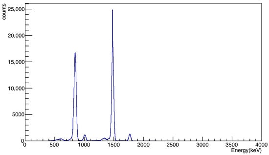
The detector was calibrated using the -particles from 6LiF and 241Am samples. Also, the signals from the 10B sample were taken into account in such channel-energy calibration. Figure 2 shows the pulse height obtained in one run with the Boron sample. The -particles and the 7Li nuclei due to the two reactions, with and without gamma emission, on 10B are clearly separated. Thus, was obtained by the integration of the peaks.

Figure 2.
Pulse height distribution of the amplitude of the signals detected in DSSSD with the Boron sample; 10B(n,)7Li reactions, with and without -ray emission, are clearly separated.
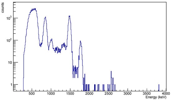
Based on previous works, a Boron contamination is expected in whatever sample is stored at the PF1B-ILL line. Therefore, before the irradiation of the 33S samples, a blank sample was irradiated. A blank sample consisted of the substrate where the 33S powder was evaporated. As mentioned in Section 2.2, the coating of the 33S samples is a key point of our experiments because the high volatility of sulphur. If sulphur would be lost during the experiments, the value of the cross-section would be unreliable. After several studies, we found that an adequate substrate consisted of m kapton foil with 25 nm Cr, a 10 nm Ti adhesion layer, and a 200 nm Cu layer. The mass thickness of each element of the sample was determined by the RBS technique before and after the present experiment at ILL confirming that no loss of 33S occurred; see all details on coating and RBS in [28]. Figure 3 shows a pulse height obtained in one run with the blank sample.

Figure 3.
Pulse height distribution of the amplitude of the signals detected in DSSSD with the blank sample which corresponds to the substrate for 33S. This histogram was acquired in two hours.
The peak around 500 keV mainly corresponds to the emitted protons by the 14N(n,p) reaction since nitrogen is a component of the kapton substrate. These signals were already detected in the experiments at the n_TOF-CERN facility [11,12]. However, signals corresponding to the same energy of the Boron sample were clearly detected. Such signals were not found in the n_TOF-CERN experiments, previously performed to the present one; therefore, a contamination of Boron was found. This contamination was located in all the samples stored at ILL. Concerning a possible contamination of Boron in the 10B sample, first, the 10B sample was not stored at ILL, it was delivered from CERN to ILL for the experiment. Secondly, it must be stressed that the data in Figure 2 were acquired in a few minutes, and the peak of the histogram of the -particle of lower energy contains more than 25,000 counts; meanwhile, the data in Figure 3 were acquired in about two hours, and the peak of histogram of the same -particle contains only 1000 counts. Therefore, if a contamination occurred it would be negligible. Signals above 2000 keV are associated to noise or possible reactions in the substrate with an extremely low cross-sections.
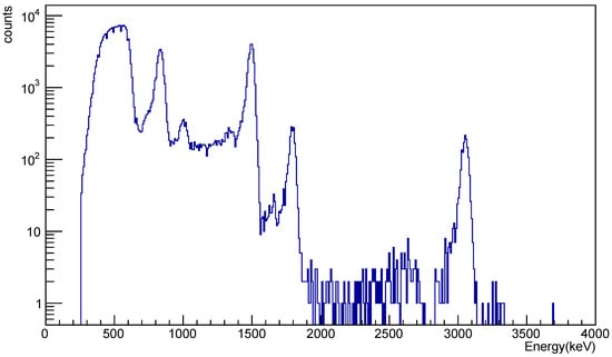
Figure 4 shows an histogram in one run with a 33S sample. The signals due to protons from 14N(n,p) reaction and the Boron contamination are clearly detected. Then, a clear peak, not present in the blank sample, appears at 3050 keV, which corresponds to the -particles of the 33S(n,)30Si reaction. This excellent separation of the -signals ensures the quality of the results. The same energy distribution was found for the second 33S sample. The energy range between 2000 and 3050 keV shows some signals that could be associated to reactions with very low cross-section as 33S(n,) studied in [18]. However, a dedicated experiment should be performed for studying such a reaction. Dead-time and pile-up effects were negligible in all the data with the different samples. Thus, the factor was obtained by the integration of the peak around 3050 keV.

Figure 4.
Pulse height distribution of the amplitude of the signals detected in DSSSD with one of the 33S samples. The signal due to the 33S(n,)30Si reaction are clearly visible at 3050 keV.
The efficiency factors, and , depend on the geometry and possible absorption or scattering of -particles and 7Li nuclei. The setup geometry and dimensions of the samples were the same as mentioned before. In order to determine possible absorption in the samples or scattering out of particles to the detector, simulations were performed. SRIM simulations [33] showed a negligible absorption due to high energy of the ions and the very thin samples used in the experiment. Regarding the scattering out of ions, MCNPX simulations were carried out considering the whole setup and isotropic emission. The simulations showed a negligible difference between 10B and 33S. Therefore, and cancelled out in formula (1).
Concerning the uncertainty, the major source was the mass thickness of the 33S samples (8–9%) as determined by RBS; see details in [28]. The uncertainty of the factors and was below 2%. The tabulated uncertainty for normalization to the standard 10B(n,)7Li reaction is lower than 1% [31]. The statistical uncertainty of the simulations was negligible. Therefore, an overall contribution of 12% was considered.
4. Results and Discussion
In the analysis of the 33S(n,)30Si thermal cross-section we considered three areas of the DSSSD detector by selecting a few strips located in the vertical direction to the neutron beam. The purpose was to obtain different results of the cross-section considering different geometries between the samples and the selected three areas of the detector. This will provide a further reliability on the geometry of the setup, the analysis, and the results. The selected areas were as follows: on the left part of the detector in Figure 1, strip 0; on central part of the detector, strips 3 to 12; on the right part of the detector in Figure 1, strips 14 and 15. Other combinations of strips provided the same results. Table 3 shows the results of the thermal cross-section with the two 33S samples for the different areas of the DSSSD detector in comparison with the other experimental values obtained at thermal energy. We provide an average value of (280 ± 33) mb.

Table 3.
33S(n,)30Si thermal cross-section in millibarns for each considered area of the DSSSD detector. The value of the present work is the average of the six values.
The comparison of our value to the previous ones shows significant discrepancies. It is significantly higher than all the previous results, and it barely overlaps the value of Münnich [16], (180 ± 80) mb, which is within the range of uncertainties.
In order to justify our higher value, we could only speculate on others’ experiments. Our works on the 33S(n,)30Si reaction at n_TOF-CERN [11] and ILL (present work) have a parallelism to the works of Wagemans et al. at GELINA [4] and ILL [20]. In our case, we used the same samples in both facilities with a radial characterization of the mass before and after each experiment [28]. Wagemans et al. used, in both facilities, samples performed with the technique described by Geerts et al. [19]. This technique solved the problems of sublimation of the sulphur in their previous works. It was based on a formvar–sulphur–formvar sandwich with a previous evaporation of enriched 33S (59.15%) onto one of the formvar foils. The characterization of the samples (60 × ) was performed with two methods: -energy loss measurements in the sandwich and destructive chemical analysis. Neither of the methods were sensible to inhomogeneities or variation of the mass along the sample. In particular, the -absorption method can only be used for thickness determinations on homogeneous foils or layers, which puts a limitation on its applicability [34]. In addition, the -energy loss measurement method is based on the knowledge of all the processes of -particles in formvar and sulphur, and in the case of Wagemans et al., the two formvar foils. Deviations up to 80% between the experimental and calculated energy loss straggling of 2.7 MeV -particles in formvar foils have been observed [35]. Such formvar foils have similar thicknesses () to those used by Geerts et al. and Wagemans et al. in their sandwiches, ≈ and . Also, the energy of the -particles was very close to the 3.183 MeV [34] used in the characterization of the samples of Wagemans et al. This should add, at least, an important source of uncertainty in the mass thickness measurements of the samples of Wagemans et al.
5. Summary
The thermal 33S(n,)30Si cross-section has been experimentally obtained using the 10B(n,)7Li standard cross-section as reference. The feasibility of the 10B sample was ensured because it was used at the n_TOF facility in comparison with the neutron flux monitor. The setup at PF1B at ILL and the analysis provide clean and separated signals of the involved reactions. The mass thickness of the 33S was determined before and after the experiments with an accurate characterization of the mass along the diameter of the samples. The details of such characterization can be found in a dedicated paper [28]. We provide an average value of (280 ± 33) mb with two 33S samples and three geometries. The value is significantly higher than the existing data and evaluations. Some of possibilities for the origin of such a discrepancy have been discussed.
The combination of the thermal result of the present work with our previous works in the 1/v and resonance regions will allow for a complete cross-section from thermal to 300 keV for the first time. These data will drive new studies on the origin of 36S in the solar system and on BNCT with 33S as cooperative target.
Author Contributions
Conceptualization, J.P. and M.S.-G.; Data curation, J.P., B.F. and M.M.; Formal analysis, J.P.; Funding acquisition, J.P., I.P. and F.A.d.S.; Investigation, B.F., M.M., I.P., H.K., M.S.-G. and F.A.d.S.; Methodology, J.P.; Resources, I.P.; Software, B.F. and M.M.; Writing—original draft, J.P.; Writing—review and editing, B.F., M.M., M.P.-R., H.K., M.S.-G. and F.A.d.S. All authors have read and agreed to the published version of this manuscript.
Funding
This work was carried out within the framework of Project No. PID2020.117969RB. i00 funded by MICIU/AEI/10.13039/5011000110 33, RTI2018-098117-B-C21 (AEI-MICIN), Spanish Association Against Cancer (AECC) (Grant PS16163811PORR), Junta de Andalucía P11-FQM-8229 and the sponsors of the University of Granada Chair Neutrons for Medicine (Fundación ACS, Capitán Antonio and La Kuadrilla) and the funding agencies of the participating institutes.
Data Availability Statement
The original contributions presented in this study are included in the article. Further inquiries can be directed to the corresponding author.
Acknowledgments
The authors are grateful to Ulli Köester for his help during the experiment.
Conflicts of Interest
Author Hanna Koivunoro was employed by the company Neutron Therapeutics, LLC. The remaining authors declare that the research was conducted in the absence of any commercial or financial relationships that could be construed as a potential conflict of interest.
Correction Statement
This article has been republished with a minor correction to the Data Availability Statement. This change does not affect the scientific content of the article.
References
- Howard, W.M.; Arnett, W.D.; Clayton, D.D.; Woosley, E.S. Nucleosynthesis of rare nuclei from seed nuclei in explosive carbon burning. Astrophys. J. 1972, 175, 201. Available online: https://adsabs.harvard.edu/pdf/1972ApJ...175..201H (accessed on 10 August 2025). [CrossRef]
- Druyts, S.; Wagemans, C.; Geltenbort, P. Determination of the 35Cl(n,p)35S reaction cross section and its astrophysical implications. Nucl. Phys. A 1994, 537, 291–305. [Google Scholar] [CrossRef]
- Auchampaugh, G.F.; Halperin, J.; Macklin, R.L.; Howard, W.M. Kilovolt S33(n,α0) and S33(n,γ) cross sections: Importance in the nucleosynthesis of the rare nucleus 36S. Phys. Rev. C 1975, 12, 1126–1133. [Google Scholar] [CrossRef]
- Wagemans, C.; Weigmann, H.; Barthelemy, R. Measurement and resonance analysis of the 33S(n,α) cross section. Nucl. Phys. A 1987, 469, 497–506. [Google Scholar] [CrossRef]
- Reifarth, R.; Schwarz, K.; Käppeler, F. The Stellar Neutron-Capture Rate of 34S: The Origin of 36S Challenged. Astrophys. J. 2000, 528, 573–581. [Google Scholar] [CrossRef]
- Porras, I. Enhancement of neutron radiation dose by the addition of sulphur-33 atoms. Phys. Med. Biol. 2008, 53, 1–9. [Google Scholar] [CrossRef] [PubMed]
- Barth, R.F.; Vicente, M.G.H.; Harling, O.K.; Kiger, W.S., III; Riley, K.J.; Binns, P.J.; Wagner, F.M.; Suzuki, M.; Aihara, T.; Kato, I.; et al. Current status of boron neutron capture therapy of high grade gliomas and recurrent head and neck cancer. Radiat. Oncol. 2012, 7, 146–166. [Google Scholar] [CrossRef]
- IAEA TECDOC, IAEA. Current Status of Neutron Capture Therapy; International Atomic Energy Agency Vienna International Centre: Bethesda, MD, USA, 2001; Available online: https://www.iaea.org/publications/6168/current-status-of-neutron-capture-therapy (accessed on 10 August 2025).
- Porras, I. Sulfur-33 nanoparticles: A Monte Carlo study of their potential as neutron capturers for enhancing boron neutron capture therapy of cancer. Appl. Radiat. Isot. 2011, 69, 1838–1841. [Google Scholar] [CrossRef] [PubMed]
- Praena, J.; Sabaté-Gilarte, M.; Porras, I.; Esquinas, P.L.; Quesada, J.M.; Mastinu, P. 33S as a cooperative capturer for BNCT. Appl. Radiat. Isot. 2014, 88, 203–205. [Google Scholar] [CrossRef] [PubMed]
- Praena, J.; Sabaté-Gilarte, M.; Porras, I.; Quesada, J.M.; Altstadt, S.; Andrzejewski, J.; Audouin, L.; Bécares, V.; Barbagallo, M.; Bečvář, F.; et al. Measurement and resonance analysis of the 33S(n,α)30Si cross section at the CERN n_TOF facility in the energy region from 10 to 300 keV. Phys. Rev. C 2018, 97, 064603. [Google Scholar] [CrossRef]
- Sabaté-Gilarte, M.; Praena, J.; Porras, I.; Quesada, J.M.; The n_TOF Collaboration. Dose effect of the 33S(n,α) 30SI reaction in BNCT using the new n_TOF-CERN data. Radiat. Prot. Dosim. 2018, 180, 342–345. [Google Scholar] [CrossRef]
- Coddens, G.P.; Salah, M.; Harvey, J.A.; Larson, H. Resonance structure of 33S+n from transmission measurements. Nucl. Phys. A 1987, 469, 480–496. [Google Scholar] [CrossRef]
- Otuka, N.; Dupont, E.; Semkova, V.; Pritychenko, B.; Blokhin, A.I.; Aikawa, M.; Babykina, S.; Bossant, M.; Chen, G.; Dunaeva, S.; et al. Towards a More Complete and Accurate Experimental Nuclear Reaction Data Library (EXFOR): International Collaboration Between Nuclear Reaction Data Centres (NRDC). Nucl. Data Sheets 2014, 120, 272. [Google Scholar] [CrossRef]
- Seppi, E.J. No Title, Private Communication. Hanford Atomic Products, Richland, WA, USA, 1956. Available online: https://www-nds.iaea.org/EXFOR/11422.004 (accessed on 10 August 2025).
- Münnich, F. Untersuchung der Energietönung und des Wirkungsquerschnittes einiger durch thermische Neutronen ausgelöster (n,α)-Prozesse. J. Z. Phys. 1958, 153, 106–123. Available online: https://link.springer.com/article/10.1007/BF01342880 (accessed on 10 August 2025). [CrossRef]
- Benisz, J.; Jasielska, A.; Panek, T. Q-values and cross-section measurement of the (n,α) reaction of some nuclei. J. Acta Phys. Pol. 1965, 28, 763. Available online: https://www-nds.iaea.org/EXFOR/31032.003 (accessed on 10 August 2025).
- Asghar, M.; Emsallem, A. Conf: 3 Symp. Neutr. Capt. Gamma Ray Spectr. Brookhaven 1978, 549. Available online: https://www-nds.iaea.org/EXFOR/21302.008 (accessed on 10 August 2025).
- Geerts, K.; van Gestel, J.; Pauwels, J. Thin 33S layers for 33S(n,α) cross-section measurements. Nucl. Inst. Met. Phys. Res. A 1985, 236, 527–529. [Google Scholar] [CrossRef]
- Wagemans, C.; D’hondt, P.; Brissot, R. Determination of the 32S(n,α)29Si and 33S(n,α)30Si reaction cross-sections. In Proceedings of the 2nd International Symposium on Nuclear Astrophysics, Karlsruhe, Germany, 6–10 July 1992; Available online: https://www-nds.iaea.org/exfor/servlet/X4sGetSubent?reqx=76751&subID=23735003 (accessed on 10 August 2025).
- Abele, H.; Dubbers, D.; Häse, H.; Klein, M.; Knöpfler, A.; Kreuz, M.; Lauer, T.; Märkisch, B.; Mund, D.; Nesvizhevsky, V. Characterization of a ballistic supermirror neutron guide. Nucl. Inst. Met. Phys. Res. A 2006, 562, 407–417. [Google Scholar] [CrossRef]
- Jentschel, M.; Blanc, A.; de France, G.; EXILL-Core Collaboration. EXILL—A high-efficiency, high-resolution setup for γ-spectroscopy at an intense cold neutron beam facility. JINST 2017, 12, P11003. [Google Scholar] [CrossRef]
- Micron Semiconductor Ltd. Available online: http://www.micronsemiconductor.co.uk/ (accessed on 10 August 2025).
- Available online: https://daq00.triumf.ca/MidasWiki (accessed on 10 August 2025).
- Watson, D.D. Simple method for making sulfur targets. Rev. Sci. Instr. 1966, 37, 1605. [Google Scholar] [CrossRef]
- Hedemann, M.A. Preparation of isotopic sulfur targets. Nucl. Inst. Met. Phys. Res. A 1977, 141, 377–379. [Google Scholar] [CrossRef]
- Schatz, H.; Jaag, S.; Linker, G.; Steininger, R.; Käppeler, F.; Koehler, P.E.; Graff, S.M.; Wiescher, M. Stellar cross sections for 33S(n,α)30Si, 36Cl(n,p)36S, and 36Cl(n,α)33P and the origin of 36S. Phys. Rev. C 1995, 51, 379. [Google Scholar] [CrossRef]
- Praena, J.; Ferrer, F.J.; Vollenberg, W.; Sabaté-Gilarte, M.; Fernández, B.; García-López, J.; Porras, I.; Quesada, J.M.; Altstadt, S.; Andrzejewski, J.; et al. Preparation and characterization of 33S samples for 33S(n,α)30Si cross-section measurements at the n_TOF facility at CERN. Nuclear Instruments and Methods in Physics Research Section A: Accelerators, Spectrometers, Detectors and Associated Equipment. Nucl Inst. Met. Phys. Res. A 2018, 890, 142–147. [Google Scholar] [CrossRef]
- Höglund, C.; Birch, J.; Andersen, K.; Bigault, T.; Buffet, J.; Correa, J.; van Esch, P.; Guerard, B.; Hall-Wilton, R.; Jensen, J.; et al. B4C thin films for neutron detection. J. Appl. Phys. 2012, 111, 104908. [Google Scholar] [CrossRef]
- Sabaté-Gilarte, M.; Barbagallo, M.; Colonna, N.; Gunsing, F.; Žugec, P.; Vlachoudis, V.; Chen, Y.H.; Stamatopoulos, A.; Lerendegui-Marco, J.; Cortés-Giraldo, M.A. High-accuracy determination of the neutron flux in the new experimental area n_TOF-EAR2 at CERN. Eur. Phys. J. A 2017, 53, 210. [Google Scholar] [CrossRef]
- Carlson, A.D.; Pronyaev, V.G.; Capote, R.; Hale, G.M.; Chen, Z.-P.; Duran, I.; Hambsch, F.-J.; Kunieda, S.; Mannhart, W.; Marcinkevicius, B. Evaluation of the Neutron Data Standards. Nucl. Data Sheets 2020, 163, 280–281. [Google Scholar] [CrossRef]
- Pelowitz, D.B. MCNPX USERS MANUAL; Version 2.5.0; LA-CP05-0369; Los Alamos National Laboratory LACP: Los Alamos, NM, USA, 2005.
- Ziegler, J.F.; Ziegler, M.D.; Biersack, J.P. SRIM—The stopping and range of ions in matter (2010). Nuc. Inst. Met. Phys. Res. B 2010, 268, 1818–1823. [Google Scholar] [CrossRef]
- Wagemans, C. On the necessity of alternative methods to determine sample thicknesses and masses. Nucl Inst. Met. Phys. Res. A 1989, 282, 4–9. [Google Scholar] [CrossRef]
- Mammeri, S.; Ammi, H.; Dib, A.; Pineda-Vargas, C.A.; Ourabah, S.; Msimanga, M.; Chekirine, M.; Guesmia, A. Stopping power and energy loss straggling of thin Formvar foil for 0.3–2.7 MeV protons and alpha particles. Radiat. Phys. Chem. 2012, 81, 1862–1866. [Google Scholar] [CrossRef]
Disclaimer/Publisher’s Note: The statements, opinions and data contained in all publications are solely those of the individual author(s) and contributor(s) and not of MDPI and/or the editor(s). MDPI and/or the editor(s) disclaim responsibility for any injury to people or property resulting from any ideas, methods, instructions or products referred to in the content. |
© 2025 by the authors. Licensee MDPI, Basel, Switzerland. This article is an open access article distributed under the terms and conditions of the Creative Commons Attribution (CC BY) license (https://creativecommons.org/licenses/by/4.0/).