
The Use of Low-Profile Angular-Stability Plates in a “Nutcracker” Tarsal Navicular Fracture Combined with a Cuboid Fracture: ORIF Experience


 ,
, 
Abstract
1. Introduction
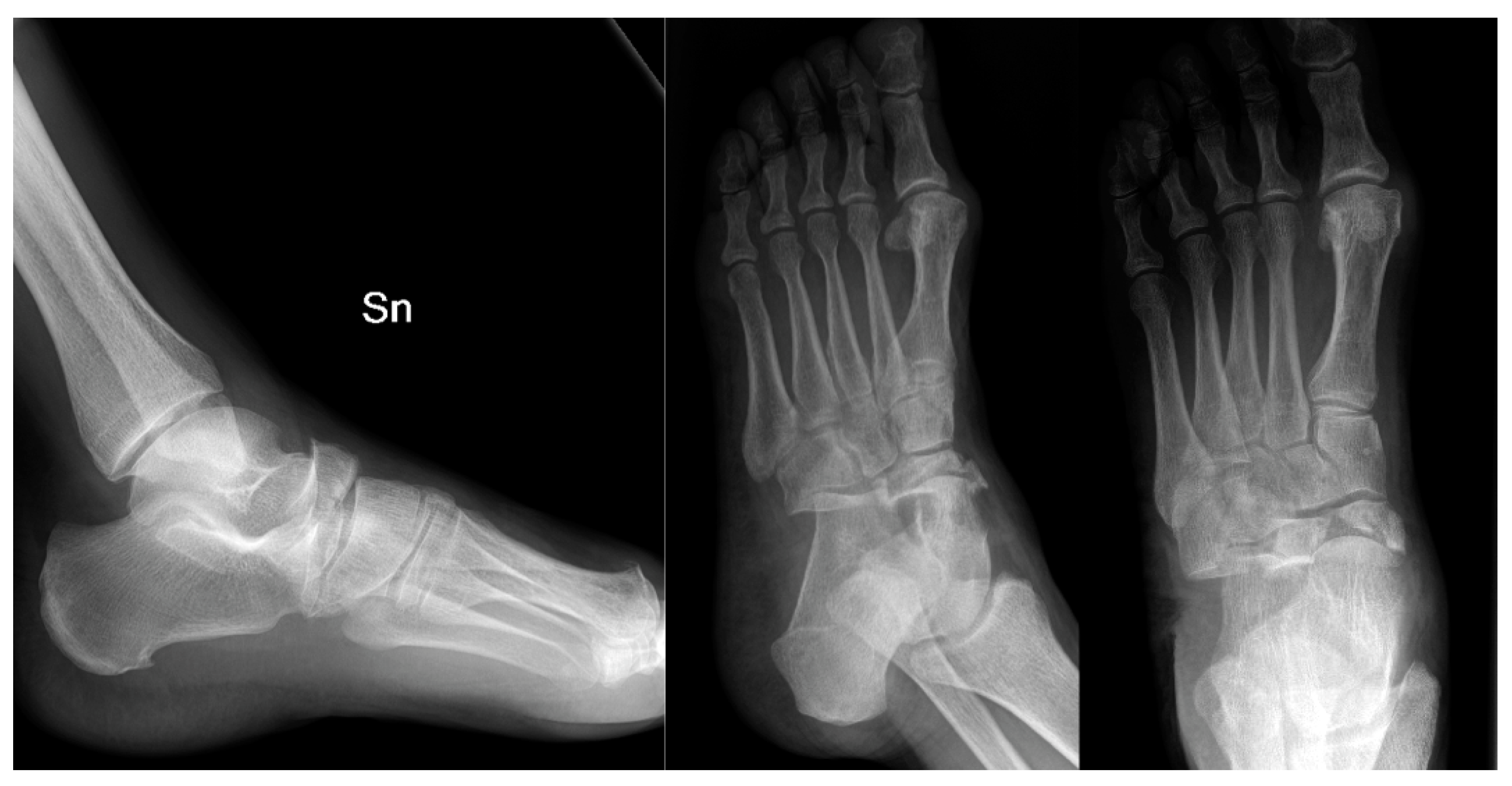
2. Case Presentation
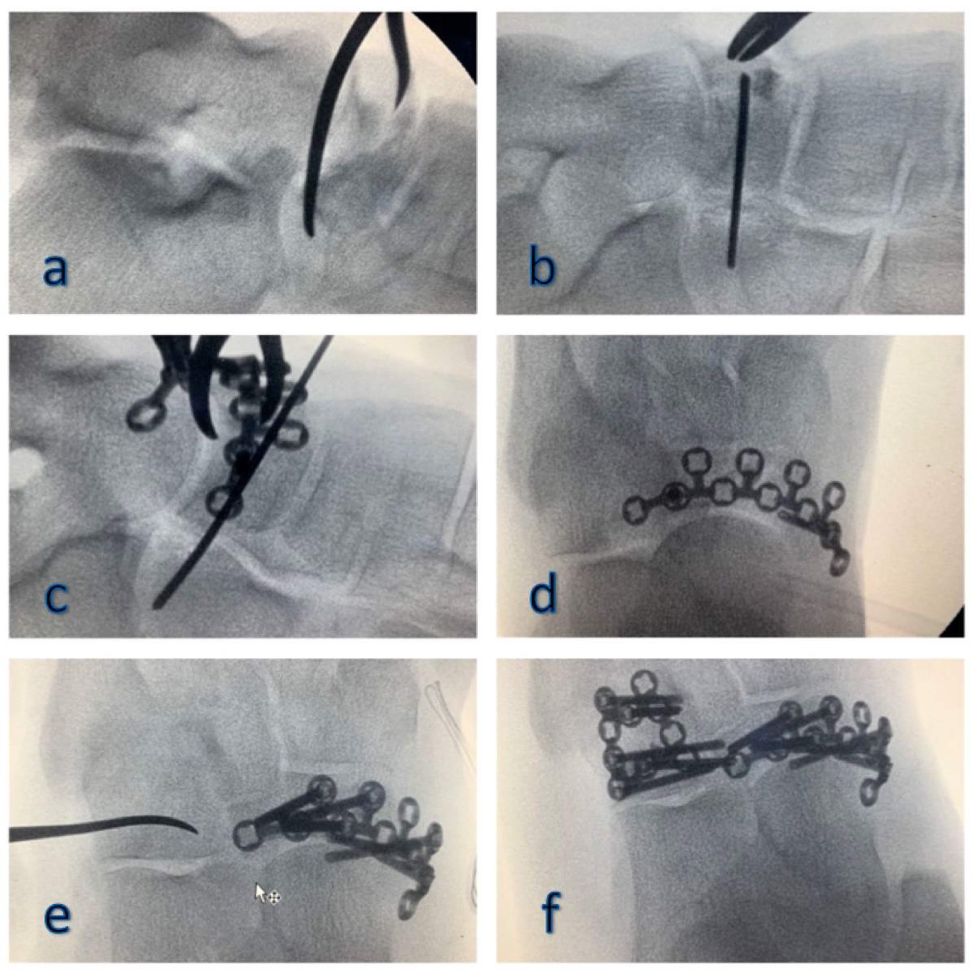
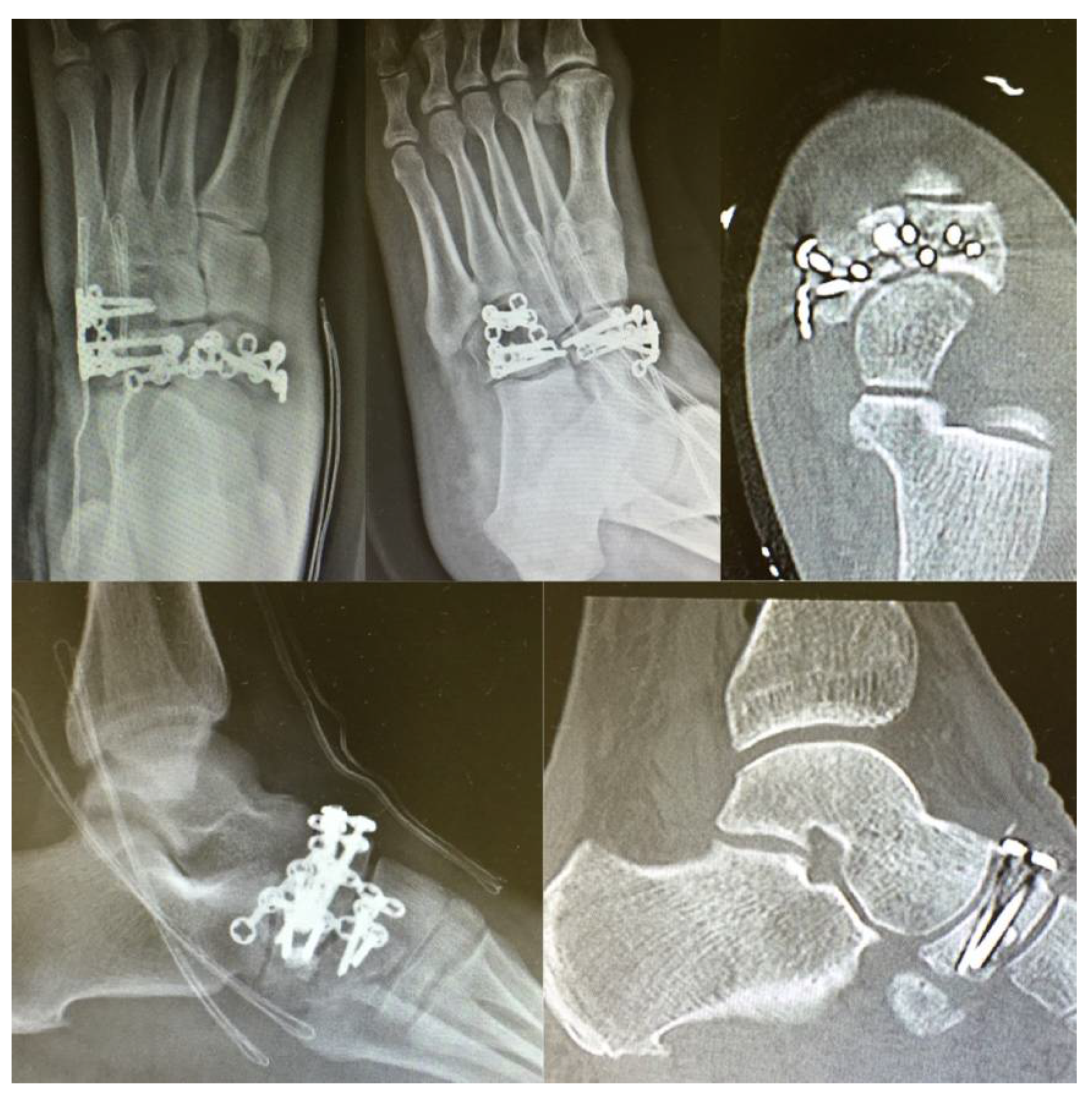
3. Surgical Procedure
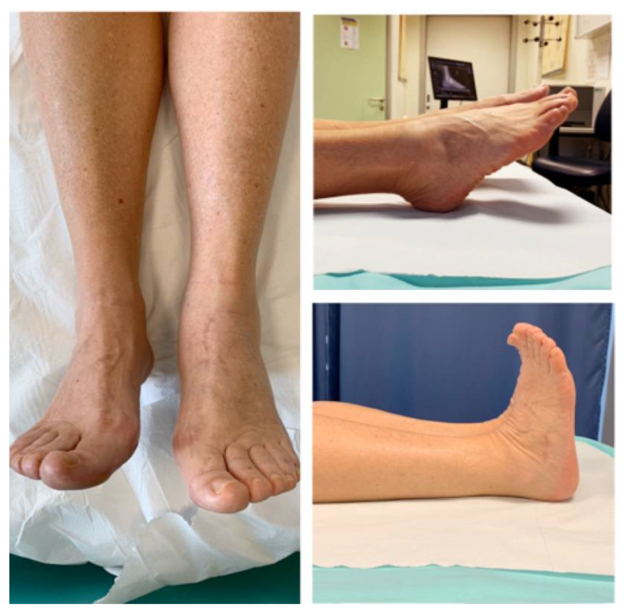
4. Discussion
5. Conclusions
Author Contributions
Funding
Institutional Review Board Statement
Informed Consent Statement
Conflicts of Interest
References
- Ponkilainen, V.T.; Laine, H.J.; Mäenpää, H.M.; Mattila, V.M.; Haapasalo, H.H. Incidence and Characteristics of Midfoot Injuries. Foot Ankle Int. 2019, 40, 105–112. [Google Scholar] [CrossRef] [PubMed]
- Klaue, K. Chopart fractures. Injury 2004, 35 (Suppl. 2), 64–70. [Google Scholar] [CrossRef]
- Richter, M.; Wippermann, B.; Krettek, C.; Schratt, H.E.; Hufner, T.; Therman, H. Fractures and fracture dislocations of the midfoot: Occurrence, causes and long-term results. Foot Ankle Int. 2001, 22, 392–398. [Google Scholar] [CrossRef] [PubMed]
- Eichenholtz, S.N.; Levine, D.B. Fractures of the tarsal navicular bone. Clin. Orthop. Relat. Res. 1964, 34, 142–157. [Google Scholar] [CrossRef]
- Main, B.J.; Jowett, R.L. Injuries of the midtarsal joint. J. Bone Jt. Surg. Br. 1975, 57, 89–97. [Google Scholar] [CrossRef]
- Nyska, M.; Margulies, J.Y.; Barbarawi, M.; Mutchler, W.; Dekel, S.; Segal, D. Fractures of the body of the tarsal navicular bone: Case reports and literature review. J. Trauma 1989, 29, 1448–1451. [Google Scholar] [CrossRef] [PubMed]
- Sangeorzan, B.J.; Benirschke, S.K.; Mosca, V.; Mayo, K.A.; Hansen, S.T., Jr. Displaced intra-articular fractures of the tarsal navicular. J. Bone Jt. Surg. Am. 1989, 71, 1504–1510. [Google Scholar] [CrossRef]
- Schmid, T.; Krause, F.; Gebel, P.; Weber, M. Operative Treatment of Acute Fractures of the Tarsal Navicular Body: Midterm Results With a New Classification. Foot Ankle Int. 2016, 37, 501–507. [Google Scholar] [CrossRef]
- Seringe, R. Le concept de bloc calcanéo-pédieux. Rev. Chir. Orthop. Réparatrice L’appareil Mot. 2007, 93, 28. [Google Scholar] [CrossRef]
- Rosenbaum, A.J.; Uhl, R.L.; DiPreta, J.A. Acute fractures of the tarsal navicular. Orthopedics 2014, 37, 541–546. [Google Scholar] [CrossRef]
- Astion, D.J.; Deland, J.T.; Otis, J.C.; Kenneally, S. Motion of the hindfoot after simulated arthrodesis. J. Bone Jt. Surg. Am. 1997, 79, 241–246. [Google Scholar] [CrossRef] [PubMed]
- Sammarco, V.J. The talonavicular and calcaneocuboid joints: Anatomy, biomechanics, and clinical management of the transverse tarsal joint. Foot Ankle Clin. 2004, 9, 127–145. [Google Scholar] [CrossRef]
- Waugh, W. The ossification and vascularisation of the tarsal navicular and their relation to Köhler’s disease. J. Bone Jt. Surg. Br. 1958, 40, 765–777. [Google Scholar] [CrossRef]
- Prathapamchandra, V.; Ravichandran, P.; Shanmugasundaram, J.; Jayaraman, A.; Salem, R.S. Vascular foramina of navicular bone: A morphometric study. Anat. Cell Biol. 2017, 50, 93–98. [Google Scholar] [CrossRef] [PubMed]
- McKeon, K.E.; McCormick, J.J.; Johnson, J.E.; Klein, S.E. Intraosseous and extraosseous arterial anatomy of the adult navicular. Foot Ankle Int. 2012, 33, 857–861. [Google Scholar] [CrossRef]
- Golano, P.; Fariñas, O.; Sáenz, I. The anatomy of the navicular and periarticular structures. Foot Ankle Clin. 2004, 9, 1–23. [Google Scholar] [CrossRef]
- Sanders, R.; Hansen, S.T., Jr. Progressive talonavicular dissociation. Orthop. Trans. 1989, 13, 572. [Google Scholar]
- Zwipp, H. Chirurgie des Fusses; Springer: Vienna, Austria, 1994. [Google Scholar]
- Sanders, R.; Papp, S. Fractures of the midfoot and forefoot. Surg. Foot Ankle 2007, 8, 2204–2211. [Google Scholar]
- Castro, M.D. Arthrodesis of the navicular. Foot Ankle Clin. 2004, 9, 73–83. [Google Scholar] [CrossRef]
- Schildhauer, T.A.; Coulibaly, M.O.; Hoffmann, M.F. Fractures and dislocations of the midfoot and forefoot. In Rockwood and Green’s Fractures in Adults, 8th ed.; Court-Brown, C., Heckman, J.D., McKee, M., McQueen, M.M., Ricci, W., Tornetta, P., III, Eds.; Wolters Kluwer: Philadelphia, PA, USA, 2015; p. 2689. [Google Scholar]
- Longino, D.; Buckley, R.E. Bone graft in the operative treatment of displaced intraarticular calcaneal fractures: Is it helpful? J. Orthop. Trauma 2001, 15, 280–286. [Google Scholar] [CrossRef] [PubMed]
- Torg, J.S.; Pavlov, H.; Cooley, L.H.; Bryant, M.H.; Arnoczky, S.P.; Bergfeld, J.; Hunter, L.Y. Stress fractures of the tarsal navicular. A retrospective review of twenty-one cases. J. Bone Jt. Surg. Am. 1982, 64, 700–712. [Google Scholar] [CrossRef]
- Van Langelaan, E.J. A kinematical analysis of the tarsal joints. An X-ray photogrammetric study. Acta Orthop. Scand. Suppl. 1983, 204, 1–269. [Google Scholar] [PubMed]
- Cronier, P.; Frin, J.M.; Steiger, V.; Bigorre, N.; Talha, A. Internal fixation of complex fractures of the tarsal navicular with locking plates. A report of 10 cases. Orthop. Traumatol. Surg. Res. 2013, 99 (Suppl. 4), S241–S249. [Google Scholar] [CrossRef]
- DiGiovanni, C.W.; Kuo, R.; Tejwani, N.; Price, R.; Hansen, S.T., Jr.; Cziernecki, J.; Sangeorzan, B.J. Isolated gastrocnemius tightness. J. Bone Jt. Surg. Am. 2002, 84, 962–970. [Google Scholar] [CrossRef]
- Pisanu, F.; Ortu, S.; Corda, M.; Andreozzi, M.; Caggiari, G.; Manunta, A.F.; Doria, C. Deltoid ligament reconstruction with autologous gracilis tendon in chronic medial ankle instability after ankle fracture surgery: A case report. Foot 2020, 6, 101714. [Google Scholar] [CrossRef]
- DiGiovanni, C.W. Fractures of the navicular. Foot Ankle Clin. 2004, 9, 25–63. [Google Scholar] [CrossRef]
- Barouk, P.; Barouk, L.S. Clinical diagnosis of gastrocnemius tightness. Foot Ankle Clin. 2014, 19, 659–667. [Google Scholar] [CrossRef] [PubMed]


Publisher’s Note: MDPI stays neutral with regard to jurisdictional claims in published maps and institutional affiliations. |
© 2021 by the authors. Licensee MDPI, Basel, Switzerland. This article is an open access article distributed under the terms and conditions of the Creative Commons Attribution (CC BY) license (https://creativecommons.org/licenses/by/4.0/).
Share and Cite
Quattrini, F.; Ciatti, C.; Gattoni, S.; Puma Pagliarello, C.; Ceccarelli, F.; Maniscalco, P. The Use of Low-Profile Angular-Stability Plates in a “Nutcracker” Tarsal Navicular Fracture Combined with a Cuboid Fracture: ORIF Experience. J. Funct. Morphol. Kinesiol. 2021, 6, 99. https://doi.org/10.3390/jfmk6040099
Quattrini F, Ciatti C, Gattoni S, Puma Pagliarello C, Ceccarelli F, Maniscalco P. The Use of Low-Profile Angular-Stability Plates in a “Nutcracker” Tarsal Navicular Fracture Combined with a Cuboid Fracture: ORIF Experience. Journal of Functional Morphology and Kinesiology. 2021; 6(4):99. https://doi.org/10.3390/jfmk6040099
Chicago/Turabian StyleQuattrini, Fabrizio, Corrado Ciatti, Serena Gattoni, Calogero Puma Pagliarello, Francesco Ceccarelli, and Pietro Maniscalco. 2021. "The Use of Low-Profile Angular-Stability Plates in a “Nutcracker” Tarsal Navicular Fracture Combined with a Cuboid Fracture: ORIF Experience" Journal of Functional Morphology and Kinesiology 6, no. 4: 99. https://doi.org/10.3390/jfmk6040099
APA StyleQuattrini, F., Ciatti, C., Gattoni, S., Puma Pagliarello, C., Ceccarelli, F., & Maniscalco, P. (2021). The Use of Low-Profile Angular-Stability Plates in a “Nutcracker” Tarsal Navicular Fracture Combined with a Cuboid Fracture: ORIF Experience. Journal of Functional Morphology and Kinesiology, 6(4), 99. https://doi.org/10.3390/jfmk6040099