Review

Sarcopenia and Exercise “The State of the Art”

by
Giuseppe Musumeci
1,2
1
Department of Biomedical and Biotechnological Sciences, Human Anatomy and Histology Section, School of Medicine, University of Catania, Via S. Sofia 87, 95123 Catania, Italy
2
Department of Health, UniPoliSI, Route de Miège, 29 CH-3968 Veyras, Switzerland
J. Funct. Morphol. Kinesiol. 2017, 2(4), 40; https://doi.org/10.3390/jfmk2040040
Submission received: 4 October 2017 / Revised: 25 October 2017 / Accepted: 4 November 2017 / Published: 7 November 2017
(This article belongs to the Special Issue Role of Exercises in Musculoskeletal Disorders)

Abstract

:
Skeletal muscle mass reduction might be a consequence of aging (sarcopenia), disease (cachexia) or inactivity (muscle atrophy). Studying the triggering factors leading to muscle loss is important in developing therapies to preserve muscle tissue function. The loss of skeletal muscle proteins is caused by an imbalance between the rate of their synthesis and degradation. Specifically, the conditions characterized by muscle loss involve an adaptation metabolism of increased protein degradation (cachexia), decreased muscle protein synthesis (inactivity), or alteration in both (sarcopenia). Sarcopenia and exercise is the main topic chosen for this review. This is a huge health problem, poorly discussed in the current literature and the aim of this review is to explain and help readers to better understand the differences between “sarcopenia”, “cachexia”, “muscle atrophy” and the relative beneficial effects of exercise used as a possible therapeutic intervention. Sarcopenia is a component of the fragility syndrome and indicates a significant health issue related to the progressive decline of muscle tissue quality and strength. Exercise is associated with improved life quality, reduced health problems, and prolonged lifespan. The latter suggests that exercise should be considered a fundamental point in the treatment of pathological skeletal muscle mass reduction. The present scientific contribution also seeks to emphasize to the scientific community the positive effects of the adapted physical activity in the elderly as a possible non-pharmacologic treatment to prevent or treat muscle atrophy.

1. Introduction

Aging is a physiological process, characterized by a decline in all physical functions leading to an impaired quality of life. Skeletal muscle mass decline occurring with aging is known as sarcopenia. However, not only the quantity, but the quality of muscle tissue should also be considered as a crucial factor [1]. Aging determines a decline in muscle force due to a progressive increase of catabolism and decrease in anabolism. These physiological events are also due to the reduced muscle regeneration ability. Indeed, the unbalanced turnover of muscle protein and tissue remodeling are associated with impaired muscle cell recruitment and cell death [1,2]. Muscle aging is a multifactorial irreversible process associated with significant decline in muscle mass and functions [3]. One of the most efficient methods to counteract age-related alterations in muscle tissue is represented by physical exercise. An alternative intervention to improve muscle structure and performance is electrical or mechanical stimulation. The present scientific contribution would like to emphasize to the scientific community the positive effects of adapted physical activity in the elderly as a possible non-pharmacologic treatment to prevent or treat muscle atrophy.

2. Sarcopenia

Skeletal muscle mass reduction occurs during several conditions such as: aging “sarcopenia”, disease “cachexia” or inactivity “muscle atrophy”. During skeletal development, the muscle fibers grow in size and number, manifesting greater body size and strength. Muscle fiber number and/or size decrease with age, disuse and illness and this is associated with a corresponding decrease in muscle capacity to generate strength. In the case of muscular atrophy following disuse, the size of muscle fibers can be restored over time through physical activity. Loss of muscle strength and mass following illness or sarcopenia is particularly problematic since the quality of muscle fiber is not so easily recoverable in these cases, producing greater fragility that tends to worsen the pathologic condition in elderly people and those suffering from chronic illnesses.
Sarcopenia is a loss of muscle mass linked to age that is responsible for the decline in muscle strength. It forms the main factor in the pathogenesis of fragility [4]. Older people with sarcopenia exhibit a reduced body mass and muscular strength. There is also accumulation of fat in the muscle, called myosteatosis, that causes a decrease in muscle strength leading to functional dysfunction and physical disability [5]. Unlike this condition, obese people often have a body fat percentage greater than normal people. These people can become fragile if they do not exercise properly and develop disabilities [6]. Muscle tissue is not static. It shows a continuous process of atrophy and hypertrophy. It is a cyclical process of death and renewal. Muscle proteins undergo degradation when developing, leading to atrophy [7]. Cells are also subject to apoptosis [1]. However, there are cell renewals after the integration of amino acids. This causes the synthesis of proteins leading to muscular hypertrophy [7]. There is also stimulation of stem cells that lead to the production of satellite cells capable of repairing damaged muscles [4]. Food intake should be adequate to maintain proper muscle function. Protein and creatine play an important role for muscle disorders [8]. The motor units decrease with the advancement of age. There is a decrease in neutrophilic ciliary factor levels and this is associated with decreased muscle strength [6]. High levels of cytokines, such as tumor necrosis factor (TNF)-α and interleukin (IL)-6, are associated with a reduction in muscle fiber strength [8]. Muscle strength decreases in different disorders (Table 1) such as diabetes mellitus. Muscle renewal is affected by the development of atherosclerosis as it causes a drop in blood supply to the muscle. Older people can become fragile. Frailty refers to a condition in which a person exhibits reduced ability to undertake essential social activities of daily life in less stressful environmental situations. There is a reduction in the reserves in the physiological function of different organs of the body to carry out important daily activities and to maintain adequate homeostasis [9]. In this context, any minor disease or adverse drug effects involve an unbalanced loss of function, an increased risk of disability and an increased risk of death for the effects of a stressor. It should be noted that such a quantity of stress does not cause disturbance in a physically fit person of the same age and sex [10]. Disadvantaged people may develop functional decline and disability following exposure to stressors such as an infection, death, and death of a spouse or the addition of a new drug to the treatment routine. These individuals do not have the resources to respond and maintain proper homeostasis. The same stresses cause small disturbances in a suitable person of the same age. Many different body systems become dislocated on anatomical, molecular and physiological levels when people get older [11]. Some of these systemic changes are more apparent in people who are psychophysically fragile. Studies have linked fragility to increased inflammation and blood clotting activity. There is a decline in humoral and cell-mediated immunity with age advancement [11]. There is also overexpression of cytokines, decreased levels of hormones, loss of muscle mass, muscle strength or sarcopenia [12].
Sarcopenia is a natural consequence of aging. Studies demonstrated that this process may be slowed down, interrupted and even inverted [13]. Although atrophy, cachexia and sarcopenia share a common feature in muscle loss, there are distinct differences in the biochemical processes that promote them. Sarcopenia is characterized by a muscle fiber loss in size and number [14,15,16,17]. In muscle atrophy, the fiber size is reduced but the fiber number is maintained [18] and characterized by a transition trend of type I fiber to type II [19,20]. The tissue loss in cachexia involves both fatty and skeletal muscle tissues [21]. The loss of muscle tissue is mainly directed towards type II fibers [22], while congestive heart failure tends to degrade contractile type I or IIA fibers [23,24]. In each of these conditions (aging, disuse and cachexia), atrophy and weakness are generated, however, it is difficult to separate them due to corresponding signaling systems.
Sarcopenia, as described by the European Working Group on Sarcopenia in 2010, is a progressive and generalized loss of the skeletal muscle mass and function [25]. Evans [26] adds that sarcopenia is age-related and is a different condition than cachexia. Although atrophy is considered the source of the resulting functional loss, some researchers believe that loss of muscle function cannot be fully explained by impressive atrophy [27,28,29]. In any case, the phenomenon of sarcopenia has devastating consequences for older populations because they experience loss of muscle mass and strength to the point of losing their independence.
Even if the term sarcopenia started to be used to define the muscle mass decline occurring with aging, today it is also associated with the severity of muscle atrophy, which is not necessarily linked to aging [30,31]. Indeed, the muscle mass decline due to several pathological conditions such as cirrhosis, cardiovascular and cardiorespiratory diseases, HIV infection, dismetabolic problems, ovariectomy, and cancer, is described by the term sarcopenia in several papers present in literature [32,33,34]. Perhaps the biggest concern for this point regards the consideration of whether muscle atrophy may be the result of processes which are distinct from aging.
Some authors use the histological analysis of aging muscle to differentiate the sarcopenia of aging from other causes of muscle atrophy, even if in these last cases it is described as sarcopenia [35]. It is interesting to note that several morphological features of sarcopenia (Figure 1) are similar to the ones observable in muscle with sporadic denervation, like in amyotrophic lateral sclerosis [36].
The existing evidence strongly implicates sporadic and repeating cycles of denervation-reinnervation in the histopathology of aging muscle, including fiber size heterogeneity, fiber type grouping, and coexpression of myosin heavy chain (MHC). Such alterations differentiate sarcopenia from cancer cachexia and may also be distinct from other clinical conditions where aging is not the cause of muscle atrophy but which are currently using the term sarcopenia. Sarcopenia has been initially defined as the decrease of muscle mass and function during aging. Hepple et al. [35] indicate that this definition has been extended to muscle atrophy conditions such as malnutrition or acute catabolic states (ACS) like sepsis and cancer. Sarcopenia is not the result of pathology and has been reported among well-nourished, healthy, physically active elderly subjects [37]. The slow erosion of muscle mass during aging is partly explained by a lower sensitivity of muscle anabolism to meal intake [38]. Therefore, the approaches used for limiting muscle loss during aging are probably not the same used in ACS due to the slow kinetics involved and differences in related mechanisms. Sarcopenia is a fragility syndrome constituent. It is often a constituent of cachexia as well. It can also exist independently of cachexia; while cachexia includes sickness and is secondary to an underlying disease (like cancer), sarcopenia can occur in healthy people and does not necessarily include sickness. In summary, whereas cachexia or muscle atrophy may be a component of sarcopenia, these conditions are not the same, and for this reason the term “sarcopenia” should be used to indicate the age-related alterations of muscle mass and function.

3. Exercise

Absence of exercise is considered a significant risk factor for sarcopenia [38]. Master class athletes, who continue to compete for their entire adult life show a progressive loss of muscle mass and strength and performances in speed and force events progressively decrease after age 30 [39]. Top class athletes maintain a high level of fitness throughout their lifetime. Even among the master athletes, the performance of marathon runners and weightlifters decreases after about 40 years of age, with peak performance rates of about 50% for 80 years of age [40]. However, a gradual loss of muscle fibers begins at about 50 years of age [40]. Exercise is very important in the treatment of sarcopenia; the test indicates superior skeletal muscle capacity and ability to synthesize proteins in response to short-term resistance exercise [40]. Diminished physical activity occurring with aging may contribute to age-related sarcopenia. The relationship between skeletal muscle mass and level of physical activity is complex. Reduction in physical activity alters body composition in different ways; muscle mass decreases while fat mass increases [41]. Cachexia constitutes a complex metabolic syndrome that is interrelated to the underlying disease and features loss of muscle with or without loss of fat mass. The most evident clinical feature is weight loss in adults or growth failure in children [42]. In order to avoid the muscle loss associated with sarcopenia and cachexia, numerous interventions such as pharmacological, non-pharmacological and nutritional ones have been used, but most with limited efficacy [43]. An alternative clinical intervention that may provide the most benefits is the exercise “Exercise is Medicine”. Indeed, a major contributor to muscle wasting in cachexia and sarcopenia is related to the reduced physical activity, frequently associated with chronic disease and age [44]. Increasing physical activity may slow, prevent, or even reverse muscle loss. However, it should also be noted that physical inactivity is only one component acting to reduce muscle mass in cachexia and sarcopenia, with exercise training further able to target numerous pathologies and relative morbidities (Table 1). Exercise training is associated with improved quality of life, reduced hospitalizations and health problems, and prolonged lifespan, suggesting that exercise should be considered a milestone in the treatment of skeletal muscle wasting [45].
Studies indicate that older adults who are less physically active are more likely to have lower skeletal muscle mass and strength and have an increased risk of developing sarcopenia [46]. Resistance exercise (RE) promotes positive functional and structural adaptive responses and is a promising tool in the treatment of sarcopenia. There is evidence showing that RE improves muscle strength among older adults, predominantly with higher intensity training. Great results have also been obtained by using different types of RE, such as flywheel, vascular occlusion, dynamic, isometric, and eccentric [47]. The morphological and functional adaptations to RE include skeletal muscles and nerves, muscle architecture and composition, and myofibrillar proteins accumulation [48]. RE induces a muscle activation and relative signaling events starting from immune/inflammatory responses, hormones and growth factors release, satellite cells proliferation and muscle fiber hypertrophy. Therefore, manipulations of RE training conditions, such as exercise choice, load, volume, rest period, lengths and exercise order, can modify the downstream cellular and molecular responses [49].
Other physical activities such as aerobic exercise (biking, running, walking, swimming), can enhance the effects of RE on skeletal muscle tissue [50]. Although aerobic exercise can more likely increase the cross-sectional area of muscle fibers, it is less likely to contribute to muscle hypertrophy. Aerobic exercise training (AET) affects skeletal muscle by improving mitochondrial bioenergetics, protein synthesis, insulin sensitivity and also decreasing oxidative stress and inflammation [51]. In addition, more evidence indicates that high-intensity interval training (HIT) may also have substantial effects on muscle metabolism. HIT involves repeated short bursts of vigorous exercise intermixed with periods of rest or recovery. The influence of HIT on sarcopenia in older adults is not well known, but it is worth considering thanks to the potent effects on peroxisome proliferator-activated receptor-gamma coactivator 1 alpha (PGC-1α), mitochondrial biogenesis, insulin sensitivity and systemic inflammation. HIT does not have a major effect on muscle size, especially when compared to RE, although there may be a modest but significant hypertrophy of both type I and type II fibers after some months [52]. Unfortunately, many forms of physical activity are either too intense or too monotonous for older adults to be maintained over a long period. Then, new exercise tools, such as whole-body vibration (WBV) and whole-body electromyostimulation (WB-EMS), are offered as alternative methods to increase or maintain muscle mass and function [53,54]. WB-EMS is known as an established skill primarily practiced as a local, passive application [55]. Quickly, during EMS, impulses are transmitted through electrodes on the skin close to the muscles in order to stimulate and improve their physical performance and strength. These impulses cause involuntary muscle contractions and recruit fast-twitch fibers that are mainly affected by age-induced muscle atrophy [56]. In the past few years, WBV was proposed as a mild approach to counteract sarcopenia and osteoporosis in the elderly [54]. Standing on an oscillating platform determines an improved response of the leg and postural muscles through the so-called tonic vibration reflex [57]. This response might be the key to long-term functional and structural neuromuscular adaptations, demonstrated in several studies. However, the potential of WBV to induce muscular strength is still unclear [58]. Many studies from Bosco et al. suggest that specially untrained or older individuals with low fitness levels benefit from WBV [59]. Both skills may be attractive especially for patients otherwise unable to exercise conventionally and will be therefore a promising option to increase patient physical activity up to a level that matches sarcopenia [60].
Programmed cell death (Apoptosis) is an organized disassembling of the cell, with defining morphological features that include plasma membrane blebbing, nuclear breakdown, and DNA fragmentation. Apoptosis is a tightly regulated by biological processes playing a crucial role in coordinating cellular proliferation and differentiation [61]. Apoptosis is a programmed mechanism of cell death, characterized by molecular, biochemical and morphological events. It is considered a possible mechanism in the skeletal muscle aging process [62]. Skeletal muscle tissue is unique: characterized by multinucleated fibers and, rather than cellular degradation, there is a reduction in the number of myonuclei per fiber, called nuclear apoptosis [63]. A reduction in number of myonuclei results in a decrease in the synthesis of nuclear gene products per unit of muscle fiber area, contributing to the muscle atrophy. Different apoptotic stimuli, such as oxidative stress, calcium and TNF-α expression, may be considered as initiators of the apoptotic signaling in aged skeletal muscle [64]. Data in literature report that during aging, the mitochondrial caspase-independent apoptotic pathway, via apoptosis inducing factor (AIF) and endonuclease G (Endo G), may play a more important role in skeletal muscle loss than caspase-mediated apoptosis, through cytochrome c, Bax/Bcl2 [65]. Degradation and resynthesize of skeletal muscle proteins are normally continuous and balanced. However, during aging this balance is disrupted by the increased oxidative stress [66]. The effects of age-related oxidative stress in skeletal muscle may also determine mitochondrial dysfunction and apoptosis by activating some major signaling pathways, leading to reduction in muscle mass and strength [67]. As reported above, age-related apoptotic pathways in skeletal muscle are many and not always clear. However, a lot of studies have been performed in the last decades, in order to establish if an appropriate lifestyle can improve the status of the musculoskeletal system during aging [68]. In this regard, physical activity and nutrition are two focuses highly considered. Physical exercise causes an increase in oxidative stress, but at the same time it stimulates the adaptive response of the body against it [69]. There are several kinds of physical exercise, and each of them differently affects the various skeletal muscle molecular mechanisms. Endurance exercise enhances protein synthesis, mitochondrial biogenesis and IL-6 release resulting in TNF-α production inhibition, it also mediates anti-inflammatory and anti-atrophy effects, including the PGC-1α upregulation of in muscle and Toll-like receptors downregulation [70]. Treadmill exercise and resistance trainings can attenuate both fiber atrophy and pro-apoptotic signaling in aging skeletal muscle [71]. Moreover, resistance training can increase mitochondrial enzymes activity, and it decreases skeletal muscle TNF-α expression in elderly humans [72].
Chronic muscle inactivity due to hind limb suspension, microgravity, immobilization, and denervation are shown to induce muscle atrophy. Denervation represents a muscle disuse paradigm causing a dramatic reduction in mass of the muscle tissue [73]. Muscle disuse, in the presence or absence of the nerve, has been shown to increase the rate of protein degradation by activating numerous well-known proteolytic pathways (i.e., ubiquitin-proteasome, lysosomal, and calpain). Denervation leads to a reduction in mitochondrial biogenesis that is related to the decrease in the mitochondrial regulators. Despite this lower total mitochondrial content, the mitochondrial driven apoptosis signaling is increased. This is due to an increased Bax-to-Bcl-2 ratio, an elevated susceptibility to pore opening, a greater reactive oxygen species (ROS) production, and a reduced antioxidant enzyme capacity [74]. Apoptosis was shown to contribute to muscle degeneration in the physiological aging process, with chronic muscle disuse and with a variety of specific muscular pathologies. Continuing to elucidate the principal apoptotic mechanisms mediating the atrophic response is important in establishing potential therapeutic approaches that could prevent and/or reduce skeletal muscle wasting and preserve physiological function [75].

4. Conclusions

Sarcopenia is an important health problem involving a progressive decline of muscle mass, quality and strength. It has been shown that it is limited to the age-related alteration of muscle mass and function. Evidence in literature suggests that low-grade chronic inflammation predisposes to the progress of sarcopenia in the elderly. The measurement of inflammatory markers may be indicative of functional limitations in older people across several diseases/health conditions. Inflammation is a potential target for interventions to avoid muscular weakness associated with ageing. However, exercise continue to be the key strategy to prevent sarcopenia. It was shown that AET or high-intensity interval training may enhance the effects of RE on skeletal muscle. Since older adults are unable or unwilling to perform exercise training programs in some cases, alternative potential treatment approaches are being developed to counter the sarcopenia. Recent clinical evidence has shown that whole-body vibration (WBV) and whole-body electromyostimulation (WB-EMS) can improve muscle exercise capacity in functionally impaired older people. The latter kind of therapeutic treatment could also be carried out at home for those patients who have monotonous chronic problems. The present scientific contribution would like to emphasize to the scientific community the positive effects of the adapted physical activity in the elderly as a possible non-pharmacologic treatment to prevent or treat muscle atrophy. Even if this scientific contribution has suggested the beneficial effects of exercise for the prevention and treatment of sarcopenia in elderly people, further studies are required to improve muscle performance in later life.

Acknowledgments

This study was supported by the University Research Project Grant (Triennial Research Plan 2016–2018), Department of Biomedical and Biotechnological Sciences (BIOMETEC), University of Catania, Italy. I would like to thank Prof. Iain Halliday for reviewing the manuscript and providing feedback for language corrections.

Conflicts of Interest

The author declares no conflict of interest.

References

  1. Musumeci, G.; Imbesi, R.; Szychlinska, M.A.; Castrogiovanni, P. Apoptosis and Skeletal Muscle in Aging. Open J. Apoptosis 2015, 4, 41–46. [Google Scholar] [CrossRef]
  2. Mobasheri, A.; Matta, C.; Zákány, R.; Musumeci, G. Chondrosenescence: Definition, hallmarks and potential role in the pathogenesis of osteoarthritis. Maturitas 2015, 80, 237–244. [Google Scholar] [CrossRef] [PubMed]
  3. Musumeci, G.; Castrogiovanni, P.; Trovato, F.M.; Imbesi, R.; Giunta, S.; Szychlinska, M.A.; Loreto, C.; Castorina, S.; Mobasheri, A. Physical activity ameliorates cartilage degeneration in a rat model of aging: A study on lubricin expression. Scand. J. Med. Sci. Sports 2015, 25, e222–e230. [Google Scholar] [CrossRef] [PubMed]
  4. Walston, J.; Fried, L.P. Frailty and the older man. Med. Clin. N. Am. 1999, 83, 1173–1194. [Google Scholar] [CrossRef]
  5. Forrest, K.Y.; Zmuda, J.M.; Cauley, J.A. Patterns and determinants of muscle strength change with aging in older men. Aging Male 2005, 8, 151–156. [Google Scholar] [CrossRef] [PubMed]
  6. Baumgartner, R.N.; Wayne, S.J.; Waters, D.L.; Janssen, I.; Gallagher, D.; Morley, J.E. Sarcopenic obesity predicts instrumental activities of daily living disability in the elderly. Obes. Res. 2004, 12, 1995–2004. [Google Scholar] [CrossRef] [PubMed]
  7. Beaudart, C.; Dawson, A.; Shaw, S.C.; Harvey, N.C.; Kanis, J.A.; Binkley, N.; Reginster, J.Y.; Chapurlat, R.; Chan, D.C.; Bruyère, O.; et al. Nutrition and physical activity in the prevention and treatment of sarcopenia: Systematic review. Osteoporos. Int. 2017, 28, 1817–1833. [Google Scholar] [CrossRef] [PubMed]
  8. Leng, S.; Chaves, P.; Koenig, K.; Walston, J. Serum interleukin-6 and hemoglobin as physiological correlates in the geriatric syndrome of frailty: A pilot study. J. Am. Geriatr. Soc. 2002, 50, 1268–1271. [Google Scholar] [CrossRef] [PubMed]
  9. Musumeci, G.; Castrogiovanni, P.; Coleman, R.; Szychlinska, M.A.; Salvatorelli, L.; Parenti, R.; Magro, G.; Imbesi, R. Somitogenesis: From somite to skeletal muscle. Acta Histochem. 2015, 117, 313–328. [Google Scholar] [CrossRef] [PubMed]
  10. Imbesi, R.; D’Agata, V.; Musumeci, G.; Castrogiovanni, P. Skeletal muscle: From development to function. Clin. Ther. 2014, 165, 47–56. [Google Scholar]
  11. Gomes, M.J.; Martinez, P.F.; Pagan, L.U.; Damatto, R.L.; Cezar, M.D.M.; Lima, A.R.R.; Okoshi, K.; Okoshi, M.P. Skeletal muscle aging: Influence of oxidative stress and physical exercise. Oncotarget 2017, 8, 20428–20440. [Google Scholar] [CrossRef] [PubMed]
  12. Trovato, F.M.; Imbesi, R.; Conway, N.; Castrogiovanni, P. Morphological and Functional Aspects of Human Skeletal Muscle. J. Funct. Morphol. Kinesiol. 2016, 1, 289–302. [Google Scholar] [CrossRef]
  13. Lynch, G.S. Overview of sarcopenia. In Sarcopenia—Age-Related Muscle Wasting and Weakness; Lynch, G.S., Ed.; Springer: Dordrecht, The Netherlands, 2012; pp. 1–7. [Google Scholar]
  14. Rosenberg, I.H. Sarcopenia: Origins and clinical relevance. J. Nutr. 1997, 127, 990S–991S. [Google Scholar] [CrossRef] [PubMed]
  15. Lexell, J. Ageing and human muscle: Observations from Sweden. Can. J. Appl. Physiol. 1993, 18, 2–18. [Google Scholar] [CrossRef] [PubMed]
  16. Lexell, J.; Downham, D.Y. The occurrence of fibre-type grouping in healthy human muscle: A quantitative study of crosssections of whole vastus lateralis from men between 15 and 83 years. Acta Neuropathol. 1991, 81, 377–381. [Google Scholar] [CrossRef] [PubMed]
  17. Brooks, S.V.; Faulkner, J.A. Skeletal muscle weakness in old age: Underlying mechanisms. Med. Sci. Sports Exerc. 1994, 26, 432–439. [Google Scholar] [CrossRef] [PubMed]
  18. Narici, M.V.; Maffulli, N. Sarcopenia: Characteristics, mechanisms and functional significance. Br. Med. Bull. 2010, 95, 139–159. [Google Scholar] [CrossRef] [PubMed]
  19. Zhang, P.; Chen, X.; Fan, M. Signaling mechanisms involved in disuse muscle atrophy. Med. Hypotheses 2007, 69, 310–321. [Google Scholar] [CrossRef] [PubMed]
  20. Ohira, Y.; Jiang, B.; Roy, R.R.; Oganov, V.; Ilyina-Kakueva, E.; Marini, J.F.; Edgerton, V.R. Rat soleus muscle fiber responses to 14 days of spaceflight and hindlimb suspension. J. Appl. Physiol. 1992, 73, 51S–57S. [Google Scholar] [PubMed]
  21. Anker, S.D.; Sharma, R. The syndrome of cardiac cachexia. Int. J. Cardiol. 2002, 85, 51–66. [Google Scholar] [CrossRef]
  22. Vandervoort, A.A. Aging of the human neuromuscular system. Muscle Nerve 2002, 25, 17–25. [Google Scholar] [CrossRef] [PubMed]
  23. Williams, A.D.; Selig, S.; Hare, D.L.; Hayes, A.; Krum, H.; Patterson, J.; Geerling, R.H.; Toia, D.; Carey, M.F. Reduced exercise tolerance in CHF may be related to factors other than impaired skeletal muscle oxidative capacity. J. Card. Fail. 2004, 10, 141–148. [Google Scholar] [CrossRef] [PubMed]
  24. Massie, B.M.; Simonini, A.; Sahgal, P.; Wells, L.; Dudley, G.A. Relation of systemic and local muscle exercise capacity to skeletal muscle characteristics in men with congestive heart failure. J. Am. Coll. Cardiol. 1996, 27, 140–145. [Google Scholar] [CrossRef]
  25. Cruz-Jentoft, A.J.; Baeyens, J.P.; Bauer, J.M.; Boirie, Y.; Cederholm, T.; Landi, F.; Martin, F.C.; Michel, J.P.; Rolland, Y.; Schneider, S.M.; et al. Sarcopenia: European consensus on definition and diagnosis: Report of the European Working Group on Sarcopenia in Older People. Age Ageing 2010, 39, 412–423. [Google Scholar] [CrossRef] [PubMed]
  26. Evans, W.J. Skeletal muscle loss: Cachexia, sarcopenia, and inactivity. Am. J. Clin. Nutr. 2010, 91, 1123S–1127S. [Google Scholar] [CrossRef] [PubMed]
  27. Delbono, O. Molecular mechanisms and therapeutics of the deficit in specific force in ageing skeletal muscle. Biogerontology 2005, 3, 265–270. [Google Scholar] [CrossRef]
  28. Lowe, D.A.; Husom, A.D.; Ferrington, D.A.; Thompson, L.V. Myofibrillar myosin ATPase activity in hindlimb muscles from young and aged rats. Mech. Ageing Dev. 2004, 125, 619–627. [Google Scholar] [CrossRef] [PubMed]
  29. Mitchell, W.K.; Williams, J.; Atherton, P.; Larvin, M.; Lund, J.; Narici, M. Sarcopenia, dynapenia, and the impact of advancing age on human skeletal muscle size and strength; a quantitative review. Front. Physiol. 2012, 3, 260. [Google Scholar] [CrossRef] [PubMed]
  30. Aubertin-Leheudre, M.; Lord, C.; Goulet, E.D.; Khalil, A.; Dionne, I.J. Effect of sarcopenia on cardiovascular disease risk factors in obese postmenopausal women. Obesity (Silver Spring) 2006, 14, 2277–2283. [Google Scholar] [CrossRef] [PubMed]
  31. Janssen, I.; Shepard, D.S.; Katzmarzyk, P.T.; Roubenoff, R. The healthcare costs of sarcopenia in the United States. J. Am. Geriatr. Soc. 2004, 52, 80–85. [Google Scholar] [CrossRef] [PubMed]
  32. Blum, D.; Omlin, A.; Baracos, V.E.; Solheim, T.S.; Tan, B.H.; Stone, P.; Kaasa, S.; Fearon, K.; Strasser, F. Cancer cachexia: A systematic literature review of items and domains associated with involuntary weight loss in cancer. Crit. Rev. Oncol. Hematol. 2011, 80, 114–144. [Google Scholar] [CrossRef] [PubMed]
  33. Buehring, B.; Kirchner, E.; Sun, Z.; Calabrese, L. The frequency of low muscle mass and its overlap with low bone mineral density and lipodystrophy in individuals with HIV-a pilot study using DXA total body composition analysis. J. Clin. Densitom. 2012, 15, 224–232. [Google Scholar] [CrossRef] [PubMed]
  34. Fonseca, H.; Powers, S.K.; Goncalves, D.; Santos, A.; Mota, M.P.; Duarte, J.A. Physical inactivity is a major contributor to ovariectomy-induced sarcopenia. Int. J. Sports Med. 2012, 33, 268–278. [Google Scholar] [CrossRef] [PubMed]
  35. Hepple, R.T. Viewpoint: Muscle atrophy is not always sarcopenia. J. Appl. Physiol. 2012, 113, 677–679. [Google Scholar] [CrossRef] [PubMed]
  36. Baloh, R.H.; Rakowicz, W.; Gardner, R.; Pestronk, A. Frequent atrophic groups with mixed-type myofibers is distinctive to motor neuron syndromes. Muscle Nerve 2007, 36, 107–110. [Google Scholar] [CrossRef] [PubMed]
  37. Hughes, V.A.; Frontera, W.R.; Wood, M.; Evans, W.J.; Dallal, G.E.; Roubenoff, R.; Fiatarone Singh, M.A. Longitudinal muscle strength changes in older adults: Influence of muscle mass, physical activity, and health. J. Gerontol. 2001, 56, B209–B217. [Google Scholar] [CrossRef]
  38. Mosoni, L.; Mirand, P.P.; Houlier, M.L.; Arnal, M. Age-related changes in protein synthesis measured in vivo in rat liver and gastrocnemius muscle. Mech. Ageing Dev. 1993, 68, 209–220. [Google Scholar] [CrossRef]
  39. Faulkner, J.A.; Larkin, L.M.; Claflin, D.R.; Brooks, S.V. Age-related changes in the structure and function of skeletal muscles. Clin. Exp. Pharmacol. Physiol. 2007, 34, 1091–1096. [Google Scholar] [CrossRef] [PubMed]
  40. Yarasheski, K.E. Exercise, aging, and muscle protein metabolism. J. Gerontol. A Biol. Sci. Med. Sci. 2003, 58, M918–M922. [Google Scholar] [CrossRef] [PubMed]
  41. Kuh, D.; Bassey, E.J.; Butterworth, S.; Hardy, R.; Wadsworth, M.E.; Musculoskeletal Study Team. Grip strength, postural control, and functional leg power in a representative cohort of British men and women: Associations with physical activity, health status, and socioeconomic conditions. J. Gerontol. A Biol. Sci. Med. Sci. 2005, 60, 224–231. [Google Scholar]
  42. Evans, W.J.; Morley, J.E.; Argiles, J.; Bales, C.; Baracos, V.; Guttridge, D.; Jatoi, A.; Kalantar-Zadeh, K.; Lochs, H.; Mantovani, G.; et al. Cachexia: A new definition. Clin. Nutr. 2008, 27, 793–799. [Google Scholar] [CrossRef] [PubMed]
  43. Makanae, Y.; Fujita, S. Role of Exercise and Nutrition in the Prevention of Sarcopenia. J. Nutr. Sci. Vitaminol. (Tokyo) 2015, 61, S125–S127. [Google Scholar] [CrossRef] [PubMed]
  44. Moses, A.W.G.; Slater, C.; Preston, T.; Barber, M.D.; Fearon, K.C.H. Reduced total energy expenditure and physical activity in cachectic patients with pancreatic cancercan be modulated by an energy and protein dense oral supplement enriched with n-3 fatty acids. Br. J. Cancer 2004, 90, 996–1002. [Google Scholar] [CrossRef] [PubMed]
  45. Belardinelli, R.; Georgiou, D.; Cianci, G.; Purcaro, A. 10-year exercise training in chronic heart failure: A randomized controlled trial. J. Am. Coll. Cardiol. 2012, 60, 1521–1528. [Google Scholar] [CrossRef] [PubMed]
  46. Burton, L.A.; Sumukadas, D. Optimal management of sarcopenia. Clin. Interv. Aging 2010, 5, 217–228. [Google Scholar] [PubMed]
  47. Nicastro, H.; Zanchi, N.E.; Da Luz, C.R.; Lancha, A.H. Functional and morphological effects of resistance exercise on disuse-induced skeletal muscle atrophy. Braz. J. Med. Biol. Res. 2011, 44, 1070–1079. [Google Scholar] [CrossRef] [PubMed] [Green Version]
  48. Roig, M.; Mac Intyre, D.L.; Narici, M.V.; Maganaris, C.N.; Reid, W.D. Preservation of eccentric strength in older adults: Evidence, mechanisms and implications for training and rehabilitation. Exp. Gerontol. 2010, 45, 400–409. [Google Scholar] [CrossRef] [PubMed]
  49. Spiering, B.A.; Kraemer, W.J.; Anderson, J.M.; Armstrong, L.E.; Nindl, B.C.; Volek, J.S.; Maresh, C.M. Resistance exercise biology: Manipulation of resistance exercise programme variables determines the responses of cellular and molecular signalling pathways. Sports Med. 2008, 38, 527–540. [Google Scholar] [CrossRef] [PubMed]
  50. Koopman, R.; Verdijk, L.B.; Van Loon, L.J.C. Exercise and nutritional interventions to combat age-related muscle loss. In Sarcopenia—Age-Related Muscle Wasting and Weakness; Lynch, G., Ed.; Springer: Berlin, Germany, 2011; pp. 289–315. [Google Scholar]
  51. Short, K.R.; Vittone, J.L.; Bigelow, M.L.; Proctor, D.N.; Nair, K.S. Age and aerobic exercise training effects on whole body and muscle protein metabolism. Am. J. Physiol. Endocrinol. Metab. 2004, 286, E92–E101. [Google Scholar] [CrossRef] [PubMed]
  52. Gibala, M.J.; Little, J.P.; Macdonald, M.J.; Hawley, J.A. Physiological adaptations to low-volume, high-intensity interval training in health and disease. J. Physiol. 2012, 590, 1077–1084. [Google Scholar] [CrossRef] [PubMed]
  53. Rogan, S.; Hilfiker, R.; Herren, K.; Radlinger, L.; De Bruin, E.D. Effects of whole-body vibration on postura control in elderly: A systematic review and meta-analysis. BMC Geriatr. 2011, 11, 72. [Google Scholar] [CrossRef] [PubMed]
  54. Musumeci, G. The Use of Vibration as Physical Exercise and Therapy. J. Funct. Morphol. Kinesiol. 2017, 2, 17. [Google Scholar] [CrossRef]
  55. Porcari, J.P.; Mclean, K.P.; Foster, C.; Kernozek, T.; Crenshaw, B.; Swenson, C. Effects of electrical muscle stimulation on body composition, muscle strength, and physical appearance. J. Strength Cond. Res. 2002, 16, 165–172. [Google Scholar] [PubMed]
  56. Lexell, J.; Taylor, C.; Sjostrom, M. What is the cause of the ageing atrophy? Total number, size and proportion of different fiber types studied in whole vastus lateralis muscle from 15- to 83-year-old men. J. Neurol. Sci. 1988, 84, 275–294. [Google Scholar] [CrossRef]
  57. Abercromby, A.F.; Amonette, W.E.; Layne, C.S.; Mcfarlin, B.K.; Hinman, M.R.; Paloski, W.H. Variation in neuromuscular responses during acute whole-body vibration exercise. Med. Sci. Sports Exerc. 2007, 39, 1642–1650. [Google Scholar] [CrossRef] [PubMed]
  58. Cochrane, D.J. Vibration exercise: The potential benefits. Int. J. Sports Med. 2011, 32, 75–99. [Google Scholar] [CrossRef] [PubMed]
  59. Cardinale, M.; Bosco, C. The use of vibration as an exercise intervention. Exerc. Sport Sci. Rev. 2003, 31, 3–7. [Google Scholar] [CrossRef] [PubMed]
  60. Kemmler, W.; Von Stengel, S. Alternative exercise technologies to fight against sarcopenia at old age: A series of studies and review. J. Aging Res. 2012, 2012, 109013. [Google Scholar] [CrossRef] [PubMed]
  61. Musumeci, G.; Castrogiovanni, P.; Trovato, F.M.; Weinberg, A.M.; Al-Wasiyah, M.K.; Alqahtani, M.H.; Mobasheri, A. Biomarkers of Chondrocyte Apoptosis and Autophagy in Osteoarthritis. Int. J. Mol. Sci. 2015, 16, 20560–20575. [Google Scholar] [CrossRef] [PubMed] [Green Version]
  62. Musumeci, G.; Castrogiovanni, P.; Loreto, C.; Castorina, S.; Pichler, K.; Weinberg, A.M. Post-traumatic caspase-3 expression in the adjacent areas of growth plate injury site: A morphological study. Int. J. Mol. Sci. 2013, 14, 15767–15784. [Google Scholar] [CrossRef] [PubMed]
  63. Sandri, M. Apoptotic signaling in skeletal muscle fibers during atrophy. Curr. Opin. Clin. Nutr. Metab. Care 2002, 5, 249–253. [Google Scholar] [CrossRef] [PubMed]
  64. Tudoraşcu, I.; Sfredel, V.; Riza, A.L.; Dănciulescu Miulescu, R.; Ianoşi, S.L.; Dănoiu, S. Motor Unit Changes in Normal Aging: A Brief Review. Rom. J. Morphol. Embryol. 2014, 55, 1295–1301. [Google Scholar] [PubMed]
  65. Marzetti, E.; Wohlgemutz, S.E.; Lees, H.A.; Chung, H.; Giovannini, S.; Leeuwenburgh, C. Age-Related Activation of Mitochondrial Caspase-Independent Apoptotic Signaling in Rat Gastrocnemius Muscle. Mech. Ageing Dev. 2008, 129, 542–549. [Google Scholar] [CrossRef] [PubMed]
  66. Koopman, R.; van Loon, L.J. Aging, Exercise and Muscle Protein Metabolism. J. Appl. Physiol. 2009, 106, 2040–2048. [Google Scholar] [CrossRef] [PubMed]
  67. Carter, C.S.; Hofer, T.; Seo, A.T.; Leeuwenburgh, C. Molecular Mechanisms of Life- and Health-Span Extension: Role of Calorie Restriction and Exercise Intervention. Appl. Physiol. Nutr. Metab. 2007, 32, 954–966. [Google Scholar] [CrossRef] [PubMed]
  68. Musumeci, G.; Trovato, F.M.; Pichler, K.; Weinberg, A.M.; Loreto, C.; Castrogiovanni, P. Extra-Virgin Olive Oil Diet and Mild Physical Activity Prevent Cartilage Degeneration in an Osteoarthritis Model: An in Vivo and in Vitro Study on Lubricin Expression. J. Nutr. Biochem. 2013, 24, 2064–2975. [Google Scholar] [CrossRef] [PubMed]
  69. Musumeci, G.; Trovato, F.M.; Imbesi, R.; Castrogiovanni, P. Effects of Dietary Extra-Virgin Olive Oil on Oxidative Stress Resulting from Exhaustive Exercise in Rat Skeletal Muscle: A Morphological Study. Acta Histochem. 2014, 116, 61–69. [Google Scholar] [CrossRef] [PubMed]
  70. Meng, S.J.; Yu, L.J. Oxidative Stress, Molecular Inflammation and Sarcopenia. Int. J. Mol. Sci. 2009, 11, 1509–1526. [Google Scholar] [CrossRef] [PubMed]
  71. Song, W.; Kwak, H.B.; Lawler, J.M. Exercise Training Attenuates Age-Induced Changes in Apoptotic Signaling in Rat Skeletal Muscle. Antioxid. Redox Signal. 2006, 8, 517–528. [Google Scholar] [CrossRef] [PubMed]
  72. Greiwe, J.S.; Cheng, B.; Rubin, D.C.; Yarasheski, K.E.; Semenkovich, C.F. Resistance Exercise Decreases Skeletal Muscle Tumor Necrosis Factor Alpha in Frail Elderly Humans. FASEB J. 2001, 15, 475–482. [Google Scholar] [CrossRef] [PubMed]
  73. Adhihetty, P.J.; O’Leary, M.F.; Chabi, B.; Wicks, K.L.; Hood, D.A. Effect of denervation on mitochondrially mediated apoptosis in skeletal muscle. J. Appl. Physiol. 2007, 102, 1143–1151. [Google Scholar] [CrossRef] [PubMed]
  74. Siu, P.M. Muscle apoptotic response to denervation, disuse, and aging. Med. Sci. Sports Exerc. 2009, 41, 1876–1886. [Google Scholar] [CrossRef] [PubMed]
  75. Cvecka, J.; Tirpakova, V.; Sedliak, M.; Kern, H.; Mayr, W.; Hamar, D. Physical Activity in Elderly. Eur. J. Transl. Myol. 2015, 25, 249–252. [Google Scholar] [CrossRef] [PubMed]
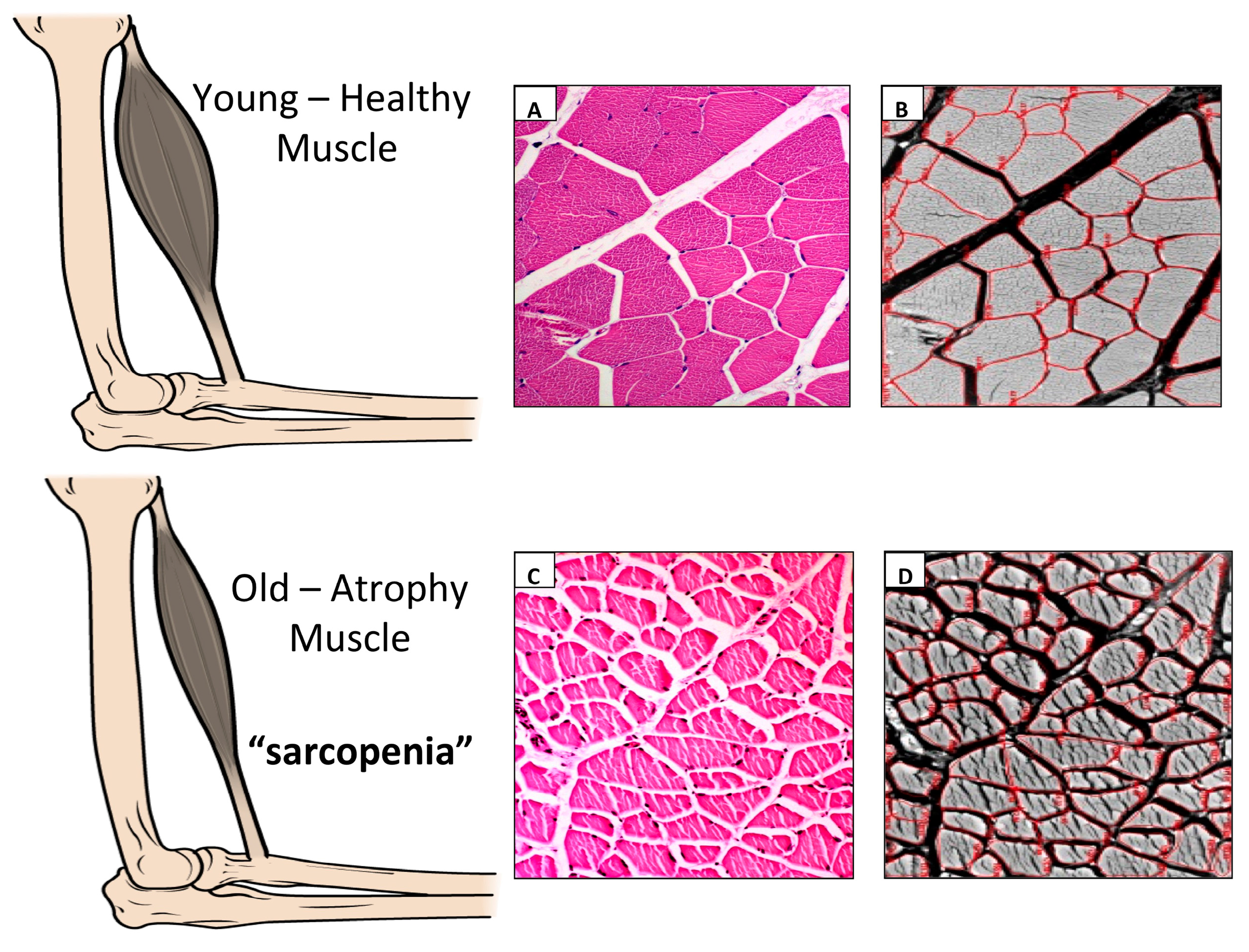
Figure 1. (A) Hematoxylin & Eosin staining in order to highlight possible structural alterations in muscle tissue from young, healthy Wistar Rats. Muscle fibers of young healthy rat did not show any damaged histological structure. The samples were examined with a Zeiss Axioplan light microscope (Carl Zeiss, Oberkochen, Germany) and a digital camera (AxioCam MRc5, Carl Zeiss) was used to take the pictures. Lens magnification: 20×. Scale bars: 50 µm; (B) Morphometric analysis of the perimeter (μm) (mean ± SD) of the muscle fibers from young healthy Wistar Rats. In the morphometric analysis of the perimeter (µm) (mean ± SD) of the muscle fibers, the young healthy rat shows normal muscle trophic. The perimeter of muscle fibers was considered and calculated using software for image acquisition (AxioVision Release 4.8.2-SP2 Software, Carl Zeiss Microscopy GmbH, Jena, Germany). Lens magnification: 20×. Scale bars: 50 µm; (C) Hematoxylin & Eosin staining in order to highlight possible structural alterations in muscle tissue from elderly Wistar Rats. Muscle fibers of elderly rat show damaged histological structure as focal perimisio fibrosis. The samples were examined with a Zeiss Axioplan light microscope (Carl Zeiss, Oberkochen, Germany) and a digital camera (AxioCam MRc5, Carl Zeiss) was used to take the pictures. Lens magnification: 20×. Scale bars: 50 µm; (D) Morphometric analysis of the perimeter (µm) (mean ± SD) of the muscle fibers from elderly Wistar Rats. In the morphometric analysis of the perimeter (µm) (mean ± SD) of the muscle fibers, the elderly rat shows a highly significant muscle fiber hypotrophy and exhibits remarkable fiber size heterogeneity. The perimeter of muscle fibers was considered and calculated using software for image acquisition (AxioVision Release 4.8.2-SP2 Software, Carl Zeiss Microscopy GmbH, Jena, Germany). Lens magnification: 20×. Scale bars: 50 µm.
Jfmk 02 00040 g001
Table 1. Possible diseases associated with sarcopenia.
Presence of angiotensin-converting enzyme D allele
Age-related loss of muscle fiber
Atherosclerosis
Diabetes mellitus
Decreased physical activity
Obesity in some individuals
Decreased food intake including protein and creatine
Decreased testosterone level
Decreased intake of vitamin D
Decreased insulin-like growth factor-1
Mechano-growth factor
Increased cytokines (tumor necrosis factor-α, interleukin-6)
Decreased motor unit acuity with a decrease in ciliary neurotrophic factor
Loss of muscle mass and strength
Overexpression of myostatin, a transforming growth factor
Fracture with low bone mass, or both
Osteoporosis and Osteoarthritis
Cancer
Cardiorespiratory and dismetabolic disorders
Chronic kidney disease
Chronic liver disease
Malnutrition
Amyotrophic lateral sclerosis
Atherosclerosis
Anorexia
Primary depression
Malabsorption
Hyperthyroidism
Inflammation
Insulin resistance