Communication

Effects of Electrical Pulse and 6-DMAP on Cleavage of Golden Hamster Oocytes—Morphological and Phisiological Observations

Jilin Province Key Laboratory of Animal Embryo Engineering, College of Animal Science and Veterinary Medicine, Jilin University, Changchun 130062, China
*
Authors to whom correspondence should be addressed.
J. Funct. Morphol. Kinesiol. 2016, 1(2), 240-248; https://doi.org/10.3390/jfmk1020240
Submission received: 28 April 2016 / Revised: 23 May 2016 / Accepted: 25 May 2016 / Published: 30 May 2016

Abstract

:
The golden hamster is a well-established model system for studies of morphology, reproductive physiology, oncology, genetics and virology. The aim of this study was to establish experimental protocols necessary for cloning the golden hamster; we examined and optimized conditions for parthenogenesis and cleavage of its oocytes. We tested oocytes of different ages, including 15 h after Human chorionic gonadotropin (hCG), with two treatments: (1) an electrical pulse ranging from 10 to 600 V/mm and (2) incubation for 2 to 6 h in 2 mM 6-dimethylaminopurine (6-DMAP). These two conditions were tested both separately and in combination. We found that (i) in oocytes of different ages, cleavage exhibits a strength-dependent increase; (ii) 6-DMAP stimulates oocyte cleavage, but the cleavage rates are significantly low; and (iii) a combined treatment is more effective than a treatment with 6-DMAP alone, and is comparable to those achieved with high pulse stimuli. These results elucidate certain parameters important for the cloning of the golden hamster species.

Graphical Abstract

1. Introduction

The golden hamster is an excellent model organism for many research fields. It represents an attractive species for studies ranging from morphology, reproductive physiology, oncology, genetics and virology. In order to establish experimental protocols necessary for cloning golden hamsters, several studies related to hamster oocyte enucleation have been reported [1,2]. In this study, we optimized the conditions for parthenogenesis and cleavage of golden hamster oocytes. Previous studies have reported that parthenogenetic activation of oocytes from several species may be induced by chemical reagents, including calcium ionophore [3], strontium (Sr2+) [4], ethanol [5], cycloheximide [6], 6-dimethylaminopurine (6-DMAP) [7], as well as by electrical stimulation [8]. Despite these findings, the activation and cleavage induction of oocytes remains one of the least efficient steps in the nuclear transplantation process.
Hamster oocytes can be activated spontaneously when cultured in vitro [9,10]. Gwatkin [11] found that electrical stimulation was highly effective in activation of hamster oocytes. However, activated oocytes are not guaranteed to undergo cleavage. In order to elucidate the parameters important for golden hamster cloning, we studied in detail the effect of different intensities of electrical pulse stimulations on the cleavage of oocytes. 6-DMAP are known to accelerate and enhance the formation of pronuclei [12]. Most mammalian oocytes, including those of ferrets [13], pig [14] and mice [5], can be activated, and cleavage may be induced by electrical stimuli combined with 6-DMAP treatments. However, the efficiency of 6-DMAP activation remains unknown in hamster oocytes. We describe the cleavage-inducing effects of 6-DMAP and electrical stimulation, both alone and in combination.

2. Materials and Methods

2.1. Chemicals and Animals

All chemicals were purchased from Sigma Chemical Co. (St. Louis, MO, USA) and Invitrogen Co. (Grand Island, NY, USA) unless otherwise noted. The golden hamster specimens (female, 6 weeks of age) were purchased from Changchun Hi-Tech Laboratory Animal Research Center (Changchun, China) and housed at a density of three animals per cage under controlled light cycle conditions (14 L:10 D). Animal treatment complied with a protocol approved by the Jilin University Institutional Animal Care and Use Committee.

2.2. Collection of Oocytes

The hamsters were superovulated with pregnant mare serum gonadotrpin (PMSG, 30 IU, ip) followed by human chorion gonadotropin (hCG, 30 IU, ip) at intervals of 72 h. Both PMSG and hCG were obtained from Ningbo Hormone Product Co., Ltd., Ningbo, China. The superovulated hamsters were sacrificed at different times after hCG injection (13.5, 15, 17, 19 h), and the oviductal ampullae were broken to release the cumulus-oocyte complexes (COCs). Cumulus cells were subsequently removed from the COCs by pipetting with a thin pipette in M2 [15] containing 0.1% hyaluronidase (Sigma, H4272). The oocytes were activated immediately after washing, rinsed, and then kept at 37.5 °C under 5% CO2.

2.3. Culture Media

The medium used for collection and culturing was M199TE [16], which contained TCM199 (with Earle salt, 26 mM sodium bicarbonate, and 25 mM Hepes; Gibco BRL, Grand Island, NY, USA) supplemented with 5% heat-inactivated fetal bovine serum (Gibco BRL), 5 mM taurine and 25 μM EDTA.

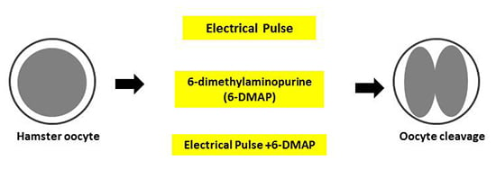
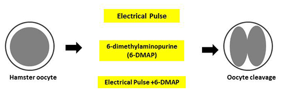
2.4. Activation and Cleavage

Electrical stimulation: Hamster oocytes collected at different times post-hCG injection were transferred to activation medium (0.3 M mannitol, 0.1 mM MgCl2, 0.1 mM CaCl2, and 0.01% (w/v) BSA) and placed between parallel electrodes (spacing, 1 mm) in the chamber of a BTX Electro-cell Manipulator 2001 (BTX, San Diego, CA, USA). The duration of the electrical stimulation was 10 μs. To test the tolerance and cleavage of the oocytes, we first treated 15 h post-hCG oocytes with different pulse intensities (10, 30, 50, 100, 200, 300, 400, 500, 550 and 600 V). We then tested the cleavage rate of oocytes of different ages (13.5, 15, 17, 19 h post-hCG) under different electrical intensities (50, 100, 300, 500 V). The oocytes of control groups were incubated in activation medium for 2 min before being transferred to the culture medium.
Chemical stimulation: We first tested 15 h post-hCG oocytes for incubation at different durations (0.5, 1, 2, 4, 6 and 8 h) in M199TE containing with 2 mM of 6-DMAP (Sigma D-2629). We then tested the cleavage rate of different ages of oocytes (13.5, 15, 17, 19 h post-hCG) over a range of 6-DMAP treatment durations (2, 4 and 6 h). Effects of 6-DMAP on oocyte activation were compared to the control groups incubated in M199TE without chemical supplement.
Combined electrical and chemical stimulation: In order to maximize the cleavage rate, oocytes were subjected to a combination of electrical and chemical activation stimuli. Oocytes were treated with electrical stimulation (300 V/mm, 10 μs) followed by a treatment with 6-DMAP (2 or 4 h).

2.5. Assessment of Oocyte Cleavage

After culturing overnight following treatment, the oocytes were examined under a microscope to assess activation and cleavage. Only oocytes with two nucleated cells were considered cleaved.

2.6. Staining and Observation of the Oocyte Cleavage Process

Oocytes were cultured in M199TE after being treated with 2 mM 6-DMAP for 2 h. Every 4 h the oocytes were collected and fixed in PBS with 4% paraformaldehyde (pH 7.4) for 40 min at room temperature (RT). Fixed samples were permeabilized by transferring into PBS supplemented with 0.1% (w/v) Triton-X100 and 0.3% BSA for 30 to 40 min at RT. After washing twice in PBS with 0.01% Triton-X100, samples were incubated in block solution (PBS containing 1% BSA) for 1 h at RT. The microtubules were localized by incubation for 1 h at RT with a fluorescein isothiocyanate-labeled mouse monoclonal antibody against α-tubulin (Sigma, F-2168) diluted 1:100 in blocking solution. The nuclear status of the samples was evaluated by staining with 10 µg/mL propidium iodide (PI) in PBS for 10 min. After thorough washing, samples were mounted on slides with antifluorescence-fade medium (1,4-diazobicyclo-[2,2,2]-octane, DABCO, Sigma, D-2522). Samples were subsequently observed under a confocal laser scanning microscope (Olympus, Tokyo, Japan).

2.7. Data Analysis

Three replicate trials were conducted for each treatment. The mean percentage (±SEM) was calculated for each experimental group. Data were analyzed by one-way analysis of variance (ANOVA), using SPSS (Statistics Production for Service Solution, Version 12.0, Chicago, IL, USA) software after being transformed via LSD. The difference was considered significant when p < 0.05.

3. Results

3.1. Cleavage of Oocytes at 15 h Post-hCG after Electrical Stimulus

Oocytes at 15 h post-hCG were stimulated for 10 μs with different electrical pulses ranging from 10 to 600 V/mm. The proportion of cleaved oocytes was observed after an overnight culture. A 10 V/mm pulse was insufficient to induce cleavage, but increasing pulse intensity correlated with increasing proportions of cleaved oocytes. When subjected to a pulse of 300 V/mm, 81.00% of oocytes induced cleavage, and after a pulse of 500 V/mm, all of oocytes developed to the 2-cell stage. The cleavage rate was low and relatively consistent after 300, 400 and 500 V/mm stimulation. With a pulse of 550 V/mm and above, oocytes began to die immediately following treatment (see Table 1).

3.2. Cleavage of Oocytes at 13.5–19 h Post-hCG after Electrical Stimulus

Oocytes at 13.5, 15, 17 and 19 h post-hCG were electrically stimulated with pulses of 50, 100, 300 and 500 V/mm. The cleavage rates of differentially-aged oocytes increased with pulse intensity. Oocytes at 13.5 h post-hCG could not withstand the 500 V/mm pulse, and all of them died immediately following stimulation. The pulse of 300 V/mm could induce cleavage in 13.5, 17 or 19 h post-hCG injection oocytes. Comparing results from every pulse group, we found that oocytes at 15 h post-hCG showed the highest rate of cleavage, and a pulse of 500 V/mm could fully activate oocytes 15 h post-hCG, with a cleavage rate up to 100% (see Table 2).

3.3. Cleavage of Oocytes at 15 h Post-hCG after 6-Dimethylaminopurine (6-DMAP) Treatment

Oocytes at 15 h post-hCG were treated with 2 mM 6-DMAP for 0.5, 1, 2, 4, 6 or 8 h in M199TE. Oocytes were then transferred to M199TE and cultured overnight. Treatment for 0.5 h with 6-DMAP induced very few oocyte cleavages. Treatment with 6-DMAP for 4 h induced the highest rate of oocyte cleavages, significantly higher than the rate induced by the 0.5 h 6-DMAP treatment group. However, treatments lasting for more than 4 h saw a decreasing rate of cleavage, diminishing to 24.23% when oocytes were treated for 8 h (see Table 3).

3.4. Cleavage of Oocytes at 13.5–19 h Post-hCG after 6-DMAP Treatment

Oocytes of different ages were treated with 2 mM of 6-DMAP for 2, 4 and 6 h. As Table 4 shows, oocytes at 13.5 and 15 h post-hCG had a higher cleavage rate no matter how long the treatment. When they became older, very few of oocytes at 17 and 19 h post-hCG could be cleaved when the 6-DMAP duration was longer than 2 h (see Table 4).

3.5. Cleavage of Oocytes after Combined Treatment of Electrical Pulse and 6-DMAP

When oocytes were treated with a 300 V/mm 10 μs electrical pulse followed by 2 or 4 h of 6-DMAP incubation, more than 90% of the samples achieved cleavage. This rate was somewhat higher than the most effective low electrical stimulus (300 V/mm) alone, and was significantly higher than the most effective 6-DMAP stimulus alone (see Table 5).

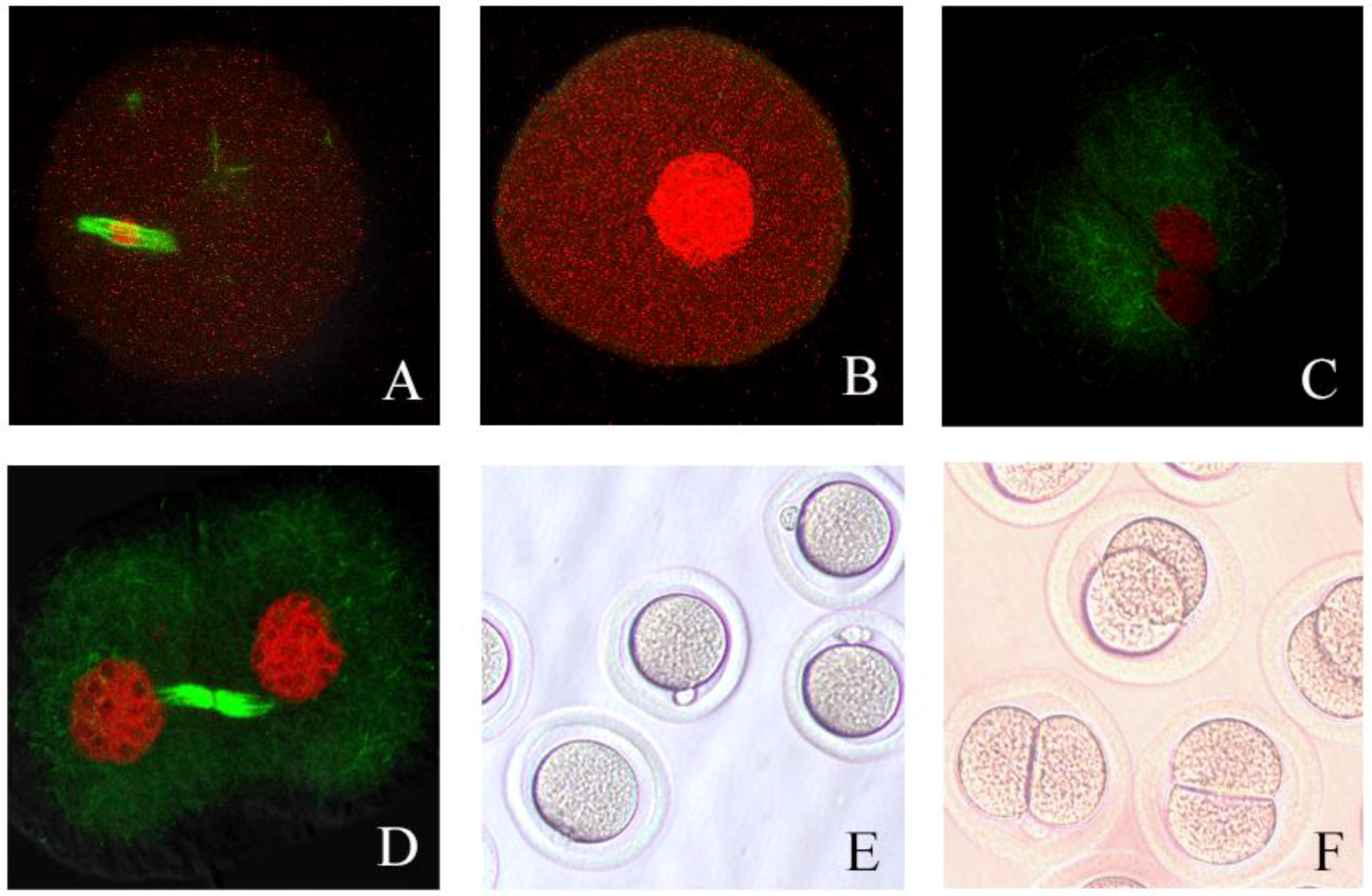
3.6. Process of Hamster Oocyte Cleavage

Oocyte spindles and chromosomes were stained at different time points following treatments. Cleaved oocytes were stained at the second meiotic metaphase (see Figure 1A; Figure 1E was unstained). After treatment, oocytes initiated the process of cleavage. Initially, the spindle disappeared and the chromosomes agglutinated into the nucleus (see Figure 1B). Subsequently, the nucleus cleaved into two nuclei and the oocyte cleaved into two cell blastomeres (Figure 1 C,D,F).

4. Discussion

Activation of oocytes is essential for successful embryo production following nuclear transfer (NT) or intracytoplasmic sperm injection (ICSI). Electric pulses have been shown to permeabilize the cell membrane, inducing an influx of extracellular calcium into the cytoplasm [17] and a rise in calcium oscillations within the oocyte [18]. Therefore, it is not surprising that electric stimulation is one of most effective methods of activating mammalian oocytes. Treatment with 6-DMAP has been used in activating oocytes of many species, including mice [5], pigs [19], cattle [20], dromedary camels [21], as well as others. But to our knowledge, this is the first report addressing the effect of 6-DMAP treatment on hamster oocyte cleavage.
Gwatkin [11] repored that electrical stimulation could activate hamster oocytes. Ogura [22] found that hamster spermatid-oocyte fusion could be induced through direct current pulse (300 V/mm, 10 μs). We therefore used a pulse of 300 V/mm, 10 μs and 15 h-post-hCG oocytes as a standard, and modulated both voltage and oocyte age to optimize this treatment. Following electrical stimulation, the cleavage of ferret oocytes [13] showed an age-dependent increase in the occurrence of cleavage and development to the morula and blastocyst stages. Bovine oocytes activate in response to ionophore A23187 treatment, electrical pulse, or ethanol treatment in an age-dependent manner [23], and electrically stimulated porcine oocytes show a similar trend [24]. We found that the hamster oocyte cleavage rate also increased with age, with a greater proportion of 15 h post-hCG oocytes cleaving than 13.5 h post-hCG oocytes. However, cleavage rates decreased with age in oocytes older than 15 h (17 and 19 h post-hCG). According to Yanagimachi and Chang [25], golden hamster oocytes begin spontaneous activation in vivo between 12 and 18 h post-ovulation. Although older oocytes are easily activated, spontaneous activation could induce cleavage. Our results showed that cleavage rates of oocytes at different ages increased with voltage. Oocytes of different ages also showed different tolerances to electrical treatments. Oocytes at 13.5 h post-hCG could not withstand treatments of 500 V/mm, and many 15 h-post-hCG oocytes could not withstand 550V/mm. Older oocytes showed greater tolerance, and the electrical stimulus condition chosen to treat hamster oocytes should take into account the age of the oocytes.
6-DMAP is an inhibitor of protein kinases, and is widely used for activation of mammalian oocytes due to its ability to enhance activation stimuli and accelerate the formation of pronuclei [26]. Studies in pig oocytes showed that 6-DMAP treatments may impair parthenogenetic development of the activated oocytes. Grupen [16] showed that treatment with 2 or 5 mM 6-DMAP for 3 h after electrical pulse increased the incidence of blastocyst formation, whereas treatment with 2 or 5 mM 6-DMAP for 5 h did not. In our study, hamster oocytes collected at 15 h post-hCG injection were treated directly with 2 mM 6-DMAP for different durations (0.5–8 h). Oocytes treated with 6-DMAP for 2 and 4 h had higher rates of cleavage (45.21% and 46.82%) than those treated with 6-DMAP for 0.5, 1, 6 and 8 h. This indicated that sufficient 6-DMAP treatment duration was necessary to induce oocyte cleavage, but excessively long treatments with 6-DMAP induced detrimental effects on the development of parthenogenetic hamster embryos. Numerous abnormalities were observed when oocytes were activated with 6-DMAP for longer durations, and these abnormalities included inhibited spindle rotation and impaired exchange of protamines into histones [27]. In golden hamster 17 and 19 h post-hCG oocytes, treatment with 6-DMAP for 2 h induced only 13.89% and 16.27% oocyte cleavage, respectively. When treated for 6 h, however, none of the oocytes were induced to cleave. We conclude that the rate of cleavage after 6-DMAP treatment is age-dependent, with younger oocytes (13.5 and 15 h post-hCG) being easier to induce than older oocytes (17 and 19 h). The older oocytes were difficult to induce to cleave, and a longer duration of treatment with 6-DMAP may cause the accumulation of excessive abnormalities.
We found that 6-DMAP treatments alone had limited efficacy in inducing oocyte cleavage, with a maximum cleavage rate of 47.12%. Consistent with previous studies, oocyte activation rate was low when 6-DMAP was used alone without any initiating stimulus [28,29]. The percentage of cleaved porcine oocytes and the rate of blastocyst formation [12] was higher in the ethanol + cycloheximide + cytochalasin B + 6-DMAP treatment (66.4%) than in other separate treatments (24.9%–57.6%). Ferret oocytes cleaved at a rate of 85.8% when treated with combined electrical and chemical (cycloheximide and 6-DMAP) stimulations [13]. We found that chemical stimulation (6-DMAP, 2 h) combined with electrical stimulation (300 V/mm, 10 μs) achieved cleavage rates of 96.49%, higher than the rate stimulated by either treatment alone. We furthermore found that oocytes of different ages showed different tolerance levels to treatments, and higher voltage often caused lysis in younger oocytes. Therefore, lower pulse electrical stimulation combined with 6-DMAP treatment may be the best option in treating hamster oocytes of every age.
We conclude that the cleavage of golden hamster oocytes exhibits a strength-dependent increase when treated with electrical pulses from 10 to 500 V/mm. We further found that 6-DMAP can stimulate the oocyte cleavage, but the cleavage percentages are significantly lower than those stimulated under high-pulse electrical treatment. A combined treatment is superior to a 6-DMAP only treatment, and is nearly identical to the responses achieved by the high-pulse stimulus. These results define the optimal conditions for oocyte activation, and should facilitate cloning attempts of the golden hamster, furthering the system as an animal model for human disease.

Acknowledgments

This work was supported by the National Natural Science Foundation of China (No. 30671510) and Jinlin University Research Starting Fund of China.

Author Contributions

Lingyan Wang and Ziyi Li conceived and designed the experiments; Lingyan Wang and Han Jiang performed the experiments; Lingyan Wang analyzed the data; Ziyi Li contributed reagents/materials/analysis tools; Lingyan Wang and Ziyi Li wrote the paper.

Conflicts of Interest

The authors declare no conflict of interest.

References

  1. Wang, L.; Jiang, H.; Su, L.; Tang, B.; Li, D.; Li, Z. Effects of colchicine or demecolcine on cytoplasmic protrusions and assisted enucleation of golden hamster oocytes. Cell Biol. Int. 2009, 33, 1274–1279. [Google Scholar] [CrossRef] [PubMed]
  2. Wang, L.; Li, D.; Li, Z. Changes in the reciprocal position of the first polar body and oocyte chromosome set in golden hamsters. Biosci. Rep. 2009, 29, 315–320. [Google Scholar] [CrossRef] [PubMed]
  3. Ware, C.B.; Barnes, F.L.; Maiki-Laurila, M.; First, N.L. Age dependence of bovine oocyte activation. Gamete Res. 1989, 22, 265–275. [Google Scholar] [CrossRef] [PubMed]
  4. Marcus, G.J. Activation of cumulus-free mouse oocytes. Mol. Reprod. Dev. 1990, 26, 159–162. [Google Scholar] [CrossRef] [PubMed]
  5. Lan, G.C.; Ma, S.F.; Wang, Z.Y.; Luo, M.J.; Chang, Z.L.; Tan, J.H. Effects of post-treatment with 6-dimethylaminopurine (6-DMAP) on ethanol activation of mouse oocytes at different ages. J. Exp. Zool. A Comp. Exp. Biol. 2004, 301, 837–843. [Google Scholar] [CrossRef] [PubMed]
  6. Presicce, G.A.; Yang, X. Parthenogenetic development of bovine oocytes matured in vitro for 24 h and activated by ethanol and cycloheximide. Mol. Reprod. Dev. 1994, 38, 380–385. [Google Scholar] [CrossRef] [PubMed]
  7. Bodart, J.F.; Bechard, D.; Bertout, M.; Gannon, J.; Rousseau, A.; Vilain, J.P.; Flament, S. Activation of xenopus eggs by the kinase inhibitor 6-DMAP suggests a differential regulation of cyclin B and p39mos proteolysis. Exp. Cell Res. 1999, 253, 413–421. [Google Scholar] [CrossRef] [PubMed]
  8. Escriba, M.J.; Garcia-Ximenez, F. Use of a variable electrical pulsing sequence in rabbit oocyte activation. Reprod. Nutr. Dev. 2000, 40, 261–269. [Google Scholar] [CrossRef] [PubMed]
  9. Jiang, H.; Wang, C.; Guan, J.; Wang, L.; Li, Z. Changes of spontaneous parthenogenetic activation and development potential of golden hamster oocytes during the aging process. Acta Histochem. 2015, 117, 104–110. [Google Scholar] [CrossRef] [PubMed]
  10. Sun, X.S.; Yue, K.Z.; Zhou, J.B.; Chen, Q.X.; Tan, J.H. In vitro spontaneous parthenogenetic activation of golden hamster oocytes. Theriogenology 2002, 57, 845–851. [Google Scholar] [CrossRef]
  11. Gwatkin, R.B.; Williams, D.T.; Hartmann, J.F.; Kniazuk, M. The zona reaction of hamster and mouse eggs: Production in vitro by a trypsin-like protease from cortical granules. J. Reprod. Fertil. 1973, 32, 259–265. [Google Scholar] [CrossRef] [PubMed]
  12. Yi, Y.J.; Park, C.S. Parthenogenetic development of porcine oocytes treated by ethanol, cycloheximide, cytochalasin B and 6-dimethylaminopurine. Anim. Reprod. Sci. 2005, 86, 297–304. [Google Scholar] [CrossRef] [PubMed]
  13. Li, Z.; Jiang, Q.; Rezaei Sabet, M.; Zhang, Y.; Ritchie, T.C.; Engelhardt, J.F. Conditions for in vitro maturation and artificial activation of ferret oocytes. Biol. Reprod. 2002, 66, 1380–1386. [Google Scholar] [CrossRef] [PubMed]
  14. Grupen, C.G.; Mau, J.C.; McIlfatrick, S.M.; Maddocks, S.; Nottle, M.B. Effect of 6-dimethylaminopurine on electrically activated in vitro matured porcine oocytes. Mol. Reprod. Dev. 2002, 62, 387–396. [Google Scholar] [CrossRef] [PubMed]
  15. Nagy, A.; Gertsenstein, M.; Vintersten, K.; Behringer, R. Manipulating the Mouse Embryo: A Laboratory Manual, 3rd ed.; Cold Spring Harbor Laboratory Press: Cold Spring Harbor, NY, USA, 2003. [Google Scholar]
  16. Yamauchi, Y.; Yanagimachi, R.; Horiuchi, T. Full-term development of golden hamster oocytes following intracytoplasmic sperm head injection. Biol. Reprod. 2002, 67, 534–539. [Google Scholar] [CrossRef] [PubMed]
  17. Zimmermann, U.; Vienken, J. Electric field-induced cell-to-cell fusion. J. Membr. Biol. 1982, 67, 165–182. [Google Scholar] [CrossRef] [PubMed]
  18. Swann, K.; Ozil, J.P. Dynamics of the calcium signal that triggers mammalian egg activation. Int. Rev. Cytol. 1994, 152, 183–222. [Google Scholar] [PubMed]
  19. Cheng, W.M.; Sun, X.L.; An, L.; Zhu, S.E.; Li, X.H.; Li, Y.; Tian, J.H. Effect of different parthenogenetic activation methods on the developmental competence of in vitro matured porcine oocytes. Anim. Biotechnol. 2007, 18, 131–141. [Google Scholar] [CrossRef] [PubMed]
  20. Meo, S.C.; Yamazaki, W.; Ferreira, C.R.; Perecin, F.; Saraiva, N.Z.; Leal, C.L.; Garcia, J.M. Parthenogenetic activation of bovine oocytes using single and combined strontium, ionomycin and 6-dimethylaminopurine treatments. Zygote 2007, 15, 295–306. [Google Scholar] [CrossRef] [PubMed]
  21. Wani, N.A. Chemical activation of in vitro matured dromedary camel (Camelus dromedarius) oocytes: Optimization of protocols. Theriogenology 2008, 69, 591–602. [Google Scholar] [CrossRef] [PubMed]
  22. Ogura, A.; Yanagimachi, R. Round spermatid nuclei injected into hamster oocytes from pronuclei and participate in syngamy. Biol. Reprod. 1993, 48, 219–225. [Google Scholar] [CrossRef] [PubMed]
  23. Nagai, T. Parthenogenetic activation of cattle follicular oocytes in vitro with ethanol. Gamete Res. 1987, 16, 243–249. [Google Scholar] [CrossRef] [PubMed]
  24. Hagen, D.R.; Prather, R.S.; First, N.L. Response of porcine oocytes to electrical and chemical activation during maturation in vitro. Mol. Reprod. Dev. 1991, 28, 70–73. [Google Scholar] [CrossRef] [PubMed]
  25. Yanagimachi, R.; Chang, M.C. Fertilizable life of golden hamster ova and their morphological changes at the time of losing fertilizability. J. Exp. Zool. 1961, 148, 185–203. [Google Scholar] [CrossRef] [PubMed]
  26. Moses, R.M.; Kline, D.; Masui, Y. Maintenance of metaphase in colcemid-treated mouse eggs by distinct calcium- and 6-dimethylaminopurine (6-DMAP)-sensitive mechanisms. Dev. Biol. 1995, 167, 329–337. [Google Scholar] [CrossRef] [PubMed]
  27. Ledda, S.; Loi, P.; Bogliolo, L.; Moor, R.M.; Fulka, J., Jr. The effect of 6-dimethylaminopurine (6-DMAP) on DNA synthesis in activated mammalian oocytes. Zygote 1996, 4, 7–9. [Google Scholar] [CrossRef] [PubMed]
  28. Liu, H.L.; Fan, B.Q.; Wang, H.H.; Cheng, Y.F.; Wang, G.J. [effect of 6-dimethylaminopurine on the resumption of meiosis and parthenogenetic activation in mouse oocytes]. Shi Yan Sheng Wu Xue Bao 1997, 30, 403–406. [Google Scholar] [PubMed]
  29. Moses, R.M.; Masui, Y. Enhancement of mouse egg activation by the kinase inhibitor, 6-dimethylaminopurine (6-DMAP). J. Exp. Zool. 1994, 270, 211–218. [Google Scholar] [CrossRef] [PubMed]
Figure 1. Confocal micrographs of oocyte activation process. (A) Matured oocyte used for activation, showing the morphology of the spindle (green) and chromosomes (red); (B) spindle disappeared and chromosomes condensed to one nuclear package; (C) nucleus divided into two pronuclei, then immediately (D) cleaved into two approximately equal-size blastomeres. Magnification: 600×; green: spindle; red: chromosomes; (E,F) normal oocytes and cleaved blastomeres observed under phase-contrast microscope, Magnification: 200×.
Jfmk 01 00240 g001
Table 1. Effect of different electrical pulses on cleavage of 15 h post-hCG oocytes.
Electrical Pulse (V/mm)No. of Oocytes ExaminedNo. of Oocytes Cleaved% of Cleavage (mean ± SE)
103200.00 ± 0.00 a
30381231.00 ± 7.57 b
50271141.33 ± 8.25 b
100332475.00 ± 7.00 c
200302064.00 ± 14.18 c
300604881.00 ± 9.71 c,d
400282796.33 ± 3.67 d
5004545100.00 ± 0.00 d
550272074.33 ± 5.46 c
600240 *0.00 ± 0.00 a
* Oocytes died after electrical stimulus; ad Values with different superscripts within the same column are significantly different (p < 0.05).
Table 2. Effect of different electrical pulses on cleavage of different ages of oocytes.
Electrical Pulse (V/mm)Hours Post-hCGNo. of Oocytes ExaminedNo. of Oocytes Cleaved% of Cleavage (mean ± SE)
5013.536612.67 ± 12.67 a,b
15271141.33 ± 8.25 a
1727414.00 ± 7.37 a,b
192127.33 ± 7.33 b
10013.53337.33 ± 3.84 a,c
15332475.00 ± 7.00 b
1729360.33 ± 11.69 b
19291813.67 ± 9.94 c
30013.531721.67 ± 11.28 a
15604881.00 ± 9.71 b
17281663.33 ± 9.28 b,c
1920839.00 ± 3.22 a,c
50013.5330 *0.00 ± 0.00 a,*
154545100.00 ± 0.00 b
1722941.00 ± 14.29 c
19381431.67 ± 9.28 c
* Oocytes died after electrical stimulus; ac Values with different superscripts within one column of the same electrical pulse are significantly different (p < 0.05).
Table 3. Cleavage of oocytes (15 h post hCG) after different duration of 6-dimethylaminopurine (6-DMAP) treatment.
Duration of 6-DMAP (h)No. of Oocytes ExaminedNo. of Cleavage% of Cleavage (mean ± SE)
0.539819.44 ± 4.24 a
1401234.33 ± 11.37 a,b
2512345.21 ± 10.65 a,b
4502346.82 ± 5.40 b
6501835.89 ± 13.33 a,b
839924.23 ± 2.25 a,b
a,b Values with different superscripts are significantly different (p < 0.05).
Table 4. Cleavage of oocytes in different age after different duration of 6-DMAP treatment.
Hours Post-hCGDuration of 6-DMAP (h)No. of Oocytes ExaminedNo. of Oocytes Cleaved% of Cleavage (mean ± SE)
13.52431847.12 ± 15.44 a
4431439.23 ± 17.12 a
6411946.21 ± 9.04 a
152502345.21 ± 10.65 a
4512346.82 ± 5.40 a
6511835.89 ± 13.33 a
17232513.89 ± 7.35 a
43214.17 ± 4.17 a
63200.00 ± 0.00 a
19234616.27 ± 12.87 a
43412.78 ± 2.78 a
63400.00 ± 0.00 a
a Values with the same superscripts within one column of oocytes at same age are not significantly different (p > 0.05).
Table 5. Cleavage of oocytes after 10 μs, 300 V/mm electrical stimulus and different duration of 6-DMAP treatment.
Duration of 6-DMAP (h)Electrical StimulusNo. of Oocytes ExaminedNo. of Oocytes Cleaved% of Cleavage (mean ± SE)
0+604881.00 ± 9.71 a
2+545296.49 ± 3.51 a
2512345.21 ± 10.65 b
4+6054 90.44 ± 1.55 a
4502346.82 ± 5.40 b
a, b Values with different superscripts are significantly different (p < 0.05). +, oocytes were treated with 10 μs, 300 V/mm electrical stimulus; −, oocytes were not treated with 10 μs, 300 V/mm electrical stimulus.

