Brief Report

Myoelectric Manifestations of Fatigue after ACL Reconstruction: A Cross-Sectional Study after Postoperative Rehabilitation

1
Rehabilitation Research Laboratory 2rLab, Department of Business Economics, Health and Social Care, University of Applied Sciences and Arts of Southern Switzerland, 6928 Manno, Switzerland
2
Department of Business Economics, Health and Social Care, University of Applied Sciences and Arts of Southern Switzerland, 7302 Landquart, Switzerland
3
Ars Medica Clinic, 6929 Gravesano, Switzerland
*
Author to whom correspondence should be addressed.
J. Funct. Morphol. Kinesiol. 2016, 1(2), 193-199; https://doi.org/10.3390/jfmk1020193
Submission received: 21 March 2016 / Revised: 11 April 2016 / Accepted: 13 April 2016 / Published: 15 April 2016

Abstract

:
An increased fatigability associated with anterior cruciate ligament (ACL) injury may persist several months after surgery. The purpose of this study was to investigate the behavior of muscle fiber conduction velocity (CV) as a descriptor of myoelectric fatigue, at different stages after rehabilitation, post-ACL reconstruction. Nineteen subjects acted as control group (CG), 10 patients had undergone surgery within 12 months (R12), and 23 patients were more than 24 months post-surgery (R24+). Surface electromyography (EMG) signals were detected from the quadriceps femoris using bidimensional arrays during isometric contractions at 20% and 60% maximal voluntary contraction (MVC). We observed a lower fatigability in the R24+ group during the 60% MVC contraction, with respect to the other groups. Lower fatigability of quadriceps muscle after ACL reconstruction in the long term may be linked to a recovery from a transitory altered motor unit recruitment strategy due to surgery, observed in the R12 group. Therefore, the findings of this study do not suggest an impaired fatigability of the quadriceps muscle during sustained isometric contractions in active patients in the long term.

Graphical Abstract

1. Introduction

Anterior cruciate ligament reconstruction (ACLr) is known to induce neuromuscular alterations of the quadriceps leading to substantial muscle weakness [1], deficits in muscle voluntary activation [2], and atrophy [3]. In addition to a history of ACLr, fatigue can also induce changes in the neuromuscular control of the quadriceps. In fact, fatigue associated with ACL injury, may reduce the ability of the muscle to generate strength and may induce changes in lower extremity biomechanics and deficits in postural control, increasing the risk of repeated ACL injury [4,5,6,7]. Furthermore, patients who have undergone ACLr, even after rehabilitation, tend to exhibit a deficit in strength and neuromuscular control [8,9,10,11].
Combined central and peripheral processes contribute to neuromuscular fatigue. Central fatigue can originate from any structure above the neuromuscular junction from the central nervous system to the peripheral nerves, resulting in a progressive reduction in voluntary activation. Peripheral fatigue reflects local changes in the muscle and hampers the execution of descending central commands [12].
Continuous monitoring of muscle fatigue during a task is possible by measuring its myoelectric activity by surface electromyography (sEMG). Biochemical and physiological changes in muscles during fatiguing contractions are, namely, reflected also in properties of myoelectric signals recorded on the surface of the skin above the muscle(s) concerned [13]. For instance, during isometric constant force contractions muscle fiber conduction velocity (CV) decreases [14], mainly related to a decrease in the intracellular pH [15,16]. Therefore, the evaluation of peripheral fatigue may be obtained by estimating CV slope (i.e., rate of change) during an isometric task [17], and this procedure is considered the most robust index [18,19,20]. Moreover, if the motor unit pool is stable, this variable correlates with fiber size and type [21,22].
In ACLr patients, central fatigue has been demonstrated to impair quadriceps activation (e.g., [23,24]); however, little is known about the peripheral contributions to muscle fatigue.
Therefore, the purpose of this study was to investigate the behavior of CV, estimated by multichannel sEMG, in two distinct patients groups at different stages after post-operative rehabilitation.

2. Experimental Section

2.1. Participants

Fifty-two subjects (21 women and 31 men; age 29 ± 10 years, height 174 ± 8 cm and weight 71 ± 12 kg) participated in the study and were enrolled according to the presence of ACL injuries and to the time elapsed since surgery (quadrupled hamstrings autografts).
  • Group CG: 19 subjects with no previous history of knee injury or painful conditions of the lower limb acted as the control group;
  • Group R12: 10 patients, up to 12 months after ACLr (10 ± 4 months);
  • Group R24+: 23 patients, more than 24 months after ACLr (59 ± 38 months).
All the patients of group R12 and group R24+ completed their rehabilitation program, which lasted about 6 months.
All the subjects were moderately active (≥3 days of moderate trainings per week) according to the International Physical Activity Questionnaire (short form) [25]. The study protocol was approved by the local ethical committee and written informed consent forms were signed prior to participation. All procedures were conducted according to the Declaration of Helsinki.

2.2. Experimental Set-Up

The experimental protocol has been previously published [26]: briefly, subjects were positioned on a custom developed ergometer chair (Figure 1A) with the knee joint at 60° of flexion the trunk-thigh angle at approximately 100°. After two maximal voluntary contractions (MVCs) of 2–3 s separated by a 2-min rest, a low-level isometric contraction (20% MVC) for 90 s and a high-level endurance isometric contraction (60% MVC), separated by a 5-min rest, were recorded. During the 60% MVC subjects were verbally encouraged to keep the force level for as long as possible, until the force value decreased to below 90% of the target. Visual feedback was provided. For each subject, left and right side were randomly presented.

2.3. EMG Measurement and Signal Processing

Myoelectric signals were detected from the vastus medialis (VM) muscle in a single-differential configuration, using an adhesive array of 30 electrodes (3 mm diameter, 6 × 5 grid, 8 mm IED; Spes Medica, Battipaglia, Italy). Adhesive arrays were applied between the distal tendon and the innervation zone, identified with a dry linear array, as has been previously described [27] (Figure 1B). The electromyography (EMG) signals were amplified (EMG-USB2; OT Bioelettronica, Turin, Italy), band-pass filtered (10–750 Hz), sampled at 2048 Hz, and stored on a computer. A custom-developed ergometer (SUPSI; OT Bioelettronica) was used to measure knee torque with a load cell operating linearly in the range 0–1000 Nm. The torque signal was amplified (MISO II; OT Bioelettronica) and stored on a computer with the sEMG data.
CV was estimated using a multichannel algorithm on single differential signals [28] (Figure 1C). CV values outside the physiological range (2–8 m/s) were excluded from the analysis. Given that the patients groups included both unilaterally or bilaterally reconstructed ACL, CV was estimated from the most recently operated side. In the CG, CV was estimated arbitrarily from the right side.

2.4. Statistical Analysis

Basic descriptive statistics were calculated for absolute MVC torque. An independent samples t-test was run to determine whether there was a statistical difference between mean MVC torques in the three groups, using SPSS (IBM; Armonk, NY, USA). Linear regression over time was applied to CV in order to extract the normalized slopes. Since the data were not normally distributed, a Mann-Whitney U test was used to observe differences in normalized slopes of CV at 20% and 60% MVC in the three groups. Eventually, a one-sample Wilcoxon signed-rank test was run to determine whether CV slopes were different from zero. Statistical significance was set to α = 0.05. Results are reported as median and interquartile range.

3. Results

Mean MVC torque for groups CG, R12, and R24+ was 338 ± 90 Nm, 377 ± 96 Nm, and 376 ± 100 Nm, respectively. No statistical differences were observed.
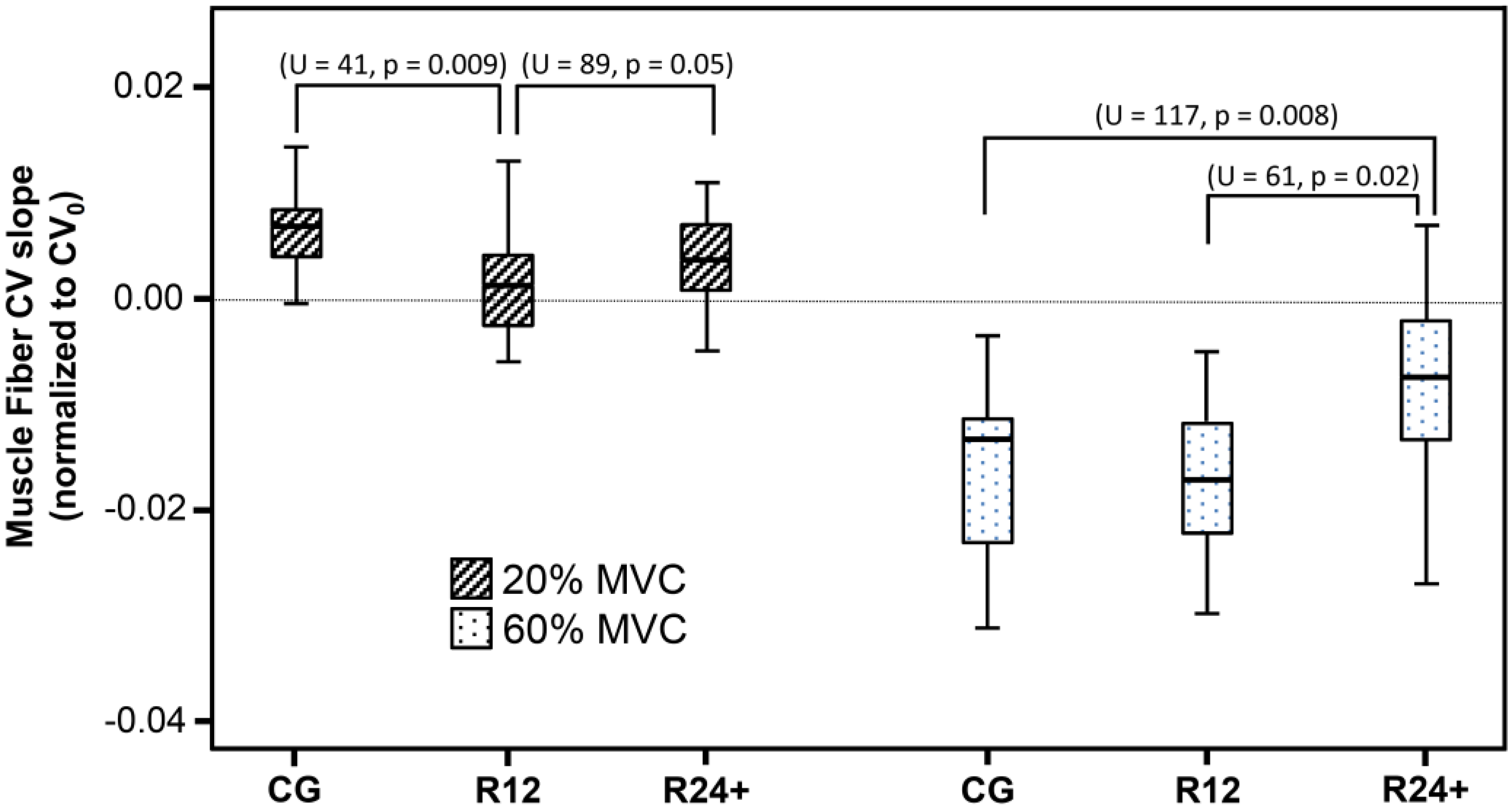
Positive CV slopes were found during the low intensity contraction in the three groups, whereas, during the higher intensity contraction, the CV slopes became negative.
At 20% MVC, we observed a lower CV slope in group R12 (not different from zero, p = 0.93) with respect to the groups CG (p = 0.009) and R24+ (p = 0.05), respectively (Figure 2). Interestingly, at 60% MVC, the group R24+ showed a less negative CV slope compared to the CG (p = 0.008) and to the R12 (p = 0.002) groups.

4. Discussion

In the present study, we observed positive CV slopes in all groups during the 20% MVC contraction, which suggested the absence of significant peripheral fatigue at low force output, in accordance with previous published data [26]. Notably, the R12 group showed a CV slope not significantly different from zero but significantly lower with respect to the CG. This suggests that the knee surgery induced alterations in motor unit (MU) recruitment strategies during low-level contractions, which tended to lessen in the R24+ group. Several studies have demonstrated a fatigue resistance in ACLr patients, attributed to selective atrophy of fast-twitch muscle fibers: McNair and Wood [29] and McHugh and colleagues [30] found lower median and mean frequency during MVC in ACL deficient patients, 4–5 weeks after surgery. A decrease in these spectral parameters primarily reflects a decrease in muscle fiber CV [8]. Moreover, Snyder-Mackler and colleagues [31] found fatigue resistance 4 weeks post-ACLr, attributed to selective atrophy of type IIx fibers during electrically elicited knee extension contractions, set to produce 20% MVC. Therefore, since CV is positively related to the muscle fiber size and type [24], a possible hypothesis for lower CV slope in group R12 may be linked to an alteration of (higher threshold) MU recruitment strategies. A reduction in the discharge rate of active MUs may also contribute to the observed effect [32].
During the high level isometric contraction, fatigability of VM was observed in all the groups, especially in the R12 group, probably because type II MUs were inhibited. Moreover, patients of group R24+, which presumably completed their rehabilitation programs and were again active, showed lower fatigability with respect to the other groups. This observation may be also explained as a reduction in the inhibition effect on type II MUs in the long term.
The current study has potential limitations: Firstly, our results should be interpreted with caution, since ACLr patients may show a different range of severity of neuromuscular alterations, according to the extent of joint damage. Furthermore, we did not analyze the behavior of sEMG frequency to assess muscle fatigue; however, since CV is a direct physiological parameter, its use is to be preferred over mean or median spectral frequency, when computed with a multi-channel approach [33]. Lastly, it is important to remark that the cross-sectional design with three distinct groups, does not provide strong evidence for the clinical course of muscle fatigue after ACLr.

5. Conclusions

This study investigated for the first time the behavior of muscle fiber CV as a descriptor of peripheral fatigue in distinct groups of patients after ACLr. The findings of this study do not suggest an impaired fatigability of the quadriceps muscle during sustained isometric contractions in active patients in the long term. Thus, further studies are needed to verify whether muscle fatigue caused by ACL injury and reconstruction may be linked to a temporary alteration in MU recruitment strategies.

Acknowledgments

This study was supported by the Yellow P Foundation, Switzerland and by the Thim van der Laan Foundation, Switzerland.

Author Contributions

Marco Barbero and Danilo Togninalli conceived the study and obtained the funding. Matteo Beretta-Piccoli, Alessandro Schneebeli, Corrado Cescon, Ron Clijsen, and Marco Barbero participated actively in data gathering. All authors contributed to the analysis, which was mainly done by Matteo Beretta-Piccoli, Michele Egloff, and Corrado Cescon. Matteo Beretta-Piccoli, Alessandro Schneebeli, and Marco Barbero wrote the first draft, and all authors contributed to the writing of the final report.

Conflicts of Interest

The authors declare no conflict of interest.

Abbreviations

The following abbreviations are used in this manuscript:
ACLranterior cruciate ligament reconstruction
CVconduction velocity
MUmotor unit
EMGelectromyography
MVCmaximal voluntary contraction
CGcontrol group
R12patients that were up to 12 months after ACLr
R24+patients that were more than 24 months after ACLr

References

  1. Snyder-Mackler, L.; de Luca, P.F.; Williams, P.R.; Eastlack, M.E.; Bartolozzi, A.R., 3rd. Reflex inhibition of the quadriceps femoris muscle after injury or reconstruction of the anterior cruciate ligament. J. Bone Jt. Surg. Am. 1994, 76, 555–560. [Google Scholar]
  2. Williams, G.N.; Buchanan, T.S.; Barrance, P.J.; Axe, M.J.; Snyder-Mackler, L. Quadriceps weakness, atrophy, and activation failure in predicted noncopers after anterior cruciate ligament injury. Am. J. Sports Med. 2005, 33, 402–407. [Google Scholar] [CrossRef] [PubMed]
  3. Thomas, A.C.; Wojtys, E.M.; Brandon, C.; Palmieri-Smith, R.M. Muscle atrophy contributes to quadriceps weakness after ACL reconstruction. J. Sci. Med. Sport 2016, 19, 7–11. [Google Scholar] [CrossRef] [PubMed]
  4. Frank, B.S.; Gilsdorf, C.M.; Goerger, B.M.; Prentice, W.E.; Padua, D.A. Neuromuscular fatigue alters postural control and sagittal plane hip biomechanics in active females with anterior cruciate ligament reconstruction. Sports Health 2014, 6, 301–308. [Google Scholar] [CrossRef] [PubMed]
  5. Webster, K.E.; Santamaria, L.J.; McClelland, J.A.; Feller, J.A. Effect of fatigue on landing biomechanics after anterior cruciate ligament reconstruction surgery. Med. Sci. Sports Exerc. 2012, 44, 910–916. [Google Scholar] [CrossRef] [PubMed]
  6. Borotikar, B.S.; Newcomer, R.; Koppes, R.; McLean, S.G. Combined effects of fatigue and decision making on female lower limb landing postures: Central and peripheral contributions to ACL injury risk. Clin. Biomech. 2008, 23, 81–92. [Google Scholar] [CrossRef] [PubMed]
  7. Lessi, G.C.; Serrao, F.V. Effects of fatigue on lower limb, pelvis and trunk kinematics and lower limb muscle activity during single-leg landing after anterior cruciate ligament reconstruction. Knee Surg. Sports Traumatol. Arthrosc. 2015. [Google Scholar] [CrossRef] [PubMed]
  8. Decker, M.J.; Torry, M.R.; Noonan, T.J.; Riviere, A.; Sterett, W.I. Landing adaptations after ACL reconstruction. Med. Sci. Sports Exerc. 2002, 34, 1408–1413. [Google Scholar] [CrossRef] [PubMed]
  9. Gokeler, A.; Hof, A.L.; Arnold, M.P.; Dijkstra, P.U.; Postema, K.; Otten, E. Abnormal landing strategies after ACL reconstruction. Scand. J. Med. Sci. Sports 2010, 20, e12–e19. [Google Scholar] [CrossRef] [PubMed]
  10. Hantes, M.E.; Tsarouhas, A.; Giakas, G.; Spiropoulos, G.; Sideris, V.; Christel, P.; Malizos, K.N. Effect of fatigue on tibial rotation after single- and double-bundle anterior cruciate ligament reconstruction: A 3-dimensional kinematic and kinetic matched-group analysis. Am. J. Sports Med. 2012, 40, 2045–2051. [Google Scholar] [CrossRef] [PubMed]
  11. Paterno, M.V.; Schmitt, L.C.; Ford, K.R.; Rauh, M.J.; Myer, G.D.; Hewett, T.E. Effects of sex on compensatory landing strategies upon return to sport after anterior cruciate ligament reconstruction. J. Orthop. Sports Phys. Ther. 2011, 41, 553–559. [Google Scholar] [CrossRef] [PubMed]
  12. Gandevia, S.C. Spinal and supraspinal factors in human muscle fatigue. Physiol. Rev. 2001, 81, 1725–1789. [Google Scholar] [PubMed]
  13. Merletti, R.; Parker, P.J. Electromyography: Physiology, Engineering, and Noninvasive Applications; IEEE Press: Piscataway, NJ, USA, 2004; p. 494S. [Google Scholar]
  14. Buchthal, F.; Guld, C.; Rosenfalck, P. Innervation zone and propagation velocity in human muscle. Acta Physiol. Scand. 1955, 35, 174–190. [Google Scholar] [CrossRef] [PubMed]
  15. Bouissou, P.; Estrade, P.Y.; Goubel, F.; Guezennec, C.Y.; Serrurier, B. Surface EMG power spectrum and intramuscular pH in human vastus lateralis muscle during dynamic exercise. J. Appl. Physiol. 1989, 67, 1245–1249. [Google Scholar] [PubMed]
  16. Brody, L.R.; Pollock, M.T.; Roy, S.H.; de Luca, C.J.; Celli, B. pH-induced effects on median frequency and conduction velocity of the myoelectric signal. J. Appl. Physiol. 1991, 71, 1878–1885. [Google Scholar] [PubMed]
  17. Gonzalez-Izal, M.; Malanda, A.; Gorostiaga, E.; Izquierdo, M. Electromyographic models to assess muscle fatigue. J. Electromyogr. Kinesiol. 2012, 22, 501–512. [Google Scholar] [CrossRef] [PubMed]
  18. Bilodeau, M.; Goulet, C.; Nadeau, S.; Arsenault, A.B.; Gravel, D. Comparison of the EMG power spectrum of the human soleus and gastrocnemius muscles. Eur. J. Appl. Physiol. Occup. Physiol. 1994, 68, 395–401. [Google Scholar] [CrossRef] [PubMed]
  19. Kollmitzer, J.; Ebenbichler, G.R.; Kopf, A. Reliability of surface electromyographic measurements. Clin. Neurophysiol. 1999, 110, 725–734. [Google Scholar] [CrossRef]
  20. Linssen, W.H.; Stegeman, D.F.; Joosten, E.M.; van’t Hof, M.A.; Binkhorst, R.A.; Notermans, S.L. Variability and interrelationships of surface EMG parameters during local muscle fatigue. Muscle Nerve 1993, 16, 849–856. [Google Scholar] [CrossRef] [PubMed]
  21. Sadoyama, T.; Masuda, T.; Miyata, H.; Katsuta, S. Fibre conduction velocity and fibre composition in human vastus lateralis. Eur. J. Appl. Physiol. Occup. Physiol. 1988, 57, 767–771. [Google Scholar] [CrossRef] [PubMed]
  22. Kupa, E.J.; Roy, S.H.; Kandarian, S.C.; de Luca, C.J. Effects of muscle fiber type and size on EMG median frequency and conduction velocity. J. Appl. Physiol. 1995, 79, 23–32. [Google Scholar] [PubMed]
  23. Maffiuletti, N.A.; Barbero, M.; Cescon, C.; Clijsen, R.; Beretta-Piccoli, M.; Schneebeli, A.; Preiss, S.; Togninalli, D. Validity of the twitch interpolation technique for the assessment of quadriceps neuromuscular asymmetries. J. Electromyogr. Kinesiol. 2016, 28, 31–36. [Google Scholar] [CrossRef] [PubMed]
  24. Thomas, A.C.; Lepley, L.K.; Wojtys, E.M.; McLean, S.G.; Palmieri-Smith, R.M. Effects of neuromuscular fatigue on quadriceps strength and activation and knee biomechanics in individuals post-anterior cruciate ligament reconstruction and healthy adults. J. Orthop. Sports Phys. Ther. 2015, 45, 1042–1050. [Google Scholar] [CrossRef] [PubMed]
  25. Craig, C.L.; Marshall, A.L.; Sjostrom, M.; Bauman, A.E.; Booth, M.L.; Ainsworth, B.E.; Pratt, M.; Ekelund, U.; Yngve, A.; Sallis, J.F.; et al. International physical activity questionnaire: 12-country reliability and validity. Med. Sci. Sports Exerc. 2003, 35, 1381–1395. [Google Scholar] [CrossRef] [PubMed]
  26. Beretta-Piccoli, M.; D’Antona, G.; Barbero, M.; Fisher, B.; Dieli-Conwright, C.M.; Clijsen, R.; Cescon, C. Evaluation of central and peripheral fatigue in the quadriceps using fractal dimension and conduction velocity in young females. PLoS ONE 2015, 10, e0123921. [Google Scholar] [CrossRef] [PubMed]
  27. Beretta-Piccoli, M.; Rainoldi, A.; Heitz, C.; Wuthrich, M.; Boccia, G.; Tomasoni, E.; Spirolazzi, C.; Egloff, M.; Barbero, M. Innervation zone locations in 43 superficial muscles: Toward a standardization of electrode positioning. Muscle Nerve 2014, 49, 413–421. [Google Scholar] [CrossRef] [PubMed]
  28. Farina, D.; Merletti, R. A novel approach for estimating muscle fiber conduction velocity by spatial and temporal filtering of surface EMG signals. IEEE Trans. Biomed. Eng. 2003, 50, 1340–1351. [Google Scholar] [CrossRef] [PubMed]
  29. McNair, P.J.; Wood, G.A. Frequency analysis of the EMG from the quadriceps of anterior cruciate ligament deficient individuals. Electromyogr. Clin. Neurophysiol. 1993, 33, 43–48. [Google Scholar] [PubMed]
  30. McHugh, M.P.; Tyler, T.F.; Nicholas, S.J.; Browne, M.G.; Gleim, G.W. Electromyographic analysis of quadriceps fatigue after anterior cruciate ligament reconstruction. J. Orthop. Sports Phys. Ther. 2001, 31, 25–32. [Google Scholar] [CrossRef] [PubMed]
  31. Snyder-Mackler, L.; Binder-Macleod, S.A.; Williams, P.R. Fatigability of human quadriceps femoris muscle following anterior cruciate ligament reconstruction. Med. Sci. Sports Exerc. 1993, 25, 783–789. [Google Scholar] [CrossRef] [PubMed]
  32. Farina, D.; Merletti, R.; Enoka, R.M. The extraction of neural strategies from the surface EMG. J. Appl. Physiol. 2004, 96, 1486–1495. [Google Scholar] [CrossRef] [PubMed]
  33. Farina, D.; Merletti, R. Estimation of average muscle fiber conduction velocity from two-dimensional surface EMG recordings. J. Neurosci. Methods 2004, 134, 199–208. [Google Scholar] [CrossRef] [PubMed]
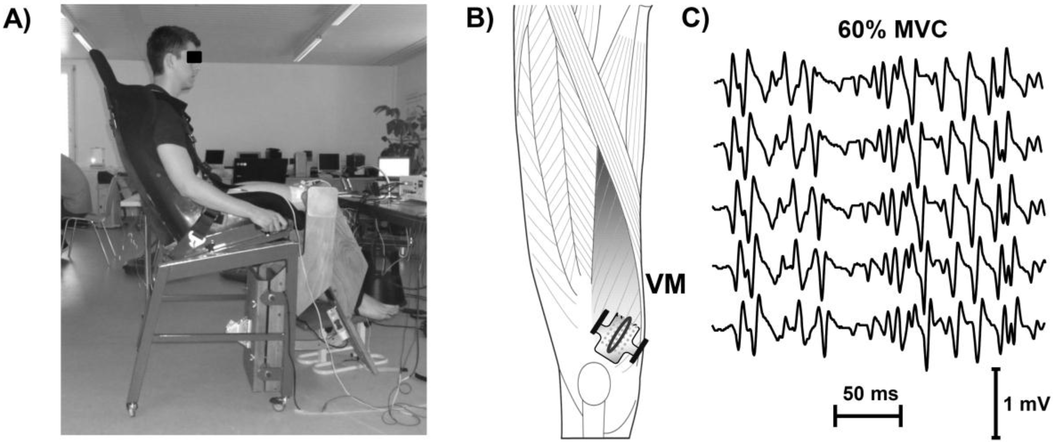
Figure 1. Experimental setup and signal processing. (A) Subject position; (B) electrode array position on vastus medialis (VM) and representation of surface electromyography (EMG) signals (C). Myoelectric signals were detected in single differential configuration, using bidimensional arrays, positioned along the length of the muscles, between the innervation zone and the distal tendon. Channels chosen by visual analysis, for the subsequent conduction velocity (CV) estimation, are indicated by the oval.
Jfmk 01 00193 g001
Figure 2. Box-and-whisker plot of the slopes of conduction velocity (CV), normalized with respect to their initial values during 20% and 60% isometric maximal voluntary contractions (MVCs) of the vastus medialis muscle. CG, control group; R12, patients up to 12 months after ACLr; R24+, patients more than 24 months after ACLr.
Jfmk 01 00193 g002