Review

Sex Differences in Autonomic Blood Pressure Regulation: Sex Chromosome Complement and Hormonal Involvement

Instituto de Investigación Médica Mercedes y Martín Ferreyra, INIMEC-CONICET-Universidad Nacional de Córdoba, Friuli 2434, Córdoba 5016, Argentina
*
Author to whom correspondence should be addressed.
Sexes 2023, 4(4), 536-554; https://doi.org/10.3390/sexes4040035
Submission received: 8 June 2023 / Revised: 7 October 2023 / Accepted: 10 October 2023 / Published: 18 October 2023

Abstract

:
Although several lines of evidence from different studies highlight sex differences in cardiovascular diseases, to date, most studies have been focused on males, with the idea that males and females are similar, differing only in the magnitude of the response. However, the principles learned in male models cannot and should not be extrapolated to women and, therefore, it is important to study in greater detail not only the differences between the sexes but also the physiological intertwining of the underlying genetic and hormonal mechanisms of sexual dimorphism. This review explores the sex disparities in the autonomic nervous system regulation of blood pressure (particularly baroreceptor function), with special emphasis on sex hormones and sex chromosome complement factors involved in sexually dimorphic autonomic blood pressure regulation. A more detailed understanding of the sources of physiological disparities between the sexes may also help in understanding the differences between the sexes in rates of cardiovascular disease and may also aid in designing future improvements for sex-tailored therapeutic treatments.

1. Introduction

Although it is not surprising to state that men and women are different, for a long time, most clinical and experimental studies involved male subjects, assuming that males and females are similar, differing only in the magnitude of the response. This introduced bias into clinical concepts, drug development, and translational findings.
Cardiovascular diseases are the primary cause of death worldwide [1]. Men develop cardiovascular diseases earlier than women until midlife, but after menopause, this sex equation is reversed, with women’s deaths due to cardiovascular disease surprisingly increasing [2,3]. Furthermore, cardiovascular diseases manifest clear sex disparities in terms of susceptibility, symptoms, and therapy outcomes [4]. Although, for a long time, sex differences were largely neglected by the scientific community (females being under-represented in experimental studies and clinical trials), considerable efforts have been made in recent years to remind clinicians and researchers that sex matters [2,5,6].
This review will discuss sex differences in autonomic blood pressure regulation over the lifetime, in particular analyzing the effects of sex chromosome complement (SCC) and sex hormones.

2. Sexual Dimorphism in Autonomic and Baroreflex Blood Pressure Regulation

The autonomic nervous system modulates short-term blood pressure regulation through the sympathetic and parasympathetic arms. Baroreflexes are not static but, on the contrary, vary according to the behavioral state and are thus highly effective in regulating sympathovagal activity and arterial blood pressure during different physiological states [7]. The factors influencing baroreflex function, including sex, may have an important role in cardiovascular diseases.
Gender-related changes in sympathetic and parasympathetic autonomic blood pressure regulation that occur throughout life have been extensively described. A meta-analysis study in children and adolescents under the age of 18 demonstrated significant sex differences in vagal activity, with girls displaying lower resting cardiac parasympathetic tone and greater mean heart rate than boys [8]. In contrast, higher vagal activity and decreased cardiac sympathetic tones were described in adult young women compared with age-matched men [9,10,11], pointing to age- and sex-dependent changes in vagal/sympathetic autonomic cardiovascular control during life. Moreover, around the age of 50, a crucial turning point for sexual dimorphism in baroreflex sensitivity are also observed, with women exhibiting a decrease in the parasympathetic regulation of heart rate, thus showing an earlier decline in baroreflex sensitivity with age than men [12,13]. Thus, while young women show a main parasympathetic control in blood pressure regulation, sympathetic tone dominates in postmenopausal women [14,15], which may contribute to increased cardiovascular morbidity with aging in women. However, after the age of 60, no differences have been found in sympathovagal influence on heart rate parameters between men and women [13].
Experimental and clinical studies have provided ambiguous results concerning sexual dimorphism in baroreflex sensitivity. Studies have demonstrated a greater as well as lower baroreflex sensitivity in females than in males and some have even found no differences among sexes [14,16,17,18,19,20,21]. The differences among the studies, including different ages, races, hormonal status, and hormonal therapies in the analysis of sexual dimorphism in autonomic blood pressure regulation, may explain these widely divergent observations. This highlights the importance of studying and dissecting in more detail the factors involved in sexual dimorphism in autonomic blood pressure regulation.

3. But Why Do Males and Females Show Differences in Autonomic Blood Pressure Regulation?

For years, there has been considerable evidence demonstrating the influence of sex hormones on sexual dimorphism on autonomic blood pressure regulation. However, SCCs may also be involved in inducing differences among sexes [22]. Males and females carry different SCCs (XY and XX, respectively) and are therefore influenced throughout life by X and Y chromosome genes. Some genes in the sex chromosomes act in a specific way to differentiate gonadal sex and establish the hormonal profile for life. Such is the case of the Sry gene (testis-determining gene within the Y chromosome), which is expressed during early embryogenesis in gonadal primordia to cause testis differentiation in XY individuals. However, in XX embryos, certain autosomal chromosomes and other X-linked genes (which are silenced by the Sry gene during male differentiation) induce bipotential gonads to differentiate into ovaries [23,24]. Thus, sex chromosomes play a dominant role in establishing sex-specific gonadal secretions, which in turn exert differential effects and influence sexual dimorphism. Sex hormones can exert organizational and activational effects. Organizational effects induce lasting or permanent changes and are due to exposure to sex steroids during critical periods of development, while activational effects are temporary or reversible and are manifested at different times of life during neonatal and peripubertal development, as well as in adulthood, causing sex differences in the phenotype [25,26,27]. Furthermore, gonadal sex hormones have been shown to modulate epigenetic modifications (through DNA methylation and histone modifications) of autosomal gene expression, thus inducing sex-related differences in gene expression [28,29]. During early embryonic development in women, one of the X chromosomes in the XX-SCC is inactivated (Xi) via an epigenetic mechanism, which leads to a dose compensation of X chromosome genes between men and women [30]. However, some genes escape X-chromosome inactivation and are expressed on both the “active (Xa)” and “inactive (Xi)” X chromosomes, and this may generate differences in addition to those due to the differential effects of the genes that reside on the X and Y sex chromosomes [31,32,33]. Furthermore, several lines of evidence have also demonstrated that some genes exhibit a tissue-specific X-chromosome inactivation escape pattern. This difference in tissue dependence, as well as the differences among species [31,34,35,36,37], may thus induce additional specific differences in phenotypes among males and females. Thus, genetic and/or hormonal pathways (organizational and activational) could act independently or interact synergistically or antagonistically to modulate sexual dimorphic development [38,39].

4. Female Autonomic Blood Pressure Regulation during Lifetime

Studies have demonstrated an activational hormonal sex effect on cardiovascular regulatory mechanisms by altering sympathetic–parasympathetic activity and thus influencing baroreflex sensitivity and blood pressure regulation [15,40]. As previously mentioned, a shift from lower vagal activity in girls under 18 years old [8] to higher vagal activity in healthy young women [9,10,11] may be linked to changes in hormonal sex hormones associated with pubertal development. Furthermore, during menopause, the switch from high (in premenopausal women) to low (in menopausal women) parasympathetic activity [12,14,15] may also be associated with changes in activational hormonal effects.
Fluctuations in sex hormones throughout the menstrual cycle can contribute to the study of the influence of physiological levels of sex hormones on autonomic blood pressure regulation. Clinical studies evaluating cardiac autonomic parameters during the menstrual cycle (in the follicular and luteal phases) have yielded contradictory results. Studies in healthy young women aged 20–38 demonstrated increased sympathetic activity in the luteal phase with a higher low-to-high-frequency power ratio (LF/HF) than in the follicular phase, suggesting an ovarian hormonal activational effect on the cardiovascular sympathetic–parasympathetic ratio during the menstrual cycle [41]. However, although the LF/HF ratio is higher during the luteal phase, it is not independent of lifestyle factors such as diet, physical activity, and sleep, which are otherwise altered during the menstrual cycle [42]. In contrast, studies in naturally menstruating young healthy women have shown an increased parasympathetic drive during the early luteal phase [43,44].
In agreement with the clinical evidence, experimental studies have demonstrated changes in baroreflex sensitivity during the rat estrous cycle [45]. Estrogen increases the synthesis of acetylcholine and the density and affinity of muscarinic receptors, enhancing cholinergic muscarinic activity, which induces a facilitator effect on cardiac vagal outflow in females [46,47,48]. Supporting these findings, De Melo et al. (2016) [49] demonstrated that ovariectomy resulted in a decrease in bradycardic baroreflex response, an increase in sympathetic outflow, and reduced parasympathetic activity in heart rate regulation.
Furthermore, experimental studies evaluated the effects of early and physiological menopause on autonomic cardiac physiology in elderly female Wistar rats. For this, 10-week-old female Wistar rats were assigned to two groups (gonadectomized and sham-operated), and after 12 and 72 weeks, autonomic cardiovascular regulation was evaluated. It should be noted that young female rats, regardless of whether they belonged to the sham-operated or ovariectomized groups, showed a predominant role of the vagal component in heart rate regulation, while both elderly groups (sham and ovariectomized operated) had a predominance of the sympathetic autonomic component, which highlights the influence of aging on autonomic cardiovascular regulation irrespective of hormonal status [50].

4.1. Activational Effect of Estrogen on Autonomic Blood Pressure Regulation

In line with these previous results, studies have also shown that the intravenous and intracerebral administration of estrogen increases vagal tone and suppresses sympathetic efferent activity in ovariectomized female and male rats [51,52], with an enhancement in arterial baroreceptor reflex in both sexes [17,51,52,53,54]. They demonstrate that estradiol induces an activational hormonal effect, increasing cardiovagal parasympathetic activity while diminishing sympathetic drive. As for the influence of activational estradiol on brainstem autonomic circuits, it is important to note that estrogen receptors are expressed throughout the brain stem’s autonomic regulatory centers [55,56,57,58]. Central estrogen administration increases both vagal nerve activity and baroreflex sensitivity. Phenylephrine (PE)-evoked changes in heart rate are significantly enhanced following the injection of estrogen into the nucleus of the solitary tract (NTS), with an enhanced baroreflex response [59]. Furthermore, estrogen injection into the parabrachial nucleus induces changes in autonomic and cardiovascular parameters, reducing the sympathetic tone while increasing the parasympathetic tone [60]. A detailed description of autonomic blood pressure and the influence of sex hormones in brain stem circuits is provided in the study of Littlejohn et al. [61].
Studies by Pamidimukkala et al. in ovariectomized wild-type female mice have demonstrated that estrogen exerts a facilitating activational effect on the bradycardic response for both angiotensin II (Ang II) and PE infusions when compared to ovariectomized female mice without estrogen replacement [17]. Furthermore, they also showed [62] that the estradiol-facilitated baroreflex response may be mediated by the estrogen receptor alpha subtype (Erα) since no activational estradiol hormonal effect on bradycardic baroreflex response was observed in ovariectomized ERαKO mice. Furthermore, as 17-estradiol production is dependent on enzymatic cytochrome P450 aromatase, knockout mice for this enzyme show high circulating levels of testosterone but are deficient in estrogen, which could explain the impaired baroreflex sensitivity in this mouse strain and thus highlight the importance of enzymatic aromatase in autonomic blood pressure regulation [63].
Clinical studies have also indicated an activational estradiol effect on baroreflex heart rate regulation; however, differences among females with low and high orthostatic intolerance have been described. Patients with orthostatic intolerance have a baroreflex function impairment, with a lower cardiac vagal baroreflex response [64]. Young women with regular menstrual cycles, assigned to low orthostatic and normal/high orthostatic tolerance groups (depending on baseline orthostatic response), demonstrated differences in sex hormone influence on baroreflex response. In the female low orthostatic tolerance group, estradiol replacement levels as those observed during natural menstrual cycling attenuated peripheral vasoconstriction while improving baroreflex heart rate regulation. However, in the normal/high orthostatic tolerance group, no changes in the baroreflex control of heart rate due to estradiol replacement treatment were observed [65]

4.2. Activational Effect of Progesterone on Autonomic Blood Pressure Regulation

Studies in young women with normal ovulatory menstrual cycles have demonstrated an increase in sympathetic baroreflex sensitivity activity during the mid-luteal phase [66,67], while no differences in parasympathetic activity were observed during the menstrual cycle [68]. However, in other studies, no differences in cardiovagal baroreflex sensitivity were observed among women in the early follicular and mid-luteal phase [69].
Taking into consideration that, during the secretory phase, both estrogen and progesterone levels are high and that they may have counteractive effects on baroreflex function (thus canceling each other’s effects), studies by Brunt et al. sought to analyze the isolated effects of both hormones on baroreflex function due to stimulated neck pressure hypotension in young women aged 21.5 ± 0.4 years old [70]. Hormone suppression treatment followed by a 3- to 4-day oral progesterone treatment (200 mg/day) resulted in an attenuated fall in femoral vascular conductance, while the opposite was observed in the E2-treated women, thus indicating that progesterone (in isolation of estrogen effects) attenuates carotid–vasomotor baroreflex sensitivity.

4.3. Pregnancy, Progesterone Metabolites, and Baroreflex Function

Normal pregnancy is characterized by a hemodynamic-specific profile. While blood volume and cardiac output increase by 30–40%, arterial blood pressure falls due to a greater decrease in total peripheral resistance. During pregnancy, the impairment of arterial baroreceptor reflex has been described in different species [71]. While pregnant animals maintain their ability of sympathoinhibition response to a hypertensive stimulus, they show an attenuated reflex response in sympathoexcitation outflow in response to hypotensive states [71,72,73]. This depressed baroreflex physiological adaptation has important adverse consequences during pregnancy, including orthostatic hypotension and increased hypotension susceptibility due to hemorrhage, which may lead to undesirable complications during pregnancy and delivery [71,73,74].
This attenuation in baroreflex response to hypotensive stimuli during pregnancy may be induced by the mediated effect of 3-hydroxydihydroprogesterone (3-OH-DHP)-GABAA in the rostral ventrolateral medulla (RVLM) [71,75]. During pregnancy, progesterone increase is accompanied by the augmentation of 3-OH-DHP levels, which is not only the major metabolite of progesterone but also a potent endogenous positive GABAA receptor modulator, thus potentiating GABA-mediated attenuation on baroreflex sympathoexcitatory responses [72,73,76,77]. Studies have demonstrated that GABAA receptor sensitivity to 3-OH-DHP is in part dependent on the subunit GABAA receptors’ composition, which may also change during pregnancy under the influence of ovarian hormones. Furthermore, 3α-OH-DHP levels may also be locally modulated by the 5-reductase and 3-OH steroid oxidoreductase enzymes, responsible for the synthesis from progesterone, which are augmented at the final stages of gestation and thus may be responsible for modulating GABAA receptor activation [71,74].

4.4. Polycystic Ovarian Syndrome and Autonomic Cardiovascular Dysfunction

Polycystic ovary syndrome is an endocrine disorder characterized by polycystic ovary, hyperandrogenism, and menstrual irregularities with an increase in adverse cardiometabolic risk factors [78,79]. Although polycystic ovarian syndrome has been associated with autonomic cardiac dysfunction, differences due to age cohorts and especially cardiovascular risk factors have been shown to influence autonomic dysfunction. Studies in young females (mean age 22.8 ± 3.9) with polycystic ovarian syndrome (but without cardiovascular risk factors), showed no differences in heart rate variability (HRV) when compared to healthy females [80]. Nonetheless, when HRV was evaluated in aged woman (46 years old) with polycystic ovarian syndrome, a reduction in parasympathetic activity (for which HRV was analyzed) was observed. However, the multivariate regression analysis revealed that this reduction in HRV was associated with metabolic abnormalities (high blood pressure, dyslipidemia, and insulin resistance) rather than with polycystic ovarian syndrome per se, thus highlighting the influence of these cardiovascular risk factors in autonomic cardiovascular dysfunction [81].

5. Male Autonomic Blood Pressure Regulation during Lifetime

Taking into account that previous clinical studies [82] had demonstrated an age-dependent decrease in cardiovagal baroreflex sensitivity, Balcock et al. evaluated whether this decline in the cardiovascular function in men may be related to decreased testosterone levels and thus analyzed cardiovagal baroreflex sensitivity in different groups: young men with normal testosterone levels (age = 31 ± 4 yr, testosterone = 535 ± 60 ng/dL); middle-aged/older men with normal testosterone levels (age 56 ± 3 yr; testosterone = 493 ± 85 ng/dL); and middle-aged/older men with low testosterone (age = 57 ± 6 yr; testosterone = 262 ± 31 ng/dL). As previously reported, the results not only demonstrated that middle-aged/older men had lower cardiovagal baroreflex sensitivity than young men, but also that baroreflex sensitivity was facilitated in middle-aged/older men with normal testosterone levels compared with the low testosterone group [83].
Hypogonadism in men is a condition characterized by low serum testosterone levels in association with clinical symptoms of hormonal deficiency; however, its association with hypertension and an increased risk of cardiovascular diseases is still controversial [84]. The increase in life expectancy and the fact that hypogonadism is a syndrome that has a high prevalence in the middle-aged and older male population, as well as the evidence from longitudinal and cross-sectional studies pointing to a gradual decrease in serum testosterone levels after the third decade of life (approximately 1% per year of life), highlight the need to study the risks and benefits of testosterone treatment in patients with hypogonadism in detail [85]. Following this line of study, Di Lodovico et al. [86] analyzed the relationship between chronic heart failure and hypogonadism in which they demonstrated a correlation between low levels of testosterone with the severity of chronic heart failure, which suggests that symptomatic patients with hypogonadism could benefit from testosterone hormone replacement treatments in the short and long term.

Activational Effect of Testosterone on Autonomic Blood Pressure Regulation

As previously mentioned, a facilitatory cardiovagal baroreflex sensitivity in men has been demonstrated [83]; however, in experimental animal models, contradictory evidence on the activational hormonal effects of testosterone on autonomic blood pressure regulation has been described. Studies have demonstrated that chronic treatment with a synthetic androgen (nandrolone decanoate) in male Wistar rats reduces cardiac vagal activity, and this effect is reversed with the administration of angiotensin II AT1 receptor blockade and aldosterone receptor blockade [87]. On the other hand, orchidectomy [88,89] or androgen receptor blockade [89] attenuated baroreflex-mediated bradycardia in conscious rats. Although PE induced similar rises in mean arterial pressure in sham-operated and orchidectomized male rats, an attenuated bradycardic baroreflex response was observed in the latter group. Furthermore, androgen receptor blockade attenuated the bradycardic baroreflex response. In coincidence with these results, studies analyzing the effect of testosterone replacement on the bradycardic baroreflex response in orchidectomized young conscious male rats demonstrated a testosterone-facilitating effect [90] with an enhancement in the cardiac vagal autonomic response [88].

6. Effect of Hormonal Therapies on Autonomic Blood Pressure Regulation

6.1. Contraceptive Treatment Effects and Autonomic Blood Pressure Regulation

In view of the evidence of an activational hormonal effect on the influence of the sympathetic–parasympathetic system on baroreflex sensitivity, clinical studies sought to evaluate in young healthy women the influence of contraceptive treatment on autonomic blood pressure regulation. After 2 weeks of receiving combined ethinyl estrogen–progestin oral contraceptives, women showed a lower cardiovagal and sympathetic baroreflex sensitivity than the placebo [91]. However, studies in women who had been under contraceptive treatment for at least 6 months (placebo for 7 days and 21 days under ethinyl estrogen and low-dose progestin treatment) [92] showed no differences in the LF/HF ratio during the placebo and high-hormone phases, thus canceling out the cardiac vagal differences observed during the menstrual cycle. Furthermore, studies by Schueller et al. analyzed the impact of synthetic progestagen contraceptive treatments on HRV and baroreceptor sensitivity in young healthy females in which no differences in autonomic cardiovascular responses were observed among women under progestogen contraceptive treatments and control groups [93].

6.2. Fertility Treatments and Autonomic Blood Pressure Regulation

During fertility treatments in women, ovulation induction causes estrogen levels to oscillate between low values followed by extremely high levels in a short period of time (about two weeks). Bearing this in mind, and given the previous evidence of a modulating effect of estrogen on autonomic blood pressure regulation, Weissman et al. [94] sought to analyze how these acute variations in estrogen in young women were capable of modulating the cardiovascular autonomic response. HRV parameters in this group of patients showed that the increase in estrogen levels induced greater vagal activation, with no significant effects on sympathetic activity, thus clearly demonstrating activational estradiol parasympathetic autonomic response.

6.3. Hormone Replacement Treatment in Menopause on Autonomic Blood Pressure Regulation

The decrease in hormone levels that occurs during menopause in women affects many tissues and their functions, including the cardiovascular system. This has led to the implementation of different hormonal replacement therapies for which controversial results have been described. Yildirir et al. studied the impact on the cardiac autonomic function of 6 months of hormone replacement therapy in postmenopausal women and found that both estrogen replacement and estrogen-plus-progesterone replacement protocols resulted in an improvement in cardiac autonomic function (with a significant decrease in the average LF/HF ratio and an increase in HF power) [95]. Moreover, in healthy perimenopausal and early postmenopausal women aged 45–60 years old, transdermal estradiol plus intermittent micronized progesterone treatment also resulted in higher baroreflex sensitivity after 6 and 12 months of treatment, highlighting the beneficial cardiovascular effects of initiating this treatment around the time of menopause onset [96].
Furthermore, in another study cohort, differences in LF/HF ratios were observed among males and premenopausal females, with the latter showing lower LF/HF levels, suggesting a preponderance of vagal over sympathetic response. Furthermore, men and postmenopausal women without hormonal treatment, as well as postmenopausal women with estradiol-plus-progesterone hormonal treatment, showed similar results, but estrogen hormonal treatment reduced LF/HF levels to those reported for young women, indicating an activational hormonal effect of estrogen on autonomic cardiovascular regulation [97]. Moreover, studies analyzing 8 weeks of transdermal estrogen replacement treatment in postmenopausal women demonstrated a decrease in sympathetic nerve discharge compared to control postmenopausal women, but no changes in baroreflex sensitivity were described for either transdermal or oral estradiol replacement protocols [98]. These studies thus indicate that, although estradiol hormonal treatment results in an improvement in cardiac autonomic function in postmenopausal women, differences among transdermal and oral estradiol treatments should be taken into consideration.

7. Organizational Sex Hormonal and SCC Effects on Sexually Dimorphic Blood Pressure Regulation

Epigenetic hormonal imprinting during critical periods of development induces persistent and heritable changes in gene expression and function (without inducing changes in DNA base sequence), influencing the hormone–receptor interplay and thus inducing sexual dimorphic profiles during lifetime [99,100]. Thus, hormonal imprinting during critical periods may have lifetime consequences inducing changes in development to adjust to the environmental conditions in the long term [101].
As cardiovascular regulation is influenced by the interactions of genetic, hormonal (organizational and activational) and environmental factors, and given the fact that in both pre-eclampsia and pregnant women with polycystic ovary syndrome, testosterone levels are increased during gestation [102,103,104], different experimental and clinical studies have sought to analyze the influence of high prenatal testosterone exposure on blood pressure regulation in adult offspring. Studies in animal models have demonstrated that prenatal exposure to higher levels of testosterone during development may induce an increase in the incidence of hypertensive profiles in the offspring later in life [105,106]. Studies also demonstrated that a higher level of testosterone exposure during pregnancy induced an increase in testosterone and blood pressure levels in adult male and female offspring; however, male offspring showed higher levels of blood pressure when compared to females. Furthermore, they also demonstrated that gonadectomy in offspring abolished the increase in blood pressure; however, testosterone replacement in males restored the hypertensive state, while estradiol in females had no effect on blood pressure. Thus, these results demonstrate that prenatal exposure to testosterone induces a hormonal-dependent increase in blood pressure later in life, thus demonstrating an interaction of organizational and activational hormonal effects on blood pressure regulation [107].
In coincidence with the latter in a cohort study, Le-ha et al. found an association between prenatal testosterone at birth (from the umbilical blood cord) and blood pressure levels after 20–27 years old. Higher prenatal testosterone levels were found to be associated with higher blood pressure levels in both men and women, thus demonstrating a testosterone programming effect during critical periods of development on both diastolic and systolic blood pressure during adulthood [108].
Differences in the SCC may lead to a genetic imbalance in males and females, acting independently or interacting (synergistically/antagonistically) with organizational and activational hormonal effects in modulating sexually dimorphic blood pressure phenotypes [38,39]. Previous studies in patients and spontaneously hypertensive rats demonstrated an association between the Y chromosome and high blood pressure [54,109,110]. In a male cohort of more than 400 patients, Ellis et al. [110] demonstrated a significant association between higher diastolic blood pressure and Y-chromosome polymorphism in the non-recombining region. Furthermore, studies in the consomic strains of spontaneously hypertensive (SHR) and normotensive Wistar Kyoto (WKY) rats demonstrated a higher increase in blood pressure in male (but not female) offspring from SHR fathers and WKY mothers, which links the Y-chromosome effect with higher blood pressure levels in SHR male rats [109]. Later, Wiley et al. showed an important reduction in blood pressure reduction in consomic adult SHR/y male rats that were neonatally sympathectomized during the first three weeks of age [111]. Moreover, studies have demonstrated that the Syr gene in the Y chromosome not only initiates gonad determination during early development in males but can also modulate norepinephrine synthesis in males. Studies have demonstrated that the SRY factor in the midbrain in males induces catecholamine synthesis [112], while in PC12 cells, it was shown to regulate TH transcription [112,113].
Sex chromosomes can cause sex differences in non-gonadal tissues independent of the effects of gonadal hormones [114]. Recent studies have demonstrated that SCC may induce cardiac sex-biased protein expression before gonadal development and hormonal influence. Thus, cardiac sex disparities may be driven by both hormonal and SCC effects which may induce sex-related differences in cardiovascular diseases later in life. In recent years, the four-core genotype mouse model has provided insights into sex chromosomes and sex hormone effects as well as their interaction [115]. In this mouse model, the Y chromosome and the Sry gene segregate independently. This transgenic line arose from the Sry gene deletion of the Y chromosome and subsequent insertion of the Sry transgene (testes-determining gene) into an autosome. Regardless of their SCC, all individuals with the Sry transgene will develop testes and male secondary external sexual characteristics, while those lacking the transgene will develop ovaries with a secondary external female phenotype. The XY-Sry male mice (without the Sry gene in the Y chromosome but with the Sry transgene in an autosomal chromosome) are fully fertile. Thus, crossing the former with XX females then results in four genotypes: XX and XY-females (without the Sry gene on the Y chromosome), and XX-Sry and XY-Sry male mice (both with Sry on an autosome). The following designations are applied for each genotype: XY-Sry male mice as XY-male; XX-Sry as XX-male, XX as XX-female, and XY- as XX-female mice. By comparing these genotypes, it is thus possible to dissociate the role of (a) SCC (by comparing individuals with the same gonadal type but with different SCC), (b) gonadal sex (studying males vs. females regardless of SCC), as well as (c) the interaction of SCC and hormonal influence (activational and/or organizational) (Figure 1).

SCC and Organizational Sex Hormonal Effects on Sexually Dimorphic Bradycardic Baroreflex Response

Ang II exerts differential effects on the bradycardic baroreflex response in males and females [116]. Clinical studies demonstrate a sexually dimorphic profile in the baroreflex bradycardic angiotensinergic response, which is attenuated in men compared to that observed in women [117]. Studies by Pamidimukkala et al. [17] have also demonstrated a sex-dependent bradycardic profile in mice. In males, although the infusion of PE and Ang II resulted in blood pressure increases of equal magnitude, the Ang II-baroreflex bradycardic response was significantly less than that induced by PE. However, in females, both PE and Ang II induced a similar baroreflex decrease in heart rate [16,17]. Studies analyzing the role of the SCC on the sexually dimorphic response using the four-core transgenic mouse model have shown that a bolus infusion of Ang II induces a differential baroreflex response depending on the SCC. Regardless of being male or female, gonadectomized mice with XY-SCC (XY-female and XY-male mice), showed an attenuated baroreflex response compared with those with XX-SCC (XX-male and XX-female mice). Thus, these data demonstrate that, in the absence of hormonal activation effects, there is a facilitatory role of the XX-SCC in the regulation of the Ang II bradycardic baroreflex response. Furthermore, experimental studies also revealed an interaction of organizational hormonal and SCC factors on the dimorphic bradycardic response due to PE infusion. Gonadectomized XY-female mice showed an attenuated PE-bradycardic baroreflex response compared to XX-female, XY-male, and XX-male mice, thus demonstrating an interaction of genetic factors within the SCC and organizational hormonal effects during critical periods of development [22].
Among the brain areas underlying the angiotensinergic baroreflex bradycardic sexual dimorphism, the evidence highlights an important role of the area postrema (AP). This nucleus sends projections to different brain areas of the medulla oblongata involved in cardiovascular regulation, including the dorsal vagal complex, the NTS, and the RVLM, modulating the parasympathetic and sympathetic drive and thus regulating the baroreflex short-term blood pressure mechanisms [118,119]. AP injury not only blocks the hypertensive response to chronic Ang II infusion but also prevents the decreased baroreflex sensitivity observed after the acute administration of this peptide in males [120,121,122]. Regarding short-term blood pressure regulation, Ang II has been shown to induce an AT1-receptor-mediated attenuation of baroreflex bradycardic response, while the activation of AT2 and Mas receptors may facilitate the baroreflex heart rate response [123,124,125]. The AT2 receptor gene (Agtr2), which is part of the dilator arm of the renin–angiotensin system (RAS) and that facilitates the baroreflex bradycardic response, is located on the X chromosome [126]. Taking this into account, as well as the evidence indicating that there are genes capable of escaping X chromosome inactivation [31,32,33], Ang receptor expressions in brain nuclei were analyzed in the four-core genotype model, in which the facilitating effect of Ang II SCC-XX on the baroreflex bradycardic response had already been described [22]. In the absence of an activational hormonal influence, a sex chromosome effect on angiotensin receptor gene expression at the AP was identified. Regardless of gonadal sex, mice with XX-SCC (XX-female and XX-male mice), compared with those with XY-SCC (XY-female and XY-male mice), showed an increase in Mas1 receptor expression in association with a diminished Agtr1a expression and Agtr1a/Agtr2 ratio [127]. These results are in line with previous studies in which an XX-SCC facilitator was shown to affect the angiotensinergic bradycardic baroreflex response [22]. In addition to the modulating effect of the angiotensinergic system on short-term blood pressure regulation, many studies have highlighted the important role of the RAS on long-term regulation [128]. As regards the modulatory effect of SCC on blood pressure, Ji et al. [129] studied the four-core genotype mouse model and demonstrated that, after 2 weeks of Ang II infusion, the mean arterial pressure was greater in gonadectomized -XX than in -XY mice regardless of whether the mice were born with testes or with ovaries. All these findings demonstrate that, in the absence of activational hormonal effects, SCC differentially modulates sexually dimorphic Ang II-hypertensive and bradycardic baroreflex responses, while its interaction with organizational hormonal effects is implicated in the phenylephrine-induced bradycardic baroreflex response [22,129].

8. Transsexual Sympathovagal Imbalances during Hormonal Treatments—Interaction of SCC, Organizational and Activational Hormonal Effects

Taking into account previous studies that addressed the influence of gender and sexual hormones on autonomic blood pressure regulation, Resmini et al. [11] aimed to evaluate the sympathovagal balance in male-to-female (MtoF) transsexual subjects, female-to-male (FtoM) transsexuals, as well as in males and females of the same age group. MtoF transsexual patients under oral or transdermal estradiol therapy showed a decrease in both sympathetic and parasympathetic activity compared to the other groups. Thus, estrogen treatment would not only reduce sympathetic activity compared to that described for men but would induce HRV indices similar to those observed in middle-aged women. However, in contrast to the latter, MtoF transsexual patients showed a decreased parasympathetic influence, differing from both men and women with regard to sympathovagal cardiac autonomic balance. On the other hand, FtoM transsexual subjects (who underwent testosterone treatment) presented a lower parasympathetic profile than that described for women, which could have an impact on the risk of cardiovascular events in the future [11]. Although a large number of clinical and experimental studies indicate a protective effect of estrogen on cardiovascular regulation, MtoF transsexual subjects treated with estrogens show higher risks of cardiovascular events than testosterone-treated FtoM transsexuals [130]. However, it is important to highlight that both the dosage rate of two or four times of oral contraceptives for cross-sex hormone treatment as well as the route of administration may have an impact on increased cardiovascular events in MtoF subjects, which highlights the importance of examining the cardiovascular physiological implications of sex hormone treatments in detail [131].

9. SCC and Cardiovascular Diseases

Cardiovascular diseases are the leading cause of mortality in both men and women, with clear sex differences in risks and outcomes in cardiac events [1]. Increased sympathetic and/or diminished parasympathetic tones induce autonomic sympathetic/vagal imbalances that have been associated with increases in cardiovascular risks, outcomes, and mortality [132,133,134,135]
Sex chromosome genes have been implicated in inherited forms of cardiovascular diseases. Supporting clinical evidence of sex chromosome involvement in cardiovascular diseases demonstrates that female patients with Turner syndrome (monosomy X) have resting tachycardia and higher blood pressure levels, with hypertension manifesting at an earlier age [136,137]. Turner syndrome is associated with the dysregulation of the sympathetic nervous system, leading to tachycardia and high blood pressure, increased resting norepinephrine levels, and a compromised response to sympathetic stimulation, thus reflecting overactivity of basal sympathetic tone [137].
Moreover, androgen insensitivity syndrome is an X-linked recessive genetic disorder caused by inactivating mutations in the androgen receptor gene in patients with a 46,XY karyotype. In consequence, these patients develop a female phenotype due to partial or complete resistance to androgen effects, thus interfering with the normal XY-male organizational masculinization effect during development [138]. Various hormonal replacement therapies have been evaluated in patients with androgen insensitivity syndrome, which is why, among other factors, the cardiovascular risks of hormonal treatments should be investigated in the future [139].
Clinical studies have demonstrated an increase in cardiovascular mortality in patients with Klinefelter syndrome (47,XXY) [140,141]. Patients show chronotropic incompetence, defined as a diminished heart rate response, that seems to result from the disruption of the sympathetic–parasympathetic balance of the autonomic nervous system due to increased sympathetic drive. Chromosomal abnormality in Klinefelter patients also appears to play a major role in inducing cardiovascular phenotype, as testosterone therapy does not normalize cardiovascular abnormalities despite leading to normal testosterone levels [142]. Taking this evidence into account and data indicating a decrease in testosterone levels during childhood in patients with Klinefelter syndrome [143], a series of preliminary studies have evaluated the effect of androgen treatment between 1 and 3 months of age on the potential organizational effects of testosterone on development [144,145]. Although these retrospective clinical descriptive studies of cohorts demonstrated an improvement in cognitive and psychosocial functions, future long-term studies including the cardiovascular profile need to be conducted.
In conjunction with Klinefelter syndrome, Jacobs syndrome (also known as 47,XYY male), is defined as a “sex chromosome trisomy” condition [146]. The analysis of prenatal status of amniotic fluid from 16 to 20 weeks of gestation in both 47,XXY and 47, XYY male fetus demonstrated no differences in testosterone levels when compared to 46,XY male [147]. Notably, 47,XXY male patients showed slight fertility problems [148]; however, unlike 47,XXY males, in whom a reduction in testosterone levels was observed, no differences in testosterone levels during puberty were reported in 46,XY male patients with Jacobs syndrome [149,150]. As regards cardiac parameters in 47,XYY males, although no differences in blood pressure were reported for these patients, differences in electrocardiogram measurements were found, with prolonged P–R intervals and shorter QRS complexes [151]. Furthermore, in a case report study of a 4-month-old child with double trisomy 48,XXY, +21 (Klinefelter–Down syndrome), pulmonary hypertension and mild tricuspid regurgitation were reported [152].

10. Conclusions

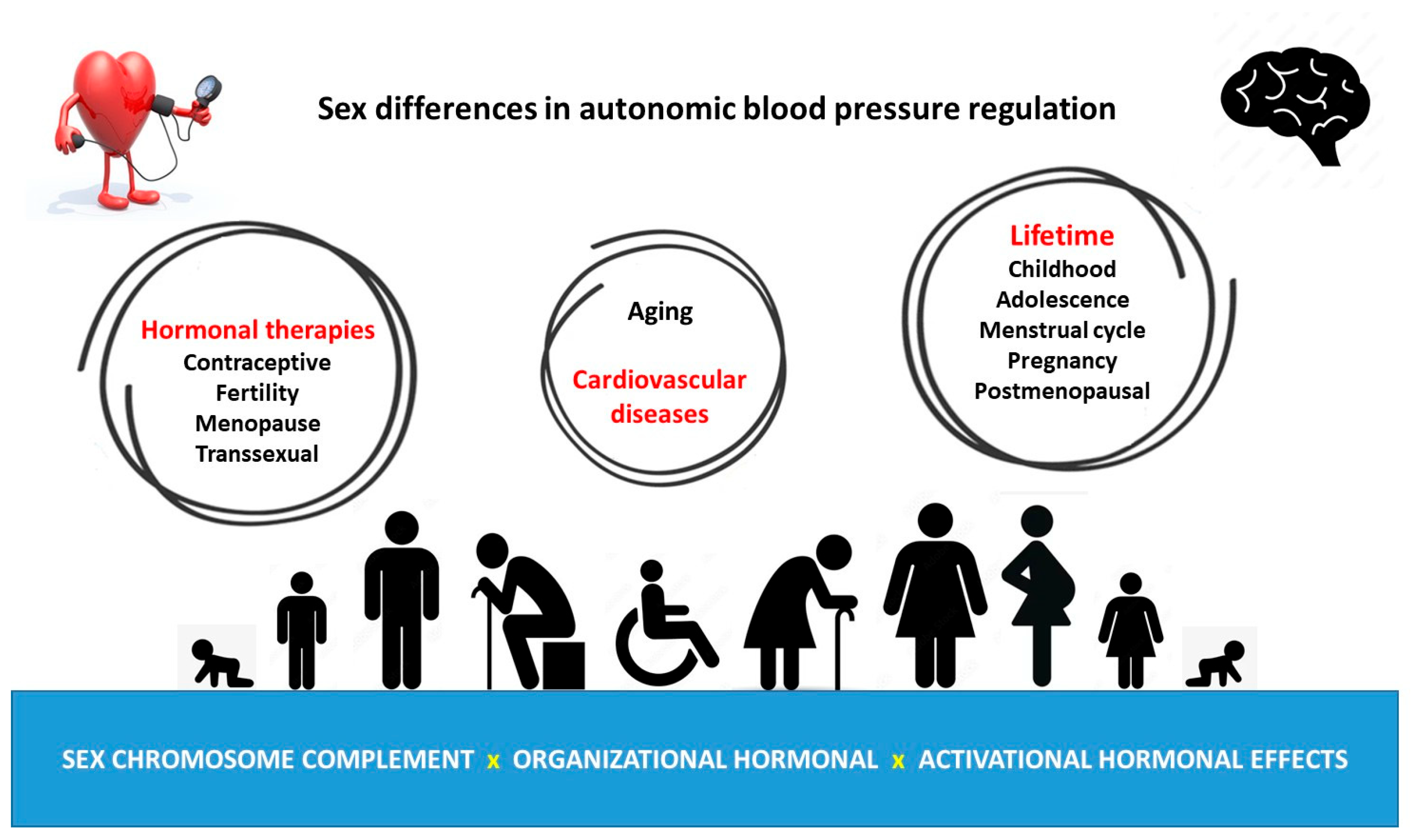
As people age, hypertension and cardiovascular risk increase in a gender- and age-dependent manner. This provides further insight into the complex influence of age- and gender-dependent regulation on cardiovascular autonomic blood pressure control and highlights the importance of studying in more detail the physiological and pathophysiological basis of gender differences in autonomic blood pressure regulation over the lifetime, with special attention to SCC and hormonal sex influences (Figure 2).
Considering the fact that sex hormone therapies are commonly used in contraceptive and fertility treatments, in patients with sex chromosome syndromes, transgender patients, and in aging women and males, it is important to increase our understanding of the influence of sex hormones and sex chromosomes (as well as their interplay) in the physiology and pathophysiology of autonomic blood pressure regulation.
Clarifying the physiological bases of the similarities and differences between the sexes in terms of the mechanisms involved in blood pressure regulation may contribute to understanding how men and women regulate blood pressure differently and thus help to influence sex-directed therapeutic approaches in the future.

Limitations and Perspectives

Several limitations should be taken into account when drawing conclusions on the influence of sex hormones and SCC on autonomic blood pressure regulation. In most clinical and experimental studies, it is not possible to dissociate the effects of SCC from organizational hormonal effects since the Sry testes-determining gene is located on the Y chromosome, and therefore all individuals with XY-SCC will develop testes, while those with the XX-SCC will develop ovaries and therefore will be exposed (during critical periods of development) to organizational hormonal effects, which, in combination with differential expression of SCC genes, may determine sexually dimorphic phenotypes.
It should also be noted that when analyzing the activational influence of sex hormones on cardiac autonomic blood pressure regulation, different physiological stages during the lifetime (puberty, menstrual cycle, menopause, and hypogonadism), as well as hormonal treatments (used for contraception, fertility, menopause, and hypogonadism; as well as in transsexual patients) are been included. However, differences in age, race, age at the time of gonadectomy or the onset of menopause, age at the onset of hormonal treatments and their duration, differential hormonal treatments as well as routes of administration (oral, transdermal, intramuscular, and subcutaneous), and different dosage types (pharmacological versus physiological), makes the results interpretation difficult to dissociate the influence of sex hormones as well as their interaction with genetic factors of SCC in sexual dimorphic autonomic blood pressure regulation.

Author Contributions

Conceptualization, X.E.C. and A.G.; investigation, X.E.C., A.G. and G.V.S.; writing—original draft preparation, X.E.C., A.G. and G.V.S.; writing—review and editing, X.E.C. and A.G.; supervision, X.E.C.; funding acquisition, X.E.C. All authors have read and agreed to the published version of the manuscript.

Funding

This research was funded in part by a grant from the Agencia Nacional de Promoción Científica y Tecnológica (ANPCyT-BID-PICT 2018-03361) to XEC.

Institutional Review Board Statement

Not applicable.

Informed Consent Statement

Not applicable.

Data Availability Statement

No new data were created or analyzed in this study. Data sharing is not applicable to this article.

Conflicts of Interest

The authors declare no conflict of interest.

Abbreviations

Ang IIAngiotensin II
APArea postrema
Agtr1aAT1 type a receptor gene
Agtr2AT2 receptor type 2 gene
AT1Angiotensinergic type 1 receptor
AT2Angiotensinergic type 2 receptor
E2Estradiol
ERαEstrogen receptor alpha
ERαKOEstrogen receptor alpha knockout mice
FtoMFemale-to-male transsexual
HRVHeart rate variability
LF/HFLow-to-high-frequency power ratio
MasAngiotensinergic Mas receptor
MtoFMale-to-female transsexual
NTSNucleus of the solitary tract
PEPhenylephrine
RASRenin–angiotensin system
RVLMRostro ventrolateral medulla
SCCSex chromosome complement
WKYWistar Kyoto

References

  1. Stramba-Badiale, M.; Fox, K.M.; Priori, S.G.; Collins, P.; Daly, C.; Graham, I.; Jonsson, B.; Schenck-Gustafsson, K.; Tendera, M. Cardiovascular diseases in women: A statement from the policy conference of the European Society of Cardiology. Eur. Heart J. 2006, 27, 994–1005. [Google Scholar] [CrossRef]
  2. Colafella, K.M.M.; Denton, K.M. Sex-specific differences in hypertension and associated cardiovascular disease. Nat. Rev. Nephrol. 2018, 14, 185–201. [Google Scholar] [CrossRef]
  3. Sandberg, K.; Ji, H. Sex differences in primary hypertension. Biol. Sex. Differ. 2012, 3, 7. [Google Scholar] [CrossRef]
  4. Mercuro, G.; Deidda, M.; Piras, A.; Dessalvi, C.C.; Maffei, S.; Rosano, G.M. Gender determinants of cardiovascular risk factors and diseases. J. Cardiovasc. Med. 2010, 11, 207–220. [Google Scholar] [CrossRef]
  5. Arain, F.A.; Kuniyoshi, F.H.; Abdalrhim, A.D.; Miller, V.M. Sex/gender medicine. The biological basis for personalized care in cardiovascular medicine. Circ. J. 2009, 73, 1774–1782. [Google Scholar] [CrossRef]
  6. Beery, A.K.; Zucker, I. Sex bias in neuroscience and biomedical research. Neurosci. Biobehav. Rev. 2011, 35, 565–572. [Google Scholar] [CrossRef]
  7. Dampney, R.A.L. Resetting of the Baroreflex Control of Sympathetic Vasomotor Activity during Natural Behaviors: Description and Conceptual Model of Central Mechanisms. Front. Neurosci. 2017, 11, 461. [Google Scholar] [CrossRef]
  8. Koenig, J.; Rash, J.A.; Campbell, T.S.; Thayer, J.F.; Kaess, M. A Meta-Analysis on Sex Differences in Resting-State Vagal Activity in Children and Adolescents. Front. Physiol. 2017, 8, 582. [Google Scholar] [CrossRef]
  9. Evans, J.M.; Ziegler, M.G.; Patwardhan, A.R.; Ott, J.B.; Kim, C.S.; Leonelli, F.M.; Knapp, C.F. Gender differences in autonomic cardiovascular regulation: Spectral, hormonal, and hemodynamic indexes. J. Appl. Physiol. 2001, 91, 2611–2618. [Google Scholar] [CrossRef]
  10. Ramesh, S.; Wilton, S.B.; Holroyd-Leduc, J.M.; Turin, T.C.; Sola, D.Y.; Ahmed, S.B. Testosterone is associated with the cardiovascular autonomic response to a stressor in healthy men. Clin. Exp. Hypertens. 2015, 37, 184–191. [Google Scholar] [CrossRef]
  11. Resmini, E.; Casu, M.; Patrone, V.; Rebora, A.; Murialdo, G.; Minuto, F.; Ferone, D. Sympathovagal imbalance in transsexual subjects. J. Endocrinol. Investig. 2008, 31, 1014–1019. [Google Scholar] [CrossRef]
  12. Yeh, C.-H.; Kuo, T.B.J.; Li, J.-Y.; Kuo, K.-L.; Chern, C.-M.; Yang, C.C.H.; Huang, H.-Y. Effects of age and sex on vasomotor activity and baroreflex sensitivity during the sleep-wake cycle. Sci. Rep. 2022, 12, 22424. [Google Scholar] [CrossRef]
  13. Kuo, T.B.; Lin, T.; Yang, C.C.; Li, C.L.; Chen, C.F.; Chou, P. Effect of aging on gender differences in neural control of heart rate. Am. J. Physiol. 1999, 277, H2233–H2239. [Google Scholar] [CrossRef]
  14. Sachse, C.; Trozic, I.; Brix, B.; Roessler, A.; Goswami, N. Sex differences in cardiovascular responses to orthostatic challenge in healthy older persons: A pilot study. Physiol. Int. 2019, 106, 236–249. [Google Scholar] [CrossRef]
  15. Lavi, S.; Nevo, O.; Thaler, I.; Rosenfeld, R.; Dayan, L.; Hirshoren, N.; Gepstein, L.; Jacob, G. Effect of aging on the cardiovascular regulatory systems in healthy women. Am. J. Physiol. Regul. Integr. Comp. Physiol. 2007, 292, R788–R793. [Google Scholar] [CrossRef]
  16. Matsumura, Y.; Hasser, E.M.; Bishop, V.S. Central effect of angiotensin II on baroreflex regulation in conscious rabbits. Am. J. Physiol. 1989, 256 Pt 2, R694–R700. [Google Scholar] [CrossRef]
  17. Pamidimukkala, J.; Taylor, J.A.; Welshons, W.V.; Lubahn, D.B.; Hay, M. Estrogen modulation of baroreflex function in conscious mice. Am. J. Physiol. Regul. Integr. Comp. Physiol. 2003, 284, R983–R989. [Google Scholar] [CrossRef]
  18. Abdel-Rahman, A.R.; Merrill, R.H.; Wooles, W.R. Gender-related differences in the baroreceptor reflex control of heart rate in normotensive humans. J. Appl. Physiol. 1994, 77, 606–613. [Google Scholar] [CrossRef]
  19. Convertino, V.A. Gender differences in autonomic functions associated with blood pressure regulation. Am. J. Physiol. 1998, 275, R1909–R1920. [Google Scholar] [CrossRef]
  20. Huikuri, H.V.; Pikkuja¨msa¨, S.M.; Airaksinen, K.J.; Ika¨heimo, M.J.; Rantala, A.O.; Kauma, H.; Lilja, M.; Kesa¨niemi, Y.A. Sex-related differences in autonomic modulation of heart rate in middle-aged subjects. Circulation 1996, 94, 122–125. [Google Scholar] [CrossRef]
  21. Tank, J.; Diedrich, A.; Szczech, E.; Luft, F.C.; Jordan, J. Baroreflex regulation of heart rate and sympathetic vasomotor tone in women and men. Hypertension 2005, 45, 1159–1164. [Google Scholar] [CrossRef]
  22. Caeiro, X.E.; Mir, F.R.; Vivas, L.M.; Carrer, H.F.; Cambiasso, M.J. Sex chromosome complement contributes to sex differences in bradycardic baroreflex response. Hypertension 2011, 58, 505–511. [Google Scholar] [CrossRef]
  23. Goodfellow, P.N.; Lovell-Badge, R. SRY and sex determination in mammals. Annu. Rev. Genet. 1993, 27, 71–92. [Google Scholar] [CrossRef]
  24. Mäkelä, J.A.; Koskenniemi, J.J.; Virtanen, H.E.; Toppari, J. Testis Development. Endocr. Rev. 2019, 40, 857–905. [Google Scholar] [CrossRef]
  25. Arnold, A.P.; Gorski, R.A. Gonadal steroid induction of structural sex differences in the central nervous system. Annu. Rev. Neurosci. 1984, 7, 413–442. [Google Scholar] [CrossRef] [PubMed]
  26. McCarthy, M.M.; Arnold, A.P.; Ball, G.F.; Blaustein, J.D.; De Vries, G.J. Sex differences in the brain: The not so inconvenient truth. J. Neurosci. 2012, 32, 2241–2247. [Google Scholar] [CrossRef] [PubMed]
  27. Morris, J.A.; Jordan, C.L.; Breedlove, S.M. Sexual differentiation of the vertebrate nervous system. Nat. Neurosci. 2004, 7, 1034–1039. [Google Scholar] [CrossRef] [PubMed]
  28. Dkhil, M.A.; Al-Quraishy, S.; Abdel-Baki, A.-A.; Ghanjati, F.; Arauzo-Bravo, M.J.; Delic, D.; Wunderlich, F. Epigenetic modifications of gene promoter DNA in the liver of adult female mice masculinized by testosterone. J. Steroid Biochem. Mol. Biol. 2015, 145, 121–130. [Google Scholar] [CrossRef]
  29. Reue, K.; Wiese, C.B. Illuminating the Mechanisms Underlying Sex Differences in Cardiovascular Disease. Circ. Res. 2022, 130, 1747–1762. [Google Scholar] [CrossRef] [PubMed]
  30. Augui, S.; Nora, E.P.; Heard, E. Regulation of X-chromosome inactivation by the X-inactivation centre. Nat. Rev. Genet. 2011, 12, 429–442. [Google Scholar] [CrossRef]
  31. Carrel, L.; Willard, H.F. X-inactivation profile reveals extensive variability in X-linked gene expression in females. Nature 2005, 434, 400–404. [Google Scholar] [CrossRef] [PubMed]
  32. Wolstenholme, J.T.; Rissman, E.F.; Bekiranov, S. Sexual differentiation in the developing mouse brain: Contributions of sex chromosome genes. Genes. Brain Behav. 2013, 12, 166–180. [Google Scholar] [CrossRef]
  33. Yang, X.; Schadt, E.E.; Wang, S.; Wang, H.; Arnold, A.P.; Ingram-Drake, L.; Drake, T.A.; Lusis, A.J. Tissue-specific expression and regulation of sexually dimorphic genes in mice. Genome Res. 2006, 16, 995–1004. [Google Scholar] [CrossRef]
  34. Berletch, J.B.; Yang, F.; Xu, J.; Carrel, L.; Disteche, C.M. Genes that escape from X inactivation. Hum. Genet. 2011, 130, 237–245. [Google Scholar] [CrossRef] [PubMed]
  35. Carrel, L.; Willard, H.F. Heterogeneous gene expression from the inactive X chromosome: An X-linked gene that escapes X inactivation in some human cell lines but is inactivated in others. Proc. Natl. Acad. Sci. USA 1999, 96, 7364–7369. [Google Scholar] [CrossRef]
  36. Zhang, Y.; Castillo-Morales, A.; Jiang, M.; Zhu, Y.; Hu, L.; Urrutia, A.O.; Kong, X.; Hurst, L.D. Genes that escape X-inactivation in humans have high intraspecific variability in expression, are associated with mental impairment but are not slow evolving. Mol. Biol. Evol. 2013, 30, 2588–2601, Erratum in Mol. Biol. Evol. 2016, 33, 302. [Google Scholar] [CrossRef] [PubMed]
  37. Yang, F.; Babak, T.; Shendure, J.; Disteche, C.M. Global survey of escape from X inactivation by RNA-sequencing in mouse. Genome Res. 2010, 20, 614–622. [Google Scholar] [CrossRef]
  38. Arnold, A.P.; Xu, J.; Grisham, W.; Chen, X.; Kim, Y.H.; Itoh, Y. Minireview: Sex chromosomes and brain sexual differentiation. Endocrinology 2004, 145, 1057–1062. [Google Scholar] [CrossRef]
  39. Cabrera Zapata, L.E.; Garcia-Segura, L.M.; Cambiasso, M.J.; Arevalo, M.A. Genetics and Epigenetics of the X and Y Chromosomes in the Sexual Differentiation of the Brain. Int. J. Mol. Sci. 2022, 23, 12288. [Google Scholar] [CrossRef]
  40. Dart, A.M.; Du, X.J.; Kingwell, B.A. Gender, sex hormones and autonomic nervous control of the cardiovascular system. Cardiovasc. Res. 2002, 53, 678–687. [Google Scholar] [CrossRef] [PubMed]
  41. Yildirir, A.; Kabakci, G.; Akgul, E.; Tokgozoglu, L.; Oto, A. Effects of menstrual cycle on cardiac autonomic innervation as assessed by heart rate variability. Ann. Noninvasive Electrocardiol. 2002, 7, 60–63. [Google Scholar] [CrossRef]
  42. Tada, Y.; Yoshizaki, T.; Tomata, Y.; Yokoyama, Y.; Sunami, A.; Hida, A.; Kawano, Y. The Impact of Menstrual Cycle Phases on Cardiac Autonomic Nervous System Activity: An Observational Study Considering Lifestyle (Diet, Physical Activity, and Sleep) among Female College Students. J. Nutr. Sci. Vitaminol. 2017, 63, 249–255. [Google Scholar] [CrossRef] [PubMed]
  43. Blake, E.F.; Eagan, L.E.; Ranadive, S.M. Heart rate variability between hormone phases of the menstrual and oral contraceptive pill cycles of young women. Clin. Auton. Res. 2023, 33, 533–537. [Google Scholar] [CrossRef] [PubMed]
  44. Hirshoren, N.; Tzoran, I.; Makrienko, I.; Edoute, Y.; Plawner, M.M.; Itskovitz-Eldor, J.; Jacob, G. Menstrual cycle effects on the neurohumoral and autonomic nervous systems regulating the cardiovascular system. J. Clin. Endocrinol. Metab. 2002, 87, 1569–1575. [Google Scholar] [CrossRef] [PubMed]
  45. Goldman, R.K.; Azar, A.S.; Mulvaney, J.M.; Hinojosa-Laborde, C.; Haywood, J.R.; Brooks, V.L. Baroreflex sensitivity varies during the rat estrous cycle: Role of gonadal steroids. Am. J. Physiol. Regul. Integr. Comp. Physiol. 2009, 296, R1419–R1426. [Google Scholar] [CrossRef]
  46. Du, X.J.; Dart, A.M.; Riemersma, R.A. Sex differences in the parasympathetic nerve control of rat heart. Clin. Exp. Pharmacol. Physiol. 1994, 21, 485–493. [Google Scholar] [CrossRef]
  47. Olsen, K.L.; Edwards, E.; Schechter, N.; Whalen, R.E. Muscarinic receptors in preoptic area and hypothalamus: Effects of cyclicity, sex and estrogen treatment. Brain Res. 1988, 448, 223–229. [Google Scholar] [CrossRef] [PubMed]
  48. Rainbow, T.C.; Degroff, V.; Luine, V.N.; McEwen, B.S. Estradiol 17 beta increases the number of muscarinic receptors in hypothalamic nuclei. Brain Res. 1980, 198, 239–243. [Google Scholar] [CrossRef]
  49. De Melo, V.U.; Saldanha, R.R.M.; Dos Santos, C.R.; Cruz, J.D.C.; Lira, V.A.; Santana-Filho, V.J.; Michelini, L.C. Ovarian Hormone Deprivation Reduces Oxytocin Expression in Paraventricular Nucleus Preautonomic Neurons and Correlates with Baroreflex Impairment in Rats. Front. Physiol. 2016, 7, 461. [Google Scholar] [CrossRef]
  50. Tezini, G.C.; Becari, C.; Zanotto, C.Z.; Salgado, M.C.; Passaglia Rde, C.; Souza, H.C. Ageing is the main determinant of haemodynamics and autonomic cardiac changes observed in post-menopausal female rats. Auton. Neurosci. 2013, 174, 36–41. [Google Scholar] [CrossRef]
  51. Mohamed, M.K.; El-Mas, M.M.; Abdel-Rahman, A.A. Estrogen enhancement of baroreflex sensitivity is centrally mediated. Am. J. Physiol. 1999, 276, R1030–R1037. [Google Scholar] [CrossRef] [PubMed]
  52. Saleh, M.C.; Connell, B.J.; Saleh, T.M. Medullary and intrathecal injections of 17beta-estradiol in male rats. Brain Res. 2000, 867, 200–209. [Google Scholar] [CrossRef]
  53. El-Mas, M.M.; Abdel-Rahman, A.A. Estrogen enhances baroreflex control of heart rate in conscious ovariectomized rats. Can. J. Physiol. Pharmacol. 1998, 76, 381–386. [Google Scholar] [CrossRef]
  54. Saleh, T.M.; Connell, B.J. 17beta-estradiol modulates baroreflex sensitivity and autonomic tone of female rats. J. Auton. Nerv. Syst. 2000, 80, 148–161. [Google Scholar] [CrossRef]
  55. Merchenthaler, I.; Lane, M.V.; Numan, S.; Dellovade, T.L. Distribution of estrogen receptor alpha and beta in the mouse central nervous system: In vivo autoradiographic and immunocytochemical analyses. J. Comp. Neurol. 2004, 473, 270–291. [Google Scholar] [CrossRef]
  56. Schlenker, E.H.; Hansen, S.N. Sex-specific densities of estrogen receptors alpha and beta in the subnuclei of the nucleus tractus solitarius, hypoglossal nucleus and dorsal vagal motor nucleus weanling rats. Brain Res. 2006, 1123, 89–100. [Google Scholar] [CrossRef]
  57. Simerly, R.B.; Chang, C.; Muramatsu, M.; Swanson, L.W. Distribution of androgen and estrogen receptor mRNA-containing cells in the rat brain: An in situ hybridization study. J. Comp. Neurol. 1990, 294, 76–95. [Google Scholar] [CrossRef] [PubMed]
  58. Spary, E.J.; Maqbool, A.; Batten, T.F. Oestrogen receptors in the central nervous system and evidence for their role in the control of cardiovascular function. J. Chem. Neuroanat. 2009, 38, 185–196. [Google Scholar] [CrossRef] [PubMed]
  59. Saleh, M.C.; Connell, B.J.; Saleh, T.M. Autonomic and cardiovascular reflex responses to central estrogen injection in ovariectomized female rats. Brain Res. 2000, 879, 105–114. [Google Scholar] [CrossRef] [PubMed]
  60. Saleh, T.M.; Connell, B.J.; McQuaid, T.; Cribb, A.E. Estrogen-induced neurochemical and electrophysiological changes in the parabrachial nucleus of the male rat. Brain Res. 2003, 990, 58–65. [Google Scholar] [CrossRef]
  61. Littlejohn, E.L.; Fedorchak, S.; Boychuk, C.R. Sex-steroid-dependent plasticity of brain-stem autonomic circuits. Am. J. Physiol. Regul. Integr. Comp. Physiol. 2020, 319, R60–R68. [Google Scholar] [CrossRef]
  62. Pamidimukkala, J.; Xue, B.; Newton, L.G.; Lubahn, D.B.; Hay, M. Estrogen receptor-alpha mediates estrogen facilitation of baroreflex heart rate responses in conscious mice. Am. J. Physiol. Heart Circ. Physiol. 2005, 288, H1063–H1070. [Google Scholar] [CrossRef]
  63. Head, G.A.; Obeyesekere, V.R.; Jones, M.E.; Simpson, E.R.; Krozowski, Z.S. Aromatase-deficient (ArKO) mice have reduced blood pressure and baroreflex sensitivity. Endocrinology 2004, 145, 4286–4291. [Google Scholar] [CrossRef]
  64. Farquhar, W.B.; Taylor, J.A.; Darling, S.E.; Chase, K.P.; Freeman, R. Abnormal baroreflex responses in patients with idiopathic orthostatic intolerance. Circulation 2000, 102, 3086–3091. [Google Scholar] [CrossRef]
  65. Wenner, M.M.; Haddadin, A.S.; Taylor, H.S.; Stachenfeld, N.S. Mechanisms contributing to low orthostatic tolerance in women: The influence of oestradiol. J. Physiol. 2013, 591, 2345–2355. [Google Scholar] [CrossRef]
  66. Minson, C.T.; Halliwill, J.R.; Young, T.M.; Joyner, M.J. Influence of the menstrual cycle on sympathetic activity, baroreflex sensitivity, and vascular transduction in young women. Circulation 2000, 101, 862–868. [Google Scholar] [CrossRef] [PubMed]
  67. De, K.; Chatterjee, S.; Patra, S.; Mishra, J. A study of sympathetic autonomic function in different phases of menstrual cycle among young adult females. Natl. J. Physiol. Pharm. Pharmacol. 2019, 9, 579. [Google Scholar]
  68. Kammar, C.K.F.; Medabala, T.; Patil, P.; Sayana, S.B. A Comparative Study of Cardiovascular Autonomic Function Tests during Different Phases of Menstrual Cycle. Int. J. Health Sci. Res. 2013, 6, 34–40. [Google Scholar]
  69. Fu, Q.; Ogoh, S. Sex differences in baroreflex function in health and disease. J. Physiol. Sci. 2019, 69, 851–859. [Google Scholar] [CrossRef] [PubMed]
  70. Brunt, V.E.; Miner, J.A.; Kaplan, P.F.; Halliwill, J.R.; Strycker, L.A.; Minson, C.T. Short-term administration of progesterone and estradiol independently alter carotid-vasomotor, but not carotid-cardiac, baroreflex function in young women. Am. J. Physiol. Heart Circ. Physiol. 2013, 305, H1041–H1049. [Google Scholar] [CrossRef] [PubMed]
  71. Brooks, V.L.; Dampney, R.A.; Heesch, C.M. Pregnancy and the endocrine regulation of the baroreceptor reflex. Am. J. Physiol. Regul. Integr. Comp. Physiol. 2010, 299, R439–R451. [Google Scholar] [CrossRef]
  72. Heesch, C.M.; Rogers, R.C. Effects of pregnancy and progesterone metabolites on regulation of sympathetic outflow. Clin. Exp. Pharmacol. Physiol. 1995, 22, 136–142. [Google Scholar] [CrossRef] [PubMed]
  73. Heesch, C.M.; Foley, C.M. CNS effects of ovarian hormones and metabolites on neural control of circulation. Ann. N. Y. Acad. Sci. 2001, 940, 348–360. [Google Scholar] [CrossRef]
  74. Brooks, V.L.; Cassaglia, P.A.; Zhao, D.; Goldman, R.K. Baroreflex function in females: Changes with the reproductive cycle and pregnancy. Gend. Med. 2012, 9, 61–67. [Google Scholar] [CrossRef]
  75. Heesch, C.M. Neurosteroid modulation of arterial baroreflex function in the rostral ventrolateral medulla. Auton. Neurosci. 2011, 161, 28–33. [Google Scholar] [CrossRef] [PubMed]
  76. Laiprasert, J.D.; Rogers, R.C.; Heesch, C.M. Neurosteroid modulation of arterial baroreflex-sensitive neurons in rat rostral ventrolateral medulla. Am. J. Physiol. 1998, 274, R903–R911. [Google Scholar] [CrossRef] [PubMed]
  77. Masilamani, S.; Heesch, C.M. Effects of pregnancy and progesterone metabolites on arterial baroreflex in conscious rats. Am. J. Physiol. 1997, 272 Pt 2, R924–R934. [Google Scholar] [CrossRef]
  78. Malachias, M.V.B. Polycystic Ovary Syndrome and Cardiovascular Diseases: Still an Open Door. Arq. Bras. Cardiol. 2019, 112, 430–431. [Google Scholar] [CrossRef] [PubMed]
  79. Zhuang, C.; Luo, X.; Wang, W.; Sun, R.; Qi, M.; Yu, J. Cardiovascular Risk According to Body Mass Index in Women of Reproductive Age with Polycystic Ovary Syndrome: A Systematic Review and Meta-Analysis. Front. Cardiovasc. Med. 2022, 9, 822079, Erratum in Front. Cardiovasc. Med. 2023, 10, 1186990. [Google Scholar] [CrossRef]
  80. Ozkececi, G.; Unlu, B.S.; Dursun, H.; Akci, O.; Koken, G.; Onrat, E.; Avsar, A. Heart rate variability and heart rate turbulence in patients with polycystic ovary syndrome. Anatol. J. Cardiol. 2016, 16, 323–327. [Google Scholar] [CrossRef]
  81. Ollila, M.-M.; Kiviniemi, A.; Stener-Victorin, E.; Tulppo, M.; Puukka, K.; Tapanainen, J.; Franks, S.; Morin-Papunen, L.; Piltonen, T. Effect of polycystic ovary syndrome on cardiac autonomic function at a late fertile age: A prospective Northern Finland Birth Cohort 1966 study. BMJ Open 2019, 9, e033780. [Google Scholar] [CrossRef]
  82. Monahan, K.D. Effect of aging on baroreflex function in humans. Am. J. Physiol. Regul. Integr. Comp. Physiol. 2007, 293, R3–R12. [Google Scholar] [CrossRef]
  83. Babcock, M.C.; DuBose, L.E.; Hildreth, K.L.; Stauffer, B.L.; Cornwell, W.K.; Kohrt, W.M.; Moreau, K.L. Age-associated reductions in cardiovagal baroreflex sensitivity are exaggerated in middle-aged and older men with low testosterone. J. Appl. Physiol. 2022, 133, 403–415. [Google Scholar] [CrossRef] [PubMed]
  84. Surampudi, P.N.; Wang, C.; Swerdloff, R. Hypogonadism in the aging male diagnosis, potential benefits, and risks of testosterone replacement therapy. Int. J. Endocrinol. 2012, 2012, 625434. [Google Scholar] [CrossRef]
  85. Rodrigues Dos Santos, M.; Bhasin, S. Benefits and Risks of Testosterone Treatment in Men with Age-Related Decline in Testosterone. Annu. Rev. Med. 2021, 72, 75–91. [Google Scholar] [CrossRef] [PubMed]
  86. Di Lodovico, E.; Facondo, P.; Delbarba, A.; Pezzaioli, L.C.; Maffezzoni, F.; Cappelli, C.; Ferlin, A. Testosterone, Hypogonadism, and Heart Failure. Circ. Heart Fail. 2022, 15, e008755. [Google Scholar] [CrossRef]
  87. Marques Neto, S.R.; da HSilva, A.; dos Santos, M.C.; Ferraz, E.F.; Nascimento, J.H. The blockade of angiotensin AT1 and aldosterone receptors protects rats from synthetic androgen-induced cardiac autonomic dysfunction. Acta Physiol. 2013, 208, 166–171. [Google Scholar] [CrossRef] [PubMed]
  88. El-Mas, M.M.; Afify, E.A.; Mohy El-Din, M.M.; Omar, A.G.; Sharabi, F.M. Testosterone facilitates the baroreceptor control of reflex bradycardia: Role of cardiac sympathetic and parasympathetic components. J. Cardiovasc. Pharmacol. 2001, 38, 754–763. [Google Scholar] [CrossRef]
  89. Ward, G.R.; Abdel-Rahman, A.A. Orchiectomy or androgen receptor blockade attenuates baroreflex-mediated bradycardia in conscious rats. BMC Pharmacol. 2006, 6, 2. [Google Scholar] [CrossRef]
  90. Ward, G.R.; Abdel-Rahman, A.A. Effect of testosterone replacement or duration of castration on baroreflex bradycardia in conscious rats. BMC Pharmacol. 2005, 5, 9. [Google Scholar] [CrossRef]
  91. Minson, C.T.; Halliwill, J.R.; Young, T.M.; Joyner, M.J. Sympathetic activity and baroreflex sensitivity in young women taking oral contraceptives. Circulation 2000, 102, 1473–1476. [Google Scholar] [CrossRef]
  92. Teixeira, A.L.; Ramos, P.S.; Vianna, L.C.; Ricardo, D.R. Heart rate variability across the menstrual cycle in young women taking oral contraceptives. Psychophysiology 2015, 52, 1451–1455. [Google Scholar] [CrossRef]
  93. Schueller, P.O.; Feuring, M.; Sharkova, Y.; Grimm, W.; Christ, M. Effects of synthetic progestagens on autonomic tone, neurohormones and C-reactive protein levels in young healthy females in reproductive age. Int. J. Cardiol. 2006, 111, 42–48. [Google Scholar] [CrossRef]
  94. Weissman, A.; Lowenstein, L.; Tal, J.; Ohel, G.; Calderon, I.; Lightman, A. Modulation of heart rate variability by estrogen in young women undergoing induction of ovulation. Eur. J. Appl. Physiol. 2009, 105, 381–386. [Google Scholar] [CrossRef] [PubMed]
  95. Yildirir, A.; Kabakci, G.; Yarali, H.; Aybar, F.; Akgul, E.; Bukulmez, O.; Tokgozoglu, L.; Gurgan, T.; Oto, A. Effects of hormone replacement therapy on heart rate variability in postmenopausal women. Ann. Noninvasive Electrocardiol. 2001, 6, 280–284. [Google Scholar] [CrossRef] [PubMed]
  96. Gordon, J.L.; Rubinow, D.R.; Watkins, L.; Hinderliter, A.L.; Caughey, M.C.; Girdler, S.S. The Effect of Perimenopausal Transdermal Estradiol and Micronized Progesterone on Markers of Risk for Arterial Disease. J. Clin. Endocrinol. Metab. 2020, 105, e2050–e2060. [Google Scholar] [CrossRef] [PubMed]
  97. Yang, S.G.; Mlček, M.; Kittnar, O. Estrogen can modulate menopausal women’s heart rate variability. Physiol. Res. 2013, 62 (Suppl. 1), S165–S171. [Google Scholar] [CrossRef]
  98. Vongpatanasin, W.; Tuncel, M.; Mansour, Y.; Arbique, D.; Victor, R.G. Transdermal estrogen replacement therapy decreases sympathetic activity in postmenopausal women. Circulation 2001, 103, 2903–2908. [Google Scholar] [CrossRef] [PubMed]
  99. Gilbert, J.S.; Nijland, M.J. Sex differences in the developmental origins of hypertension and cardiorenal disease. Am. J. Physiol. Regul. Integr. Comp. Physiol. 2008, 295, R1941–R1952. [Google Scholar] [CrossRef] [PubMed]
  100. Csaba, G. Hormonal Imprinting: The First Cellular-level Evidence of Epigenetic Inheritance and its Present State. Curr. Genomics 2019, 20, 409–418. [Google Scholar] [CrossRef] [PubMed]
  101. Jones, J.E.; Jurgens, J.A.; Evans, S.A.; Ennis, R.C.; Villar, V.A.; Jose, P.A. Mechanisms of fetal programming in hypertension. Int. J. Pediatr. 2012, 2012, 584831. [Google Scholar] [CrossRef] [PubMed]
  102. Ghorashi, V.; Sheikhvatan, M. The relationship between serum concentration of free testosterone and pre-eclampsia. Endokrynol. Pol. 2008, 59, 390–392. [Google Scholar]
  103. Serin, I.S.; Kula, M.; Başbuğ, M.; Unlühizarci, K.; Güçer, S.; Tayyar, M. Androgen levels of preeclamptic patients in the third trimester of pregnancy and six weeks after delivery. Acta Obstet. Gynecol. Scand. 2001, 80, 1009–1013. [Google Scholar] [CrossRef]
  104. Sir-Petermann, T.; Maliqueo, M.; Angel, B.; Lara, H.E.; Pérez-Bravo, F.; Recabarren, S.E. Maternal serum androgens in pregnant women with polycystic ovarian syndrome: Possible implications in prenatal androgenization. Hum. Reprod. 2002, 17, 2573–2579. [Google Scholar] [CrossRef] [PubMed]
  105. King, A.J.; Olivier, N.B.; Mohankumar, P.S.; Lee, J.S.; Padmanabhan, V.; Fink, G.D. Hypertension caused by prenatal testosterone excess in female sheep. Am. J. Physiol. Endocrinol. Metab. 2007, 292, E1837–E1841. [Google Scholar] [CrossRef] [PubMed]
  106. Blesson, C.S.; Chinnathambi, V.; Hankins, G.D.; Yallampalli, C.; Sathishkumar, K. Prenatal testosterone exposure induces hypertension in adult females via androgen receptor-dependent protein kinase Cδ-mediated mechanism. Hypertension 2015, 65, 683–690. [Google Scholar] [CrossRef] [PubMed]
  107. Chinnathambi, V.; Balakrishnan, M.; Yallampalli, C.; Sathishkumar, K. Prenatal testosterone exposure leads to hypertension that is gonadal hormone-dependent in adult rat male and female offspring. Biol. Reprod. 2012, 86, 137. [Google Scholar] [CrossRef]
  108. Le-Ha, C.; Beilin, L.J.; Burrows, S.; Keelan, J.A.; Hickey, M.; Mori, T.A. Prenatal Testosterone Associates with Blood Pressure in Young Adults: A Prospective Cohort Study. Hypertension 2021, 77, 1756–1764. [Google Scholar] [CrossRef] [PubMed]
  109. Ellis, J.A.; Stebbing, M.; Harrap, S.B. Association of the human Y chromosome with high blood pressure in the general population. Hypertension 2000, 36, 731–733. [Google Scholar] [CrossRef] [PubMed]
  110. Ely, D.L.; Turner, M.E. Hypertension in the spontaneously hypertensive rat is linked to the Y chromosome. Hypertension 1990, 16, 277–281. [Google Scholar] [CrossRef]
  111. Wiley, D.H.; Dunphy, G.; Daneshvar, H.; Salisbury, R.; Neeki, M.; Ely, D.L. Neonatal sympathectomy reduces adult blood pressure and cardiovascular pathology in Y chromosome consomic rats. Blood Press. 1999, 8, 300–307. [Google Scholar] [CrossRef] [PubMed]
  112. Czech, D.P.; Lee, J.; Sim, H.; Parish, C.L.; Vilain, E.; Harley, V.R. The human testis-determining factor SRY localizes in midbrain dopamine neurons and regulates multiple components of catecholamine synthesis and metabolism. J. Neurochem. 2012, 122, 260–271. [Google Scholar] [CrossRef] [PubMed]
  113. Milsted, A.; Serova, L.; Sabban, E.L.; Dunphy, G.; Turner, M.E.; Ely, D.L. Regulation of tyrosine hydroxylase gene transcription by Sry. Neurosci. Lett. 2004, 369, 203–207. [Google Scholar] [CrossRef]
  114. Shi, W.; Sheng, X.; Dorr, K.M.; Hutton, J.E.; Emerson, J.I.; Davies, H.A.; Andrade, T.D.; Wasson, L.K.; Greco, T.M.; Hashimoto, Y.; et al. Cardiac proteomics reveals sex chromosome-dependent differences between males and females that arise prior to gonad formation. Dev. Cell 2021, 56, 3019–3034.e7. [Google Scholar] [CrossRef] [PubMed]
  115. Arnold, A.P.; Chen, X. What does the “four core genotypes” mouse model tell us about sex differences in the brain and other tissues? Front. Neuroendocrinol. 2009, 30, 1–9. [Google Scholar] [CrossRef]
  116. Cox, B.F.; Bishop, V.S. Neural and humoral mechanisms of angiotensin-dependent hypertension. Am. J. Physiol. 1991, 261 Pt 2, H1284–H1291. [Google Scholar] [CrossRef]
  117. Gandhi, S.K.; Gainer, J.; King, D.; Brown, N.J. Gender affects renal vasoconstrictor response to Ang I and Ang II. Hypertension 1998, 31, 90–96. [Google Scholar] [CrossRef] [PubMed]
  118. Blessing, W.W.; Hedger, S.C.; Joh, T.H.; Willoughby, J.O. Neurons in the area postrema are the only catecholamine-synthesizing cells in the medulla or pons with projections to the rostral ventrolateral medulla (C1-area) in the rabbit. Brain Res. 1987, 419, 336–340. [Google Scholar] [CrossRef]
  119. Shapiro, R.E.; Miselis, R.R. The central neural connections of the area postrema of the rat. J. Comp. Neurol. 1985, 234, 344–364. [Google Scholar] [CrossRef] [PubMed]
  120. Fink, G.D.; Bruner, C.A.; Mangiapane, M.L. Area postrema is critical for angiotensin-induced hypertension in rats. Hypertension 1987, 9, 355–361. [Google Scholar] [CrossRef]
  121. Matsukawa, S.; Reid, I.A. Role of the area postrema in the modulation of the baroreflex control of heart rate by angiotensin II. Circ. Res. 1990, 67, 1462–1473. [Google Scholar] [CrossRef]
  122. Xue, B.; Gole, H.; Pamidimukkala, J.; Hay, M. Role of the area postrema in angiotensin II modulation of baroreflex control of heart rate in conscious mice. Am. J. Physiol. Heart Circ. Physiol. 2003, 284, H1003–H1007. [Google Scholar] [CrossRef] [PubMed]
  123. Chaves, G.Z.; Caligiorne, S.M.; Santos, R.A.; Khosla, M.C.; Campagnole-Santos, M.J. Modulation of the baroreflex control of heart rate by angiotensin-(1-7) at the nucleus tractus solitarii of normotensive and spontaneously hypertensive rats. J. Hypertens. 2000, 18, 1841–1848. [Google Scholar] [CrossRef] [PubMed]
  124. Gao, X.-Y.; Zhang, F.; Han, Y.; Wang, H.-J.; Zhang, Y.; Guo, R.; Zhu, G.-Q. AT1 receptor in rostral ventrolateral medulla mediating blunted baroreceptor reflex in spontaneously hypertensive rats. Acta Pharmacol. Sin. 2004, 25, 1433–1438. [Google Scholar] [PubMed]
  125. Sakima, A.; Averill, D.B.; Gallagher, P.E.; Kasper, S.O.; Tommasi, E.N.; Ferrario, C.M.; Diz, D.I. Impaired heart rate baroreflex in older rats: Role of endogenous angiotensin-(1-7) at the nucleus tractus solitarii. Hypertension 2005, 46, 333–340. [Google Scholar] [CrossRef]
  126. Koike, G.; Horiuchi, M.; Yamada, T.; Szpirer, C.; Jacob, H.J.; Dzau, V.J. Human type 2 angiotensin II receptor gene: Cloned, mapped to the X chromosome, and its mRNA is expressed in the human lung. Biochem. Biophys. Res. Commun. 1994, 203, 1842–1850. [Google Scholar] [CrossRef]
  127. Dadam, F.M.; Cisternas, C.D.; Macchione, A.F.; Godino, A.; Antunes-Rodrigues, J.; Cambiasso, M.J.; Vivas, L.M.; Caeiro, X.E. Sex chromosome complement involvement in angiotensin receptor sexual dimorphism. Mol. Cell Endocrinol. 2017, 447, 98–105. [Google Scholar] [CrossRef]
  128. Hall, J.E.; Brands, M.W.; Henegar, J.R. Angiotensin II and long-term arterial pressure regulation: The overriding dominance of the kidney. J. Am. Soc. Nephrol. 1999, 10 (Suppl. S12), S258–S265. [Google Scholar]
  129. Ji, H.; Zheng, W.; Wu, X.; Liu, J.; Ecelbarger, C.M.; Watkins, R.; Arnold, A.P.; Sandberg, K. Sex chromosome effects unmasked in angiotensin II-induced hypertension. Hypertension 2010, 55, 1275–1282. [Google Scholar] [CrossRef]
  130. Gooren, L.J.; Wierckx, K.; Giltay, E.J. Cardiovascular disease in transsexual persons treated with cross-sex hormones: Reversal of the traditional sex difference in cardiovascular disease pattern. Eur. J. Endocrinol. 2014, 170, 809–819. [Google Scholar] [CrossRef]
  131. Wiik, A.; Andersson, D.P.; Brismar, T.B.; Chanpen, S.; Dhejne, C.; Ekström, T.J.; Flanagan, J.N.; Holmberg, M.; Kere, J.; Lilja, M.; et al. Metabolic and functional changes in transgender individuals following cross-sex hormone treatment: Design and methods of the GEnder Dysphoria Treatment in Sweden (GETS) study. Contemp. Clin. Trials Commun. 2018, 10, 148–153. [Google Scholar] [CrossRef]
  132. De Andrade, P.E.; Amaral, J.A.T.D.; Paiva, L.d.S.; Adami, F.; Raimudo, J.Z.; Valenti, V.E.; de Abreu, L.C.; Raimundo, R.D. Reduction of heart rate variability in hypertensive elderly. Blood Press. 2017, 26, 350–358. [Google Scholar] [CrossRef] [PubMed]
  133. Kleiger, R.E.; Miller, J.P.; Bigger, J.T., Jr.; Moss, A.J. Decreased heart rate variability and its association with increased mortality after acute myocardial infarction. Am. J. Cardiol. 1987, 59, 256–262. [Google Scholar] [CrossRef] [PubMed]
  134. Tsuji, H.; Larson, M.G.; Venditti, F.J.; Manders, E.S.; Evans, J.C.; Feldman, C.L.; Levy, D. Impact of reduced heart rate variability on risk for cardiac events. The Framingham Heart Study. Circulation 1996, 94, 2850–2855. [Google Scholar] [CrossRef] [PubMed]
  135. Malliani, A.; Lombardi, F.; Pagani, M.; Cerutti, S. Power spectral analysis of cardiovascular variability in patients at risk for sudden cardiac death. J. Cardiovasc. Electrophysiol. 1994, 5, 274–286. [Google Scholar] [CrossRef]
  136. Nathwani, N.C.; Unwin, R.; Brook, C.G.; Hindmarsh, P.C. Blood pressure and Turner syndrome. Clin. Endocrinol. 2000, 52, 363–370. [Google Scholar] [CrossRef]
  137. Zuckerman-Levin, N.; Zinder, O.; Greenberg, A.; Levin, M.; Jacob, G.; Hochberg, Z. Physiological and catecholamine response to sympathetic stimulation in turner syndrome. Clin. Endocrinol. 2006, 64, 410–415. [Google Scholar] [CrossRef]
  138. Lanciotti, L.; Cofini, M.; Leonardi, A.; Bertozzi, M.; Penta, L.; Esposito, S. Different Clinical Presentations and Management in Complete Androgen Insensitivity Syndrome (CAIS). Int. J. Environ. Res. Public. Health 2019, 16, 1268. [Google Scholar] [CrossRef]
  139. Tyutyusheva, N.; Mancini, I.; Baroncelli, G.I.; D’elios, S.; Peroni, D.; Meriggiola, M.C.; Bertelloni, S. Complete Androgen Insensitivity Syndrome: From Bench to Bed. Int. J. Mol. Sci. 2021, 22, 1264. [Google Scholar] [CrossRef]
  140. Bojesen, A.; Juul, S.; Birkebaek, N.; Gravholt, C.H. Increased mortality in Klinefelter syndrome. J. Clin. Endocrinol. Metab. 2004, 89, 3830–3834. [Google Scholar] [CrossRef]
  141. Swerdlow, A.J.; Higgins, C.D.; Schoemaker, M.J.; Wright, A.F.; Jacobs, P.A.; United Kingdom Clinical Cytogenetics Group. Mortality in patients with Klinefelter syndrome in Britain: A cohort study. J. Clin. Endocrinol. Metab. 2005, 90, 6516–6522. [Google Scholar] [CrossRef]
  142. Pasquali, D.; Arcopinto, M.; Renzullo, A.; Rotondi, M.; Accardo, G.; Salzano, A.; Esposito, D.; Saldamarco, L.; Isidori, A.M.; Marra, A.M.; et al. Cardiovascular abnormalities in Klinefelter syndrome. Int. J. Cardiol. 2013, 168, 754–759. [Google Scholar] [CrossRef] [PubMed]
  143. Lahlou, N.; Fennoy, I.; Carel, J.C.; Roger, M. Inhibin B and anti-Müllerian hormone, but not testosterone levels, are normal in infants with nonmosaic Klinefelter syndrome. J. Clin. Endocrinol. Metab. 2004, 89, 1864–1868. [Google Scholar] [CrossRef] [PubMed]
  144. Samango-Sprouse, C.A.; Sadeghin, T.; Mitchell, F.L.; Dixon, T.; Stapleton, E.; Kingery, M.; Gropman, A.L. Positive effects of short course androgen therapy on the neurodevelopmental outcome in boys with 47,XXY syndrome at 36 and 72 months of age. Am. J. Med. Genet. A 2013, 161, 501–508. [Google Scholar] [CrossRef]
  145. Samango-Sprouse, C.; Stapleton, E.J.; Lawson, P.; Mitchell, F.; Sadeghin, T.; Powell, S.; Gropman, A.L. Positive effects of early androgen therapy on the behavioral phenotype of boys with 47,XXY. Am. J. Med. Genet. C Semin. Med. Genet. 2015, 169, 150–157. [Google Scholar] [CrossRef] [PubMed]
  146. Sood, B.; Clemente Fuentes, R.W. Jacobs Syndrome. In StatPearls; StatPearls Publishing: Treasure Island, FL, USA, 2022. [Google Scholar]
  147. Ratcliffe, S.G.; Read, G.; Pan, H.; Fear, C.; Lindenbaum, R.; Crossley, J. Prenatal testosterone levels in XXY and XYY males. Horm. Res. 1994, 42, 106–109. [Google Scholar] [CrossRef]
  148. Tartaglia, N.; Howell, S.; Davis, S.; Kowal, K.; Tanda, T.; Brown, M.; Boada, C.; Alston, A.; Crawford, L.; Thompson, T.; et al. Early neurodevelopmental and medical profile in children with sex chromosome trisomies: Background for the prospective eXtraordinarY babies study to identify early risk factors and targets for intervention. Am. J. Med. Genet. C Semin. Med. Genet. 2020, 184, 428–443. [Google Scholar] [CrossRef]
  149. Davis, S.M.; Bloy, L.; Roberts, T.P.; Kowal, K.; Alston, A.; Tahsin, A.; Truxon, A.; Ross, J.L. Testicular function in boys with 47,XYY and relationship to phenotype. Am. J. Med. Genet. C Semin. Med. Genet. 2020, 184, 371–385. [Google Scholar] [CrossRef]
  150. Aksglaede, L.; Skakkebaek, N.E.; Juul, A. Abnormal sex chromosome constitution and longitudinal growth: Serum levels of insulin-like growth factor (IGF)-I, IGF binding protein-3, luteinizing hormone, and testosterone in 109 males with 47,XXY, 47,XYY, or sex-determining region of the Y chromosome (SRY)-positive 46,XX karyotypes. J. Clin. Endocrinol. Metab. 2008, 93, 169–176. [Google Scholar] [CrossRef]
  151. Boisen, E.; Owen, D.R.; Rasmussen, L.; Sergeant, J. Cardiac functioning and blood pressure of 47,XYY and 47,XXY men in a double-blind, double-matched population survey. Am. J. Hum. Genet. 1981, 33, 77–84. [Google Scholar]
  152. Shen, Z.; Zou, C.C.; Shang, S.Q.; Jiang, K.W. Down-Klinefelter syndrome (48,XXY,+21) in a child with congenital heart disease: Case report and literature review. Intern. Med. 2012, 51, 1371–1374. [Google Scholar] [CrossRef] [PubMed]
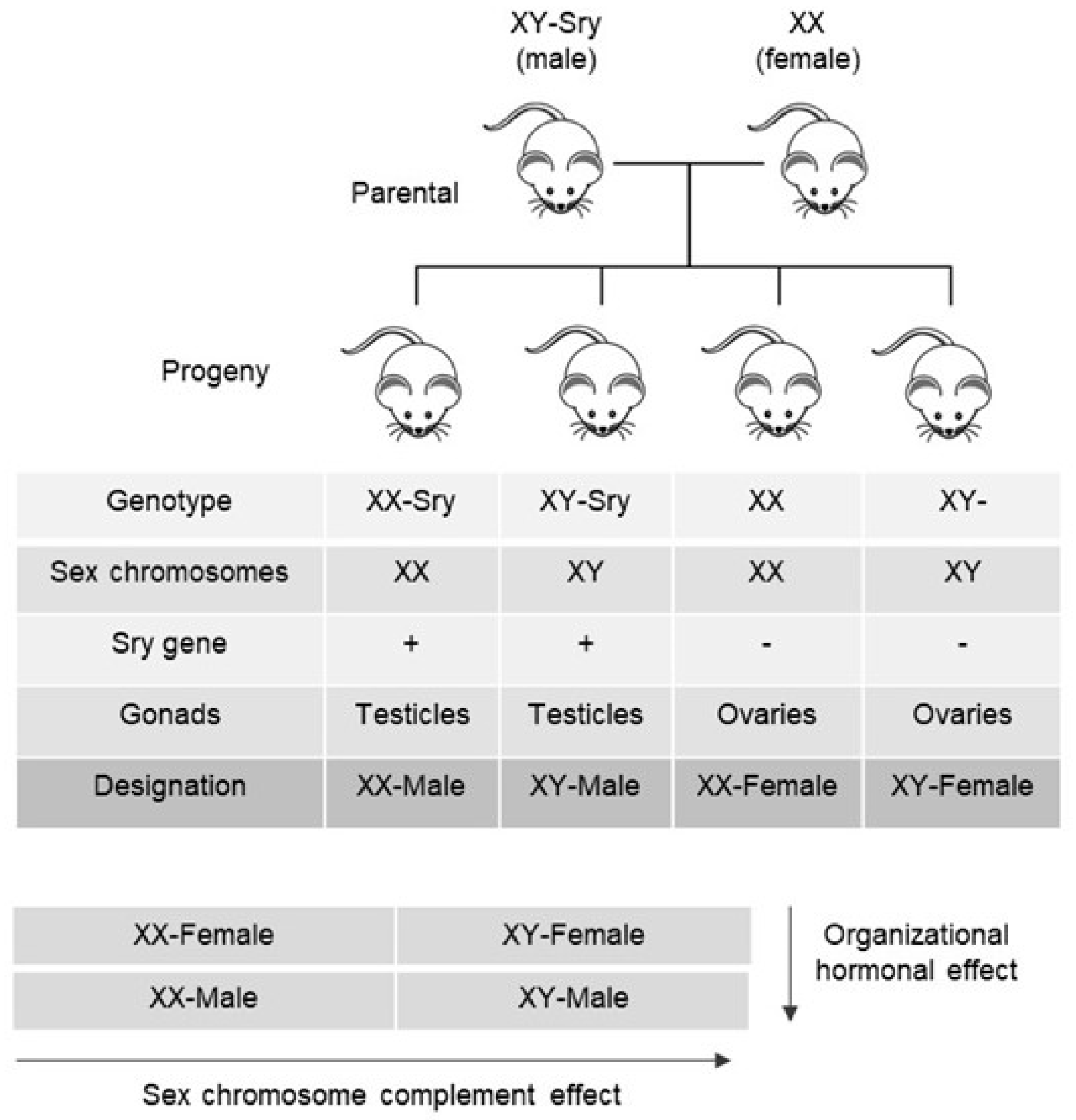
Figure 1. Four-core genotype mouse model. By breeding XY-Sry fertile males to XX females, four types of offspring (genotypes) are produced: XX and XY-female mice (without Sry on the Y chromosome) and XX-Sry and XY-Sry male mice (both with Sry transgene in an autosome). Regardless of their SCC, all individuals possessing the Sry transgene develop testes and have a male external phenotype, while mice lacking the transgene have ovaries and external female secondary sex characteristics. The following designations are applied for each genotype: XY-Sry male mice as XY-male; XX-Sry as XX-male, XX female as XX-female, and XY- female as XX-female.
Sexes 04 00035 g001
Figure 2. Schematic representation of the influence of SCC, the organizational and activational hormonal effects on sexually dimorphic autonomic blood pressure regulation during the lifetime.
Sexes 04 00035 g002
Disclaimer/Publisher’s Note: The statements, opinions and data contained in all publications are solely those of the individual author(s) and contributor(s) and not of MDPI and/or the editor(s). MDPI and/or the editor(s) disclaim responsibility for any injury to people or property resulting from any ideas, methods, instructions or products referred to in the content.