Article

Measurement of the Beam Energy Distribution of a Medical Cyclotron with a Multi-Leaf Faraday Cup

by
Konrad P. Nesteruk
*,†,
Luca Ramseyer
,
Tommaso S. Carzaniga
and
Saverio Braccini
Albert Einstein Center for Fundamental Physics (AEC), Laboratory for High Energy Physics (LHEP), University of Bern, Sidlerstrasse 5, CH-3012 Bern, Switzerland
*
Author to whom correspondence should be addressed.
Current address: Paul Scherrer Institut, Forschungsstrasse 111, 5232 Villigen PSI, Switzerland.
Instruments 2019, 3(1), 4; https://doi.org/10.3390/instruments3010004
Submission received: 18 December 2018 / Revised: 30 December 2018 / Accepted: 1 January 2019 / Published: 4 January 2019
(This article belongs to the Special Issue Instruments and Methods for Cyclotron Produced Radioisotopes)

Abstract

:
Accurate knowledge of the beam energy distribution is crucial for particle accelerators, compact medical cyclotrons for the production of radioisotopes in particular. For this purpose, a compact instrument was developed, based on a multi-leaf Faraday cup made of thin aluminum foils interleaved with plastic absorbers. The protons stopping in the aluminum foils produce a measurable current that is used to determine the range distribution of the proton beam. On the basis of the proton range distribution, the beam energy distribution is assessed by means of stopping-power Monte Carlo simulations. In this paper, we report on the design, construction, and testing of this apparatus, as well as on the first measurements performed with the IBA Cyclone 18-MeV medical cyclotron in operation at the Bern University Hospital.

1. Introduction

Beam energy is a key parameter for all particle accelerators. In the case of medical cyclotrons for radioisotope production, the yield and the purity of the produced radioisotopes strongly depend on the cross-section of both the desired radioisotope and the possible impurities. This is particularly crucial in the case of the bombardment of thin targets by means of solid target stations.
Compact medical cyclotrons for the production of radioisotopes have an energy in the range of 15–25 MeV and are characterized by a considerable potential for fundamental and applied research, especially if they are equipped with a beam transfer with independent access to the beam area [1]. They are usually installed in hospitals, where space constraints severely limit this possibility. For all activities beyond the production of 18 F—the main radioisotope for Positron Emission Tomography (PET)—accurate knowledge of the beam energy distribution is often crucial.
Along this line, a compact, easy-to-use, and cost-effective apparatus for the measurement of the beam energy distribution of the compact medical cyclotron has been designed, built, and tested. In this paper, we report on the first measurements obtained with this instrument performed with the Bern medical cyclotron.

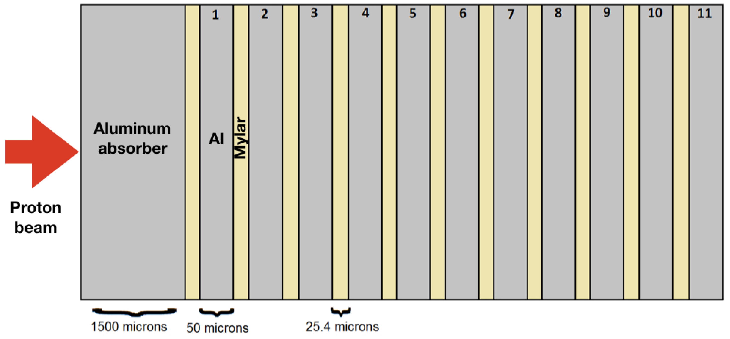
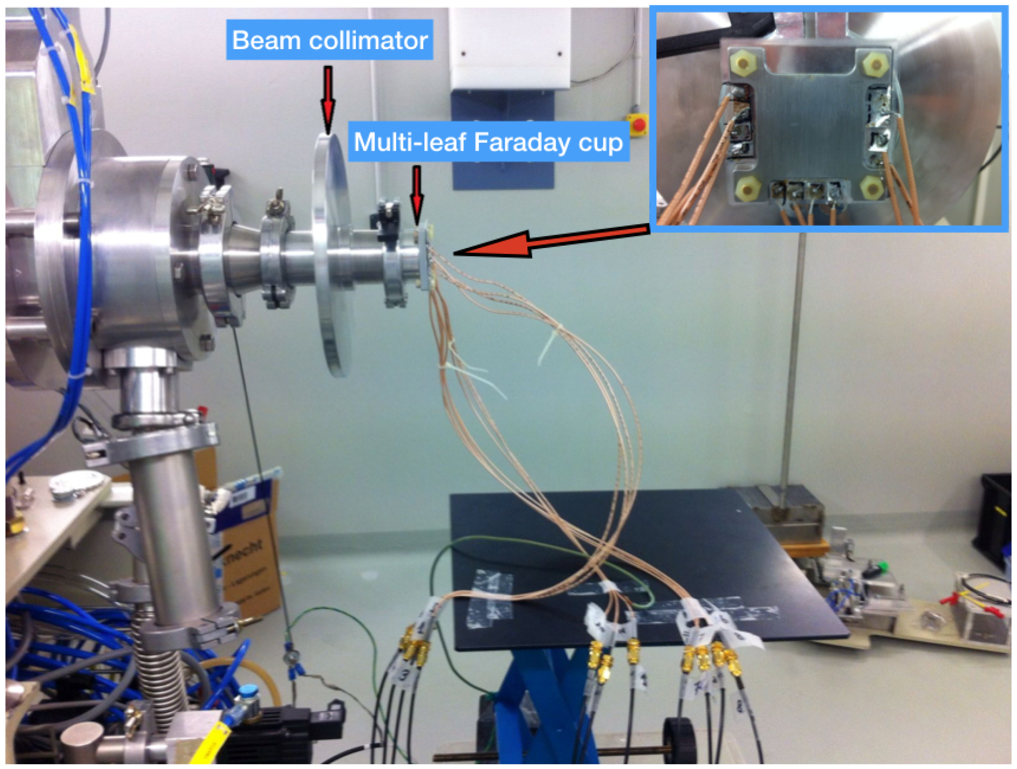
2. Materials and Methods
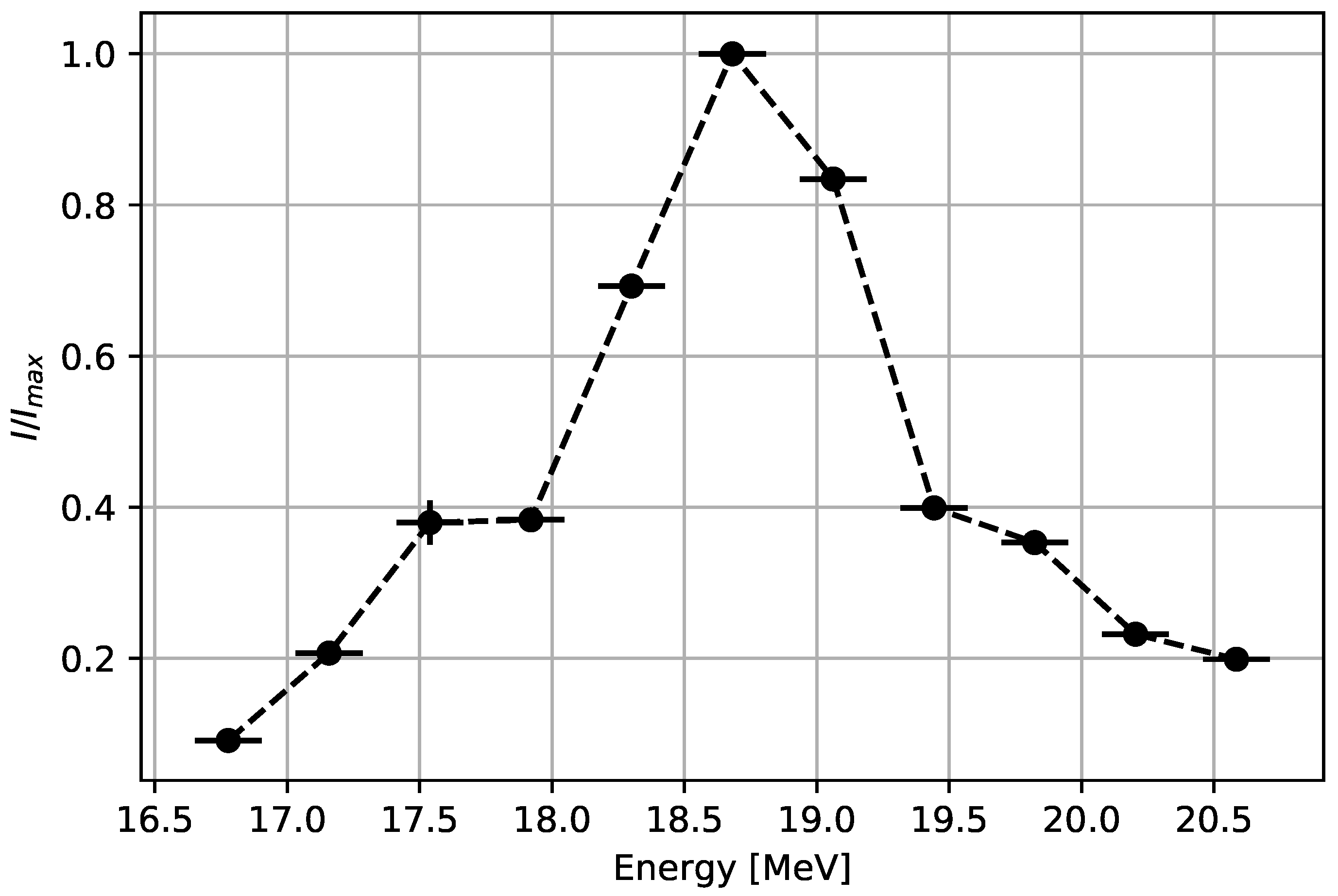
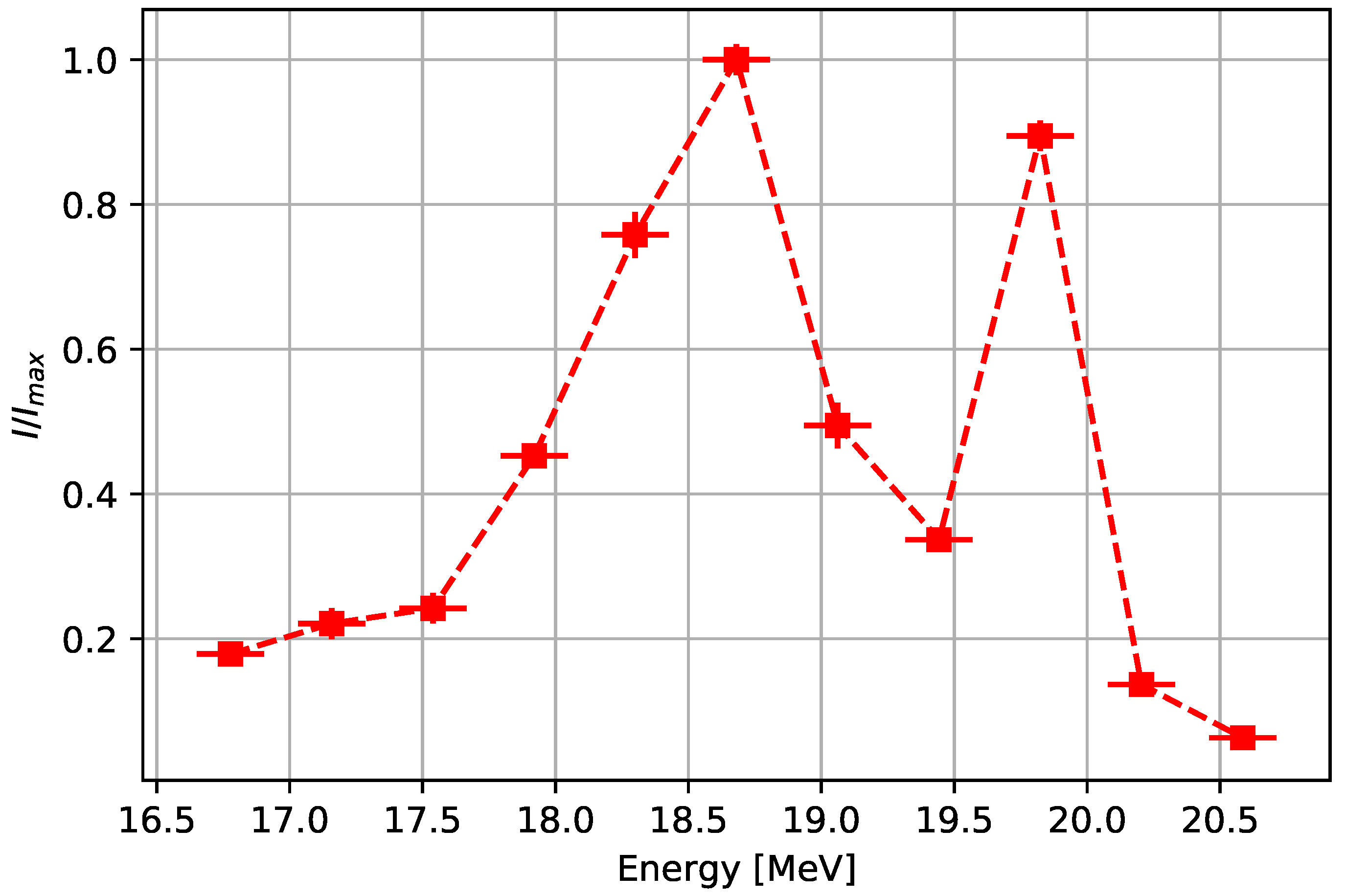
The laboratory at the Bern University Hospital (Inselspital) [2] features an IBA Cyclone 18/18 high-current cyclotron and two bunkers with independent access. This accelerator is used daily for the production of 18 F for PET diagnostics. The beam is brought to the second bunker by means of a 6 m-long Beam Transport Line (BTL), which is used for multi-disciplinary research activities. The BTL was used for the measurements presented in this paper.
The core of the experimental setup is a multi-leaf Faraday cup composed of aluminum foils interleaved with a non-conductive material. Protons have a range within this Faraday cup according to their energy and stop either in a foil or in the material. The number of protons stopping within one aluminum foil is assessed by measuring the current they produce by means of an electrometer. A similar, but more complex apparatus was developed to measure the energy distribution of a 68-MeV proton beam for eye melanoma hadrontherapy [3].
The multi-leaf Faraday cup we designed and constructed allows assessing the proton beam energy distribution by measuring the current collected by successive foils. The resolution of the measurement depends on the thickness of the absorber material and of the aluminum foils. The thickness and number of layers were chosen according to the outcome of a simulation performed with the SRIM [4] software, with which the transport of ions in matter can be accurately calculated. A previous measurement of the beam energy and energy spread of the Bern medical cyclotron [5] was used as input to the simulation to calculate the proton range within the multi-leaf Faraday cup. On the basis of simulations and material availability, 50 μ m ± 10%-thick aluminum sheets and 25.4 μ m ± 10%-thick Mylar ® polyester foils were chosen. Following the SRIM calculations, the protons in the range of 16.5–20.5 MeV were able to be measured by interleaving 11 aluminum foils with 11 Mylar sheets and preceding such a stack by a 1.5 mm-thick aluminum absorber, as shown in Figure 1.
The complete apparatus installed on the BTL is shown in Figure 2. The beam collimator, visible in the figure, was designed to shape the beam, and its surrounding disk was conceived of to reduce undesirable effects caused by single off-aperture scattered protons.
The measurements were performed by recording the mean current induced by protons in each aluminum foil. Each foil was connected to a switch by means of a coaxial cable. The switch was placed outside the BTL bunker and connected to an electrometer (Keysight B2987A) that read out the 11 channels in sequence. The fast sampling mode of the electrometer was used in order to reduce possible beam instabilities occurring during the read-out of the 11 channels to a negligible level in the order of statistical fluctuations.

3. Results

The measurements were taken in two different conditions of cyclotron operation. The first mode of operation corresponds to the optimal isochronism condition and is determined by maximizing the transmission of the beam from the ion source to the stripper. This mode of operation is used for radioisotope production and for some experiments requiring a high beam current. For most of the research activities performed with the BTL, the cyclotron is operated in the regime of non-optimal isochronism in order to obtain stable beam currents from the nA to the pA range [6].
The first series of measurements was performed for the optimal isochronism condition. The ion source arc current was always set to a value corresponding to induced currents in the nA range in all the channels of the multi-leaf Faraday cup. In this current range, the electrometer shows the most stable readings. The measured beam energy distribution in the condition of the optimal isochronism is shown in Figure 3. The probability density is expressed in units of normalized induced current I / I m a x , where I m a x corresponds to the channel with the highest measured current. The distribution presents one peak at a beam energy of ( 18.68 ± 0.13 ) MeV. The measurement was repeated for two slightly different currents set in the main coil of the cyclotron, but remaining in the vicinity of the optimal isochronism. A good agreement was found, and the peak was localized at the same beam energy value within the uncertainty. In order to compare the obtained results with the measurement performed with a different method and reported in [5], the mean energy and RMS of the distribution were also evaluated and found to be ( 18.7 ± 0.3 ) MeV and ( 0.9 ± 0.2 ) MeV, respectively. The values reported in [5] were ( 18.3 ± 0.3 ) MeV and ( 0.4 ± 0.2 ) MeV for the beam energy and RMS, respectively. The beam energy values are therefore in agreement within 1 σ and the RMS values within 1.8 σ . The peak energy of ( 18.76 ± 0.02 ) MeV, found in [5], is in a good agreement with the results presented in this paper. It has to be noted that the two compared methods are characterized by a different resolution and were performed in slightly different conditions. The region of the optimal isochronism, corresponding to our definition based on a maximum beam transmission, spans a certain range of main coil settings, and the resulting energy distributions can vary to some extent. Another possible reason for the observed difference is the fact that the method reported in [5] gives the probabilities of finding beam energy within histogram bins, while the measurements described in this paper characterize the distribution by the evaluation of discrete points. Furthermore, the main coil of the cyclotron warms up during operation, making the operation conditions slightly different.
The second series of measurements was conducted in the non-optimal isochronism regime. The main coil current was set to a value corresponding to a low transmission of the beam from the ion source to the stripper. The ion source arc current was again adjusted to provide electrometer readings in the nA range. The obtained beam energy distribution is shown in Figure 4. This time, two peaks are visible. The first one corresponds to a beam energy of ( 18.68 ± 0.13 ) MeV, while the second to an energy of ( 19.82 ± 0.13 ) MeV. Also for the non-optimal isochronism condition, the distribution was measured for two different values of the main coil currents, and a good agreement was found.
The results obtained in both series for the optimal and non-optimal isochronism conditions present a peak at the beam energy of ( 18.68 ± 0.13 ) MeV. An additional peak of the distribution corresponding to the non-optimal isochronism condition can be explained by the fact that the beam size and shape on the stripper are optimized for the isochronous condition such that the majority of the accelerated beam is stripped in one turn. Operation of the machine far from the optimum can lead to an effect in which a significant number of ions are stripped after making extra turns in the cyclotron, which is manifested by the second peak in the energy distribution corresponding to a 1.1-MeV higher beam energy. This energy difference is equivalent to about 18 extra turns. Since the radial separation between particle orbits at the energies over 18 MeV is very small (below 1 mm) and there is no optimal horizontal focusing provided in this mode of operation, a large beam spot at the stripper location is likely, which leads to those extra turns. Steering effects in the quadrupoles along the BTL might also play a role in making the transport efficiency energy dependent.

4. Conclusions

In this paper, we presented a simple and efficient method to measure the energy distribution of an ion beam extracted from a medical cyclotron based on a multi-leaf Faraday cup. On the basis of simulations, a specific apparatus was designed, built, and tested at the Bern medical cyclotron laboratory to assess the energy distribution at the end of the Beam Transport Line (BTL) connected to an IBA Cyclone 18-MeV cyclotron. Measurements were performed for two modes of cyclotron operation. The first one corresponds to the optimal isochronism condition and applies to radioisotope production and to experiments requiring a high beam current. The second mode is used for most of the research activities with currents in the nA and pA range and corresponds to the regime of non-optimal isochronism. As expected, the energy distributions of the extracted proton beam for the two modes of operation were found to be different. Although for both modes, a peak at an energy of ( 18.68 ± 0.13 ) MeV is observed, the distribution corresponding to the non-optimal isochronism shows an additional peak at ( 19.82 ± 0.13 ) MeV. This effect is mostly due to the beam extraction by stripping. For the optimal isochronism, the results were compared to measurements previously performed, obtained with a different method, and a good agreement was found. The obtained results are higher with respect to the nominal beam energy of 18 MeV. This is consistent with the fact that for the BTL, the stripper is located at a radius ∼5 mm larger with respect to the nominal one for beam transport optimization purposes, making the beam energy higher [2].
The proposed method can be applied to any cyclotron to assess the beam energy precisely for scientific and industrial purposes, in particular to optimize radioisotope production with solid target stations.

Author Contributions

S.B. and K.P.N. conceived of and designed the experiment; K.P.N., T.S.C., and L.R. performed the experiment; K.P.N. and L.R. analyzed the data; K.P.N. and S.B. wrote and revised the paper; S.B. coordinated the project.

Acknowledgments

We acknowledge contributions from the LHEP engineering and technical staff. One of the authors (T.S.C.) was financially supported by the Swiss National Science Foundation (Grant CR23I2_156852).

Conflicts of Interest

The authors declare that there is no conflict of interest.

References

  1. Braccini, S. Compact medical cyclotrons and their use for radioisotope production and multi-disciplinary research. In Proceedings of the Cyclotrons2016 TUD01, Zurich, Switzerland, 11–16 September 2016; p. 229. [Google Scholar]
  2. Braccini, S. The new Bern PET cyclotron, its research beam line, and the development of an innovative beam monitor detector. AIP Conf. Proc. 2013, 1525, 144. [Google Scholar] [CrossRef]
  3. Seidel, S.; Bundesmann, J.; Damerow, T.; Denker, A.; Kunert, C.; Weber, A. A multi-leaf Faraday cup especially for proton therapy of ocular tumors. In Proceedings of the Cyclotrons2016, Zurich, Switzerland, 11–16 September 2016; Volume MOE02, pp. 118–120. [Google Scholar]
  4. Ziegler, J.F.; Ziegler, M.D.; Biersack, J.P. SRIM—The stopping and range of ions in matter. Nucl. Instrum. Methods 2010, 268, 1818–1823. [Google Scholar] [CrossRef]
  5. Nesteruk, K.P.; Auger, M.; Braccini, S.; Carzaniga, T.S.; Ereditato, A.; Scampoli, P. A system for online beam emittance measurements and proton beam characterization. J. Instrum. 2018, 13, P01011. [Google Scholar] [CrossRef]
  6. Auger, M.; Braccini, S.; Ereditato, A.; Nesteruk, K.P.; Scampoli, P. Low current performance of the Bern medical cyclotron down to the pA range. Meas. Sci. Technol. 2015, 26, 094006. [Google Scholar] [CrossRef]
Figure 1. Scheme of the multi-leaf Faraday cup for the beam energy measurements of the Bern medical cyclotron.
Instruments 03 00004 g001
Figure 2. The multi-leaf Faraday cup installed on the Beam Transport Line (BTL) of the Bern cyclotron. A close-up is shown in the inset.
Instruments 03 00004 g002
Figure 3. Beam energy distribution measured for the optimal isochronism condition of the Bern medical cyclotron. The dashed line is a guide to the eye.
Instruments 03 00004 g003
Figure 4. Beam energy distribution measured for the non-optimal isochronism condition of the Bern medical cyclotron. The dashed line is a guide to the eye.
Instruments 03 00004 g004

