1. Introduction
One of the most quoted methods to perform high energy resolution X-ray measurements both in laboratory experiment and synchrotron radiation facility is provided by the Bragg spectroscopy. The requirement on the size of the target not to exceed tens of microns represents the major hindrance in its use when photons emitted from extended sources (millimetric) need to be measured [
1]. In addition, the typical very low efficiencies of Bragg spectrometers prevent them from being used in several applications. The prototype of a high resolution Von Hamos X-ray spectrometer using HAPG (Highly Annealed Pyrolytic Graphite) mosaic crystals developed by the VOXES collaboration at INFN National Laboratories of Frascati offers the possibility to achieve few eV energy resolution for energies going from 2 keV up to 10 of keV and to measure not only collimated sources, but also extended ones. Highly Annealed Pyrolitic Graphite (HAPG) is a mosaic crystal consisting in a large number of nearly perfect small crystallites. The crystallites distribution can be described by a Gaussian function and the mosaicity is defined as the FWHM of this distribution. It makes it possible that a photon can find a crystallite plane at the right Bragg angle and be reflected unless if it is slightly deviated from reaching the crystal with the exact Bragg relation [
2,
3]. This property together with a lattice spacing constant
d = 3.514 Å, enables them to be highly efficient in the 2–20 keV energy range. The peak reflectivity is always lower than one and depends on the diffraction volume and on the crystal thickness. It is equal to unity for perfect crystals only. The mosaic crystals technology is suitable to be used in the Von Hamos configuration combining the standard dispersion of a flat crystal with the focusing properties of cylindrically bent crystals.
Several studies aimed to analyze the effect on the energy resolution of the precision of the mosaicity and thickness of the crystal have been already performed, and the capability of the spectrometer to be optimized in order to achieve the best precision has been also demonstrated [
4,
5].
The performance of the spectrometer has been lately studied in terms of reflection efficiency [
6] and in this work we want to focus on the ray-tracing simulations implemented in order to check the agreement with the experimental results. The achievement of consistent results is fundamental to evaluate the efficiency of the spectrometer. In the next sections the description of the experimental setup and procedure, of the ray-tracing simulations, and the comparison of the experimental and simulated data are presented.
2. Setup
In the spectrometer configuration used in our measurements the X-ray source and the position detector are placed on the axis of a cylindrical crystal. This configuration is known as Von Hamos and allows to increase the reflection efficiency due to the vertical focusing. This geometry permits to determine the source-crystal and the source-detector distances,
and
, respectively, by means of the Bragg angle (
) and of the curvature radius of the crystal (
):
where
=
.
Adding a pair of slits to this configuration, it is possible to shape the beam of the X-rays emitted by an extended target modifying the position (
and
) and the apertures (
and
) of the slits to create a virtual point-like source (
), an angular acceptance
and an effective source
(see
Figure 1 for the horizontal plane of the beam) [
5].
The experimental apparatus consists of a XTF-5011 Tungsten anode X-ray tube produced by OXFORD INSTRUMENTS, located on the top of an aluminum box where a 125 μm thick target foil is contained. The center of the foil is the source position and it is placed on a 45° target holder.
The two adjustable motorized slits (STANDA 10AOS10-1) with 1 mm sensitivity are arranged after the circular exit window of the aluminum box of 5.9 mm diameter. The HAPG crystal used for the measurements has a thickness of 100 μm, a curvature radius (
) of 206.7 mm and mosaicity (
) of 0.1°. The position detector, also equipped with a positioning motorized system, is a commercial MYTHEN2-R-1D 640 channels strip detector produced by DECTRIS (Zurich, Switzerland). The active area is 32 × 8 mm
2; strip width and thickness are, respectively, 50 μm and 420 μm; further details can be found in [
5].
3. Experimental Procedure
The spectrometer setup was optimized for the measurement of the two Cu and Fe
lines. The target foil is activated by the X-ray tube and the
lines, isotropically emitted, are collimated by the slits system to simulate a point-like source. An example of the resulting spectrum, after performing the calibration and fitting procedure reported in our previous work [
5], is shown in the
Figure 2. In this case the slits are set to maintain an angular divergence
of 0.7° and the distances between the source-to-HAPG crystal and crystal-to-detector are of 900.54 mm. The alignment of the optical system has been performed by using a laser.
These kind of spectra are used in the evaluation of the HAPG crystal reflection efficiency. Usually this quantity is defined as the percentage of Bragg reflected X-rays of a given energy
for different impinging angles and is obtained from X-ray beam emitted from a monochromatic point-like source. Instead, the quantity we want to measure is the following:
It represents the ratio between the number of
or
X-rays reflected from the crystal (
) and that of those impinging on it (
) for different source sizes
and beam divergence
pairs [
6]. The numerator of Equation (3) is obtained from the Bragg energy spectrum
, as the one shown in
Figure 2, taking into account the X-ray transmission in air (
) and the MYTHEN2 detection efficiency (
):
The
coefficient is evaluated, for a given energy and HAPG-MYTHEN2 distance, from the CXRO database [
7] while
is provided by the producers. In the
Figure 3 and the resulting behaviour of the transmission coefficient with the energy is shown; the fit carried out in the energy range of interest for our measurements is represented in red.
The determination of the number of X-rays impinging on the crystal (
) required a longer procedure. First of all, for each
combination, a measurement with the MYTHEN2 detector in place of the HAPG crystal has been performed in order to quantify the X-rays reaching the HAPG position that we defined as MYTHEN2 direct measurements
. However, since with the MYTHEN2 detector we can set only an energy threshold and we have to find the numbers of pure
or
signal, we needed of the help of a Silicon PinDiode to extract the ratio
between the
or
lines and all the other background photons, produced by Bremmstrahlung or other processes occurring in the source box and reaching the crystal, having also exceeded the set energy threshold. The spectrum is bin by bin corrected accounting for the different efficiencies of the PinDiode and of the MYTHEN2 detector (see
Figure 4):
The number of X-rays impinging on the HAPG crystal can then be evaluated from the following relation:
where the ratio between the beam vertical dimension
and 8 mm accounts for the difference between the MTYTHEN2 strip height and the XRF beam spot vertical size on the HAPG.
is evaluated, following the procedure described in [
5], from the slits positions and dimensions.
The reflection efficiencies, obtained with a
mm HAPG crystal for both
XRF lines, from Equations (4) and (6), are estimated as [
6]:
and resulted to vary between 0.15–0.35 and 0.1–0.3 for copper and iron, respectively.
4. Ray-Tracing Simulations
In this section we present the comparison of the performances of the VOXES spectrometer with the ones obtained from the ray-tracing simulations. We used the XOP+SHADOW3 software [
8,
9,
10] implemented in the Oasys enviroment [
11]. The XOP package includes all the codes that are necessary to evaluate the interaction of the X-rays with the optical components defined by the user. In particular, we choose an extension, the
SHADOWVUI package that provides a Visual User Interface for the SHADOW ray-tracing program. The elements of the optical system are defined by objects that are called
widgets within a canvas that represents our workspace. Each widget contains the user-defined parameters by double-clicking on it and has to be connected with others by connectors. The
Loop Point widget allows to increase the statistics performing several runs of smaller size and can be used also to start the simulation from the source widget which we refer to as
target.
The
target is generated by means of Shadow Geometrical Source and we defined it as a
mm rectangular 0.1 mm thick to prevent any losses. In the same widget we provide as input the energy spectrum of the copper (or iron) X-rays. It is sampled with a double Lorentzian function, the widths of which are obtained from the paper [
12]. In the snapshot of the
Figure 5 from the Geometrical Source widget, an example of the copper spectrum input is reported; on the left we inserted the energy range of the spectrum, the binning and the path of the input file while on the right the histogram statistics is listed. After that, we defined other two widgets that include two python codes to take into account the target rotation of 45° and to consider the X-ray divergence.
Then, we implement the description of the box hole and the two slits system in order to be able to set their positions and apertures. These last two widgets have a very important role in the simulation because they define the effective source size
and the angular divergence
. The snapshot of the
Figure 6 is referred to the widget of the first slit used in the simulation of the copper Bragg spectra for
= 0.7° and
= 1.1 mm; in the right part the setting of the position are shown while on the center, the two-dimensional XZ plot, together with the corresponding projections of the beam dimensions, are reported. In these plots, it is possible to notice the effective source size of 1.1 mm. The following step consists in the simulation of the HAPG crystal by using the Shadow Spherical Crystal widget. Here, we defined the geometry of the crystal, using the cylindrical one, and we introduced the value of the curvature radius (
= 206 mm), the mosaicity (
= 0.1
), usually provided by the producer, the position and the nominal Bragg angle at which the reflection occurs as shown in the snapshot reported in the
Figure 7 on the left column; on the center instead, the XZ plot of the XRF photons emitted from the effective source and reflected by the HAPG crystal is shown in case of a copper target
= 0.7° and
= 1.1 mm, together with the projections, in arbitrary units, of the Z and X coordinates on the right and on the bottom of the two-dimensional plot respectively. The Z-plot converted in energy and fitted with a Gaussian function is shown in the
Figure 8 where it is possible to appreciate the good matching of the two XRF emission lines
both in terms of energy and resolution with the experimental one of the
Figure 2. The small difference between the resolution values is attributable to small deviation, at micrometer level, of the position and/or aperture of the slits from the nominal ones.
To evaluate the reflection efficiencies from the simulations we used the following ratio [
6]:
where
refers to the number of photons emitted by the source and reaching the MYTHEN2 detector, while
accounts also for the HAPG crystal reflectivity. These quantities are displayed in the right part of the snapshot of the
Figure 7.
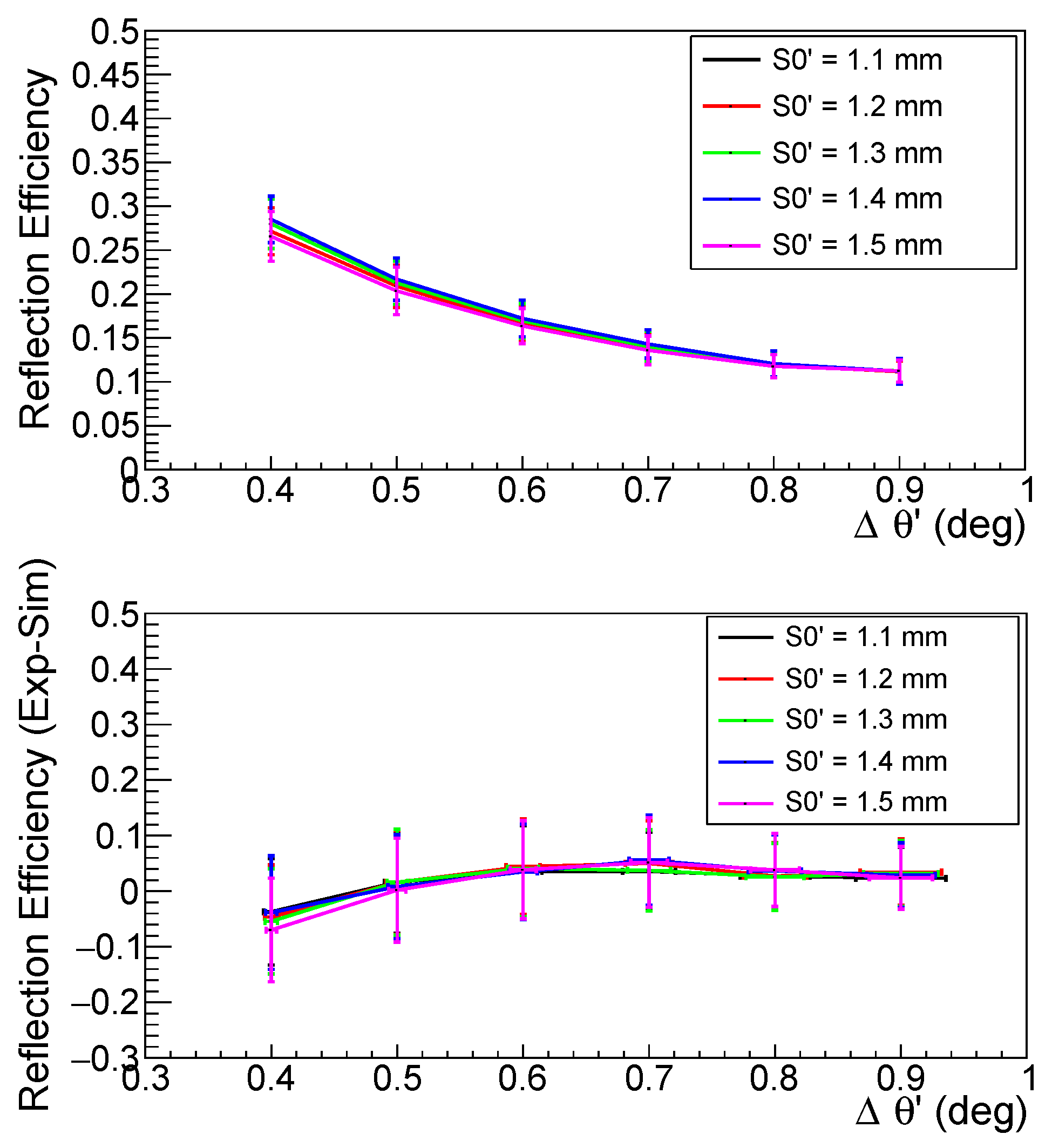
The results obtained in terms of reflection efficiency for different effective source sizes,
, as a function of the angular divergence
from simulated data are shown for copper and iron in
Figure 9 and
Figure 10, respectively, in the top pads, while the differences between the experimental and the simulated efficiency are given in the bottom pads.
The results highlight how the simulation well reproduces the experimental data within the associated errors. For each point of the simulated reflection efficiencies plots, the error bars are the result of the sum in quadrature of the statistical (almost negligible) and systematic errors. To evaluate the systematic uncertainty we performed simulations fixing the and values and varying different parameters as the position of the slits, crystal and MYTHEN detector (±5 mm), the opening of the slits (±0.05 mm), their possible misalignment in the x-coordinate (±5 mm) and in the z-coordinate (±0.1 mm), and finally the mosaicity (±0.01°).
The consequent variation of the reflection efficiency values has been accounted as systematic error. In the
Figure 11 we reported the study carried out in case of use of a copper target for
= 0.7°,
= 1.1 mm. Since the expected uncertainty on the mosaicity value of the used HAPG crystal is at the level of 0.01°, we performed the systematics analysis studying its variation between 0.09° and 0.11°. In particular, on top the reflection efficiencies obtained for mosaicity values 0.09°, 0.10° and 0.11° are represented in blue, cyan and magenta, respectively, while on bottom the systematic uncertainties referred to all analyzed parameters are summarized. As one can see, most parameters gave a contribution less than 5 per mill while the variation of the mosaicity resulted of the order of 2%. This value has been taken as systematic uncertainty associated to the reflection efficiency.
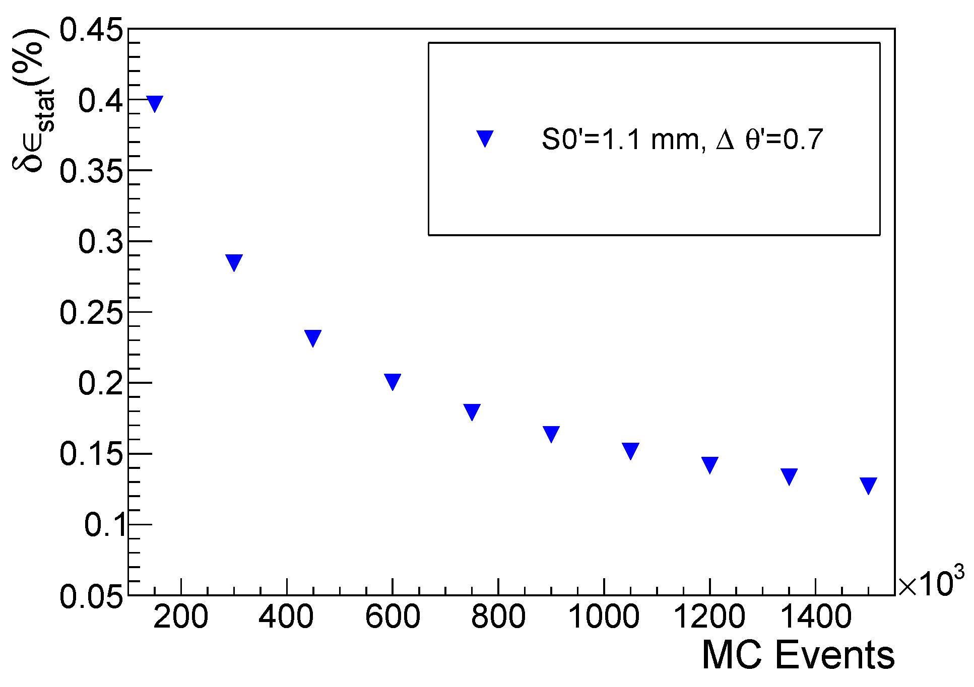
The number of events used as input in the simulations has been chosen according to the plot shown in
Figure 12, where the statistical error on the reflection efficiency (
) as a function of the MC simulated events, as example, for the
m, and
copper case, is reported. In our final simulations, we used
events not to affect the precision of the efficiency simulations by low statistics.


