Challenging X-ray Fluorescence Applications for Environmental Studies at XLab Frascati
Abstract
:1. Introduction
2. XLab Frascati
3. TXRF Results
3.1. Environmental Analysis
4. Conclusions
Supplementary Materials
Author Contributions
Funding
Acknowledgments
Conflicts of Interest
Abbreviations
| PolyCO | Polycapillary Optics |
| XRF | X-ray Fluorescence |
| XRF | micro X-ray Fluorescence |
| TXRF | Total Reflection X-ray Fluorescence |
| SR | Synchrotron Radiation |
| UCC | Upper Continental Crust |
References
- Tsuji, k.; Nakano, K.; Hayashi, H.; Hayashi, K.; Ro, C. X-ray Spectrometry. Anal. Chem. 2008, 80, 4421–4454. [Google Scholar] [CrossRef] [PubMed]
- Hampai, D.; Dabagov, S.B.; Cappuccio, G.; Longoni, A.; Frizzi, T.; Cibin, G.; Guglielmotti, V.; Sala, M. Elemental Mapping and Micro-Imaging by X-Ray Capillary Optics. Opt. Lett. 2008, 33, 2743–2745. [Google Scholar] [CrossRef] [PubMed]
- Giannoncelli, A. Proceedings of the 24th International Congress on X-Ray Optics and Microanalysis, Trieste, Italy, 25–29 September 2017.
- Croudace, I.W.; Rothwell, R.G. MIcro-XRF Studies of Sediment Cores; Springer: Berlin, Germany, 2015. [Google Scholar]
- Shackley, M.S. X-ray Fluorescence Spectrometry (XRF) in Geoarchaeology; Spinger: Berlin, Germany, 2011. [Google Scholar]
- Cibin, G.; Marcelli, A.; Maggi, V.; Sala, M.; Marino, F.; Delmonte, B.; Albani, S.; Pignotti, S. First combined total reflection X-ray fluorescence and grazing incidence X-ray absorption spectroscopy characterization of aeolian dust archived in Antarctica and Alpine deep ice cores. Spectrochim. Acta B 2008, 63, 1503–1510. [Google Scholar] [CrossRef]
- Marcelli, A.; Cibin, G.; Hampai, D.; Giannone, F.; Sala, M.; Pignotti, S.; Maggi, V.; Marino, F. XANES characterization of deep ice core insoluble dust in the ppb range. J. Anal. At. Spectrom. 2012, 22, 33–37. [Google Scholar] [CrossRef]
- Dabagov, S.B. Channeling of neutral particles in micro- and nanocapillaries. Phys. Uspekhi 2003, 46, 1053–1075. [Google Scholar] [CrossRef]
- Hampai, D.; Cherepennikov, Y.M.; Liedl, A.; Cappuccio, G.; Capitolo, E.; Iannarelli, M.; Azzutti, C.; Gladkikh, Y.P.; Marcelli, A.; Dabagov, S.B. Polycapillary based μXRF station for 3D colour tomography. JINST 2018, 13, C04024. [Google Scholar] [CrossRef]
- Hampai, D.; Liedl, A.; Cappuccio, G.; Capitolo, E.; Iannarelli, M.; Massussi, M.; Tucci, S.; Sardella, R.; Sciancalepore, A.; Polese, C.; et al. 2D-3D μXRF elemental mapping of archeological samples. Nucl. Instrum. Methods B 2017, 402, 274–277. [Google Scholar] [CrossRef]
- Zoeger, N.; Streli, C.; Wobrauschek, P.; Jokubonis, C.; Pepponi, G.; Roschger, P.; Hofstaetter, J.; Berzlanovich, A.; Wegrzynek, D.; Chinea-Cano, E.; et al. Determination of the elemental distribution in human joint bones by SR micro XRF. X-Ray Spectrom. 2008, 37, 3–11. [Google Scholar] [CrossRef] [Green Version]
- Nakano, K.; Tanaka, K.; Ding, X.; Tsuji, K. Development of a new total reflection X-ray fluorescence instrument using polycapillary X-ray lens. Spectrochim. Acta B 2006, 61, 1105–1109. [Google Scholar] [CrossRef]
- Bernini, R.; Pelosi, C.; Carastro, I.; Venanzi, R.; Di Filippo, A.; Piovesan, G.; Ronchi, B.; Danieli, P.P. Dendrochemical investigation on hexachlorocyclohexane isomers (HCHs) in poplars by an integrated study of micro-Fourier transform infrared spectroscopy and gas chromatography. Trees Struct. Funct. 2016, 30, 1455–1463. [Google Scholar] [CrossRef]
- Petit, J.R.; Jouzel, J.; Raynaud, R.; Barkov, N.I.; Barnola, J.M.; Basile, I.; Bender, M.; Chappellaz, J.; Davis, M.; Delaygue, G.; et al. Climate and atmospheric history of the past 420,000 years from the Vostok ice core, Antarctica. Nature 1999, 399, 429–436. [Google Scholar] [CrossRef]
- Community Members EPICA. Eight glacial cycles from an Antarctic ice core. Nature 2004, 429, 623–628. [Google Scholar] [CrossRef] [PubMed] [Green Version]
- Maggi, V. Mineralogy of atmospheric microparticles deposited along the Greenland Ice Core Project ice core. J. Geophys. Res. 1997, 102, 26725–26734. [Google Scholar] [CrossRef] [Green Version]
- Sawidis, T.; Breuste, T.; Mitrovic, M.; Pavlovic, P.; Tsigaridas, K. Trees as bioindicator of heavy metal pollution in three European cities. Environ. Pollut. 2011, 159, 3560–3570. [Google Scholar] [CrossRef] [PubMed]
- Hampai, D.; Dabagov, S.B.; Cappuccio, G. Advanced studies on the Polycapillary Optics use at XLab Frascati. Nucl. Instrum. Methods B 2015, 355, 264–267. [Google Scholar] [CrossRef]
- Nanoray, a Portable X-ray Machine, FP7 Project N 222426 (2008–2011). Available online: https://www.parlementairemonitor.nl/9353000/1/j9tvgajcor7dxyk_j9vvij5epmj1ey0/vj4871mm6efo?ctx=vg9pjpw5wsz1&start_tab0=1160 (accessed on 18 October 2018).
- Hampai, D.; Dabagov, S.B.; Cappuccio, G.; Cibin, G.; Sessa, V. X-ray micro-imaging by capillary optics. Spectrochim. Acta Part B 2009, 64, 1180–1184. [Google Scholar] [CrossRef]
- Cherepennikov, Y.; Miloichikova, I.; Gogolev, A.; Stuchebrov, S.; Hampai, D.; Dabagov, S.; Liedl, A. Application of polycapillary optics for dual energy spectroscopy based on a laboratory source. Nucl. Instrum. Methods B 2017, 402, 278–281. [Google Scholar] [CrossRef]
- Liedl, A.; Dabagov, S.B.; Hampai, D.; Polese, C.; Tsuji, K. On X-ray channeling in a vibrating capillary. Nucl. Instrum. Methods B 2015, 355, 289–292. [Google Scholar] [CrossRef]
- Marchitto, L.; Hampai, D.; Dabagov, S.B.; Allocca, L.; Alfuso, S.; Polese, C.; Liedl, A. GDI spray structure analysis by polycapillary X-ray μ-tomography. Int. J. Multiph. Flow 2015, 70, 15–21. [Google Scholar] [CrossRef]
- Bonfigli, F.; Hampai, D.; Dabagov, S.B.; Montereali, R.M. Characterization of X-ray polycapillary optics by LiF crystal radiation detectors through confocal fluorescence microscopy. Opt. Mater. 2016, 58, 398–405. [Google Scholar] [CrossRef]
- Gogolev, A.S.; Hampai, D.; Khusainov, A.K.; Zhukov, M.P.; Dabagov, S.B.; Potylitsyn, A.P.; Liedl, A.; Polese, C. Results of testing the energy dispersive Si detector with large working area. Nucl. Instrum. Methods B 2015, 355, 268–271. [Google Scholar] [CrossRef]
- Available online: http://www.oxford-instruments.com (accessed on 18 October 2018).
- Available online: http://www.xglab.it (accessed on 18 October 2018).
- Available online: http://www.photonic-science.com (accessed on 18 October 2018).
- Available online: https://www.physikinstrumente.com/en/ (accessed on 18 October 2018).
- Available online: http://www.axo-dresden.de/products/highprecision/reference.htm (accessed on 18 October 2018).
- Bostick, B.C.; Theissen, K.M.; Dunbar, R.B.; Vairavamurthy, A. Record of redox status in laminated sediments from Lake Titicaca: A sulfur K-edge X-ray absorption near edge structure (XANES) study. Chem. Geol. 2005, 219, 163–174. [Google Scholar] [CrossRef]
- Moy, C.M.; Dunbar, R.B.; Guilderson, T.P.; Waldmann, N.; Mucciarone, D.A.; Recasens, C.; Ariztegui, D.; Austin, J.A., Jr.; Anselmetti, F.S. A geochemical and sedimentary record of high southern latitude Holocene climate evolution from Lago Fagnano, Tierra del Fuego. Earth Planet. Sci. Lett. 2011, 302, 1–13. [Google Scholar] [CrossRef] [Green Version]
- Polyak, V.J.; Asmerom, Y. Late Holocene Climate and Cultural Changes in the Southwestern United States. Science 2001, 294, 148–151. [Google Scholar] [CrossRef] [PubMed]
- Di Filippo, A.; Biondi, F.; Cûfar, K.; De Luis, M.; Grabner, M.; Maugeri, M.; Presutti Saba, E.; Schirone, B.; Piovesan, G. Bioclimatology of beech (Fagus sylvatica L.) in the Eastern Alps: Spatial and altitudinal climatic signals identified through a tree-ring network. J. Biogeogr. 1892, 34, 1873–1892. [Google Scholar] [CrossRef]
- Macis, S.; Cibin, G.; Maggi, V.; Baccolo, G.; Hampai, D.; Delmonte, B.; D’Elia, A.; Marcelli, A. Microdrop Deposition Technique: Preparation and Characterization of Diluted Suspended Particulate Samples. Condens. Matter 2018, 3, 21. [Google Scholar] [CrossRef]
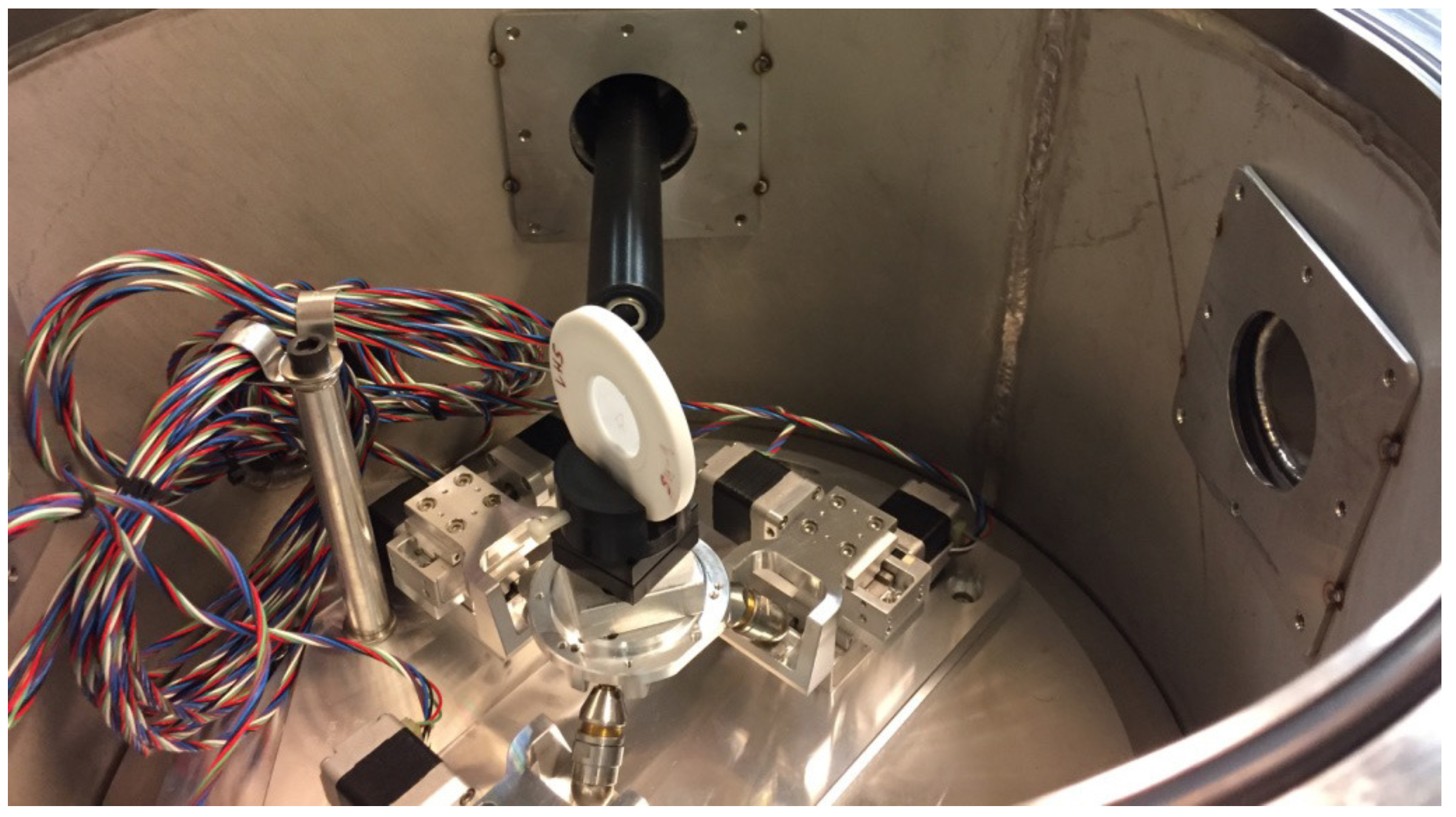
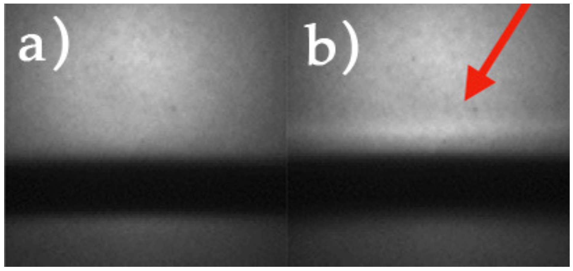
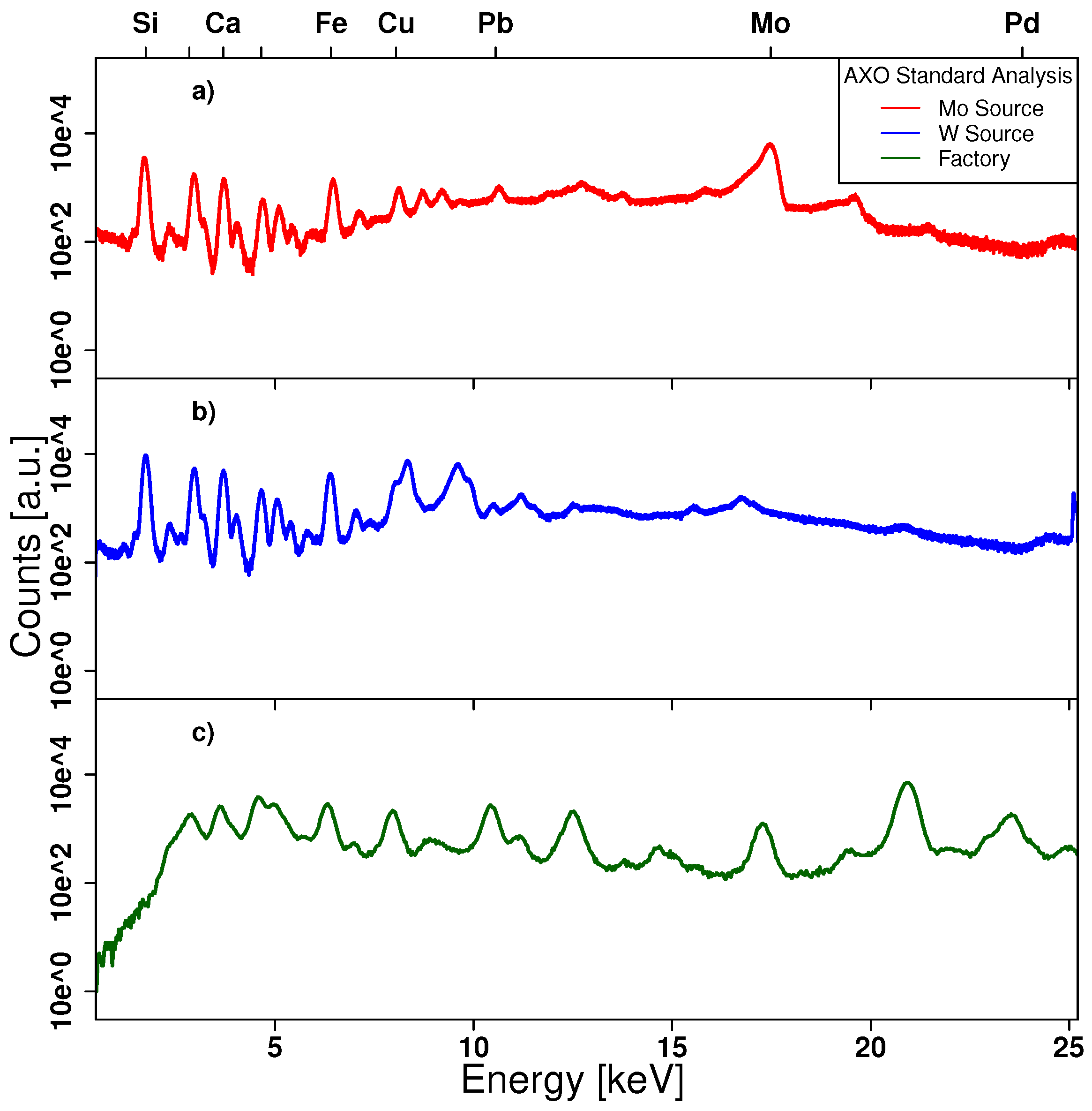
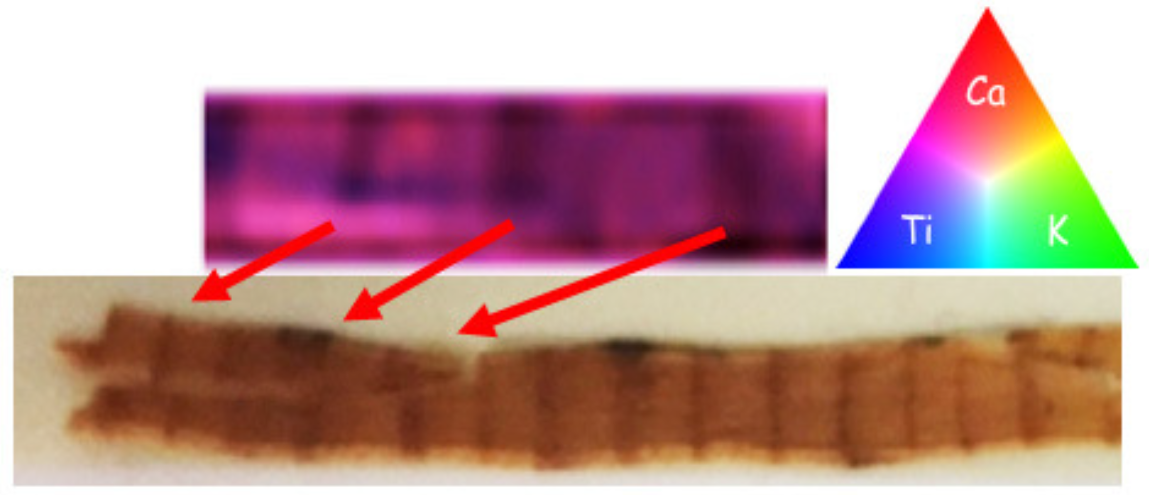
| XENA | RXR | |
|---|---|---|
| Station | X-ray Elemental station | Rainbow X-ray |
| for Non-destructive Analysis | ||
| Analysis | (1) High Resolution Imaging | (1) XRF (2D and 3D mapping) |
| (2) CT | ||
| (3) X-ray Optics Characterization | (2) TXRF | |
| (4) Detector Characterization | ||
| (5) Novel Sources | ||
| Resolution | (1) <1 m [24] | (1) ∼ m [9] |
| (2) < m [23] | ∼ m [9] | |
| (2) 25 ± 1.25 ng/g concentrations [20] |
| 2 X-ray Tubes [26] | Oxford Apogee 5000 |
| - MoK | |
| - W bremsstrahlung inelastic radiation | |
| source spot <50 m | |
| power: 50 W | |
| Silicon Drift Detector [27] | active area: 30 mm with AP3.7 window |
| CCD Camera | pixel resolution: m |
| (FDI 1:1.61) [28] | |
| Polycapillary semi-lens [20] | Focal Distance: 59 mm |
| transmission ∼60% | |
| residual divergence: 1.4 mrad |
© 2018 by the authors. Licensee MDPI, Basel, Switzerland. This article is an open access article distributed under the terms and conditions of the Creative Commons Attribution (CC BY) license (http://creativecommons.org/licenses/by/4.0/).
Share and Cite
Cappuccio, G.; Cibin, G.; Dabagov, S.B.; Di Filippo, A.; Piovesan, G.; Hampai, D.; Maggi, V.; Marcelli, A. Challenging X-ray Fluorescence Applications for Environmental Studies at XLab Frascati. Condens. Matter 2018, 3, 33. https://doi.org/10.3390/condmat3040033
Cappuccio G, Cibin G, Dabagov SB, Di Filippo A, Piovesan G, Hampai D, Maggi V, Marcelli A. Challenging X-ray Fluorescence Applications for Environmental Studies at XLab Frascati. Condensed Matter. 2018; 3(4):33. https://doi.org/10.3390/condmat3040033
Chicago/Turabian StyleCappuccio, Giorgio, Giannantonio Cibin, Sultan B. Dabagov, Alfredo Di Filippo, Gianluca Piovesan, Dariush Hampai, Valter Maggi, and Augusto Marcelli. 2018. "Challenging X-ray Fluorescence Applications for Environmental Studies at XLab Frascati" Condensed Matter 3, no. 4: 33. https://doi.org/10.3390/condmat3040033
APA StyleCappuccio, G., Cibin, G., Dabagov, S. B., Di Filippo, A., Piovesan, G., Hampai, D., Maggi, V., & Marcelli, A. (2018). Challenging X-ray Fluorescence Applications for Environmental Studies at XLab Frascati. Condensed Matter, 3(4), 33. https://doi.org/10.3390/condmat3040033


