Article

Exposure of Zebrafish Embryos to Morphine and Cocaine Induces Changes in the Levels of Dopamine and of Proteins Related to the Reward Pathway

by
Andres Angel Calderon-Garcia
1,2,3,
Mercedes Sánchez-Barba
4,5 and
Veronica Gonzalez-Nunez
2,3,5,*
1
Department of Health Science, Universidad Católica de Ávila (UCAV), Calle Canteros s/n, 05005 Ávila, Spain
2
Department of Biochemistry and Molecular Biology, Faculty of Medicine, University of Salamanca, 37007 Salamanca, Spain
3
Instituto de Neurociencias de Castilla y León (INCYL), University of Salamanca, 37007 Salamanca, Spain
4
Department of Statistics, Faculty of Medicine, University of Salamanca, 37007 Salamanca, Spain
5
Institute of Biomedical Research of Salamanca (IBSAL), 37007 Salamanca, Spain
*
Author to whom correspondence should be addressed.
Fishes 2024, 9(7), 268; https://doi.org/10.3390/fishes9070268
Submission received: 18 June 2024 / Revised: 1 July 2024 / Accepted: 3 July 2024 / Published: 5 July 2024
(This article belongs to the Section Physiology and Biochemistry)

Abstract

:
Morphine, a drug of abuse used to treat moderate-to-severe pain, elicits its actions by binding to the opioid receptors. Cocaine is an example of a recreational drug that inhibits dopamine reuptake. The molecular effects of morphine and cocaine have been described in different specific brain regions. However, the systemic outcome of these drugs on the whole organism has not been fully addressed. The aim of this study is to analyse the global effects of morphine (10 μM) and cocaine (15 μM) in the expression of proteins related to the reward pathway. Zebrafish embryos were exposed to these drugs from 5 hpf (hours post-fertilisation) to 6 dpf (days post-fertilisation). Dopamine levels were determined by ELISA, and the expression of Fos proteins, Creb, its activated form p-Creb and tyrosine hydroxylase (Th) were examined by Western blot. Both drugs decreased Th levels at 72 hpf and 6 dpf and modified the expression of Fos family members, pCreb and Creb in a time-dependent manner. Morphine and cocaine exposure differentially modified dopamine levels in 72 hpf and 6 dpf zebrafish embryos. Our results indicate that drugs of abuse modify the expression of several proteins and molecules related to the activation of the reward pathway.
Key Contribution: Morphine and cocaine alter dopamine levels and modify the expression of Th and of Creb and Fos transcription factors in zebrafish embryos.

1. Introduction

Drugs of abuse are considered a serious public health problem due to their addictive properties. Morphine is the most widely used opioid for the treatment of moderate and severe pain [1,2] since it elicits analgesia by blocking the transmission of nociceptive signals after the activation of the opioid receptors [3,4]. Morphine presents high affinity for the μ opioid receptor, and it activates intracellular signalling cascades mediated by Go/i proteins [5]. Acute morphine administration mediates analgesia, as well as respiratory depression and gastrointestinal adverse effects [6], while chronic exposure induces long-term changes at the cellular and molecular level, thus leading to the phenomenon of addiction. Chronic morphine use causes physical dependence and psychological addiction and is associated with a number of Central Nervous System (CNS) disorders, such as mood and conduct disorders, attention deficit hyperactivity disorder (ADHD) and anxiety [7,8].
Cocaine is a tropane alkaloid used as a recreational drug, and its pharmacological actions are characterised by alterations in noradrenergic and dopaminergic activity. Cocaine acts as an inhibitor of dopamine (DA), serotonin and noradrenaline reuptake, leading to an increase in the concentration of these neurotransmitters in the synaptic cleft and enhancing their effects on the post-synaptic neuron [9,10]. Acute cocaine primarily targets the cardiovascular and CNS systems, and repeated cocaine exposure reduces the response in the reward pathway [11] and increases its side effects, including the risk of stroke [12,13] or sudden death due to cardiotoxicity [9]. Cocaine use is also related to several neurological diseases, such as motor disorders, including Parkinson’s disease [12,14], cognitive impairment and (ADHD) [15].
The reward pathway is a group of brain nuclei responsible for associative learning, motivation and reward, as well as positive emotions or pleasure [16]. The main brain structures are the ventral tegmental area (VTA), the nucleus accumbens (NAc), the olfactory tubercle and the prefrontal and cingulate cortex [17,18,19]. This pathway is activated by natural stimuli that are essential for survival (food or sex) and by addictive cues, including drugs of abuse such as morphine and cocaine [20]. Dopamine (DA), the main neurotransmitter in the reward pathway [21], is synthesised in the VTA after the stimulation of this system. The rate-limiting step of DA biosynthesis is the biotransformation of L-Tyr in L-3,4-dihydroxyphenylalanine (L-DOPA) by tyrosine hydroxylase (tyrosine 3-monooxygenase, EC 1.14.16.2) [22]. Any substance that provides pleasure stimulates the mesolimbic dopaminergic system, activating the VTA and leading to the release of DA in the NAc, which is responsible for the rewarding effects of drugs, as well as for the development of addiction [23].
The rewarding effects of morphine are related to the activation of µ-opioid receptors located on GABAergic neurons in the VTA. This activation inhibits GABA release, disinhibiting dopaminergic neurons in the VTA and consequently promoting DA release in the NAc [24,25]. An increase in DA levels in the synaptic cleft leads to enhanced activation of its post-synaptic receptors and downstream signalling, resulting in the characteristic feeling of euphoria. In the NAc, drugs of abuse alter the expression of several protein kinases that phosphorylate Creb (cAMP-responsive element binding protein). Phosphorylated Creb (pCreb), the active form of Creb, is a transcription factor that induces the expression of immediate early genes (IEGs), which are also transcription factors that modulate the expression of late-response genes, the effectors responsible for the long-term molecular changes induced by drugs of abuse. Thus, short- and long-term exposure to drugs of abuse can alter gene expression and disrupt the proper functioning of neurons [26,27,28]. One of the main changes after chronic exposure to drugs of abuse is an increase in the expression of Fos proteins [29]. This family of proteins binds Jun to form the AP-1 (Activating Protein-1 transcription factor) heterodimer. The Fos gene family consists of c-fos, fra1, fra2 and fosB, which in turn codes for two proteins, FosB and ΔFosB [29]. Opioids and cocaine induce Creb and Fos expression in the NAc, and these changes are related to tolerance and dependence, leading to an increased use of these substances [19,30].
Morphine activates several phosphorylation cascades mediated by PKA, PKC, MAP kinases and Akt kinases [31,32], ultimately leading to Creb phosphorylation [33,34] and also increases the levels of the stable form ΔFosB, which mediates motor and reward responses to drugs [29,35]. Acute cocaine induces striatal expression of c-Fos, FosB, Jun B, or NGFI-A [28] and Creb phosphorylation, which also induces a rapid increase in the expression of c-Fos, FosB, JunB, NGFI-A, NGFI-B, NF-kB, NGFI-B, NF-kB, Akt and Cdk5 [19]. Chronic cocaine administration induces ΔFosB expression in NAc neurons and increases its levels for a long period of time [36]. Indeed, ΔFosB could be responsible for most of the long-term changes in gene expression related to the instatement of addiction. Therefore, prolonged expression of ΔFosB in the NAc enhances the reinforcing effects of cocaine [35,36].
There are several studies on the differential regulation of gene expression induced by drugs of abuse in specific brain areas or nuclei, but little is known about the systemic effects of these drugs. In previous work from our research group, we analysed the transcriptomic and epigenetic changes elicited by morphine and cocaine in 72 hpf (hours post-fertilisation) zebrafish embryos [37]. In this paper, we aim to analyse the effects of morphine and cocaine on the expression of proteins related to the reward pathway. Rewarding behaviour is conserved in all vertebrates, and these neurotransmitter pathways are also conserved among different species [38] so that our research using zebrafish embryos can be easily extrapolated to higher vertebrates.

2. Materials and Methods

2.1. Chemicals

Morphine and cocaine chlorhydrate were kindly provided by Prof. Raquel E. Rodríguez Rodríguez. These compounds were dissolved in water to obtain stock solutions of 1 mM of morphine and 1.5 mM of cocaine, which were stored at −20 °C until use.

2.2. Zebrafish Care and Breeding

Zebrafish were maintained in racks (Aquaneering, San Diego, CA, USA) at the zebrafish facility at INCyL on a 14 h/10 h light/dark cycle. Embryos were obtained by natural breeding, distributed in 6-well plates and kept at 28.5 °C in an incubator with E3 medium (NaCl 5 mM, KCl 0.17 mM, CaCl2 0.33 mM, MgSO4 0.33 mM dissolved in ddH2O with methylene blue and pH = 7). All protocols and experimental procedures followed the current guidelines and regulations approved by the Spanish legislation (RD53/2013, BOE34, of 8 February 2013, pp. 11370–11421), the European Communities Council Directive 2010/63/EU, and are in accordance/agree with the Declaration of Helsinki, and the Guide for the Care and Use of Laboratory Animals, adopted and promulgated by the U.S. National Institutes of Health. All the experiments performed were approved by the Bioethics Committee at the University of Salamanca (registration number 0099).

2.3. Experimental Design

20, 50 or 100 embryos per well of multiwell plates were exposed to 10 μM morphine or 15 μM cocaine from 5 hpf to 24 hpf, 72 hpf or 6 dpf (days post-fertilisation), and a control group was run in parallel. Every day medium was renewed, embryo viability was checked, and dead embryos were removed. No significant increase in the mortality rate was observed in any of the experiments (the maximum acceptable limit was set at 5% mortality), and no variation in the pH and temperature of the medium was found along the exposures. Embryo viability was evaluated using a stereomicroscope (Zeiss Stereomicroscope Discovery.V8, Oberkochen, Germany) by observing the presence of a heartbeat, embryo movements or swimming after hatching. In all cases, no morphological alterations were observed. At 24 hpf, 72 hpf and/or 6 dpf, embryos were euthanised and transferred to Eppendorf tubes. E3 medium was removed, and tissue was frozen in N2(l) and stored at −80 °C until use.

2.4. Determination of Morphine, Cocaine and Benzoylecgonine Levels

Five different concentrations were used from 10 nM to 100 μM for morphine (1/10 dilutions: 10 nM, 100 nM, 1 μM, 10 μM and 100 μM) and from 15 nM to 150 μM for cocaine (1/10 dilutions: 15 nM, 150 nM, 1.5 μM, 15 μM and 150 μM). These concentrations were administered to 20 embryos from 5 hpf to 72 hpf, placed in 12-well plates with 2 mL E3 medium. At 72 hpf, embryos were transferred to Eppendorf tubes; the supernatant was removed, and embryos were washed in ddH20 and euthanised. Tissue was frozen in N2(l) and stored at −80 °C until use.
Embryos were mechanically homogenised in 60 μL 10 mM NH4HCOO pH 9.3, the homogenate was centrifuged at 6000× g 4 °C for 20 min, and the supernatants were transferred to new Eppendorf tubes. Samples were analysed in the “Servicio de Análisis Elemental, Cromatografía y Masas de la Plataforma Nucleus” of the University of Salamanca. The amount of morphine and cocaine absorbed by the embryos was determined by HPLC/ESI-MS (ESI+ mode), as well as the presence of benzoylecgonine (a cocaine metabolite and indicator of cocaine consumption in humans).

2.5. Western Blot Analysis

Fifty zebrafish embryos from each condition were mechanically homogenised and lysed in RIPA buffer (50 mM Tris pH 8, 150 mM NaCl, 1% Igepal, 0.5% sodium deoxycholate and 0.1% sodium dodecyl sulphate, SDS) with protease inhibitors (Sigma-Aldrich, Darmstadt Germany). Samples were centrifuged at 10,000× g for 10 min at 4 °C to eliminate debris. Supernatants were transferred to new Eppendorf tubes, and protein quantification was performed by Bradford (BioRad, Hercules, CA, USA).
Lysates were resuspended in 2× Laemmli buffer (4% SDS, 20% glycerol, 10% β-mercaptoethanol, 0.004% bromophenol blue and 0.125 M Tris-HCl pH 6.8) and denatured at 94 °C for 5 min. Next, 80 µg total protein was electrophoresed in 10% polyacrylamide SDS-PAGE gels. Proteins were transferred to polyvinylidene difluoride (PVDF) membranes (Amersham), and membranes were blocked with 3% bovine serum albumin (BSA) in TBST (Tris-HCl 20 mM, NaCl 150 mM and 0.05% Tween®-20) and incubated with specific antibodies (Table 1) overnight (O.N.) at 4 °C. After three washes with TBST for 10 min, membranes were incubated with horseradish peroxidase (HRP)-conjugated secondary antibody for 1 h at room temperature. HRP signal was developed using chemiluminescence (ECL) detection system (16101705; WesternBright ECL HRP substrate, Advansta, San Jose, CA, USA) in MicroChemi 4.2 (DNR Bio-Imaging Systems, Jerusalem, Israel). β-actin antibody was used as a loading control. Images were quantified using Adobe Photoshop CS6, and total integrated density was obtained for each band, either target protein or β-actin, and background integrated density was subtracted.

2.6. Dopamine (DA) Assay

One hundred zebrafish embryos per experimental group were mechanically homogenised and lysed in 120 µL of PBS at 4 °C. The homogenate was centrifuged at 5000× g for 5 min at 4 °C, the supernatants were transferred to clean Eppendorf tubes and protein concentration was quantified by Bradford.
Dopamine (DA) levels were determined using a DA ELISA kit (Elabscience, Catalogue No: E-EL-0046. Lot: AK0017FEB18049, Houston, TX, USA) following the manufacturer’s instructions. ODs were measured at 405 nm using a Labtech LT-4000 Plate Reader (Labtech, Sorisole, Italy). Samples were assayed in triplicate, and a standard curve was included in each experiment.

2.7. In Silico Determination of Binding Sites for Transcription Factors (TFBSs) at the Regulatory Promoter

The 3000 bp sequence of the regulatory promoter of the target genes (located between the −3000 and −1 bp from the transcription start site) was retrieved from ENSEMBL GRCz10 zebrafish genome alignment (https://www.ensembl.org/ (accessed on 12 September 2022). PhysBinder [39] and Jaspar [40] software were used to determine the Creb and/or AP-1 binding sites in the regulatory promoter. These software indicate the probability of the presence of binding sequences for a transcription factor, as well as the position in the sequence.
Synteny analysis was additionally performed to increase the reliability of the in silico predictions. For each target gene, the orthologous gene in the human genome was retrieved from the GRCh37 genome alignment from the ENSEMBL database. Both zebrafish and human genomic regions were aligned to assess the conservation of the genes comprised in these chromosomal segments. The TFBSs for the human orthologous gene were compared with those obtained for zebrafish to determine the conservation of these regulatory sites between these two species.

2.8. Statistical Analysis

All experiments were performed at least five times (biological replicates). Statistical analyses were performed using Microsoft Excel (Microsoft Corporation, Redmond, WA, USA) or GraphPad Prism (GraphPad Software Inc. Windows 7, San Diego, CA, USA) software. One-way analysis of variance (one-way ANOVA) followed by Tukey’s post hoc test was used to assess differences among experimental groups. Statistical significance was represented as: * p-value < 0.05; **—p-value < 0.01; ***—p-value < 0.001. In all graphical representations, results are presented as the mean, and the standard error of the mean (±SEM) and the number of biological replicates (independent experiments) are indicated. Graphs summarising the results were generated using GraphPad Prism software.

3. Results

3.1. Amount of Morphine and Cocaine Absorbed by Zebrafish Embryos

Zebrafish embryos were exposed from 5 hpf to 72 hpf to 5 different concentrations of morphine and cocaine (10–100 µM for morphine and 15–150 µM for cocaine) to determine the amount of drug assimilated by the embryos. Next, 23.11 ± 2.91 pmol morphine was detected in those embryos exposed to 10 µM morphine (Supplementary Figure S1), which represents 0.12 ± 0.01% of the initial concentration dissolved in the E3 medium. In the case of cocaine, there is a dose-response relationship between the amount of drug found in the lysates and the initial amount dissolved in the E3 medium. The percentage of cocaine assimilated was between 0.2% and 1% for all concentrations used, except for 15 nM (as it was probably below the limit of detection of 8 ng/mL). Small amounts of benzoylecgonine (2.160 ± 0.330 pmol) were also detected after 15 μM cocaine administration, which is 4.701 ± 0.373% of the amount of cocaine assimilated by the embryos.

3.2. Expression of IEGs, Fos and Creb after Morphine or Cocaine Treatment

One of the main changes after chronic exposure is an altered expression of the transcription factors FOS and CREB, two IEGs that control the expression of other late-response genes. Thus, the expression of these proteins was assessed in lysates from zebrafish embryos chronically exposed to morphine and cocaine.

3.2.1. Fos Expression Analysis

Zebrafish embryos were exposed to 10 μM morphine or 15 μM cocaine from 5 hpf until 24 hpf, 72 hpf and 6 dpf. Protein lysates were obtained, and the expression of Fos family transcription factors was analysed by Western Blot. The results obtained show that chronic exposure to morphine and cocaine modifies the expression of the Fos family of proteins (Figure 1). For the 56 KDa band (c-Fos) (Figure 1A), a significant increase in Fos levels was observed in both 24 hpf and 72 hpf embryos treated with morphine. For the 43 KDa band (Fra-1 and Fra-2) (Figure 1B), there is a significant increase in the levels of these proteins in embryos of 24 hpf and 72 hpf treated with cocaine. In the case of FosB (34 KDa band) (Figure 1C), there is a significant increase in the expression of this protein in embryo lysates of 24 hpf and 72 hpf treated with cocaine. For the 26 KDa band (Figure 1D), a significant increase was observed in embryos of 24 hpf treated with cocaine and in embryos of 72 hpf exposed to morphine. At 6 dpf, morphine caused a significant increase in the levels of the 43 KDa and 34 KDa proteins, whereas cocaine only increased the expression of 34 KDa proteins.

3.2.2. pCreb and Creb Expression Analysis

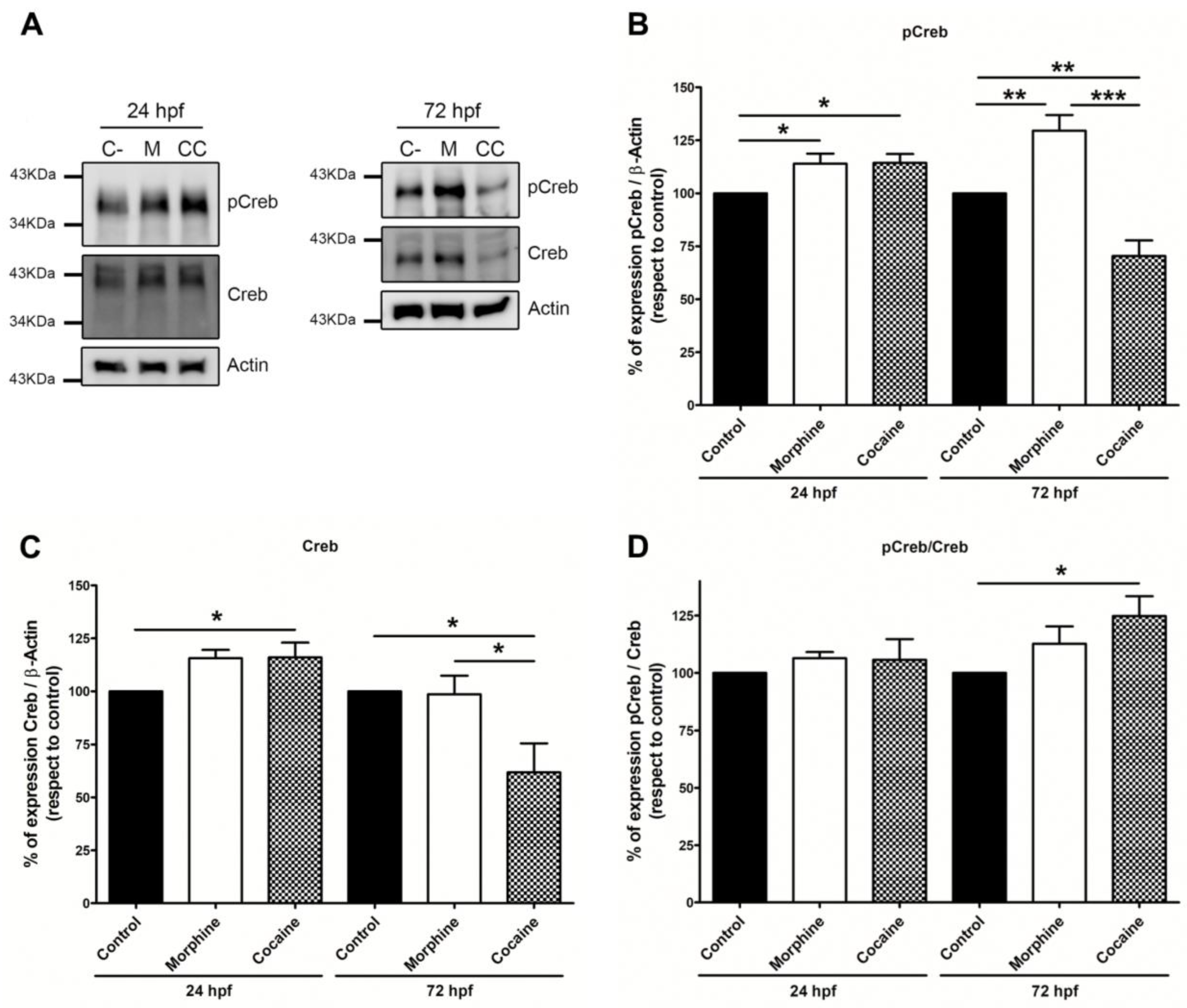
The expression of Creb and its phosphorylated form (pCreb in Ser133) was analysed by Western Blot (Figure 2) using the same membranes for both epitopes. Once the quantifications were made, the pCreb/Creb ratio was determined. Our results indicate that increased levels of pCreb were found in 24 hpf embryos exposed to drugs of abuse, but there was only an increase in total Creb levels after exposure to cocaine. On the other hand, chronic morphine increased pCreb levels in 72 hpf embryos but not Creb. Cocaine produced a significant decrease in both pCreb and Creb expression at this stage of development.

3.3. Fos and Creb TFBSs in Target Genes

In order to find a possible link between the dysregulated genes after morphine and cocaine exposure [37] and the TFs Creb and Fos (also affected by drug exposure), a bioinformatic study was carried out to determine the presence of TFBSs for AP-1 and Creb in the regulatory promoter of the previously identified genes. Bdnf was also included in this analysis, as it is related to the addictive process, but its function still remains unclear. PhysBinder and JASPAR software found at least one binding site for the transcription factors Creb and/or AP-1 in most of the promoters analysed (Table 2). The degree of conservation of the genomic regions that comprise these genes was analysed, and in all cases, they showed synteny within the zebrafish and the human genomes.

3.4. Dopamine (DA) Assays

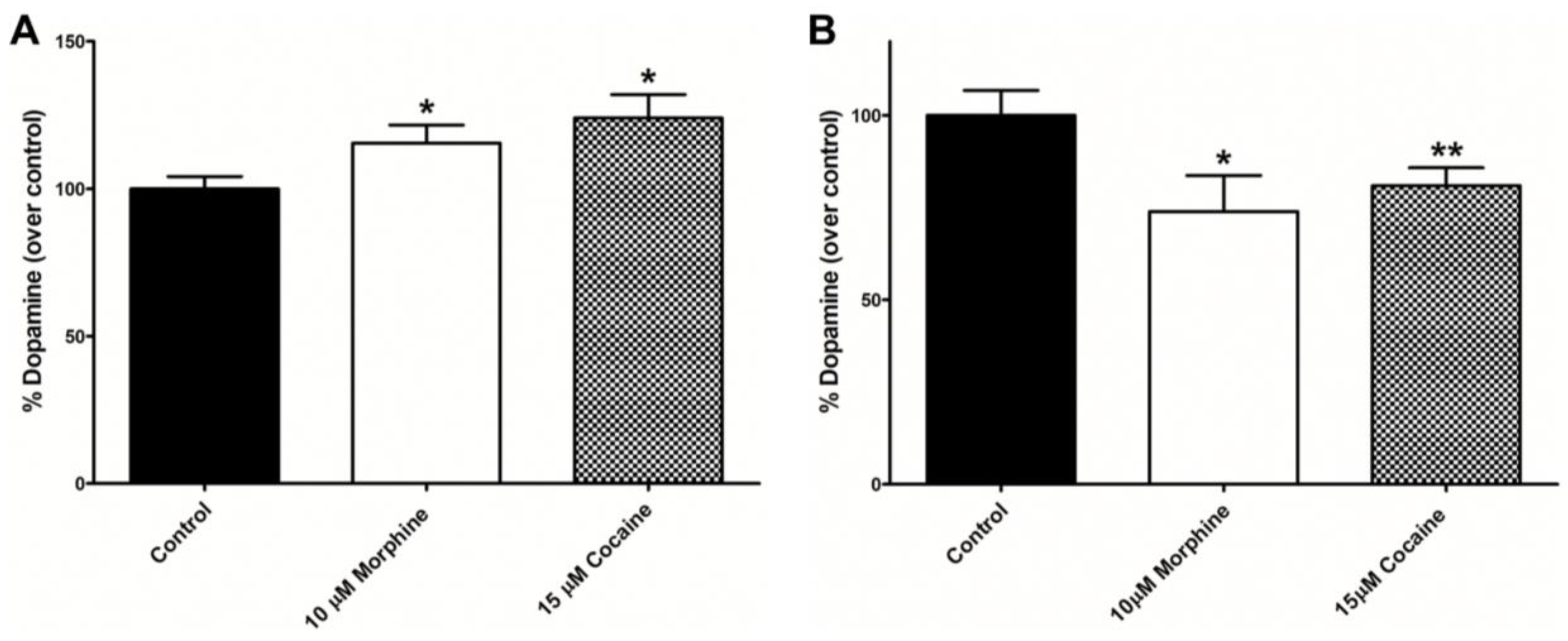
We have determined the amount of dopamine (DA) in embryos treated with drugs of abuse, and the obtained results indicate that exposure to morphine and cocaine produces a biphasic effect: a significant increase in total DA levels is observed in 3 dpf embryos, whereas total DA levels decrease in 6 dpf drug-treated embryos (Figure 3).

3.5. Analysis of the Effect of Morphine and Cocaine on the Expression of Tyrosine Hydroxylase

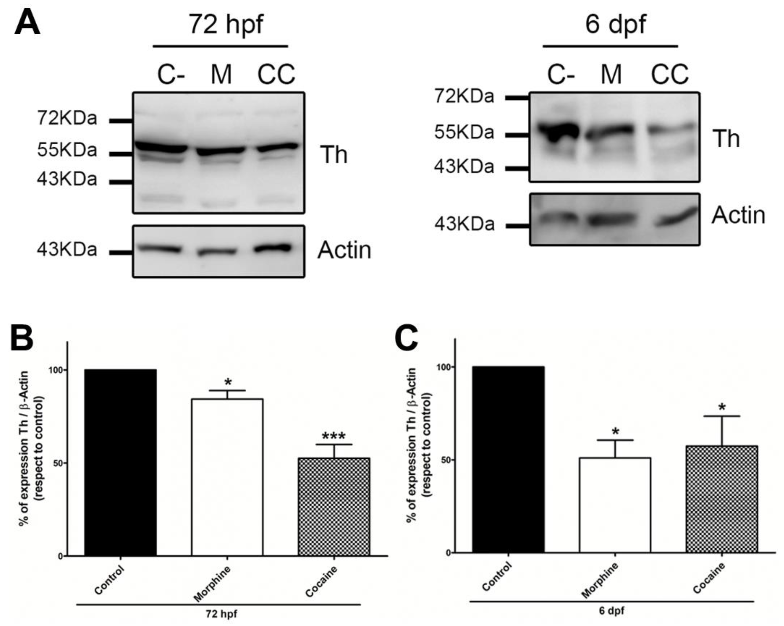
Tyrosine hydroxylase (Th) is the rate-limiting enzyme in catecholamine synthesis, so its expression levels could be directly related to the amount of DA synthesised. We analysed the expression of Th in zebrafish embryos chronically exposed to morphine or cocaine (Figure 4), and both drugs produced a statistically significant decrease in Th levels at 72 hpf and 6 dpf.

4. Discussion

Most studies with drugs of abuse in murine models have focused on specific brain areas. For example, Sadat-Shirazi et al. have studied the role of D1 dopaminergic receptors of the NAc on the development of morphine-induced place conditioning in the offspring of abstinent rats [41]. Schaefer et al. have studied the effects of chronic morphine administration on glycoprotein trafficking in rat brain capillaries [42]. There are also studies with cocaine in rats that analyse its effects on cannabinoid receptors [42] or the effects of cocaine in combination with other substances (such as heroin) on the prelimbic cortex [43].
The zebrafish is an animal model that allows us to study the effects of different substances in the whole organism, so it is possible to determine the complex relationships between different tissues and systems. Current research is mainly focused on toxicology and embryonic development. For example, Mersereau et al. have determined the effects of cocaine administration during embryonic development on cardiovascular physiology in adult zebrafish [44]. As we have also observed, these authors found no significant morphological changes in embryos from 24 hpf to 96 hpf, except for a reduction in the size of the telencephalon in those embryos treated with the highest concentrations of cocaine (10 mg/L and 20 mg/L). Parolini et al. have analysed the effects of cocaine and its main metabolites in zebrafish using trace concentrations equivalent to those that can be found in freshwater as a result of environmental pollution [45]. Regarding the study of morphine effects in zebrafish, some research has focused on the effects of this drug on microRNAs and opioid receptors [46].
First, we performed a toxicological analysis using different concentrations of morphine and cocaine, following a protocol similar to The Fish Embryo Test (ZFET) [47]. Our results showed that neither morphine nor cocaine treatment induced macroscopic malformations or higher mortality rates as compared to the control group at the concentrations used (from 1 nM to 100 µM). We have determined the amount of drug assimilated by the embryos. In the case of 10 μM morphine, 23.11 ± 2.91 pmol morphine was found in embryo lysates, which represents 0.12 ± 0.01% of the initial concentration dissolved in the E3 medium. In the case of cocaine, the absorption was dose-dependent, with a linear relationship between the initial amount of cocaine and the amount detected in the embryo extract. The percentage of cocaine assimilated from the initial concentration was similar for all the concentrations used. According to our results, 10 µM morphine was chosen as the working concentration because it is similar to the concentration commonly administered in murine models (between 0.1 mg/kg to 10 mg/kg) [48]. 15 µM cocaine was chosen because it is also a similar concentration to the concentrations administered in rats (between 15 mg/kg and 50 mg/kg) [49]. In addition to this, benzoylecgonine, the cocaine metabolite used as a biomarker of cocaine use in humans, was detected in embryo extracts treated with this concentration of cocaine. This finding indicates that zebrafish are able to metabolise cocaine and that the metabolic pathway is conserved between these two species. Thus, we have validated a protocol for administering compounds to zebrafish embryos, which have the capacity to assimilate morphine and cocaine, as well as to metabolise the latter by the same detoxification pathway as in humans.
It is well known that drugs of abuse modify dopamine synthesis. We have shown that morphine and cocaine exposure increased dopamine levels in embryos of 72 hpf but decreased in 6 dpf embryos. These effects may be caused due to changes in the synthesis and release of DA. In contrast, after 6 days of exposure, it is possible that DA synthesis cannot compensate for the loss of total DA, thus decreasing total DA levels, or that other regulatory mechanisms (e.g., at the transcriptional level) could be activated, as described in chronic exposure to drugs of abuse [50]. Most studies related to DA release have focused on small brain nuclei. Acute cocaine administration caused an increase in extracellular DA levels, as reported by [51] in the NAc and ventral pallidum using the dual-probe microdialysis technique, and [52] in the striatum of both in control rats and in a rat model of ADHD using in vivo chronoamperometry. Thus, it is possible that after chronic drug treatment, DA release cannot be compensated by endogenous synthesis or reuptake, thus reducing total DA levels.
Changes in DA levels could be due to the differential expression of tyrosine hydroxylase (Th), the rate-limiting enzyme in DA biosynthesis, and we found reduced levels at both 3 dpf and 6 dpf. However, Th activity is regulated by multiple mechanisms, including allosteric effector binding and phosphorylation [53]. Th is activated by phosphorylation by different kinases (PKA, PKC, ERK1 or ERK2) that phosphorylate different Serine (Ser) residues; for example, phosphorylation at Ser40 increases the activity of the enzyme, whereas phosphorylation at Ser31, Ser8 or Ser19 has no direct effect on the activity of the enzyme [54,55]. In response to acute stimulation, Th is mainly regulated by catecholamine inhibition, presenting two allosteric sites [53]. Mice treated with 3,4-methylenedioxy-methamphetamine (MDMA) and MDMA-ethanol mixture showed an increase in both total and the phosphorylated Th levels in the right ventricle after 48 h and 7 days post-exposure [56]. In our study, we have only determined total Th levels so that it is possible that Th is present in a more active form at 3 dpf, thus compensating for the decrease in total Th; consequently, the decrease in total Th would not be reflected in DA levels. However, at 6 dpf, DA stores would be depleted after prolonged drug exposure and Th downregulated; in this case, inhibition of DA biosynthesis cannot compensate for the decrease in DA levels caused by a chronic drug administration [57,58].
Drugs of abuse trigger a series of signalling cascades that induce the expression of different transcription factors (TFs) [28] called immediate expression genes (IEGs); two of them are Creb and Fos, which bind to the promoter of other genes (called late expression genes) and modulate their expression [29,59]; these genes are the final effectors and are closely related to the instatement of tolerance and dependence that lead to increased consumption of these substances [30]. We have analysed the effects of morphine and cocaine exposure on the expression of Fos family proteins (c-Fos, Fra1, Fra2, and FosB), total Creb (Creb) and its phosphorylated form pCreb in 24 hpf, 72 hpf and 6 dpf embryos. Fos proteins showed an increase in their expression, although the changes observed depended on the drug used and the exposure time (Supplementary Table S1). There was a significant increase for c-Fos (55 KDa) in both 24 hpf and 72 hpf embryos treated with morphine, in line with that described by [27] in rat NAc. In the case of FosB (34 KDa band), there is a significant increase in 24 hpf morphine-treated embryos and in 72 hpf embryos exposed to cocaine. In the case of the 26 KDa protein, a significant increase was found in 24 hpf embryos treated with cocaine and in 72 hpf embryos exposed to morphine. In embryos treated with up to 6 dpf, morphine increased the expression of Fra-1. Fra-2 and FosB, whereas cocaine only increased the expression of FosB. Total Creb and its phosphorylated form pCreb were increased by both drugs at 24 hpf. In the 72 hpf assays, a different pattern was observed for each drug: while morphine increased pCreb expression, it was decreased in cocaine-treated embryos. Total Creb was reduced in only cocaine-treated embryos of 72 hpf. The pCreb/Creb ratio was higher for both drugs, although it was only statistically significant for cocaine.
The effects of morphine on c-Fos and Creb protein expression have been studied in the NAc, amygdala, striatum and prefrontal cortex of rats [27]. These authors observed an increase in both c-Fos expression and the pCreb/Creb ratio. Similarly, Hoffmann et al. demonstrated an increase in pCreb levels in the striatum and NAc of cocaine-treated rats [26]. Other authors have found increased expression of pCreb (Ser133) in the heart of postnatal rats exposed to cocaine during the embryonic stage [60]. To our knowledge, there are few studies on the expression of the Fos and Creb transcription factors in zebrafish, particularly in relation to drug exposure, so it is very difficult to compare our results with previously published data. We cannot rule out that changes in Creb expression differ across tissues or organs, as it is known that drugs of abuse not only affect several brain regions but also other organs, causing cardiotoxicity and stroke, as well as altering the digestive system and metabolism [9,12].
In order to find a possible link between the dysregulated genes after morphine and cocaine exposure [37] and the TFs Creb and Fos (also affected by drug exposure), a bioinformatic study was carried out to determine the presence of TFBSs for AP-1 and Creb in the regulatory promoter of the previously identified genes. All of them, except rx3, present at least one binding site for the AP-1 complex or for Creb. Accordingly, morphine and cocaine modify the expression of Creb and AP-1, which are the IEGs that control the expression of the different late-response genes. Activation of Fos and Creb usually induces the expression of their target genes, although Creb and Fos also have truncated isoforms that are not fully functional. In some cases, these isoforms block the active proteins and prevent them from binding to the promoter. For example, the absence of Gln Creb isoforms is essential for their activity or the kinase-inducible domain (KID).

5. Conclusions

We have shown that zebrafish embryos are able to assimilate morphine and cocaine and that these drugs modulate the expression of several proteins related to the reward pathway, namely the transcription factors Fos, Creb and Th, the rate-limiting enzyme of dopamine. Dopamine levels are also modified after drug exposure. These molecular changes may be responsible for the physiological effects of morphine and cocaine, not only in those brain areas related to the reward pathway but also for the systemic outcomes in the entire organism.

Supplementary Materials

The following supporting information can be downloaded at https://www.mdpi.com/article/10.3390/fishes9070268/s1, Figure S1: Amount of drug assimilated by zebrafish embryos of 72 hpf; Table S1: Schematic comparative table of the results obtained in the Western Blot assays.

Author Contributions

Conceptualization, V.G.-N.; Data curation, A.A.C.-G., M.S.-B. and V.G.-N.; Formal analysis, A.A.C.-G. and V.G.-N.; Funding acquisition, V.G.-N.; Investigation, A.A.C.-G. and V.G.-N.; Methodology, A.A.C.-G., M.S.-B. and V.G.-N.; Project administration, V.G.-N.; Resources, V.G.-N.; Supervision, V.G.-N.; Validation, A.A.C.-G., M.S.-B. and V.G.-N.; Writing—original draft, A.A.C.-G. and V.G.-N.; Writing—review and editing, A.A.C.-G., M.S.-B. and V.G.-N. All authors have read and agreed to the published version of the manuscript.

Funding

This research was funded by MINECO/FEDER (AGL2015 68330-C2-2-R). Andres Angel Calderon-Garcia had a pre- and a postdoctoral fellowship, contract USAL (Convocatoria 2018 de ayudas para financiar 34 contratos predoctorales de la Universidad de Salamanca. Cofinanciadas por el Banco Santander. Programa Propio III de la USAL).

Institutional Review Board Statement

The animal study protocol was approved by the Institutional Ethics Committee of Universidad de Salamanca (protocol code 0099).

Data Availability Statement

The original contributions presented in the study are included in the article/Supplementary Material. Further inquiries can be directed to the corresponding author.

Acknowledgments

The authors would like to thank Ana P. Velasco-Criado and Cesar Raposo for their valuable comments and suggestions. We would also like to thank Lucia Moreno Verde for her extraordinary technical support.

Conflicts of Interest

The authors declare no conflicts of interest.

References

  1. Devereaux, A.L.; Mercer, S.L.; Cunningham, C.W. DARK Classics in Chemical Neuroscience: Morphine. ACS Chem. Neurosci. 2018, 9, 2395–2407. [Google Scholar] [CrossRef]
  2. Stein, C.; Schäfer, M.; Machelska, H. Why Is Morphine Not the Ultimate Analgesic and What Can Be Done to Improve It? J. Pain 2000, 1, 51–56. [Google Scholar] [CrossRef] [PubMed]
  3. Martin, W.R. Pharmacology of Opioids. Pharmacol. Rev. 1984, 35, 283–323. [Google Scholar]
  4. Pacifici, G.M. Metabolism and Pharmacokinetics of Morphine in Neonates: A Review. Clinics 2016, 71, 474–480. [Google Scholar] [CrossRef] [PubMed]
  5. Chakrabarti, S.; Chang, A.; Liu, N.; Gintzler, A.R. HHS Public Access. J. Neurochem. 2016, 139, 737–747. [Google Scholar] [CrossRef] [PubMed]
  6. Sverrisdóttir, E.; Lund, T.M.; Olesen, A.E.; Drewes, A.M.; Christrup, L.L.; Kreilgaard, M. A Review of Morphine and Morphine-6-Glucuronide’s Pharmacokinetic-Pharmacodynamic Relationships in Experimental and Clinical Pain. Eur. J. Pharm. Sci. 2015, 74, 45–62. [Google Scholar] [CrossRef]
  7. Korsgaard, H.O.; Torgersen, S.; Wentzel-Larsen, T.; Ulberg, R. Substance Abuse and Personality Disorder Comorbidity in Adolescent Outpatients: Are Girls More Severely Ill than Boys? Child Adolesc. Psychiatry Ment. Health 2016, 10, 1–9. [Google Scholar] [CrossRef] [PubMed]
  8. Fridell, M.; Bäckström, M.; Hesse, M.; Krantz, P.; Perrin, S.; Nyhlén, A. Prediction of Psychiatric Comorbidity on Premature Death in a Cohort of Patients with Substance Use Disorders: A 42-Year Follow-Up. BMC Psychiatry 2019, 19, 150. [Google Scholar] [CrossRef] [PubMed]
  9. Kim, S.T.; Park, T. Acute and Chronic Effects of Cocaine on Cardiovascular Health. Int. J. Mol. Sci. 2019, 20, 584. [Google Scholar] [CrossRef]
  10. Drake, L.R.; Scott, P.J.H. DARK Classics in Chemical Neuroscience: Cocaine. ACS Chem. Neurosci. 2018, 9, 2358–2372. [Google Scholar] [CrossRef]
  11. Mantsch, J.R.; Vranjkovic, O.; Twining, R.C.; Gasser, P.J.; Mcreynolds, J.R.; Blacktop, J.M. Neurobiological Mechanisms That Contribute to Stress-Related Cocaine Use. Neuropharmacology 2014, 76, 383–394. [Google Scholar] [CrossRef] [PubMed]
  12. Riezzo, I.; Fiore, C.; De Carlo, D.; Pascale, N.; Neri, M.; Turillazzi, E.; Fineschi, V. Side Effects of Cocaine Abuse: Multiorgan Toxicity and Pathological Consequences. Curr. Med. Chem. 2012, 19, 5624–5646. [Google Scholar] [CrossRef] [PubMed]
  13. Fonseca, A.C.; Ferro, J.M. Drug Abuse and Stroke. Curr. Neurol. Neurosci. Rep. 2013, 13, 325. [Google Scholar] [CrossRef] [PubMed]
  14. Buttner, A. Neuropathological Alterations in Cocaine Abuse. Curr. Med. Chem. 2012, 19, 5597–5600. [Google Scholar] [CrossRef] [PubMed]
  15. Spronk, D.B.; van Wel, J.H.P.; Ramaekers, J.G.; Verkes, R.J. Characterizing the Cognitive Effects of Cocaine: A Comprehensive Review. Neurosci. Biobehav. Rev. 2013, 37, 1838–1859. [Google Scholar] [CrossRef] [PubMed]
  16. Berridge, K.C.; Kringelbach, M.L. Pleasure Systems of the Brain. Neuron 2016, 86, 646–664. [Google Scholar] [CrossRef] [PubMed]
  17. Yager, L.M.; Garcia, A.F.; Wunsch, A.M.; Ferguson, S.M. The Ins and Outs of the Striatum: Role in Drug Addiction. Neuroscience 2015, 301, 529–541. [Google Scholar] [CrossRef]
  18. Adinoff, B. Neurobiologic Processes in Drug Reward and Addiction. Harv. Rev. Psychiatry 2004, 12, 305–320. [Google Scholar] [CrossRef]
  19. Nestler, E.J. Historical Review: Molecular and Cellular Mechanisms of Opiate and Cocaine Addiction. Trends Pharmacol. Sci. 2004, 25, 210–218. [Google Scholar] [CrossRef]
  20. Lau, B.; Bretaud, S.; Huang, Y.; Lin, E.; Guo, S. Dissociation of Food and Opiate Preference by a Genetic Mutation in Zebrafish. Genes Brain Behav. 2006, 5, 497–505. [Google Scholar] [CrossRef]
  21. Wenzel, J.M.; Rauscher, N.A.; Cheer, J.F.; Oleson, E.B. A Role for Phasic Dopamine Release within the Nucleus Accumbens in Encoding Aversion: A Review of the Neurochemical Literature. ACS Chem. Neurosci. 2014, 6, 16–26. [Google Scholar] [CrossRef]
  22. Musacchio, J.M. Enzymes Involved in the Biosynthesis and Degradation of Catecholamines. In Biochemistry of Biogenic Amines; Springer: New York, NY, USA, 2013; pp. 1–35. ISBN 978-1-4684-3171-1. [Google Scholar]
  23. Koob, G.F.; Ahmed, S.H.; Boutrel, B.; Chen, S.A.; Kenny, P.J.; Markou, A.; Dell, L.E.O.; Parsons, L.H.; Sanna, P.P. Neurobiological Mechanisms in the Transition from Drug Use to Drug Dependence. Neurosci. Biobehav. Rev. 2004, 27, 739–749. [Google Scholar] [CrossRef]
  24. Bodnar, R.J. Endogenous Opiates and Behavior: 2014. Peptides 2016, 124, 18–70. [Google Scholar] [CrossRef]
  25. Johnson, S.W.; North, R.A. Opioids Excite Dopamine Neurons by Hyperpolarization Interneurons. J. Neurosci. 1992, 12, 4. [Google Scholar] [CrossRef] [PubMed]
  26. Hoffmann, H.M.; Nadal, R.; Vignes, M.; Ortiz, J. Chronic Cocaine Self-Administration Modulates ERK1/2 and CREB Responses to Dopamine Receptor Agonists in Striatal Slices. Addict. Biol. 2012, 17, 565–575. [Google Scholar] [CrossRef]
  27. Haghparast, A.; Fatahi, Z.; Alamdary, S.Z.; Reisi, Z.; Khodagholi, F. Changes in the Levels of P-ERK, p-CREB, and c-Fos in Rat Mesocorticolimbic Dopaminergic System after Morphine-Induced Conditioned Place Preference: The Role of Acute and Subchronic Stress. Cell. Mol. Neurobiol. 2014, 34, 277–288. [Google Scholar] [CrossRef]
  28. Moratalla, R. Neurobiología de La Cocaína. Trastor. Adict. 2008, 10, 143–150. [Google Scholar] [CrossRef]
  29. Kelz, M.B.; Nestler, E.J. ΔFosB: A Molecular Switch Underlying Long-Term Neural Plasticity. Curr. Opin. Neurol. 2000, 13, 715–720. [Google Scholar] [CrossRef] [PubMed]
  30. Nestler, E.J. Reflections on: “A General Role for Adaptations in G-Proteins and the Cyclic AMP System in Mediating the Chronic Actions of Morphine and Cocaine on Neuronal Function”. Brain Res. 2016, 1645, 71–74. [Google Scholar] [CrossRef]
  31. Corbett, A.D.; Henderson, G.; McKnight, A.T.; Paterson, S.J. 75 Years of Opioid Research: The Exciting but Vain Quest for the Holy Grail. Br. J. Pharmacol. 2006, 147, 153–162. [Google Scholar] [CrossRef]
  32. Waldhoer, M.; Bartlett, S.E.; Whistler, J.L. Opioid Receptors. Annu. Rev. Biochem. 2004, 73, 953–990. [Google Scholar] [CrossRef] [PubMed]
  33. Macey, T.A.; Lowe, J.D.; Chavkin, C. Mu Opioid Receptor Activation of ERK1/2 Is GRK3 and Arrestin Dependent in Striatal Neurons. J. Biol. Chem. 2006, 281, 34515–34524. [Google Scholar] [CrossRef] [PubMed]
  34. Blendy, J.A.; Maldonado, R. Genetic Analysis of Drug Addiction: The Role of CAMP Response Element Binding Protein. J. Mol. Med. 1998, 76, 104–110. [Google Scholar] [CrossRef]
  35. Nestler, E.J. Molecular Basis of Long-Term Plasticity Underlying Addiction. Nat. Rev. Neurosci. 2001, 2, 119–128. [Google Scholar] [CrossRef] [PubMed]
  36. McClung, C.A.; Nestler, E.J. Regulation of Gene Expression and Cocaine Reward by CREB and ΔFosB. Nat. Neurosci. 2003, 6, 1208–1215. [Google Scholar] [CrossRef] [PubMed]
  37. Calderon-Garcia, A.A.; Perez-Fernandez, M.; Curto-Aguilera, D.; Rodriguez-Martin, I.; Sánchez-Barba, M.; Gonzalez-Nunez, V. Exposure to Morphine and Cocaine Modify the Transcriptomic Landscape in Zebrafish Embryos. Neuroscience 2022, 507, 14–27. [Google Scholar] [CrossRef] [PubMed]
  38. Webb, K.J.; Norton, W.H.J.; Trümbach, D.; Meijer, A.H.; Ninkovic, J.; Topp, S.; Heck, D.; Marr, C.; Wurst, W.; Theis, F.J.; et al. Zebrafish Reward Mutants Reveal Novel Transcripts Mediating the Behavioral Effects of Amphetamine. Genome Biol. 2009, 10, R81. [Google Scholar] [CrossRef] [PubMed]
  39. Broos, S.; Soete, A.; Hooghe, B.; Moran, R.; van Roy, F.; De Bleser, P. PhysBinder: Improving the Prediction of Transcription Factor Binding Sites by Flexible Inclusion of Biophysical Properties. Nucleic Acids Res. 2013, 41, 531–534. [Google Scholar] [CrossRef]
  40. Khan, A.; Fornes, O.; Stigliani, A.; Gheorghe, M.; Castro-Mondragon, J.A.; Van Der Lee, R.; Bessy, A.; Chèneby, J.; Kulkarni, S.R.; Tan, G.; et al. JASPAR 2018: Update of the Open-Access Database of Transcription Factor Binding Profiles and Its Web Framework. Nucleic Acids Res 2018, 46, D260–D266. [Google Scholar] [CrossRef]
  41. Sadat-shirazi, M.; Monfared, N.; Matloob, M. Possible Involvement of Nucleus Accumbens D1-like Dopamine Receptors in the Morphine-Induced Condition Place Preference in the Offspring of Morphine Abstinent Rats. Life Sci. 2019, 233, 116712. [Google Scholar] [CrossRef]
  42. Schaefer, C.P.; Arkwright, N.B.; Jacobs, L.M.; Jarvis, C.K.; Hunn, C.; Largent-milnes, T.M.; Tome, M.E.; Davis, T.P. Chronic Morphine Exposure Potentiates P-Glycoprotein Trafficking from Nuclear Reservoirs in Cortical Rat Brain Microvessels. PLoS ONE 2018, 13, e0192340. [Google Scholar] [CrossRef]
  43. Rubio, F.J.; Quintana-Feliciano, R.; Warren, B.L.; Li, X.; Witonsky, K.F.R.; del Valle, F.S.; Selvam, P.V.; Caprioli, D.; Venniro, M.; Bossert, J.M.; et al. Prelimbic Cortex Is a Common Brain Area Activated during Cue-Induced Reinstatement of Cocaine and Heroin Seeking in a Polydrug Self-Administration Rat Model. Eur. J. Neurosci. 2019, 49, 165–178. [Google Scholar] [CrossRef] [PubMed]
  44. Mersereau, E.J.; Boyle, C.A.; Poitra, S.; Espinoza, A.; Seiler, J.; Longie, R.; Delvo, L.; Szarkowski, M.; Maliske, J.; Chalmers, S.; et al. Longitudinal Effects of Embryonic Exposure to Cocaine on Morphology, Cardiovascular Physiology, and Behavior in Zebrafish. Int. J. Mol. Sci. 2016, 17, 847. [Google Scholar] [CrossRef] [PubMed]
  45. Parolini, M.; Ghilardi, A.; Della Torre, C.; Magni, S.; Prosperi, L.; Calvagno, M.; Del Giacco, L.; Binelli, A. Environmental Concentrations of Cocaine and Its Main Metabolites Modulated Antioxidant Response and Caused Cyto-Genotoxic Effects in Zebrafish Embryo Cells. Environ. Pollut. 2017, 226, 504–514. [Google Scholar] [CrossRef] [PubMed]
  46. Jimenez-Gonzalez, A.; García-Concejo, A.; López-Benito, S.; Gonzalez-Nunez, V.; Arévalo, J.C.; Rodriguez, R.E. Role of Morphine, MiR-212/132 and Mu Opioid Receptor in the Regulation of Bdnf in Zebrafish Embryos. Biochim. Biophys. Acta Gen. Subj. 2016, 1860, 1308–1316. [Google Scholar] [CrossRef] [PubMed]
  47. Braunbeck, T.; Kais, B.; Lammer, E.; Otte, J.; Schneider, K.; Stengel, D. The Fish Embryo Test (FET): Origin, Applications, and Future. Environ. Sci. Pollut. Res. Int. 2015, 22, 16247–16261. [Google Scholar] [CrossRef] [PubMed]
  48. Wang, D.; Zhang, J.; Bai, Y.; Zheng, X.; Alizamini, M.M.; Shang, W.; Yang, Q.; Li, M.; Li, Y.; Sui, N. Melanin-Concentrating Hormone in Rat Nucleus Accumbens or Lateral Hypothalamus Differentially Impacts Morphine and Food Seeking Behaviors. J. Psychopharmacol. 2020, 34, 478–489. [Google Scholar] [CrossRef] [PubMed]
  49. Zhang-james, Y.; Lloyd, D.R.; James, M.L.; Yang, L.; Richards, J.B.; Faraone, S.V. Oral Methylphenidate Treatment of an Adolescent ADHD Rat Model Does Not Alter Cocaine-Conditioned Place Preference during Adulthood: A Negative Report. J. Psychiatr. Brain Sci. 2019, 4, e190021. [Google Scholar] [CrossRef] [PubMed]
  50. NIDA. How Does Cocaine Produce Its Effects? Available online: https://www.drugabuse.gov/publications/research-reports/cocaine/how-does-cocaine-produce-its-effects (accessed on 2 March 2024).
  51. Wydra, K.; Golembiowska, K.; Zaniewska, M.; Kamińska, K.; Ferraro, L.; Fuxe, K.; Filip, M. Accumbal and Pallidal Dopamine, Glutamate and GABA Overflow during Cocaine Self-Administration and Its Extinction in Rats. Addict. Biol. 2013, 18, 307–324. [Google Scholar] [CrossRef]
  52. Womersley, J.S.; Kellaway, L.A.; Stein, D.J.; Gerhardt, G.A.; Russell, V.A. Effect of Cocaine on Striatal Dopamine Clearance in a Rat Model of Developmental Stress and Attention-Deficit/Hyperactivity Disorder. Stress 2016, 19, 78–82. [Google Scholar] [CrossRef]
  53. Dickson, P.W.; Briggs, G.D. Tyrosine Hydroxylase. Regulation by Feedback Inhibition and Phosphorylation, 1st ed.; Elsevier Inc.: Amsterdam, The Netherlands, 2013; Volume 68, ISBN 9780124115125. [Google Scholar]
  54. Daubner, S.C.; Le, T.; Wang, S. Tyrosine Hydroxylase and Regulation of Dopamine Synthesis. Arch. Biochem. Biophys. 2011, 508, 1–12. [Google Scholar] [CrossRef] [PubMed]
  55. Dunkley, P.R.; Bobrovskaya, L.; Graham, M.E.; Von Nagy-Felsobuki, E.I.; Dickson, P.W. Tyrosine Hydroxylase Phosphorylation: Regulation and Consequences. J. Neurochem. 2004, 91, 1025–1043. [Google Scholar] [CrossRef] [PubMed]
  56. Navarro-Zaragoza, J.; Ros-Simó, C.; Milanés, M.V.; Valverde, O.; Laorden, M.L. Binge Ethanol and MDMA Combination Exacerbates HSP27 and Trx-1 (Biomarkers of Toxic Cardiac Effects) Expression in Right Ventricle. Life Sci. 2019, 220, 50–57. [Google Scholar] [CrossRef] [PubMed]
  57. Beitner-Johnson, D.; Guitart, X.; Nestler, E.J. Common Intracellular Actions of Chronic Morphine and Cocaine in Dopaminergic Brain Reward Regions. Ann. N. Y. Acad. Sci. 1992, 654, 70–87. [Google Scholar] [CrossRef] [PubMed]
  58. Beitner-Johnson, D.; Guitart, X.; Nestler, E.J. Dopaminergic Brain Reward Regions of Lewis and Fischer Rats Display Different Levels of Tyrosine Hydroxylase and Other Morphine-and Cocaine-Regulated Phosphoproteins. Brain Res. 1991, 561, 147–150. [Google Scholar] [CrossRef]
  59. Han, M.; Bolaños, C.; Green, T.; Olson, V.; Neve, R.; Liu, R.; Aghajanian, G.; Nestler, E. Role of CAMP Response Element-Binding Protein in the Rat Locus Ceruleus: Regulation of Neuronal Activity and Opiate Withdrawal Behaviors. J. Neurosci. 2006, 26, 4624–4629. [Google Scholar] [CrossRef]
  60. Sun, L.S.; Quamina, A. Perinatal Cocaine Exposure Stimulates the Expression and Activation of CREB in the Neonatal Rat Heart. Pediatr. Res. 2003, 53, 500–506. [Google Scholar] [CrossRef]
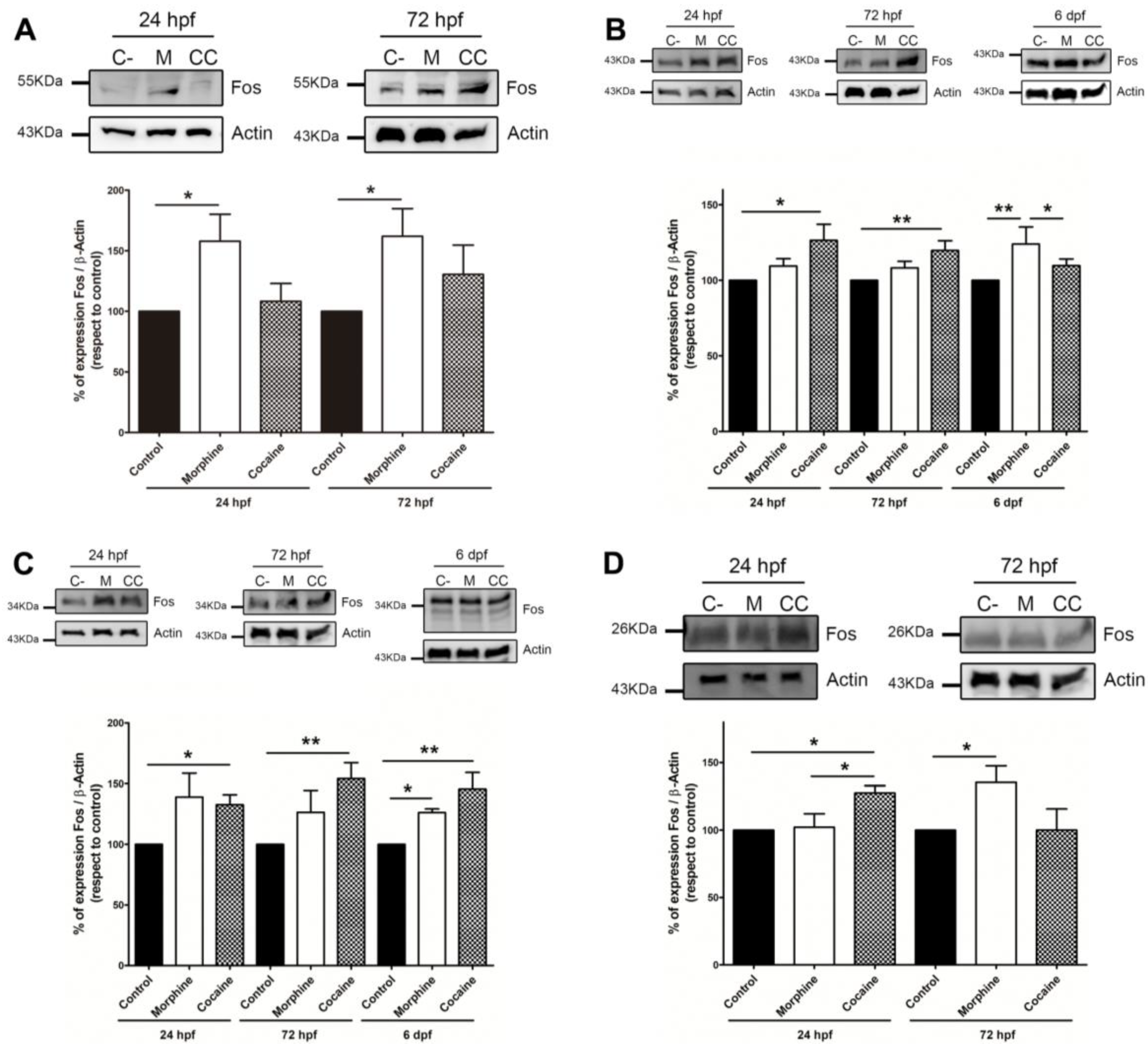
Figure 1. Expression levels of the Fos family of proteins in zebrafish embryos of 24 hpf, 72 hpf and 6 dpf treated with 10 μM morphine or 15 μM cocaine. (A) 55 KDa, (B) 43 KDa, (C) 34 KDa and (D) 26 KDa (ΔFosB) bands. For each of the bands, a representative image of the Western Blot assays using β-actin as loading control (top) and quantification data (bottom) are shown. Data correspond to the mean ± S.E.M. of the intensity of each of the bands analysed from at least four independent experiments. The results for each experimental group have been analysed with one-way ANOVA, followed by Tukey post-test. Legend: * p-value < 0.05; **—p-value < 0.01.
Fishes 09 00268 g001
Figure 2. Creb and pCreb (Ser133) protein expression levels in 24 hpf and 72 hpf zebrafish embryos treated with 10 μM morphine or 15 μM cocaine. (A) Representative image of the Western Blot assays for Creb and pCreb using β-actin as loading control. (B) pCreb quantification. (C) Creb quantification. (D) pCreb/Creb expression ratio. Data correspond to the mean ± S.E.M. of the intensity of each of the bands analysed from at least five independent experiments. The results for each experimental group have been analysed with one-way ANOVA, followed by Tukey’s post-test. Legend: * p-value < 0.05; **—p-value < 0.01; ***—p-value < 0.001.
Fishes 09 00268 g002
Figure 3. Effect of chronic morphine and cocaine on total dopamine levels in (A) 72 hpf and (B) 6 dpf zebrafish embryos. The amount of dopamine in the drug-exposed groups is expressed as a percentage of the control group. Data correspond to the mean ± S.E.M. from at least six independent experiments. The results for each experimental group have been analysed with one-way ANOVA, followed by Tukey post-test. Legend: * p-value < 0.05; **—p-value < 0.01.
Fishes 09 00268 g003
Figure 4. Tyrosine hydroxylase (Th) protein expression levels in 72 hpf and 6 dpf zebrafish embryos treated with 10 μM morphine or 15 μM cocaine. (A) Representative image of the Western Blot assays for Th using β-actin as the loading control. (B) Quantification data of Th expression at 72 hpf. (C) Quantification data of Th expression at 6 dpf. Data correspond to the mean ± S.E.M. of the intensity of each of the bands analysed from at least five independent experiments. The results for each experimental group have been analysed with one-way ANOVA, followed by Tukey post-test. Legend: * p-value < 0.05; ***—p-value < 0.001.
Fishes 09 00268 g004
Table 1. Specific antibodies that were used in Western blot assays. The table shows the antibodies used with the reference, lot number, company and dilution used.
Antibody
(Reference n.)
Host SpeciesCompanylot n.Dilution
Fos (K-25)
(sc-253)
RabbitSanta Cruz Biotechnology ®, Dallas, TX, USAJ28091:500
phospho-CREB Ser133(87G3)RabbitCell Signalling ®, Danvers, MA, USA141:1000
CREB
(48H2)
RabbitCell Signalling ®, Danvers, MA, USA161:1000
Tyrosine hydroxylase (AB152)RabbitMillipore, Burlington, MA, USANG18307491:1000
β-actin
(4967)
RabbitCell Signalling ®, Danvers, MA, USA10/20161:1000
Anti-Rabbit-HRP (111-035-003)GoatJackson InmunoResearch ®, West Grove, PA, USA 1315991:10,000
Table 2. Binding sites for CREB and AP-1 transcription factors in the regulatory promoter of morphine and/or cocaine dysregulated genes. This table shows the ENSEMBL identifier, the binding sites and the existence of synteny between the zebrafish and the human genomic fragment.
GeneENSEMBL IDAP-1 SiteCRE SiteSynteny
apoc1ENSDARG00000092170PF0007.1
−2449 a −2442
-
apoeaENSDARG00000102004PF0007.1
−1416 a −1406
−643 a −633
-
bdnfENSDARG00000018817PF0007.1
−1943 a −1933
-
cfosENSDARG00000031683PF0007.1
−2899 a −2893
MA0018.2
−1098 a −1089
cxcl11.5ENSDARG00000092423-MA0018.2
−232 a −223
cxcl11.7ENSDARG00000093779PF0007.1
−608 a −598
-
cyp1aENSDARG00000098315-MA0018.2
−73 a −64
elf3ENSDARG00000077982-MA0018.1
−1899 a −1893
fabp2ENSDARG00000006427PF0007.1
−1035 a −1028
-
gnb3aENSDARG00000004358HSA0000011.1
−1442 a −1434
MA0018.3
−154 a −143
hbzENSDARG00000045142PF0007.1
−388 a −378
-
opn1sw1ENSDARG00000045677PF0007.1
−2886 a −2876
−2308 a −2298
−371 a −361
rx3ENSDARG00000052893--
Disclaimer/Publisher’s Note: The statements, opinions and data contained in all publications are solely those of the individual author(s) and contributor(s) and not of MDPI and/or the editor(s). MDPI and/or the editor(s) disclaim responsibility for any injury to people or property resulting from any ideas, methods, instructions or products referred to in the content.