Brief Report

Molecular Cloning and Gene Expression of Type I Suppressors of Cytokine Signaling 6 and 7 (SOCS6 and SOCS7) in Whiteleg Shrimp (Litopenaeus vannamei)

by
Gunasekara Chathura Wikumpriya
,
Madhuranga Walawedurage Srinith Prabhatha
,
Jiye Lee
and
Chan-Hee Kim
*
Division of Fisheries Life Science, Pukyong National University, Busan 48513, Republic of Korea
*
Author to whom correspondence should be addressed.
Fishes 2023, 8(8), 416; https://doi.org/10.3390/fishes8080416
Submission received: 28 July 2023 / Revised: 10 August 2023 / Accepted: 11 August 2023 / Published: 14 August 2023
(This article belongs to the Section Genetics and Biotechnology)

Abstract

:
Suppressor of cytokine signaling (SOCS) genes are essential negative regulators that modulate cytokine signaling and play key roles in numerous biological processes, including immune responses. In this study, we cloned the complementary DNA (cDNA) sequences of two SOCS genes, designated as LvSOCS6 and LvSOCS7, from the whiteleg shrimp, Litopenaeus vannamei. LvSOCS6 encoded a polypeptide of 463 amino acids (aas), spanning 1392 base pairs (bps), while LvSOCS7 encoded a significantly larger polypeptide of 955 aas, encompassing 2868 bps. Both LvSOCS proteins exhibited conserved domains associated with SOCS, including a centralized Src homology 2 (SH2) domain and a C-terminal SOCS box. Phylogenetic analysis revealed that the deduced aa sequences of LvSOCS6 and LvSOCS7 clustered within the invertebrate type I SOCS family, indicating their evolutionary relatedness. Tissue distribution analysis demonstrated ubiquitous expression of both LvSOCS genes across all examined tissues, with LvSOCS6 showing heightened expression in the gills and LvSOCS7 in the gills and stomach. Notably, mRNA expression patterns of LvSOCS genes following LPS and poly (I:C) stimulations exhibited significant upregulations, while PGN stimulation yielded incongruous results across the examined tissues. Interestingly, concurrent with the diminished expression of LvSOCS6 and LvSOCS7, there was significant elevation in mRNA expression levels of LvSTAT, a vital component of the Janus kinase (JAK)/signal transducer and activator of transcription (STAT) pathway. These findings shed light on the potential involvement of the newly discovered type I SOCS genes in regulating the JAK/STAT pathways and playing pivotal roles in orchestrating the innate immune responses in L. vannamei defense mechanisms.
Key Contribution: Successfully identified novel members of the type I suppressor of cytokine signaling (SOCS) gene family, namely LvSOCS6 and LvSOCS7, in whiteleg shrimp (L. vannamei). Their phylogenetic relationships; their expression patterns in normal tissues; and, following LPS, poly (I:C) and PGN stimulation were analyzed.

1. Introduction

Cytokines, which are secretory protein molecules, are pivotal players in various biological processes, including cell proliferation, growth, and immune regulation across both vertebrates and invertebrates [1,2,3]. Most of the cytokines are involved with the Janus kinase (JAK) and the signal transducers and activators of transcription (STAT) pathways, which are known to play an essential role in immune responses [4]. Accumulating evidence suggests the presence of a number of JAKs and STATs in vertebrates, including humans and fishes, and their possible regulation by these molecules [5,6]. Hence, tight control of cytokines is vital for maintaining immune homeostasis. Consequently, various molecules have been identified that act as physiological suppressors and restrain the excessive activities of cytokines. Among them, the suppressor of cytokine signaling (SOCS) molecules hold particular significance as the “key negative regulators” of these molecules [7].
The SOCS family of vertebrates comprises eight members, namely SOCS1–7 and cytokine-inducible SH2-containing protein (CISH) [8,9]. Based on the available evolutionary analysis data, two distinctive SOCS groups have been identified as type II with SOCS1–3 and CISH and type I with SOCS4–7. All SOCS members share common structural features, including a C-terminal SOCS box and a centralized SH2 domain [1,8,9]. Meanwhile, their N-terminal regions exhibit variability which is believed to be associated with their specific functions. For instance, SOCS1 and SOCS3 have a unique N-terminal domain, the kinase inhibitory region (KIR), which is essential for the suppression of the JAK tyrosine kinase activity [10,11]. SOCS4 and SOCS5 contain a conserved region in their N-terminal (N-terminal conserved region-NLTR) which has been shown to play an important role in the regulation of epidermal growth factor receptor signaling [12,13]. Interestingly, SOCS6 and SOCS7 do not have any of these functional elements in their respective N-terminal regions [14].
Despite the well-documented immune regulatory activities of SOCS genes in vertebrates, their functions in invertebrates, particularly the type I SOCS, remain relatively undiscovered. As such, several studies have been directed toward understanding the role of SOCS in the immune regulation of invertebrates. The first identified invertebrate SOCS was SOCS-36E in fruit flies (Drosophila melanogaster), and it has revealed that it has similar characteristics and functional significance to the human SOCS5 in regulating the JAK-STAT pathway [15]. Subsequent studies revealed two additional SOCS genes in D. melanogaster, named SOCS44A and SOCS16D, which displayed sequence similarities (33–34% and 45–48%) to human SOCS6 and SOCS7, with potential regulatory capabilities in the JAK-STAT pathway [16]. In reference to crustaceans, the role of SOCS6 in the Chinese mitten crab (Eriocheir sinensis) was recently elucidated, highlighting its involvement in the mediation of both the JAK-STAT and the NF-κB (nuclear factor-kappa B) pathways [14]. Additionally, in the Pacific oyster (Crassostrea gigas), three SOCS genes (SOCS2, SOCS5, and SOCS7) have been identified, exhibiting potential for the regulation of NF-κB transcription [17]. Furthermore, the expression and functional implications of SOCS2 (which belongs to the type II SOCS family) in the innate immune responses of red swamp crayfish and whiteleg shrimp (Procambarus clarkii and L. vannamei) have been investigated following lipopolysaccharide (LPS) or bacterial challenges [18,19]. These studies conveyed the importance of SOCS genes in the immune defense mechanisms of crustaceans.
Despite the growing body of research on invertebrate SOCS, there is a notable knowledge gap regarding the presence and function of type I SOCS genes in shrimp. The present study aims to address this gap by investigating the presence of type I SOCS genes (LvSOCS6 and LvSOCS7) in whiteleg shrimp. The mRNA expression levels of the identified SOCS upon post-immune stimulations and their possible association with the JAK-STAT signaling pathway were investigated, expanding our understanding of the immune regulatory mechanisms of SOCS in crustaceans.

2. Materials and Methods

2.1. Shrimp Rearing and Tissue Collection

One-month-old, healthy juvenile whiteleg shrimp, L. vannamei (body weight, BW = 0.9 ± 0.1 g; body length, BL = 1.8 ± 0.2 cm), were purchased from a commercial shrimp farm in Muan-gun, Jeollanam-do, South Korea. All shrimp were transported live to the laboratory of Pukyong National University and reared in a recirculating aquaculture system tank (width × depth × height = 1.0 m × 3.0 m × 0.5 m) equipped with sponge-filtered and UV-sterilized seawater. The shrimp were fed four times per day with a commercially formulated shrimp diet (Jeil Feed Co., Ltd., Daejeon, Republic of Korea) on an ad libitum basis. The rearing tank was maintained under continuous aeration (dissolved oxygen, DO = 9.7 ± 0.2 mg/L) with an ambient temperature of 24 ± 1 °C and a pH of 7.6–7.8. The water chemistry parameters were measured once a day, and the concentrations of ammonia (NH3), nitrites (NO2), and nitrates (NO3) were maintained at 0.25–0.5 mg/L, 0.25 mg/L, and 20–40 mg/L, respectively, to ensure optimal rearing conditions. Whiteleg shrimp grown to a BW of 3.0 ± 0.5 g and a BL of 2.5 ± 0.3 cm were used in all experiments. To isolate hemocytes, shrimp hemolymph was drawn from the ventral region above the first abdominal segment using a sterilized syringe preloaded with a commercial anticoagulant (Alsever’s solution, A3551, Sigma-Aldrich, St. Louis, MO, USA) in a 1:1 ratio, and then immediately centrifuged at 8000× g for 15 min at 4 °C. Tissues were harvested by dissection. All tissue samples were stored in RNAlater solution (Thermo Fisher Scientific, Schwerte, Germany) at −80 °C until use. Specific approval by the local institution/ethics committee was not required for the present study using invertebrate crustaceans, and all experimental procedures were strictly conducted according to the guidelines for the care and use of laboratory animals by the Animal Ethics Committee of Pukyong National University.

2.2. Immune Challenge

The reared shrimp were randomly distributed into four 40 L experimental tanks with four groups, including three immune challenge groups and a control group, each containing 30 shrimp. For the immune challenge experiments, 10 µL of polyinosinic-polycytidylic acid (poly I:C) (Sigma-Aldrich, St. Louis, MO, USA), lipopolysaccharide (LPS) (Escherichia coli 0111: B4, Sigma-Aldrich), and peptidoglycan (PGN) (Staphylococcus aureus, Sigma-Aldrich), suspended in phosphate buffer saline (PBS, pH 7.4) at 100 mg/mL, were injected into shrimp abdominal segments Ⅲ and Ⅳ. In the control group, an equal volume of PBS was injected. Tissues (gill, heart, muscle, and stomach) were pooled from three shrimp that were randomly sampled at 0, 6, 12, 24, 48, and 72 h post-injection (hpi) and stored in RNAlater solution (Thermo Fisher Scientific) at −80 °C until use. The immune challenge experiments were performed in triplicate.

2.3. Total RNA Extraction and cDNA Synthesis

Total RNA was extracted from the RNAlater-stored tissues using an RNA extraction kit following the manufacturer’s protocol (Bioneer, Daejeon, Republic of Korea), including the DNA elimination step. The quantity and quality of the obtained total RNA were assessed using a spectrophotometer (Nanophotomer NP 80, Implen, Munich, Germany), and the ratios of both 260 nm/280 nm and 260 nm/230 nm were confirmed to be higher than 1.9. Complementary DNA was synthesized through reverse transcription with oligo dT (dT18) using AccuPower® RT PreMix (Bioneer) according to the manufacturer’s instructions. The synthesized cDNA was stored at −80 °C for subsequent experiments.

2.4. Cloning of LvSOCS6 and LvSOCS7 cDNA

The nucleotide sequences for L. vannamei SOCS6 and SOCS7 (designated as LvSOCS6 and LvSOCS7) were firstly obtained from a homology search against the NCBI shrimp transcriptome shotgun assembly (TSA) database using orthologous proteins, EsSOCS6 (accession number: ATW63847.1) from the Chinese mitten crab (E. sinensis) and TmSOCS7 (accession number: QDL52635.1) from the mealworm beetle (Tenebrio molitor), as queries [14,20]. For LvSOCS6 and LvSOCS7, a single set of primers was designed based on the retrieved TSA sequences (accession numbers: GETZ01043689.1 and GETZ01051062.1). (Table S1). PCR was performed using cDNA obtained from the hepatopancreas of three individuals as a template according to the following procedures: a cycle of 94 °C for 3 min; 35 cycles of 94 °C for 30 s, 60 °C for 30, and 72 °C for 1 to 3 min; and an extension at 72 °C for 10 min. The targeted PCR product was cloned into the pTOP TA V2 vector (Enzynomics, Daejeon, Korea) and verified by sequencing.

2.5. In Silico Sequence Analysis and Molecular Phylogeny

The protein sequences of LvSOCS6 and LvSOCS7 were deduced from the DNA sequences using the NCBI open reading frame (ORF) finder (https://www.ncbi.nlm.nih.gov/orffinder/ (accessed on 11 April 2023) [21]. Domain architectures were visualized by the Simple Modular Architecture Research Tool (SMART) (http://smart.embl-heidelberg.de/ (accessed on 11 April 2023) and the CDD blast program (https://www.ncbi.nlm.nih.gov/Structure/cdd/wrpsb.cgi (accessed on 11 April 2023) [22,23]. Molecular weights and theoretical isoelectric point (pI) values of each LvSOCS were identified using the ExPASy pI/Mw tool (https://web.expasy.org/compute_pi/ (accessed on 13 April 2023) [24]. Multiple sequence alignments of LvSOCS6 and LvSOCS7 with orthologous SOCSs were conducted using Clustal Omega (http://www.ebi.ac.uk/Tools/msa/clustalo/ (accessed on 15 April 2023) and refined manually [25]. To elucidate the evolutionary relationships between LvSOCS6 and LvSOCS7 and other homologs, their molecular phylogeny was analyzed using full-length representative protein sequences in vertebrates and invertebrates available in the NCBI database. The phylogenetic tree was reconstructed using maximum likelihood (ML) methods with MEGA software (ver. 10.0.5; https://www.megasoftware.net/ (accessed on 18 April 2023). The Jones–Taylor–Thornton (JTT) model was employed as a substitution model. The confidence of the tree topology was tested with 1000 bootstrap replicates [26].

2.6. Tissue Distribution and Expression Pattern Analysis

The tissue distribution of LvSOCS mRNAs was analyzed in triplicate in seven tissues (gill, heart, hemocytes, hepatopancreas, lymphoid organs, muscle, and stomach) that were obtained by pooling from three individuals. Based on the results of the tissue distribution analysis, only the tissues (muscle, stomach, heart, and gill) with relatively high mRNA expression of LvSOCS6 and LvSOCS7 were selected for analysis of expression pattern changes following immune challenge. In addition, the expression patterns of the STAT gene (GeneBank accession number: HQ228176.1) in L. vannamei were examined to show the possible interconnection with the JAK-STAT signaling pathway according to changes in the mRNA expression patterns of LvSOCS6 and LvSOCS7 following immune challenge. To determine tissue distribution and expression changes in response to immune challenge, quantitative real-time PCR (RT-qPCR) was carried out using a LightCycler 480 Real-Time PCR System (Roche, Germany) with SYBR green premix (TOPreal qPCR 2X PreMix, Enzynomics, Daejeon, Republic of Korea). The primer pairs used for amplifying LvSOCS6, LvSOCS7, LvSTAT, and elongation factor 1α (EF1α) cDNA as a control for normalization are listed in Table S1. The relative expression was estimated based on the normalization of the expression level of each of LvSOCS6, LvSOCS7, and LvSTAT to EF1α expression using the 2−ΔΔCT method [27].

2.7. Statistical Analysis

The data are presented as means ± standard deviation. The statistical analysis for RT-qPCR data was performed using a one-way analysis of variance (ANOVA) and supported by the Tukey multiple comparison test using SPSS software (Version 25). The p values of less than 0.05 (p < 0.05) were considered statistically significant.

3. Results

3.1. Molecular Characteristics and Phylogenetic Relationships of LvSOCS6 and LvSOCS7

The initial bioinformatics analysis, employing a tblastn search utilizing reference sequences of EsSOCS6 and TmSOCS7, yielded potential TSA sequences for LvSOCS. Based on these results, two identified cDNA sequences that encoded LvSOCS6 and LvSOCS7 were cloned, sequenced, and deposited into the GenBank database under accession numbers OR030046 and OR030047, respectively. The cloned nucleotide sequence of LvSOCS6 contained a length of 1465 bps, in which a 1392 bp region encoded a 463-amino acid (aa) protein with a calculated molecular weight of 49.84 kDa and a theoretical isoelectric point of 8.88. The LvSOCS7 cDNA had a nucleotide sequence of 2994 bps, in which a 2868 bp region encoded a 955 aa protein with a molecular weight of 103.9 kDa and a theoretical isoelectric point of 7.49 (Figure 1, Figures S1 and S2). The presence of the SH2 domain and a SOCS box domain within the LvSOCS6 and LvSOCS7 proteins was confirmed through CDD blast and SMART analyses. The LvSOCS6 protein had a conserved SH2 domain (Ala301 to Tyr 400) and SOCS box (Thr423 to Tyr463); meanwhile, the LvSOCS7 protein had an SH2 domain (Ala520 to Phe618), a SOCS box domain (Lys641 to Glu687), and a comparatively extended C-terminal region. Multiple sequence alignment revealed conserved phosphotyrosine and hydrophobic binding motifs within the SH2 domain, with presence of a putative elongin B/C binding motif in the SOCS box domain in both LvSOCS proteins (Figures S3 and S4). Apparently, only LvSOCS6 exhibited a conserved pY site (Arg351 and Ser352) that was centralized in the SH2 domain, which was previously reported in E. sinensis EsSOCS6 as well [14]. Estimated percentage sequence homologies showed that LvSOCS6 and LvSOCS7 proteins were similar to invertebrate SOCS6 (69.3–33.5%) and SOCS7 (55.2–19%). In comparison, their vertebrate counterparts had a range of 51–43.9% and 26.8–15.4% sequence similarities to the aforementioned, respectively (Table S2).
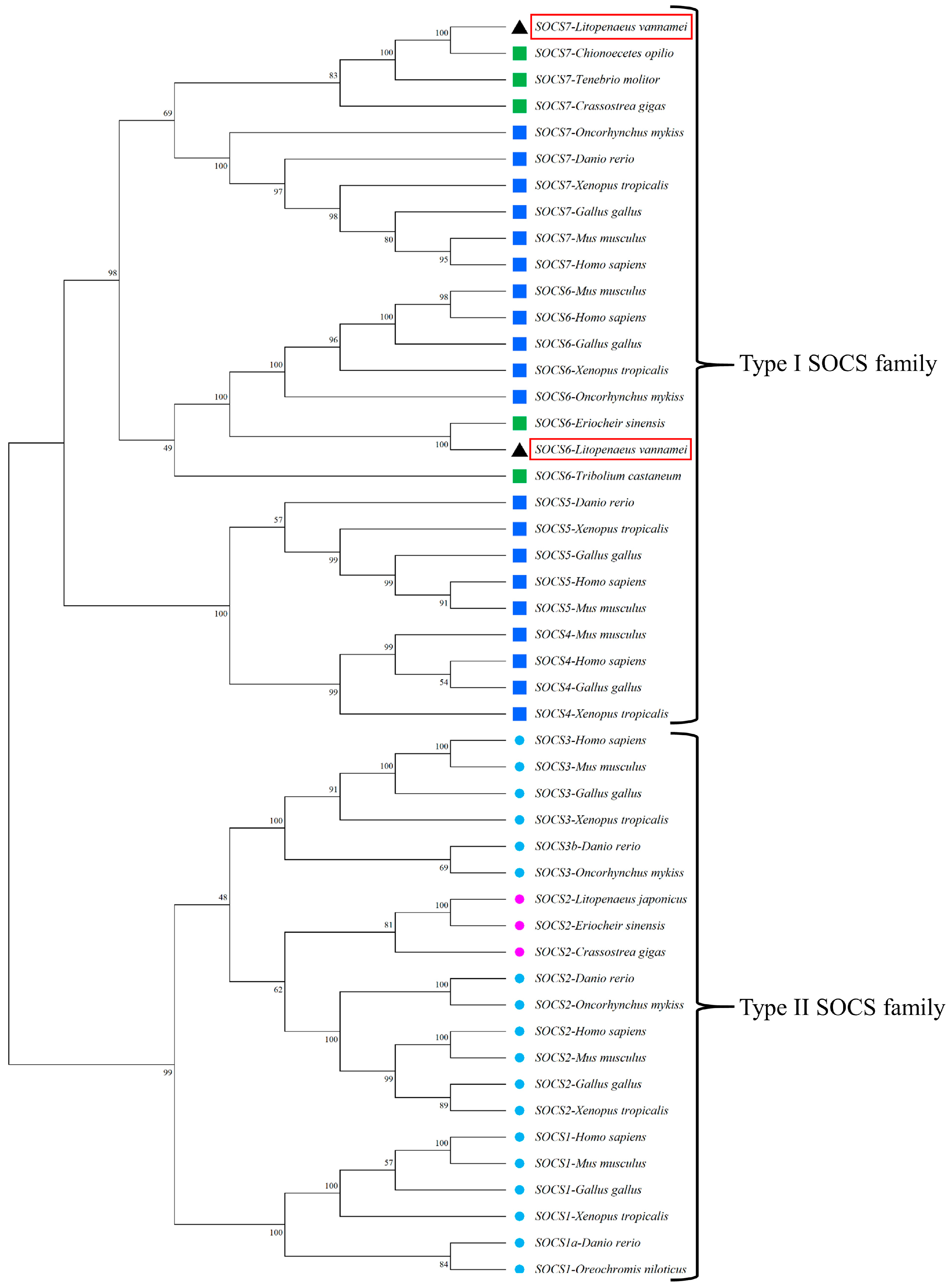
To visualize the evolutionary relationships of LvSOCS proteins, a comprehensive phylogenetic tree was constructed by integrating 48 distinct SOCS protein sequences. They were thoughtfully selected to encompass a diverse array of invertebrate taxa (including crustaceans, mollusks, and insects) as well as representative vertebrate taxa (including amphibians, fishes, mammalians, and birds). The results unveiled topological disparities between two major lineages, designated as type I SOCS and type II SOCS (Figure 2). Within these lineages, a remarkable degree of differentiation was observed, giving rise to distinct SOCS subtypes, namely SOCS1, SOCS2, SOCS3, SOCS4, SOCS5, SOCS6, and SOCS7. Notably, the identified LvSOCS6 and LvSOCS7 were assigned to the established invertebrate type I SOCS6 and SOCS7 groups, respectively, based on their positioning within the phylogenetic tree. In contrast, LvSOCS6 was phylogenetically rooted with the previously reported EsSOCS6 of the Chinese mitten crab, and LvSOCS7 displayed a robust evolutionary connection to the snow crab’s SOCS7, reflecting the close affiliation within this taxonomic group.

3.2. Tissue Distribution and mRNA Expression of LvSOCS6, LvSOCS7, and LvSTAT after LPS, Poly (I:C), and PGN Stimulation

To gain insights into the patterns of LvSOCS6 and LvSOCS7 expression across various tissues, quantitative reverse transcription PCR (qRT-PCR) was conducted on the hepatopancreas, hemocytes, heart, lymphoid organ, muscle, gills, and stomach (Figure 3). Notably, both LvSOCS6 and LvSOCS7 exhibited detectable expression levels in all examined tissues. Among them, the gills showed the highest level of expression of LvSOCS6, followed by elevated expression levels in the muscle, lymphoid organ, heart, hemocytes, and stomach. On the other hand, LvSOCS7 exhibited higher mRNA expression in the gills and stomach than LvSOCS6. The heart also demonstrated higher expression for LvSOCS7 than its levels in the hepatopancreas, hemocytes, lymphoid organ, and muscle.
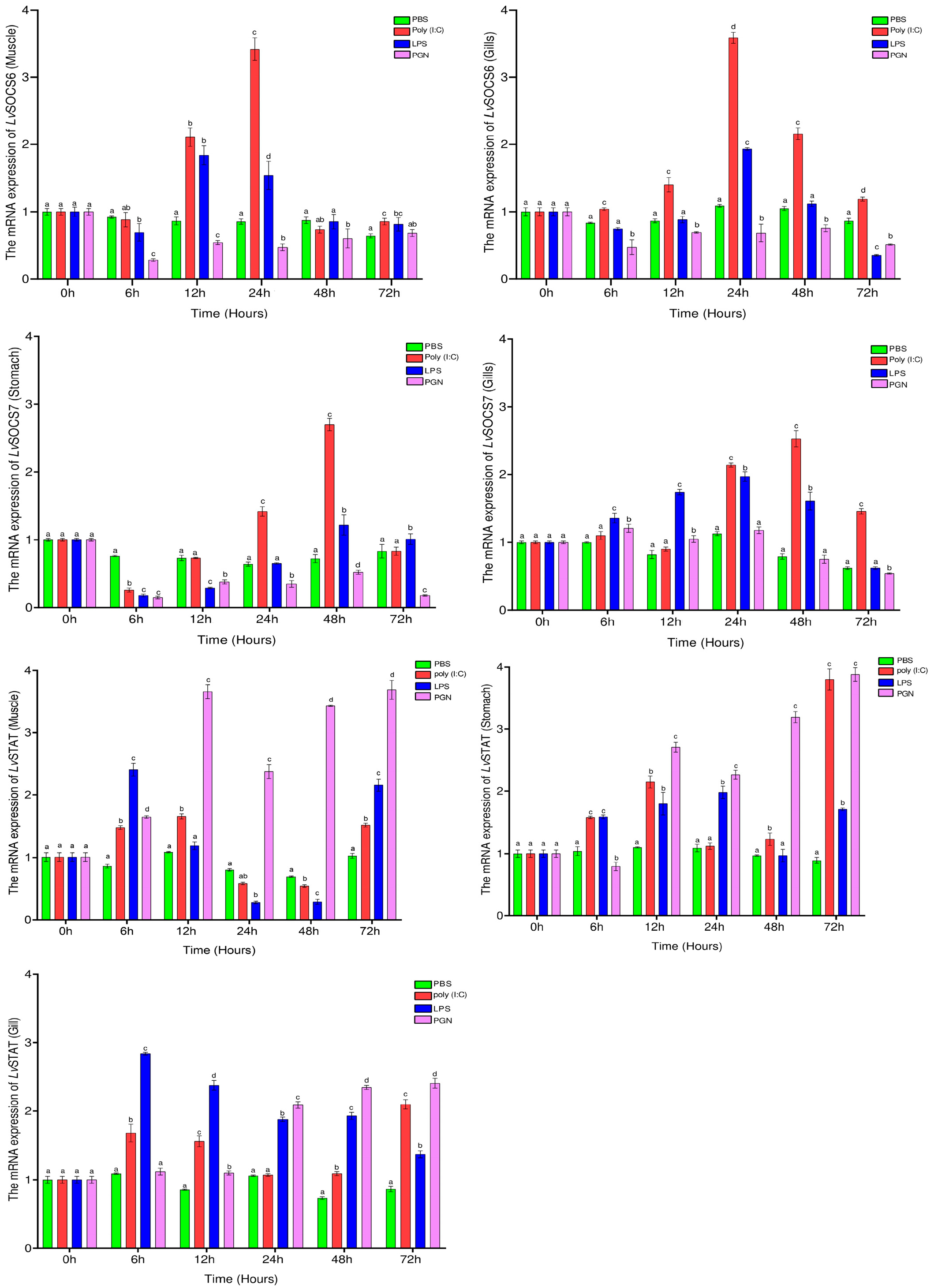
The present study further investigated the mRNA expression patterns of LvSOCS6, LvSOCS7, and LvSTAT in multiple tissues following immune induction. Initially, LvSOCS6 expression exhibited a significant decrease across all experimented tissues at 6 h post immune stimulation in all experimental groups. However, subsequent induction with LPS or poly (I:C) led to a notable increase in LvSOCS6 expression after 6 h in all examined tissues. The gills and muscle displayed the highest expression levels of LvSOCS6 at 24 h, with 3.59-fold and 3.42-fold increases, and in the heart, it peaked at 48 h (2.54-fold, p < 0.05) after poly (I:C) stimulation. LvSOCS6 also showed elevated expression in the gills, muscle, and heart at 24 h, 12 h, and 48 h, respectively, following LPS induction (1.93-, 1.84-, and 1.62-fold, p < 0.05). Interestingly, the PGN induction consistently resulted in decreased LvSOCS6 expression compared to the other experimental groups. Subsequently, LvSOCS6 expression declined by more than 1.5-fold in the LPS- and poly (I:C)- stimulated groups at 72 h. In the case of LvSOCS7, its expression significantly decreased in the heart and stomach at 6 h post-immune stimulation, while showing an increase only in the gills following LPS and PGN inductions. LvSOCS7 expression reached its highest levels in the gills, stomach, and heart at 48 h after poly (I:C) stimulation, with increases of 2.53-, 2.7-, and 2.09-fold, respectively, compared to the control group (p < 0.05). Peak expressions of LvSOCS7 were observed at 24 h and 48 h in response to LPS induction, with increases of 1.97-, 1.82-, and 1.22-fold in the gills, heart, and stomach (p < 0.05). LvSOCS7 expression increased in the heart at 24 h following PGN stimulation; however, overall, LvSOCS7 expression gradually decreased after 48 h in all experimental groups (Figure 4 and Figure S5).
Contrasting with the LvSOCS genes, the LvSTAT gene, which is involved in the JAK-STAT immune pathway, exhibited significantly higher expression at 6 h in all examined tissues. Stimulation with PGN resulted in a progressive increase in LvSTAT expression, peaking at 12, 72, and 24 h in the muscle, stomach or gills, and heart, respectively, with increases of 3.62-, 3.88- or 2.41-, and 1.47-fold compared to the control group (p < 0.05). In the LPS-stimulated group, the highest LvSTAT expression was observed at 6 h in the gills, muscle, and heart, while in the stomach, it peaked at 24 h, with increases of 2.84-, 2.39-, 1.78-, and 1.98-fold (p < 0.05). The poly (I:C)-stimulated group exhibited higher LvSTAT expression at 72 h in the stomach and gills (3.8-fold and 2.1-fold, p < 0.05), while in the muscle and heart, the peaks occurred at 12 h (1.64-fold and 2.24-fold, p < 0.05). Overall, LvSTAT demonstrated higher immune expression in all examined tissues, with a concomitant decrease in LvSOCS gene expression (Figure 4 and Figure S5).

4. Discussion

Cytokines, which are secreted polypeptide molecules, exert significant influence over a myriad of biological processes, encompassing cellular proliferation, growth, and immune regulation in both vertebrates and invertebrates [2,8,19,20]. Among the diverse array of molecules involved in cytokine signaling, the suppressor of cytokine signaling (SOCS) molecules possess noteworthy significance due to their crucial role as principal antagonists of these signaling mediators [7]. Within the SOCS gene family, a dichotomy emerges, comprising the type II SOCS and type I SOCS groups (consisting of SOCS1, SOCS2, SOCS3, SOCS4, SOCS5, SOCS6, and SOCS7) [1,8,12]. In contrast, the presence and comprehensive characterization of these SOCS genes and their immunoregulatory involvement in vertebrates have reached a well-documented stage. Nonetheless, the existence and potential functionalities of SOCS genes in invertebrates, particularly within crustaceans, lack substantive evidence. Within the scope of this investigation, two previously unreported type I SOCS genes were successfully identified in the popular food crustacean, the whiteleg shrimp, and designated as LvSOCS6 and LvSOCS7. The LvSOCS genes characterized in the present study exhibited an archetypal, centrally localized Src homology 2 (SH2) domain region, concomitant with the C-terminal suppressor of cytokine signaling (SOCS) box domain. Furthermore, there were three distinct motifs, encompassing the phosphotyrosine binding, hydrophobic binding, and putative elongin B/C binding, which are known for their involvement in mediating protein–protein interactions and modulating diverse signaling pathways [28,29]. Additionally, the investigation of phylogenetic relationships among SOCS proteins provided valuable insights into the evolutionary history and functional diversification of these identified LvSOCSs. We employed the maximum likelihood method to construct a phylogenetic tree, which integrated a comprehensive set of SOCS protein sequences from both vertebrate and invertebrate organisms. The resulting phylogenetic tree uncovered significant topological distinctions, giving rise to two major lineages as type I SOCS and type II SOCS. Within the type II SOCS lineage, further differentiation was observed, manifesting as distinct sub-branches that corresponded to specific members of SOCS1, SOCS2, and SOCS3. Similarly, the type I SOCS lineage exhibited its own differentiation, generating representative sub-branches associated with SOCS4, SOCS5, SOCS6, and SOCS7. This suggests that SOCS proteins have undergone lineage-specific evolutionary changes and emphasizes their divergent evolutionary trajectories and their unique functional attributes [8]. Importantly, our analysis also included LvSOCS6 and LvSOCS7, identified in this study, which were assigned to the invertebrate SOCS6 and SOCS7 groups within the phylogeny. The placement of LvSOCS6 and LvSOCS7 within their respective invertebrate sub-branches of the phylogenetic tree supports their classification and highlights their evolutionary relationships with other known SOCS proteins.
In whiteleg shrimp, the identified LvSOCS mRNAs exhibited a pervasive presence across all analyzed tissues, showcasing noteworthy fluctuations in expression levels. These dynamic variations potentially might be aligned with multifaceted biological functionalities intrinsic to shrimp. Comparable observations have been made in other invertebrates, such as the Chinese mitten crab, where distinct levels of EsSOCS6 gene expression were detected in the hepatopancreas and hemopoietic tissues, displaying differential patterns of higher and lower expression [14]. Likewise, in mealworms, TmSOCS6 expression had its peak in the hemocytes, whereas TmSOCS7 expression was elevated in the Malpighian tubules [20]. Similar to these findings, the silk moth (B. mori) exhibited higher BmSOCS6 expression within the fat body than in other tissues [30]. In the realm of vertebrates, such as fish species, the expression profiles of SOCS genes have also been observed to fluctuate across different tissues. For example, in rainbow trout (O. mykiss), OmSOCS6 expression levels showcased variation in the skin and gills [31]. In our investigation, both LvSOCS genes manifested significant expression in the gills, with the intriguing discovery that LvSOCS7 also exhibited prominent expression levels in the stomach. These findings suggest the potential existence of distinct and organ-specific roles fulfilled by the identified LvSOCS genes, necessitating further experimental investigation.
The JAK/STAT signaling cascade plays a pivotal role in orchestrating immune responses across the animal kingdom, primarily driven by an array of cytokines [4,14]. Extensive research has elucidated the significance of type I SOCS genes in vertebrates; notable examples include the involvement of SOCS7 in the translocation of the STAT3 gene and neural cell differentiation in humans, with SOCS6 emerging as an essential participant in this process [32,33]. In invertebrates, studies involving d. melanogaster have shed light on the regulatory roles of SOCS36E, SOCS44A, and SOCS16D (analogous to SOCS5, SOCS6, and SOCS7) in dampening the JAK-STAT pathway by influencing its regulation and also the development of wing formation [15,16]. Nevertheless, the current body of knowledge regarding the involvement of type I SOCS genes in the JAK/STAT pathway in crustaceans remains limited. Hence, to further explore the regulatory association of LvSOCS and LvSTAT genes, we investigated their mRNA expression in response to the administration of pathogen-associated molecular patterns (PAMPs), including poly (I:C), LPS, and PGN, which are known to induce inflammatory and immune regulatory responses (mimicking the activities of viruses and gram-negative and/or gram-positive bacteria), activating key signaling pathways such as JAK-STAT and NF-kB [19,31,34]. In our investigation, immune stimulation experiments revealed a significant increase in LvSOCS expression following poly (I:C) or LPS administration, peaking at or after 12 h, but not following the PGN administration. However, the highest significant increase in LvSOCS6 and LvSOCS7 expression was observed in the poly (I:C)-administrated group, suggesting that these identified genes might have more enhanced sensitivity to viral infections than to bacterial infections. On the other hand, the conspicuous downregulation of LvSOCS expression coincided with the upregulation of LvSTAT mRNA levels. Although the precise temporal pattern of this response was not uniform across all assessed time points, it implies a noteworthy correlation between diminished LvSOCS expression and the modulation of LvSTAT gene activity. It is important to acknowledge that other members of the SOCS gene family, such as LvSOCS2, have also been implicated in the multifaceted JAK/STAT pathway, thereby potentially contributing to the current regulatory network at play [19]. Moreover, given the existing evidence elucidating the involvement of type I SOCS genes in the NF-ĸB pathway, a comprehensive exploration of their sophisticated roles within the immune machinery of crustaceans becomes imperative, necessitating in-depth investigations to unravel their precise contributions [14,17]. Consequently, understanding the regulatory mechanisms of SOCS genes and their involvement in various immune pathways will significantly augment our comprehension of crustacean immunology and make notable contributions to the wider domain of invertebrate immune modulation.

5. Conclusions

In synopsis, the current investigation identified novel members of the type I suppressor of cytokine signaling (SOCS) gene family, namely LvSOCS6 and LvSOCS7, in whiteleg shrimp, thereby illuminating their involvement in the intricate orchestration of immune regulatory mechanisms. The mRNA expression patterns of LvSOCS6 and LvSOCS7 demonstrated a pervasive presence throughout diverse tissues, with noteworthy augmented levels of expression observed particularly within the gills, while LvSOCS7 exhibited conspicuous prominence in the stomach. Additionally, the administration of poly (I:C) and LPS elicited a significant induction of LvSOCS mRNA transcripts, underscoring their heightened responsiveness to immune stimulation mediated by viral and bacterial agents. Intriguingly, this immune activation coincided with a substantial decrement in LvSTAT expression, implying a potential interplay between these genes in the context of immune modulation. However, to further unravel the genomic architectures, specific roles, and underlying mechanisms of LvSOCS within the regulatory pathways of whiteleg shrimp, investigations employing RACE PCR- and RNA interference (RNAi)-based methodology are imperative. The findings of the present study establish a solid groundwork for prospective investigations aimed at deciphering the intricacies of immune regulation in invertebrates, particularly in crustaceans.

Supplementary Materials

The following supporting information can be downloaded at: https://www.mdpi.com/article/10.3390/fishes8080416/s1, Figure S1: The LvSOCS6 protein coding nucleotide region (top) and its deduced amino acid sequence (bottom). The start (ATG) and stop (TGA) codons are presented in red color. In the amino acid sequence, SH2 and SOCS-box domain positions are shown in red and blue color; Figure S2: The LvSOCS7 protein coding nucleotide region (top) and its deduced amino acid sequence (bottom). The start (ATG) and stop (TGA) codons are presented in red color. In the amino acid sequence, SH2 and SOCS-box domain positions are shown in red and blue color; Figure S3: Multiple sequence alignment (MSA) of LvSOCS6 and its counterparts. Strongly conserved and similar residues are highlighted by black and gray shading and the conserved phosphotyrosine recognition site (pY) of LvSOCS6 is indicated with (▼). The regions of the phosphotyrosine binding pocket, hydrophobic binding pocket, and putative elongin B/C binding pockets are indicated in purple, yellow, and green color respectively. The species and the GenBank accession numbers used for the MSA were as follows: SOCS6 from H. sapiens (NP_004223.2), M. musculus (NP_061291.2), G. gallus (NP_001120784.1), X. tropicalis (NP_001096240.1) D. rerio (XP_687041.2) O. mykiss (NP_001182102.1), E. sinensis (ATW63847.1) and T. castaneum (XP_008190646); Figure S4: Multiple sequence alignment (MSA) of LvSOCS7 and its counterparts. Strongly conserved and similar residues are highlighted by black and gray shading and the regions of phosphotyrosine binding motif, hydrophobic binding motif, and putative elongin B/C binding motifs are indicated in purple, yellow, and green color respectively. The species and the GenBank accession numbers used for the LvSOCS7 MSA were as follows: C. opilio (KAG0725835.1), Z. cucurbita (JAD11819), C. capitate (JAC02138), O. mykiss (CAP17279.1), D. rerio (ABM68038.1), X. tropicalis (NP_001121531.1), G. gallus (XP_040509254.1), M. musculus (NP_619598.1) and H. sapiens (NP_055413.1); Figure S5: Temporal mRNA expression analysis of (A) LvSOCS6, (B) LvSOCS7, and (C) LvSTAT in heart tissue as determined by qPCR following stimulation of the immune response. Shrimps were challenged with LPS, poly (I:C), PGN, or phosphate-buffered saline (PBS), as a control. The expression was normalized to EF1α internal control gene and analyzed using the 2(−ΔΔCT) method. Results are represented as the mean ± S.E (N = 3). Statistically significant values (p < 0.05) are denoted with different letters; Table S1: Primers used in this study; Table S2: The sequence homologies of LvSOCS6/ LvSOCS7 in relation to other SOCS6/SOCS7 proteins. Table S3: The qPCR expression dynamics of immune challenged shrimps.

Author Contributions

G.C.W.: conceptualization, methodology, investigation, data curation, formal analysis, and writing—original draft; M.W.S.P.: investigation, methodology, validation, and writing; J.L.: investigation; C.-H.K.: writing—review and editing, supervision, and resources. All authors have read and agreed to the published version of the manuscript.

Funding

This research received support from the National Research Foundation of Korea (grant number: 2022R1I1A1A01073372), a grant funded by the Korean government.

Institutional Review Board Statement

All experiment procedures were strictly conducted according to the guidelines for the care and use of laboratory animals by the Animal Ethics Committee of Pukyong National University.

Data Availability Statement

The sequences of LvSOCS6 and LvSOCS7 were submitted to NCBI with GenBank accession numbers OR030046 and OR030047.

Conflicts of Interest

All authors have read and agreed to the present version of the manuscript and declare no conflict of interest.

References

  1. Beschin, A.; Bilej, M.; Torreele, E.; De Baetselier, P. On the existence of cytokines in invertebrates. Cell. Mol. Life Sci. 2001, 58, 801–814. [Google Scholar] [CrossRef] [PubMed]
  2. Zou, J.; Secombes, C.J. The function of fish cytokines. Biology 2016, 5, 23. [Google Scholar] [CrossRef] [PubMed] [Green Version]
  3. Morris, R.; Kershaw, N.J.; Babon, J.J. The molecular details of cytokine signaling via the JAK/STAT pathway. Protein Sci. 2018, 27, 1984–2009. [Google Scholar] [CrossRef] [PubMed] [Green Version]
  4. Ortmann, R.A.; Cheng, T.; Visconti, R.; Frucht, D.M.; O’Shea, J.J. Janus kinases and signal transducers and activators of transcription: Their roles in cytokine signaling, development and immunoregulation. Arthritis Res. 2000, 2, 16–32. [Google Scholar] [CrossRef] [Green Version]
  5. Boudinot, P.; Bird, S.; Du Pasquier, L.; Collet, B. The repertoire of vertebrate STAT transcription factors: Origin and variations in fish. Dev. Comp. Immunol. 2021, 116, 103929. [Google Scholar] [CrossRef]
  6. Hou, S.X.; Zheng, Z.; Chen, X.; Perrimon, N. The JAK/STAT pathway in model organisms: Emerging roles in cell movement. Dev. Cell 2002, 3, 765–778. [Google Scholar] [CrossRef] [Green Version]
  7. Alexander, W.S.; Hilton, D.J. The role of suppressors of cytokine signaling (SOCS) proteins in regulation of the immune response. Annu. Rev. Immunol. 2004, 22, 503–529. [Google Scholar] [CrossRef]
  8. Wang, B.; Wangkahart, E.; Secombes, C.J.; Wang, T. Insights into the evolution of the suppressors of cytokine signaling (SOCS) gene family in vertebrates. Mol. Biol. Evol. 2019, 36, 393–411. [Google Scholar] [CrossRef] [Green Version]
  9. Delgado-Ortega, M.; Marc, D.; Dupont, J.; Trapp, S.; Berri, M.; Meurens, F. SOCS proteins in infectious diseases of mammals. Vet. Immunol. Immunopathol. 2013, 151, 1–19. [Google Scholar] [CrossRef]
  10. Yasukawa, H.; Misawa, H.; Sakamoto, H.; Masuhara, M.; Sasaki, A.; Wakioka, T.; Ohtsuka, S.; Imaizumi, T.; Matsuda, T.; Ihle, J.N.; et al. The JAK-binding protein JAB inhibits Janus tyrosine kinase activity through binding in the activation loop. EMBO J. 1999, 18, 1309–1320. [Google Scholar] [CrossRef]
  11. Sasaki, A.; Yasukawa, H.; Suzuki, A.; Kamizono, S.; Syoda, T.; Kinjyo, I.; Sasaki, M.; Johnston, J.A.; Yoshimura, A. Cytokine-inducible SH2 protein-3 (CIS3/SOCS3) inhibits Janus tyrosine kinase by binding through the N-terminal kinase inhibitory region as well as SH2 domain. Genes Cells 1999, 4, 339–351. [Google Scholar] [CrossRef] [PubMed]
  12. Dalpke, A.; Heeg, K.; Bartz, H.; Baetz, A. Regulation of innate immunity by suppressor of cytokine signaling (SOCS) proteins. Immunobiology 2008, 213, 225–235. [Google Scholar] [CrossRef] [PubMed]
  13. Feng, Z.P.; Chandrashekaran, I.R.; Low, A.; Speed, T.P.; Nicholson, S.E.; Norton, R.S. The N-terminal domains of SOCS proteins: A conserved region in the disordered N-termini of SOCS4 and 5. Proteins Struct. Funct. Bioinform. 2012, 80, 946–957. [Google Scholar] [CrossRef]
  14. Qu, C.; Xu, Q.; Lu, M.; Wang, F.; Liu, Z.; Liu, D.; Yang, W.; Yi, Q.; Wang, L.; Song, L. The involvement of suppressor of cytokine signaling 6 (SOCS6) in immune response of Chinese mitten crab Eriocheir sinensis. Fish Shellfish Immunol. 2018, 72, 502–509. [Google Scholar] [CrossRef]
  15. Callus, B.A.; Mathey-Prevot, B. SOCS36E, a novel Drosophila SOCS protein, suppresses JAK/STAT and EGF-R signaling in the imaginal wing disc. Oncogene 2002, 21, 4812–4821. [Google Scholar] [CrossRef] [PubMed] [Green Version]
  16. Rawlings, J.S.; Rennebeck, G.; Harrison, S.M.W.; Xi, R.; Harrison, D.A. Two Drosophila suppressors of cytokine signaling (SOCS) differentially regulate JAK and EGFR pathway activities. BMC Cell Biol. 2004, 5, 38. [Google Scholar] [CrossRef] [Green Version]
  17. Li, J.; Zhang, Y.; Zhang, Y.; Liu, Y.; Xiang, Z.; Qu, F.; Yu, Z. Cloning and characterization of three suppressors of cytokine signaling (SOCS) genes from the Pacific oyster, Crassostrea gigas. Fish Shellfish Immunol. 2015, 44, 525–532. [Google Scholar] [CrossRef]
  18. Zhu, B.; Dai, L.; Yu, Y.; Wang, D.; Peng, T.; Liu, C. A role of suppressor of cytokine signaling 2 in the regulation of ecdysteroid signaling pathway in Procambarus clarkii. J. Exp. Zool. Part A Ecol. Genet. Physiol. 2016, 325, 441–452. [Google Scholar] [CrossRef]
  19. Wang, S.; Song, X.; Zhang, Z.; Li, H.; Lǚ, K.; Yin, B.; He, J.; Li, C. Shrimp with knockdown of LvSOCS2, a negative feedback loop regulator of JAK/STAT pathway in Litopenaeus vannamei, exhibit enhanced resistance against WSSV. Dev. Comp. Immunol. 2016, 65, 289–298. [Google Scholar] [CrossRef]
  20. Patnaik, B.B.; Kim, B.B.; Jo, Y.H.; Bang, I.S. Molecular cloning and expression analysis of three suppressors of cytokine signaling genes (SOCS5, SOCS6, SOCS7) in the Mealworm beetle Tenebrio molitor. Insects 2019, 10, 76. [Google Scholar] [CrossRef] [Green Version]
  21. Wheeler, D.L.; Barrett, T.; Benson, D.A.; Bryant, S.H.; Canese, K.; Chetvernin, V.; Church, D.M.; Dicuccio, M.; Edgar, R.; Federhen, S.; et al. Database resources of the National Center for Biotechnology Information. Nucleic Acids Res. 2008, 36, D13–D21. [Google Scholar] [CrossRef] [Green Version]
  22. Marchler-Bauer, A.; Lu, S.; Anderson, J.B.; Chitsaz, F.; Derbyshire, M.K.; DeWeese-Scott, C.; Fong, J.H.; Geer, L.Y.; Geer, R.C.; Gonzales, N.R.; et al. CDD: A conserved domain database for the functional annotation of proteins. Nucleic Acids Res. 2011, 39, 225–229. [Google Scholar] [CrossRef] [PubMed] [Green Version]
  23. Letunic, I.; Khedkar, S.; Bork, P. SMART: Recent updates, new developments and status in 2020. Nucleic Acids Res. 2021, 49, D458–D460. [Google Scholar] [CrossRef] [PubMed]
  24. Gasteiger, E.; Hoogland, C.; Gattiker, A.; Duvaud, S.; Wilkins, M.R.; Appel, R.D.; Bairoch, A. protein identification and analysis tools on the ExPASy server. In The Proteomics Protocols Handbook; Humana Press: Totowa, NJ, USA, 2005; pp. 571–607. [Google Scholar]
  25. Sievers, F.; Higgins, D.G. Clustal omega for making accurate alignments of many protein sequences. Protein Sci. 2018, 27, 135–145. [Google Scholar] [CrossRef] [Green Version]
  26. Tamura, K.; Stecher, G.; Kumar, S. MEGA11: Molecular evolutionary genetics analysis version 11. Mol. Biol. Evol. 2021, 38, 3022–3027. [Google Scholar] [CrossRef]
  27. Livak, K.J.; Schmittgen, T.D. Analysis of relative gene expression data using real-time quantitative PCR and the 2−ΔΔCT method. Methods 2001, 25, 402–408. [Google Scholar] [CrossRef]
  28. Kamura, T.; Sato, S.; Haque, D.; Liu, L.; Kaelin, W.G.; Conaway, R.C.; Conaway, J.W. The elongin BC complex interacts with the conserved SOCS-Box motif present in members of the SOCS, ras, WD-40 repeat, and ankyrin repeat families. Genes Dev. 1998, 12, 3872–3881. [Google Scholar] [CrossRef] [Green Version]
  29. Bullock, A.N.; Rodriguez, M.C.; Debreczeni, J.É.; Songyang, Z.; Knapp, S. Structure of the SOCS4-elonginB/C complex reveals a distinct SOCS box interface and the molecular basis for SOCS-dependent EGFR degradation. Structure 2007, 15, 1493–1504. [Google Scholar] [CrossRef] [Green Version]
  30. Abbas, M.N.; Kausar, S.; Sun, Y.X.; Tian, J.W.; Zhu, B.J.; Liu, C.L. Suppressor of cytokine signaling 6 can enhance epidermal growth factor receptor signaling pathway in Bombyx mori (Dazao). Dev. Comp. Immunol. 2018, 81, 187–192. [Google Scholar] [CrossRef] [PubMed]
  31. Wang, T.; Gao, Q.; Nie, P.; Secombes, C.J. Identification of suppressor of cytokine signalling (SOCS) 6, 7, 9 and CISH in rainbow trout Oncorhynchus mykiss and analysis of their expression in relation to other known trout SOCS. Fish Shellfish Immunol. 2010, 29, 656–667. [Google Scholar] [CrossRef] [PubMed]
  32. Noguchi, S.; Yamada, N.; Kumazaki, M.; Yasui, Y.; Iwasaki, J.; Naito, S.; Akao, Y. Socs7, a target gene of microRNA-145, regulates interferon-β induction through STAT3 nuclear translocation in bladder cancer cells. Cell Death Dis. 2013, 4, e482. [Google Scholar] [CrossRef] [PubMed] [Green Version]
  33. Gupta, S.; Mishra, K.; Surolia, A.; Banerjee, K. Suppressor of cytokine signalling-6 promotes neurite outgrowth via JAK2/STAT5-mediated signalling pathway, involving negative feedback inhibition. PLoS ONE 2011, 6, e26674. [Google Scholar] [CrossRef] [PubMed] [Green Version]
  34. Owen, K.L.; Brockwell, N.K.; Parker, B.S. Jak-Stat signaling: A double-edged sword of immune regulation and cancer progression. Cancers 2019, 11, 2002. [Google Scholar] [CrossRef] [PubMed] [Green Version]
Figure 1. The domain architectures of LvSOCS6 and LvSOCS7 proteins. The predicted SH2 and SOCS box positions are shown in red and blue colors, respectively.
Fishes 08 00416 g001
Figure 2. Maximum likelihood tree of 48 SOCS protein sequences among vertebrates and invertebrates. The numbers at tree nodes refer to the percent bootstrap values following 1000 replications. The species and the GenBank accession numbers used for the phylogenetic analysis were as follows: The type I family includes SOCS1 from D. rerio (NP_001003467.1), H. sapiens (NP_003736.1), M. musculus (NP_034026.1), G. gallus (NP_001131120.1), X. tropicalis (NP_001011327.1), and O. niloticus (NP_001297020.1); SOCS2 from D. rerio (XP_005164804.1), H. sapiens (NP_001257400.1), M. musculus (NP_031732.1), G. gallus (NP_989871.1), X. tropicalis (NP_001120898.1), O. niloticus (XP_036813373.1), C. gigas (EKC24772.1), L. japonicus (BAI70368.1), and E. sinensis (ACU42699.1); SOCS3 from D. rerio (NP_998469.1), H. sapiens (NP_003946.3), M. musculus (NP_031733.1), G. gallus (NP_001186037.1), X. tropicalis (XP_031746340.1), and O. mykiss (NP_001139640.1); and SOCS4 from H. sapiens (NP_659198.1), M. musculus (NP_543119.2), G. gallus (NP_001186037.1), and X. tropicalis (XP_031746340.1). The type II family includes SOCS5 from H. sapiens (NP_955453.1), M. musculus (NP_062628.2), G. gallus (NP_001120786.1), X. tropicalis (NP_001016844.1), and D. rerio (ABM68036.1); SOCS6 from H. sapiens (NP_004223.2), M. musculus (NP_061291.2), G. gallus (NP_001120784.1), X. tropicalis (NP_001096240.1), O. mykiss (NP_001182102.1), E. sinensis (ATW63847.1), L. vannamei (SOCS6-OR030046), and T. castaneum (XP_008190646.1); and SOCS7 from H. sapiens (NP_055413.2), M. musculus (NP_619598.2), G. gallus (XP_040509254.1), X. tropicalis (XP_012827004.2), D. rerio (XP_009304138.1), O. mykiss (CAP17279.1), T. molitor (QDL52635.1), C. gigas (AKA59677.1), Chionoecetes opilio (KAG0725835.1), and L. vannamei (SOCS7-OR030047). The blue colors indicate vertebrate species, purple and green colors indicate invertebrates, and the red box highlights the newly identified SOCS6/SOCS7 for L. vannamei.
Fishes 08 00416 g002
Figure 3. The tissue distribution patterns of LvSOCS6 and LvSOCS7 in different tissues following qPCR analysis. The data are expressed using the 2 (−ΔCt).
Fishes 08 00416 g003
Figure 4. Temporal mRNA expression analysis of LvSOCS6 (muscle, gills), LvSOCS7 (stomach, gills), and LvSTAT (muscle, stomach, and gills) following challenge experiments with LPS, poly (I:C), PGN, or phosphate-buffered saline (PBS). All the qPCR data were normalized to those of the EF1α internal control gene. Results are represented as mean ± S.E (N = 3). Statistically significant values (p < 0.05) are denoted with different letters.
Fishes 08 00416 g004
Disclaimer/Publisher’s Note: The statements, opinions and data contained in all publications are solely those of the individual author(s) and contributor(s) and not of MDPI and/or the editor(s). MDPI and/or the editor(s) disclaim responsibility for any injury to people or property resulting from any ideas, methods, instructions or products referred to in the content.