Article

Muscle Transcriptome Analysis Reveals Molecular Mechanisms of Superior Growth Performance in Kuruma Shrimp, Marsupenaeus japonicus

1
Jiangsu Key Laboratory of Marine Bioresources and Environment, Jiangsu Ocean University, Lianyungang 222005, China
2
Jiangsu Key Laboratory of Marine Biotechnology, Jiangsu Ocean University, Lianyungang 222005, China
3
Co-Innovation Center of Jiangsu Marine Bio-Industry Technology, Jiangsu Ocean University, Lianyungang 222005, China
4
Jiangsu Institute of Marine Resources Development, Jiangsu Ocean University, Lianyungang 222005, China
5
The Jiangsu Provincial Infrastructure for Conservation and Utilization of Agricultural Germplasm, Nanjing 210014, China
6
Marine and Fishery Development Promotion Center of Lianyungang, Lianyungang 222000, China
*
Author to whom correspondence should be addressed.
These authors contributed equally to this work.
Fishes 2023, 8(7), 350; https://doi.org/10.3390/fishes8070350
Submission received: 18 May 2023 / Revised: 30 June 2023 / Accepted: 1 July 2023 / Published: 5 July 2023

Abstract

:
During the cultivation of Marsupenaeus japonicus, there are often obvious differences in the growth within the same family under the same food, water quality, and environment, which greatly affects cultivation efficiency. To explore the molecular mechanism of this growth difference, this study used RNA-seq technology to compare the transcriptomes of M. japonicus individuals with significant growth differences from the same family. A total of 1375 differentially expressed genes were identified, of which 1109 were upregulated and 266 were downregulated. Gene Ontology (GO) and Kyoto Encyclopedia of Genes and Genomes (KEGG) analyses were performed on the differentially expressed genes, and the results showed that growth-related processes, including chitin metabolism, chitin-binding amino sugar metabolism, and antioxidant processes, including response to oxidative stress, peroxidase activity, antioxidant activity, and peroxidase activity, showed significant differences between the large-size and small-size groups. The expression levels of some differentially expressed genes, such as cuticular protein, low-density lipoprotein receptor, ecdysteroid kinase, myosin heavy chain, and apoptosis inhibitor, were verified by quantitative PCR experiments. One cuticle gene was annotated, and phylogenetic analysis showed that this sequence clustered with the penaeid cuticle genes. This study provides valuable data and a scientific basis for understanding the mechanism of growth differences in M. japonicus at the molecular-genetic level.
Key Contribution: The study identified potential growth trait-related genes and provided insights revealing mechanisms underlying the growth differences in Marsupenaeus japonicus.

Graphical Abstract

1. Introduction

Marsupenaeus japonicus, also known as kuruma shrimp and Japanese tiger prawn, is widely distributed in the Indo-West Pacific waters, eastern Africa, the Red Sea, and the Mediterranean Sea [1]. In China, it is mainly found in the waters south of the mouth of the Yangtze River [2,3]. M. japonicus is one of the most important farmed shrimp species in the world. It has a high quality and a delicious taste, so it is very much loved. Compared with other prawns, M. japonicus has many advantages, such as a fast growth rate, high fecundity, and high efficiency of water-less transportation [4,5,6]. The global annual aquaculture production of M. japonicus increased from 10,000 tons in 1991 to 50,000 tons in 2004, but has remained near 50,000 tons in the past two decades [7]. Long-term stable production means that the farming industry of M. japonicus has encountered bottlenecks, among which slow growth rate and low farming yield are the key factors affecting its industrial development [8]. M. japonicus often inhabits sandy or sandy-muddy sea areas with water depth of 10–100 m and has a habit of burrowing in the sand [9]. After four to five molts, the shrimp larvae can transition to benthic life. This habit makes it impossible to significantly increase its breeding density. M. japonicus has a strong habit of cannibalism, in which larger individuals prey on smaller or weaker individuals who have just shed their exoskeleton [10].
During the culture of M. japonicus, under the same food, water quality, and environment, individual differences within the same family of shrimp seeds are obvious, and the growth rate of some shrimp seeds is slow [8]. The growth and development of animals is a process in which a large number of cells continuously grow, divide, and differentiate to make tissues, organs, and systems mature in structure and function. Although only limited genomic information is currently available for most crustacean species, many putative candidate genes have been identified that are involved in growth and muscle development in some species [11]. Zeng et al. Reference [12] conducted comparative transcriptome analysis on the muscle tissues of 3-month-old Chinese perch, Siniperca chuatsi, with significantly different body weights in the same family and obtained many differentially expressed genes related to protein synthesis, digestion, RNA transport, and other functions. Wang et al. [13] conducted a comparative transcriptome analysis of the muscle tissue of a 6-month-old common carp Cyprinus carpio in the same family with different growth rates and screened out 749 differentially expressed genes, such as myoglobin, myosin light chain 2b, and troponin type I, which are related to muscle growth. Wang et al. [14] conducted a comparative transcriptome analysis of the mantles of blacklip pearl oyster, Pinctada margaritifera of different sizes from the same family and found a total of 1921 differentially expressed genes, including cuticular growth factor receptor, cathepsin B, and insulin-like protein receptor. Huang et al. [15] conducted comparative miRNA and comparative proteomic analysis of the different sizes of disc abalone, Haliotis discus hannai, in one family and found many differentially expressed genes related to muscle growth between the two groups of samples, such as thyroid hormone signaling, bone morphogenetic protein 7, and actin cytoskeleton regulation. To screen the muscle growth-related genes regulated by the myostatin gene in Chinese shrimp, Fenneropenaeus chinensis, Yan et al. [16] performed comparative transcriptome analysis on individuals in the control group and the Mstn expression inhibition group and identified 29 Mstn-regulated genes relating to muscle growth. To reveal the molecular basis of the growth difference between fast-growing and slow-growing red swamp crayfish Procambarus clarkii, Guo et al. [17] identified 122 growth-related differentially expressed genes using RNA-Seq and Iso-seq strategies. Other studies using transcriptome techniques to study growth traits included Penaeus monodon [18], Acanthopagrus schlegelii [19], Paramisgurnus dabryanus [20], and Macrobrachium rosenbergii [21].
To further reveal the molecular mechanism of the obvious growth differences in the culture of M. japonicus, this study used comparative transcriptome sequencing technology to analyze the transcriptomes of individuals with different growth characteristics in the same family and identified potential growth trait–related genes. Functional research on the up- and downregulated genes lays the foundation for molecular marker-assisted breeding of M. japonicus.

2. Materials and Methods

2.1. Ethics Statement

This study was approved by the Animal Care and Use Committee of Jiangsu Ocean University (protocol no. 2020-37; approval date: 1 September 2019). All procedures involving animals were performed in accordance with guidelines for the Care and Use of Laboratory Animals in China.

2.2. Sample Collection

An experiment in the cultivation of M. japonicus was carried out at an aquaculture company. M. japonicus was derived from a laboratory-proposed full-sib family. About 4000 larvae were grown in conical-bottomed tanks (diameter 1.0 m, height 1.2 m) at a density of 200 individuals/m2 and fed twice a day with continuous aeration. The salinity was kept at 27–29 parts per thousand and the pH was kept at 8.1–8.3 throughout the breeding period. The filtered seawater was renewed every 2 days, and the entire cultivation lasted for 70 days.
Feeding was stopped 24 h before the formal experiment. We randomly selected 200 individuals from the same family, measured their body weight, and selected the top 30 individuals as the large individual group, and the last 30 individuals as the small individual group. Then, 18 individuals were selected from the large individual group and the small individual group as experimental samples. The average weight of individuals in the fast-growing group was 2.241 ± 0.54 g, and in the slow-growing group was 0.733 ± 0.32 g. There was a significant difference in body weight between the two groups. The experimental shrimp were euthanized with the anesthetic alcohol: eugenol = 10:1. The first abdominal muscle tissue of 18 shrimp from the two groups was snap-frozen in liquid nitrogen and then transferred to −80 °C for RNA extraction.

2.3. RNA Extraction and cDNA Synthesis

In the experiment, TRIzol reagent (TaKaRa, Dalian, China) was used to extract RNA from muscle tissue samples. One percent agarose gel electrophoresis was used to assess total RNA quantity and contamination, a spectrophotometer was used to determine RNA purity and concentration, and a bioanalyzer was used to determine total RNA integrity. The muscle RNA of nine shrimp in the fast-growing group and the slow-growing group, respectively, was randomly divided into three groups. Equal amounts of total RNA from each group (containing three individuals) were pooled. Finally, each group had three RNA samples for transcriptome and real-time quantitative PCR analysis.

2.4. Library Sequencing, Assembly, and Functional Annotation

Three micrograms of RNA per sample were taken to synthesize first- and second-strand cDNA. Six cDNA libraries were generated using Illumina kits and sequenced on the Illumina sequencing platform. High-quality data were obtained by removing linker sequences and low-quality data. High-quality data were reassembled using Trinity software (v1.0, Cambridge MA, USA) [22] with the recommended parameters. The resulting reference sequences were used for subsequent analysis. HISAT2 software (v2.0.4, Santa Cruz, CA, USA) [23] was used to quickly and accurately align the clean reads against the reference genome of M. japonicus to obtain the location information of the reads in the reference genome. StringTie software (v1.3.1, Baltimore, MD, USA) [24] was used to read the map data. Assembled transcripts were annotated against the National Center for Biotechnology Information (NCBI)-nr, NCBI-nt, Protein family (Pfam), EuKaryotic Orthologous Groups (KOG), KEGG Orthology (KO), Gene Ontology (GO), and Swiss-Prot databases.

2.5. Differentially Expressed Gene Analysis

The number of reads corresponding to each gene was determined using the featureCounts tool of the subread software (v1.5, Victoria, Australia) [25]. The transcript expression levels of the different groups were normalized to transcripts per kilobase per million fragments using RSEM software (v1.2.15, Heidelberg, Germany) [26]. Gene function annotation was done with reference to the NCBI and KEGG databases. Differentially expressed genes between the two groups (fast-growing and slow-growing groups) were screened using the DESeq2 method [27]. Genes were considered to be significantly differentially expressed when the padj < 0.05 and |log2(Fold change)| > 1. GO enrichment and Kyoto Encyclopedia of Genes and Genomes (KEGG) analysis were performed using ClusterProfiler software (v4.0, Guangzhou, China) [28].

2.6. Homologous Cloning and Sequence Analysis

Total RNA was extracted using the TRIzol reagent (Takara, Dalian, China). The purity and integrity of the RNAs were tested. First-strand cDNA was synthesized using the TransScript kit (Takara, Dalian, China) as instructed. Primers were designed based on our transcripts and sequences from closely related species in NCBI. The PCR-amplified product was ligated into the pMD19-T vector (Takara, Dalian, China), transformed into Escherichia coli DH5α cells (Takara, Dalian, China) and sequenced by paired-end sequencing. Open reading frames of genes were predicted using the ORF Finder tool (https://www.ncbi.nlm.nih.gov/orffinder/, accessed on 1 March 2023). The molecular mass and isoelectric point of the protein sequence were predicted by ExPASy software (https://www.expasy.org/, accessed on 3 March 2023). The secondary structures of protein sequences were predicted using SMART software (v1.0, Heidelberg, Germany) [29], SOPMA server [30], and PROSITE software (v1.0, Geneva, Switzerland) [31] software. Multiple sequence alignments were performed using ClustalW, and phylogenetic analysis was performed using MEGA software (v7.0, Allentown, PA, USA) [32].

2.7. Real-Time PCR Verification

To verify the reliability of the transcriptome sequencing results, the expression levels of ten differentially expressed genes between the fast-growing group and the slow-growing group were detected by real-time PCR. The Primer 5 software [33] was used to design quantitative primers based on transcript sequences (Table S1). Quantitative PCRs were run in triplicate using the SYBR kit (Takara, Dalian, China), and three biological replicates were studied. The results were normalized using EF1α as a reference gene and the expression levels of DEGs was calculated using the 2−△△Ct method. The quantitative PCR results were compared with transcriptome data. Expression data were analyzed with SPSS software (v18.0, SPSS Inc., Chicago, IL, USA) and displayed as the mean and standard deviation.

2.8. Single Nucleotide Polymorphism (SNP) Detection

We used Picard tools (https://sourceforge.net/projects/picard/, accessed on 3 March 2023) and other tools to compare the results, sort the chromosome coordinates, remove duplicate reads, etc., and finally used GATK [34] to perform SNP calling and indel calling and filter the original results. We used the SnpEff software (v3.0, San Diego, CA, USA) to annotate the variant sites and performed analysis on each variant site based on the annotation information, mainly including variant site function, variant site region, and variant site impact analysis.

3. Results

3.1. Transcriptome Sequencing and Assembly

Illumina sequencing yielded a total of 20.61 Gb of data, including 140.5 million raw data and 137.4 million clean reads for the fast-growing group (Table 1). There were a total of 20.74 Gb of data, including 141.2 million raw data and 138.2 million clean data for the slow-growing group. We obtained a total of 22,948 Unigene sequences with an average length of 1654.25 bp and predicted 1776 new genes. The mapping rates of the six cDNA libraries to the reference genome of M. japonicus ranged from 86.99% to 94.24%. Based on the FRKM (fragments per kilobase of transcript sequence per million base pairs sequenced) value, we calculated the Pearson correlation coefficient R2 value as a measure of the correlation of the samples (Figure 1). The R2 value of the sample was greater than 0.85.

3.2. Identification of Differentially Expressed Genes

By comparing the relative expression abundances of the two groups of samples, we identified differentially expressed genes. We chose padj < 0.05 and |log2(Fold change)| > 1 as the criterion for defining the differential expression of genes between the fast-growing group and the slow-growing group. A total of 1375 differentially expressed genes were identified in this study, of which 1109 genes were upregulated in the fast-growing group and 266 genes were downregulated. Among these differentially expressed genes, some may be related to muscle growth of M. japonicus, such as tubulin α-1 chain, ecdysteroid kinase, myosin heavy chain C, cuticular protein, sarcoplasmic calcium-binding protein beta chain, mitochondrial basic amino acid transporter, and actin (Table 2).

3.3. Enrichment Analysis of Differentially Expressed Genes

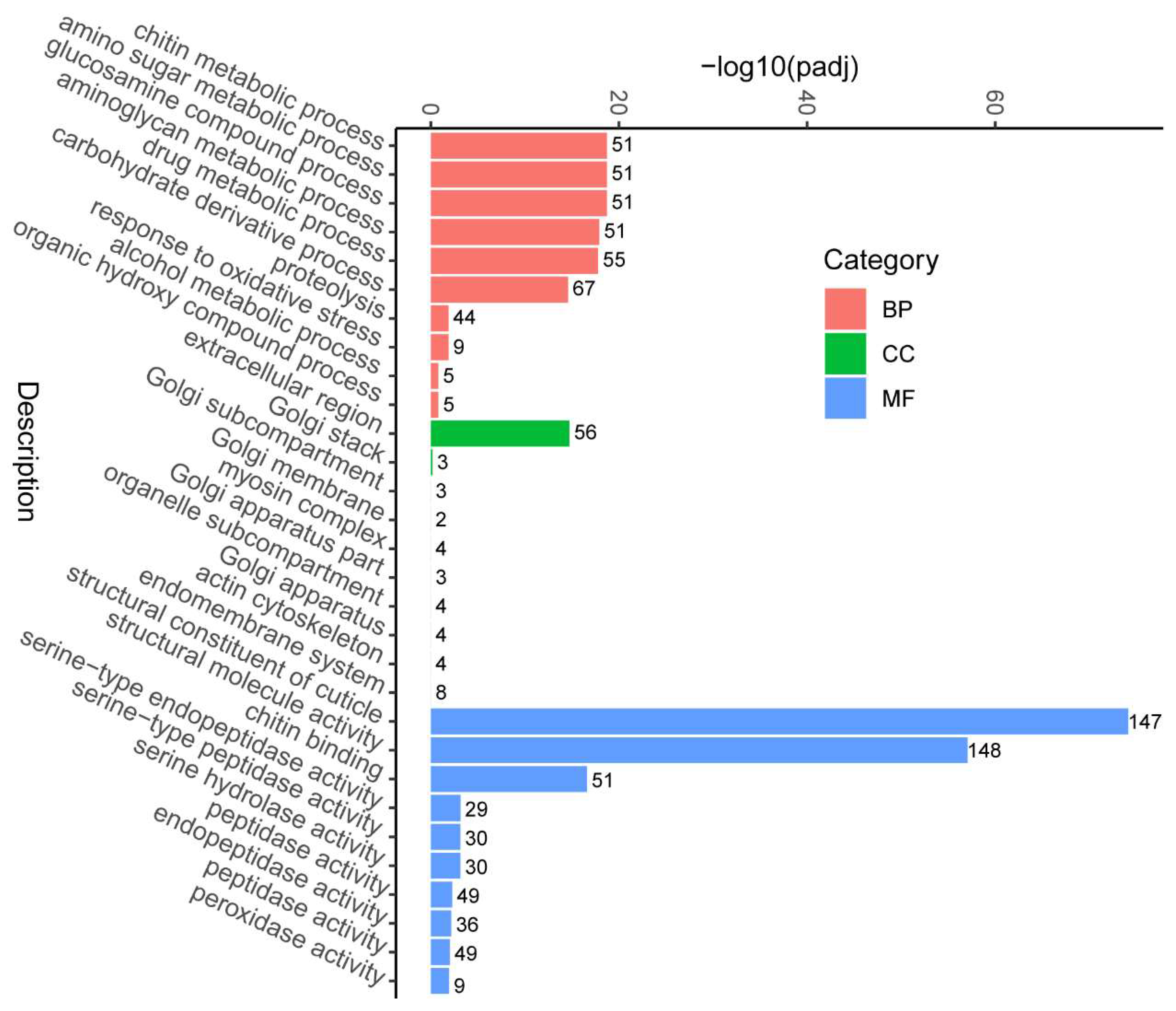
To understand which biological processes and pathways are involved in the growth regulation of M. japonicus, we performed GO and KEGG analyses on the differentially expressed genes. All differentially expressed genes were assigned to groups among the 30 functional groups in the GO annotation system (Figure 2). The significant GO molecular function entries mainly regarded the structural constituent of the cuticle. Among the genes of the biological process, most were involved in the chitin metabolic process. The extracellular region was the most significantly enriched among the GO entries for cellular components.
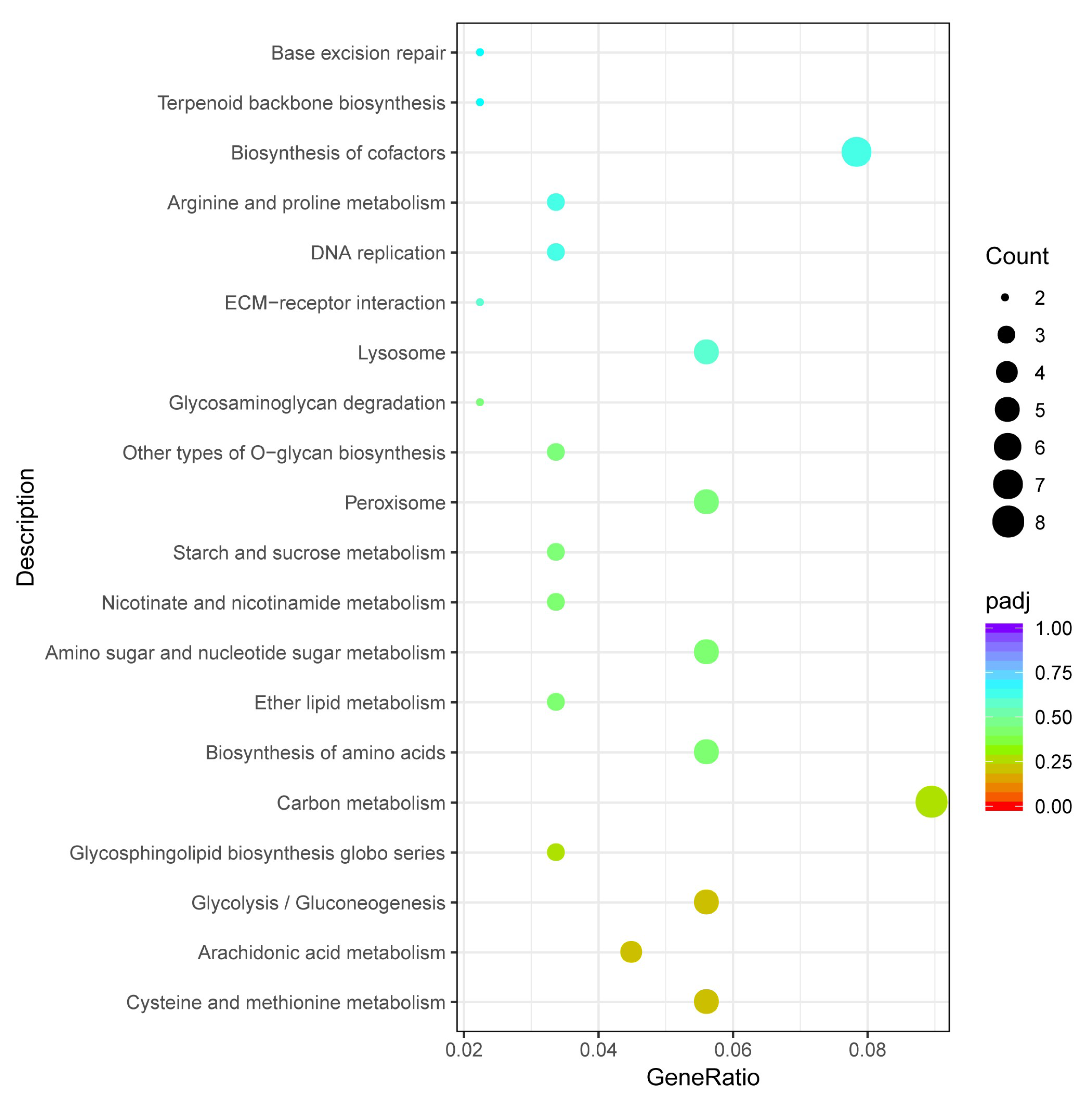
To identify biological pathways that were repressed or activated differentially during the growth of M. japonicus, these differentially expressed genes were annotated in the KEGG database. These differentially expressed genes were annotated to 72 KEGG pathways. The bottom panel of Figure 3 shows the top 20 significantly enriched pathways. Carbon metabolism (dpx01200), biosynthesis of cofactors (dpx01240), cysteine and methionine metabolism (dpx00270), and glycolysis/gluconeogenesis (dpx00010), and biosynthesis of amino acids (dpx01230) were enriched.

3.4. Quantitative PCR to Verify Differentially Expressed Genes

We screened 10 differentially expressed genes of interest from M. japonicus muscle tissue using real-time quantitative PCR analysis to validate the expression patterns in the transcriptome data. There was a good correlation between the RNA-seq data expression of candidate genes and the results of real-time PCR, and the expression trends of the same gene between the two groups were consistent (Figure 4). Therefore, the differential gene expression patterns in transcriptome sequencing were found to be reliable.

3.5. Single-Nucleotide Polymorphism (SNP) Analysis

We used SnpEff software v3.0 [35] to annotate the variant sites between the two growth groups and performed an analysis on each variant site based on the annotation information, including functional statistics of variant sites (synonymous mutations, missense mutations, and nonsense mutations), variant site region (exon, intron, or intergenic), and variation site impact (high, moderate, low, modifier). From the functional data of variant sites, missense mutation sites numbered 37,339, nonsense mutation sites 339, and synonymous mutation sites 176,640, accounting for 82.4% of the total (Figure S1). From the impact data of variant sites, the number of high-impact mutation sites was 1656, the number of low-impact mutation sites was 177,921, and the number of medium-impact mutation sites was 38,678 (Figure S2). From the regional data of variant sites, the number of intron regions was 47,095 and the number of exon regions was 216,251. They made up a proportion of approximately 31.0% (Figure S3).
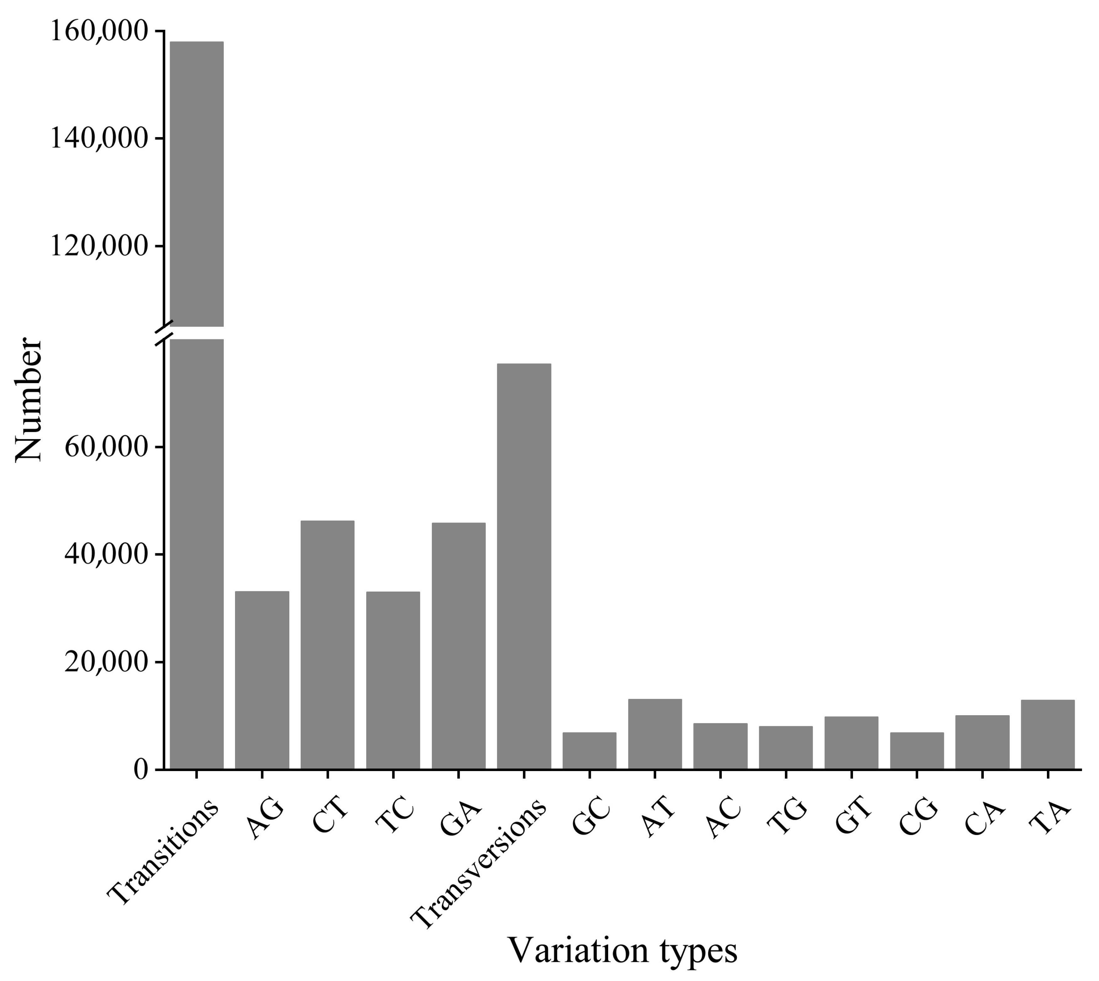
A total of 233,263 SNP sites were identified in this study, of which 157,920 were transition sites and 75,343 were transversion sites, for the ratio of transition to transversion of 2.1 (Figure 5). According to Liew et al.’s method [36], the SNP loci were divided into four categories: there were 36,054 in class 1 (CA/AC/TG/GT), 157,920 in class 2 (CT/GA/TC/AG), 13,538 in class 3 (CG/GC), and 25,751 in class 4 (AT/TA).

3.6. Structure and Phylogenetic Analysis of Cuticle Protein Genes

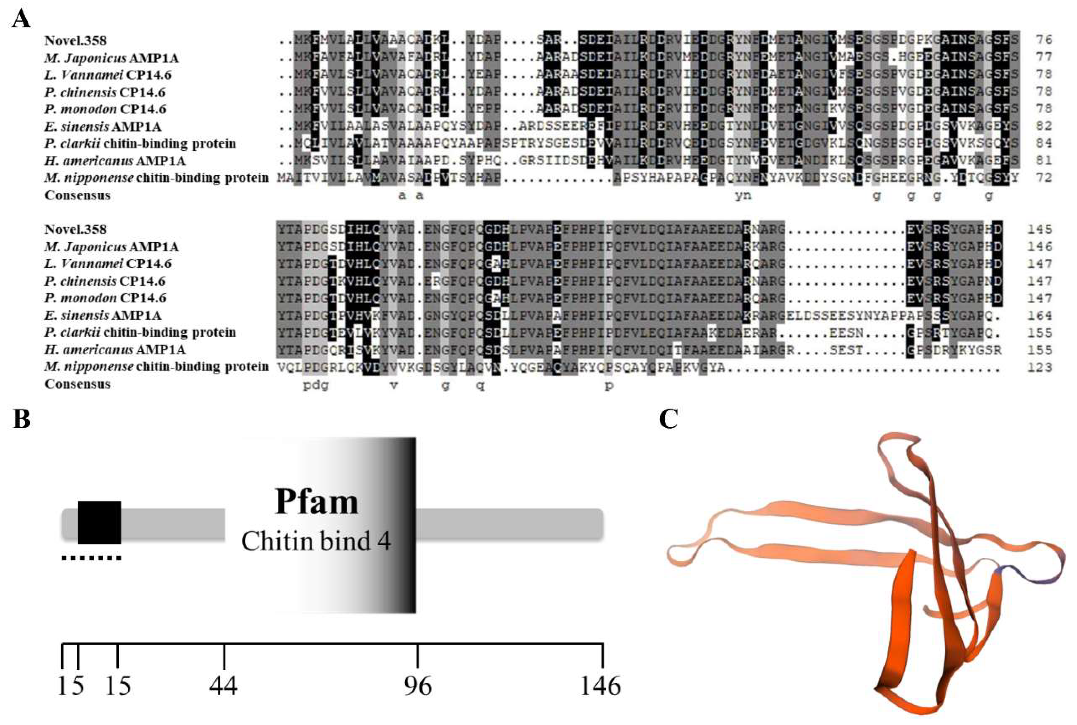
The open reading frame of the cuticle protein (novel.358) gene was 441 bp, encoding a total of 146 amino acids (Figure 6). The theoretical molecular mass of the cuticular protein was 15729.27 u, and the isoelectric point was 4.26 through analysis with the program in ExPASy. The SOPMA server was used to predict the secondary structure of cuticular proteins, which yielded 43 α-helices, 11 β-sheets, and other structures. The results using the Novopro online tool (https://www.novopro.cn/tools/signalp.html, accessed on 9 March 2023) showed that the cuticular protein had a signal peptide type SP, and the signal peptide sequence was MKFMVLALLVAAACA (Figure 6).
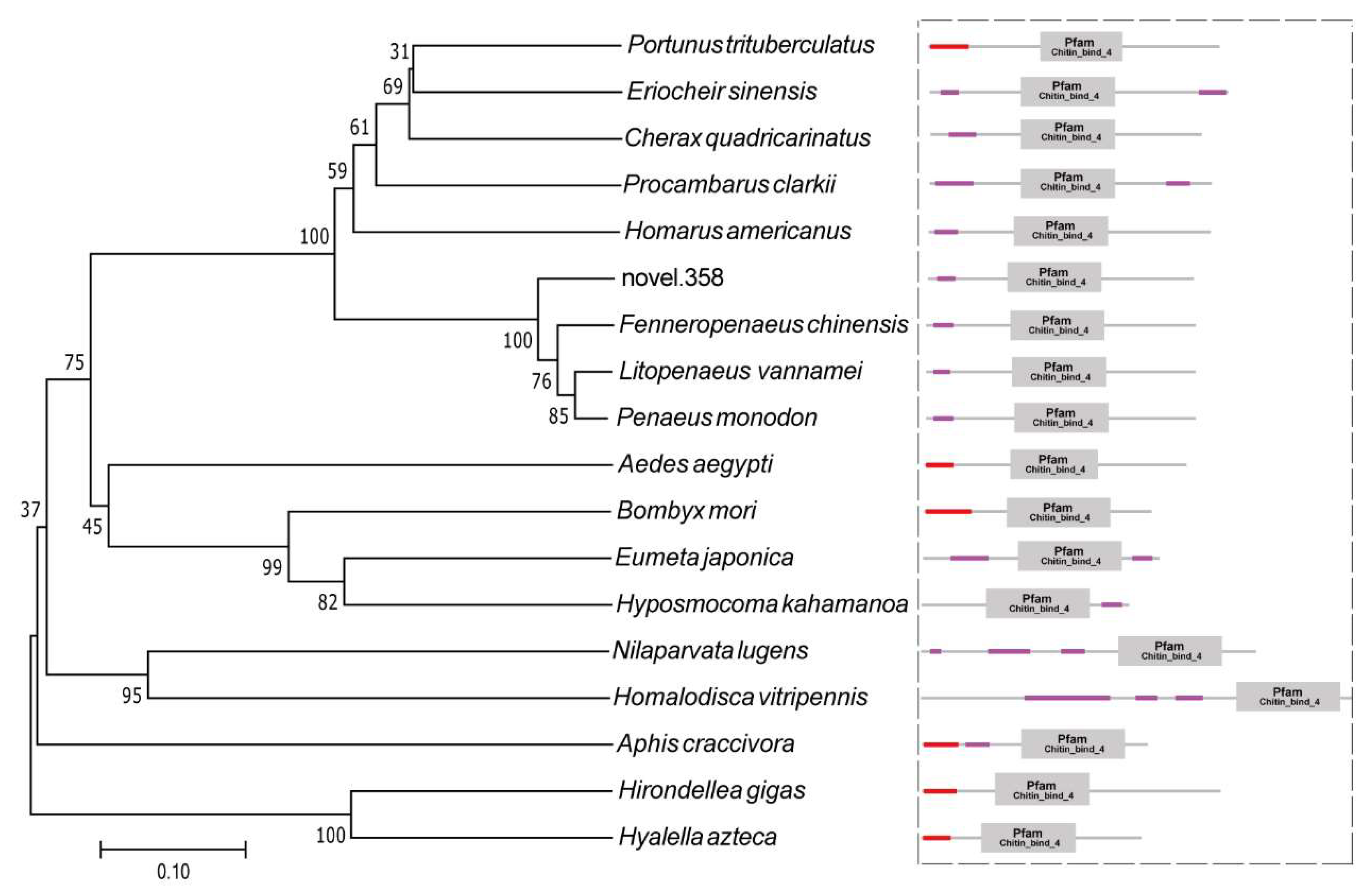
The amino acid sequences of cuticular proteins of M. japonicus and a variety of closely related organisms all show a certain degree of similarity. The results of the sequence comparison showed that the sequence similarity of M. japonicus with L. vannamei was 88.5%, and the similarity with F. chinensis was 87.2%. Phylogenetic analysis of the amino acid sequence of the novel cuticle genes was carried out. As shown in Figure 7, M. japonicus first grouped with shrimp, such as L. vannamei, F. chinensis, and P. monodon and then grouped with other decapod crustaceans, Homarus americanus, Procambarus clarkii, and Cherax quadricarinatus.

4. Discussion

The growth and development of crustaceans are represented by a discontinuous process of molting [37]. Crustacean growth is mainly concentrated on the molting stage. During the cultivation of M. japonicus of the same batch, there are often obvious differences in the growth of individual larvae, but the underlying growth regulation mechanism is not yet clear. In this study, we screened for differentially expressed genes related to growth by the comparative transcriptome analysis of M. japonicus individuals with different growth rates from one full-sib family. This study identified 1375 differentially expressed genes between fast-growing and slow-growing M. japonicus. Transcriptome and quantitative PCR analysis identified several genes associated with molting or muscle growth.
Muscle growth in crustaceans is intermittent and closely associated with the molt cycle due to the presence of the rigid calcified exoskeleton. Increases in muscle mass are restricted to the ecdysial period when the old exoskeleton is shed and the new exoskeleton expands in size [38]. Tissue growth in crustacea occurs at specific stages of the molt cycle and is influenced by a number of physical, hormonal, and environmental factors [39]. Consequently, growth is closely associated with the stages surrounding ecdysis when there is a considerable increase in the rate of water uptake and a subsequent increase in hydrostatic pressure causing the new uncalcified exoskeleton to expand, providing space for tissue growth [40]. Typically, the larger individuals at the beginning have a stronger competitive advantage and will have access to more food. Traditional selective breeding methods are used to select growth-advantaged individuals within a family or population as parents of the next generation.
Troponin, which contains three subunits: inhibitory (TnI), tropomyosin binding (TnT), and Ca2+ binding (TnC), regulates muscle contraction and relaxation [41,42]. The troponin T subunit binds to tropomyosin, while the I subunit inhibits the interaction of myosin and actin and the C subunit triggers muscle contraction through dynamic structural changes upon binding of Ca2+ [43]. In this study, the expression of the TnC gene was significantly upregulated in the large-size group, while the TnI gene was significantly upregulated in the small-size group. Zhao et al. [8] found that the TnI gene of M. japonicus was significantly upregulated in early developmental stages. Wang et al. [44] found that the expression levels of the TnT and TnC genes of E. carinicauda were significantly upregulated in larger individuals compared with smaller individuals. In our previous study about E. carinicauda, we found that TnT and TnC were significantly upregulated in the fast-growing group [45]. These data also suggest that different subunits of troponin do regulate the growth of crustaceans, but the interactions between the different subunits need to be further explored.
Myosin is an important component of muscle cells, and plays an important role in muscle movement, cytoplasmic flow, and signal transduction [46]. Haezsch et al. [47] found that the expression levels of myosin light chain (MLC) and myosin heavy chain (MHC) affect muscle growth and muscle fiber composition. The results of this study showed that MHC genes were significantly upregulated in the fast-growing individual group. MHC has ATPase activity and can bind to actin, allowing MHC to play a leading role in muscle contraction [48]. Zhao et al. [8] showed that the MHC transcription level in the muscle of the large-size group of M. japonicus was significantly increased. The expression of the myosin gene was upregulated in Asian blue crab Portunus trituberculatus with large size [49]. The mutual regulatory relationship between actin and myosin in M. japonicus remains to be further explored.
Crustaceans have a hard outer cuticle whose organic matter is mainly chitin and cuticular proteins [50,51]. The muscles and exoskeletal cuticle form the arthropod musculoskeletal system and are essential for locomotion [52]. In crustaceans, reports on the microscopic architecture of cuticle–muscle connections refer to different body regions in adult specimens, and the integrity of the connections between tendon cells and chitinous matrix of the cuticle is also maintained in the premolt [52]. Cuticular proteins are important in the formation of new cuticles before and after molting [53,54]. Different types of cuticular proteins bind to long-chain chitin and affect the structure and function of the cuticle [55]. Research on cuticular protein genes has mainly focused on insects, such as Drosophila and silkworm [56,57,58]. Cesar et al. [59] established a cDNA library of pacific white shrimp L. vannamei abdominal muscle and identified multiple cuticle protein genes. It is also possible that cuticle proteins are not specific to epidermal tissue. Tissue distribution analysis revealed that a novel cuticle protein gene, LvCPAP1, was predominantly expressed in the epidermis, stomach, and muscle [60]. In L. vannamei, 13,000 DEGs were identified in families with high and low growth performance, including genes encoding cuticle, chitin, ecdysteroids, and muscle proteins [61]. The results of this study showed that the expression of cuticular proteins in large individual M. japonicus was 5 times higher than that in small individuals. The isoelectric point of the cuticle of M. japonicus is 4.26, which is typical of acidic molecules, which was similar to the result of the cuticular protein gene in the Chinese mitten crab Eriocheir sinensis [62]. Acidic macromolecules play a crucial role in cuticular hardening [63,64]. Different types of cuticular proteins bind to long-chain chitin and affect the structure and function of the cuticle [65]. The DD4 and DD5 acidic cuticular proteins have been cloned from the cuticular tissue of the tail fan in the late molting stage of M. japonicus, which have calcium-binding ability [66,67]. Two calcification-related peptides, CAP1 and CAP2, have been isolated and purified from the cuticular matrix of red swamp crayfish, and both are rich in acidic amino acids [68,69]. Future studies should explore the expression characteristics of this cuticular protein in the premolt, intermolt, and postmolt stages of M. japonicus, and develop more SNPs for marker-assisted selection of M. japonicus.

5. Conclusions

In this study, RNA-Seq and qRT-PCR were used to find gene expression differences between fast-growing and slow-growing M. japonicus individuals. We identified several key genes belonging to pathways related to the growth of M. japonicus. The dynamic expression characteristics of these genes in the molting process and different developmental stages of M. japonicus need to be further studied. In addition, our data can be used to screen for SNP loci of growth-related genes to provide data for marker-assisted breeding. Taken together, our findings help to elucidate the molecular regulatory mechanisms of crustacean growth.

Supplementary Materials

The following supporting information can be downloaded at: https://www.mdpi.com/article/10.3390/fishes8070350/s1, Figure S1: The functional data of the variant sites; Figure S2: The impact data of variant sites; Figure S3: The regional data of variant sites; Table S1: Primer sequences for qRT−PCR.

Author Contributions

Conceptualization, P.W., F.Y. and C.X; Methodology, X.L., S.X. and J.Z.; Validation, L.W., X.Z. (Xinlei Zhou) and X.Z. (Xinyi Zhou); Writing—original draft preparation, P.W. and F.Y.; Writing—review and editing, B.Y., H.G. and C.X.; Funding acquisition, C.X. and P.W. All authors have read and agreed to the published version of the manuscript.

Funding

This study was funded by the Natural Science Foundation of Jiangsu Province (No. BK20210924); the National Natural Science Foundation of China (32200411); Natural Science Foundation of the Jiangsu Higher Education Institutions of China (21KJB240001); the Open-end Funds of Jiangsu Key Laboratory of Marine Bioresources and Environment (SH20221205); Project funded by China Postdoctoral Science Foundation (2022M721397); Project funded by Postdoctoral Science Foundation of Lianyungang (LYG20220021); the Innovation and Entrepreneurship Project of Jiangsu Ocean University (SY202211641631006, SY202311641631003, SY202311641631010); the Postgraduate Research and Practice Innovation Program of Jiangsu Province (KYCX23-3454, KYCX2023-104); “521”scientific research projects of Lianyungang (LYG06521202128); Fisheries high quality development project of Yancheng (YCSCYJ2021006); the Priority Academic Program Development of Jiangsu Higher Education Institutions (PAPD).

Institutional Review Board Statement

This study was approved by the Animal Care and Use Committee of Jiangsu University (protocol no. 2020-37; approval date: 1 September 2019). All procedures involving animals were performed in accordance with guidelines for the Care and Use of Laboratory Animals in China.

Data Availability Statement

The datasets presented in this study can be found in the online version. The Illumina sequence reads generated during the present study are available in the NCBI SRA database under the BioProject ID: PRJNA970064.

Conflicts of Interest

The authors declare no conflict of interest.

References

  1. Tsoi, K.H.; Ma, K.Y.; Wu, T.H.; Fennessy, S.T.; Chu, K.H.; Chan, T.Y. Verification of the cryptic species Penaeus pulchricaudatus in the commercially important kuruma shrimp P. japonicus (Decapoda: Penaeidae) using molecular taxonomy. Invertebr. Syst. 2014, 28, 476–490. [Google Scholar] [CrossRef] [Green Version]
  2. Wang, P.; Chen, B.; Zheng, J.; Cheng, W.; Zhang, H.; Wang, J.; Su, Y.; Xu, P.; Mao, Y. Fine-scale population genetic structure and parapatric cryptic species of kuruma shrimp (Marsupenaeus japonicus), along the northwestern Pacific coast of China. Front. Genet. 2020, 11, 118. [Google Scholar] [CrossRef] [PubMed] [Green Version]
  3. He, Y.Q.; Su, Y.Q.; Mao, Y.; Wang, J. Genetic diversity analysis of microsatellite DNA in different varieties of Marsupenaneus japonicus. J. Fish. China 2012, 36, 1520–1528. [Google Scholar] [CrossRef]
  4. Wang, P.; Mao, Y.; Su, Y.; Wang, J. Comparative analysis of transcriptomic data shows the effects of multiple evolutionary selection processes on codon usage in Marsupenaeus japonicus and Marsupenaeus pulchricaudatus. BMC Genom. 2021, 22, 1–14. [Google Scholar] [CrossRef]
  5. Duan, Y.; Zhang, J.; Dong, H.; Wang, Y.; Liu, Q.; Li, H. Effect of desiccation and resubmersion on the oxidative stress response of the kuruma shrimp Marsupenaeus japonicus. Fish Shellfish Immunol. 2016, 49, 91–99. [Google Scholar] [CrossRef] [PubMed]
  6. Hewitt, D.; Duncan, P. Effect of high water temperature on the survival, moulting and food consumption of Penaeus (Marsupenaeus) japonicus (Bate, 1888). Aquac. Res. 2001, 32, 305–313. [Google Scholar] [CrossRef]
  7. FAO (Food and Agriculture Organization of the United Nations). Global Production Statistics. 2018. Available online: http://www.fao.org/fishery/statistics/global-aquaculture-production/query/en (accessed on 30 March 2018).
  8. Zhao, J.; He, Z.; Chen, X.; Huang, Y.; Xie, J.; Qin, X.; Ni, Z.; Sun, C. Growth trait gene analysis of kuruma shrimp (Marsupenaeus japonicus) by transcriptome study. Comp. Biochem. Physiol. Part D Genom. Proteom. 2021, 40, 100874. [Google Scholar] [CrossRef]
  9. Cheng, W.; Zhang, H.; Wang, P.; Wei, Y.; Chen, C.; Hou, Y.; Deng, X.; Li, S.; Sun, S.; Cai, Q. The multiple influences of natural farming environment on the cultured population behavior of kuruma prawn, Penaeus japonicus. Animals 2022, 12, 3383. [Google Scholar] [CrossRef]
  10. Kim, S.-K.; Guo, Q.; Jang, I.-K. Effect of biofloc on the survival and growth of the postlarvae of three penaeids (Litopenaeus vannamei, Fenneropenaeus chinensis, and Marsupenaeus japonicus) and their biofloc feeding efficiencies, as related to the morphological structure of the third maxilliped. J. Crustac. Biol. 2015, 35, 41–50. [Google Scholar]
  11. Jung, H.; Lyons, R.E.; Hurwood, D.A.; Mather, P.B. Genes and growth performance in crustacean species: A review of relevant genomic studies in crustaceans and other taxa. Rev. Aquac. 2013, 5, 77–110. [Google Scholar] [CrossRef]
  12. Shuang, Z.; Xuange, L.; Pengfei, W.; Peng, X.; Lei, Z.; Lei, Z.; Qindong, T.; Zhi, C.; Guifeng, L. Muscle transcriptome of Siniperca chuatsi with different weights from a full-sib family. J. Fish. Sci. China 2020, 27, 53–61. [Google Scholar]
  13. Lanmei, W.; Wenbin, Z.; Jianjun, F.; Zaijie, D. De novo transcriptome analysis and comparison of the FFRC No. 2 strain common carp (Cyprinus carpio) associated with its muscle growth. J. Fish. China 2021, 45, 79–87. [Google Scholar]
  14. Wang, Z.; Liang, F.; Huang, R.; Deng, Y.; Li, J. Identification of the differentially expressed genes of Pinctada maxima individuals with different sizes through transcriptome analysis. Reg. Stud. Mar. Sci. 2019, 26, 100512. [Google Scholar] [CrossRef]
  15. Huang, J.; You, W.; Luo, X.; Ke, C. iTRAQ-based identification of proteins related to muscle growth in the Pacific abalone, Haliotis discus hannai. Int. J. Mol. Sci. 2017, 18, 2237. [Google Scholar] [CrossRef] [PubMed] [Green Version]
  16. Yunjie, Y.; Xia, L.; Xianhong, M.; Sheng, L.; Baolong, C.; Jie, K. Screening of genes related to muscle growth under the myostatin regulation by RNA-seq in Fenneropenaeus chinensis. Prog. Fish. Sci. 2021, 42, 55–63. [Google Scholar]
  17. Guo, X.F.; Zhou, Y.L.; Liu, M.; Wang, Z.W.; Gui, J.F. Integrated application of Iso-seq and RNA-seq provides insights into unsynchronized growth in red swamp crayfish (Procambarus clarkii). Aquac. Rep. 2022, 22, 101008. [Google Scholar] [CrossRef]
  18. Uengwetwanit, T.; Uawisetwathana, U.; Arayamethakorn, S.; Khudet, J.; Chaiyapechara, S.; Karoonuthaisiri, N.; Rungrassamee, W. Multi-omics analysis to examine microbiota, host gene expression and metabolites in the intestine of black tiger shrimp (Penaeus monodon) with different growth performance. PeerJ 2020, 8, e9646. [Google Scholar] [CrossRef]
  19. Lin, Z.; Zhang, Z.; Solberg, M.F.; Chen, Z.; Wei, M.; Zhu, F.; Jia, C.; Meng, Q.; Zhang, Z. Comparative transcriptome analysis of mixed tissues of black porgy (Acanthopagrus schlegelii) with differing growth rates. Aquac. Res. 2021, 52, 5800–5813. [Google Scholar] [CrossRef]
  20. Zhao, L.; He, K.; Xiao, Q.; Liu, Q.; Luo, W.; Luo, J.; Fu, H.; Li, J.; Wu, X.; Du, J. Comparative transcriptome profiles of large and small bodied large-scale loaches cultivated in paddy fields. Sci. Rep. 2021, 11, 4936. [Google Scholar] [CrossRef]
  21. Li, Y.; Liu, Z.; Dai, X. Transcriptome analysis of growth retardation and normal Macrobrachium rosenbergii. Genom. Appl. Biol. 2021, 40, 89–100. (In Chinese) [Google Scholar]
  22. Grabherr, M.G.; Haas, B.J.; Yassour, M.; Levin, J.Z.; Amit, I. Full-length transcriptome assembly from RNA-Seq data without a reference genome. Nat. Biotechnol. 2013, 29, 644–652. [Google Scholar] [CrossRef] [Green Version]
  23. Mortazavi, A.; Williams, B.A.; Mccue, K.; Schaeffer, L.; Wold, B. Mapping and quantifying mammalian transcriptomes by RNA-Seq. Nat. Methods 2008, 5, 621–628. [Google Scholar] [CrossRef] [PubMed]
  24. Pertea, M.; Pertea, G.M.; Antonescu, C.M.; Chang, T.C.; Mendell, J.T.; Salzberg, S.L. StringTie enables improved reconstruction of a transcriptome from RNA-seq reads. Nat. Biotechnol. 2015, 33, 290–295. [Google Scholar] [CrossRef] [PubMed] [Green Version]
  25. Yang, L.; Smyth, G.K.; Wei, S. featureCounts: An efficient general purpose program for assigning sequence reads to genomic features. Bioinformatics 2014, 30, 923–930. [Google Scholar]
  26. Simon, A.; Wolfgang, H. Differential expression analysis for sequence count data. Genome Biol. 2010, 11, 106. [Google Scholar]
  27. Love, M.I.; Huber, W.; Anders, S. Moderated estimation of fold change and dispersion for RNA-seq data with DESeq2. Genome Biol. 2014, 15, 550. [Google Scholar] [CrossRef] [PubMed] [Green Version]
  28. Wu, T.; Hu, E.; Xu, S.; Chen, M.; Guo, P.; Dai, Z.; Feng, T.; Zhou, L.; Tang, W.; Zhan, L.; et al. clusterProfiler 4.0: A universal enrichment tool for interpreting omics data. Innovation 2021, 2, 100141. [Google Scholar] [CrossRef]
  29. Letunic, I.; Doerks, T.; Bork, P. SMART 7: Recent updates to the protein domain annotation resource. Nucleic Acids Res. 2011, 40, 302–305. [Google Scholar] [CrossRef] [Green Version]
  30. Geourjon, C.; Deleage, G. SOPMA: Significant improvements in protein secondary structure prediction by consensus prediction from multiple alignments. Bioinformatics 1995, 11, 681–684. [Google Scholar] [CrossRef]
  31. Sigrist, C.J.A.; Lorenzo, C.; Edouard, D.C.; Langendijk-Genevaux, P.S.; Virginie, B.; Amos, B.; Nicolas, H. PROSITE, a protein domain database for functional characterization and annotation. Nucleic Acids Res. 2010, 38, 161–166. [Google Scholar] [CrossRef] [Green Version]
  32. Kumar, S.; Stecher, G.; Tamura, K. MEGA7: Molecular evolutionary genetics analysis version 7.0 for bigger datasets. Mol. Biol. Evol. 2016, 33, 1870–1874. [Google Scholar] [CrossRef] [PubMed] [Green Version]
  33. Lalitha, S.J. Primer Premier 5. Biotech Softw. Internet Rep. Comput. Softw. J. Sci. 2000, 1, 270–272. [Google Scholar] [CrossRef]
  34. McKenna, A.; Hanna, M.; Banks, E.; Sivachenko, A.; Cibulskis, K.; Kernytsky, A.; Garimella, K.; Altshuler, D.; Gabriel, S.; Daly, M. The Genome Analysis Toolkit: A MapReduce framework for analyzing next-generation DNA sequencing data. Genome Res. 2010, 20, 1297–1303. [Google Scholar] [CrossRef] [Green Version]
  35. Cingolani, P. A program for annotating and predicting the effects of single nucleotide polymorphisms, SnpEff: SNPs in the genome of Drosophila melanogaster strain w1118; iso-2; iso-3. Fly 2012, 6, 1–13. [Google Scholar] [CrossRef] [Green Version]
  36. Liew, M.; Pryor, R.; Palais, R.; Meadows, C.; Erali, M.; Lyon, E.; Wittwer, C. Genotyping of single-nucleotide polymorphisms by high-resolution melting of small amplicons. Clin. Chem. 2004, 50, 1156–1164. [Google Scholar] [CrossRef] [PubMed]
  37. Skov, M.W.; Hartnoll, R.G. Comparative suitability of binocular observation, burrow counting and excavation for the quantification of the mangrove fiddler crab Uca annulipes (H. Milne Edwards). Adv. Decapod Crustac. Res. 2001, 154, 201–212. [Google Scholar]
  38. El Haj, A.J.; Harrison, P.; Whiteley, N.M. Regulation of muscle gene expression in Crustacea over the moult cycle. Symp. Soc. Exp. Biol. 1992, 46, 151–165. [Google Scholar]
  39. Haj, A.J.E.; Whiteley, N.M. Molecular regulation of muscle growth in Crustacea. J. Mar. Biol. Assoc. UK 1997, 77, 95–106. [Google Scholar] [CrossRef]
  40. Mykles, D.L. The mechanism of fluid absorption at ecdysis in the American lobster, Homarus americanus. J. Exp. Biol. 2015, 84, 89–102. [Google Scholar] [CrossRef]
  41. Tobacman, L.S. Troponin revealed: Uncovering the structure of the thin filament on-off switch in striated muscle. Biophys. J. 2021, 120, 1–9. [Google Scholar] [CrossRef]
  42. Cao, T.X.; Thongam, U.; Jin, J.P. Invertebrate troponin: Insights into the evolution and regulation of striated muscle contraction. Arch. Biochem. Biophys. 2019, 666, 40–45. [Google Scholar] [CrossRef] [PubMed]
  43. Ochiai, Y.; Ozawa, H. Biochemical and physicochemical characteristics of the major muscle proteins from fish and shellfish. Fish. Sci. 2020, 86, 729–740. [Google Scholar] [CrossRef]
  44. Wang, J.; Ge, Q.; Li, J.; Chen, Z.; Li, J. Isolation and characterization of three skeletal troponin genes and association with growth-related traits in Exopalaemon carinicauda. Mol. Biol. Rep. 2019, 46, 705–718. [Google Scholar] [CrossRef]
  45. Xing, C.F.; Xiong, J.Y.; Xie, S.M.; Guo, H.X.; Hua, S.S.; Yao, Y.J.; Zhu, J.W.; Yan, B.L.; Shen, X.; Gao, H.; et al. Comparative transcriptome and gut microbiota analysis of Exopalaemon carinicauda with different growth rates from a full-sib family. Aquac. Rep. 2023, 30, 101580. [Google Scholar] [CrossRef]
  46. Heissler, S.M.; Sellers, J.R. Kinetic adaptations of myosins for their diverse cellular functions. Traffic 2016, 17, 839–859. [Google Scholar] [CrossRef] [Green Version]
  47. Harzsch, S.; Kreissl, S. Myogenesis in the thoracic limbs of the American lobster. Arthropod Struct. Dev. 2010, 39, 423–435. [Google Scholar] [CrossRef] [PubMed]
  48. Andruchov, O.; Andruchova, O.; Wang, Y.; Galler, S. Dependence of cross-bridge kinetics on myosin light chain isoforms in rabbit and rat skeletal muscle fibres. J. Physiol. 2006, 571, 231–242. [Google Scholar] [CrossRef] [Green Version]
  49. Ren, X.; Yu, X.; Gao, B.; Li, J.; Liu, P. iTRAQ-based identification of differentially expressed proteins related to growth in the swimming crab, Portunus trituberculatus. Aquac. Res. 2017, 48, 3257–3267. [Google Scholar] [CrossRef]
  50. Zhang, X.; Yuan, J.; Li, F.; Xiang, J. Chitin synthesis and degradation in crustaceans: A genomic view and application. Mar. Drugs 2021, 19, 153. [Google Scholar] [CrossRef]
  51. Abehsera, S.; Weil, S.; Manor, R.; Sagi, A. The search for proteins involved in the formation of crustacean cuticular structures. Hydrobiologia 2018, 825, 29–45. [Google Scholar] [CrossRef]
  52. Mrak, P.; Bogataj, U.; Štrus, J.; Žnidaršič, N. Cuticle morphogenesis in crustacean embryonic and postembryonic stages. Arthropod Struct. Dev. 2017, 46, 77–95. [Google Scholar] [CrossRef] [PubMed]
  53. Nagasawa, H. The crustacean cuticle: Structure, composition and mineralization. Front. Biosci. 2012, 4, 711–720. [Google Scholar] [CrossRef]
  54. Vincent, J.F. Arthropod cuticle: A natural composite shell system. Compos. Part A Appl. Sci. Manuf. 2002, 33, 1311–1315. [Google Scholar] [CrossRef]
  55. Willis, J.H.; Papandreou, N.C.; Iconomidou, V.A.; Hamodrakas, S.J. Cuticular proteins. In Insect Molecular Biology and Biochemistry; Academic Press: San Diego, CA, USA, 2012; pp. 134–166. [Google Scholar]
  56. Cornman, R.S. Molecular evolution of Drosophila cuticular protein genes. PLoS ONE 2009, 4, e8345. [Google Scholar] [CrossRef] [PubMed]
  57. Wang, X.; Xie, X.; Xie, K.; Liu, Q.; Li, Y.; Tan, X.; Dong, H.; Li, X.; Dong, Z.; Xia, Q. Chitin and cuticle proteins form the cuticular layer in the spinning duct of silkworm. Acta Biomater. 2022, 145, 260–271. [Google Scholar] [CrossRef] [PubMed]
  58. Balabanidou, V.; Grigoraki, L.; Vontas, J. Insect cuticle: A critical determinant of insecticide resistance. Curr. Opin. Insect Sci. 2018, 27, 68–74. [Google Scholar] [CrossRef]
  59. Cesar, J.R.; Zhao, B.; Yang, J. Analysis of expressed sequence tags from abdominal muscle cDNA library of the pacific white shrimp Litopenaeus vannamei. Animal 2008, 2, 1377–1383. [Google Scholar] [CrossRef] [Green Version]
  60. Yang, F.; Li, X.; Li, S.; Xiang, J.; Li, F. A novel cuticle protein involved in WSSV infection to the Pacific white shrimp Litopenaeus vannamei. Dev. Comp. Immunol. 2019, 102, 103491. [Google Scholar] [CrossRef]
  61. Santos, C.A.; Andrade, S.C.S.; Teixeira, A.K.; Farias, F.; Guerrelhas, A.C.; Rocha, J.L.; Freitas, P.D. Transcriptome differential expression analysis reveals the activated genes in Litopenaeus vannamei shrimp families of superior growth performance. Aquaculture 2021, 531, 735871. [Google Scholar] [CrossRef]
  62. Jundong, C.; Pengyun, D.; Xuguang, L.; Keran, B. Cloning and expression analysis of epidermal protein gene EsCAP from Chinese Mitten crab. Jiangsu Agric. Sci. 2021, 49, 74–80. [Google Scholar]
  63. Coblentz, F.E.; Shafer, T.H.; Roer, R.D. Cuticular proteins from the blue crab alter in vitro calcium carbonate mineralization. Comp. Biochem. Physiol. Part B Biochem. Mol. Biol. 1998, 121, 349–360. [Google Scholar] [CrossRef]
  64. Glazer, L.; Sagi, A. On the involvement of proteins in the assembly of the crayfish gastrolith extracellular matrix. Invertebr. Reprod. Dev. 2012, 56, 57–65. [Google Scholar] [CrossRef]
  65. Andersen, S.O. Insect cuticular sclerotization: A review. Insect Biochem. Mol. Biol. 2010, 40, 166–178. [Google Scholar] [CrossRef] [PubMed]
  66. Endo, H.; Persson, P.; Watanabe, T. Molecular cloning of the crustacean DD4 cDNA encoding a Ca2+-binding protein. Biochem. Biophys. Res. Commun. 2000, 276, 286–291. [Google Scholar] [CrossRef] [PubMed]
  67. Endo, H.; Takagi, Y.; Ozaki, N.; Kogure, T.; Watanabe, T. A crustacean Ca2+-binding protein with a glutamate-rich sequence promotes CaCO3 crystallization. Biochem. J. 2004, 384, 159–167. [Google Scholar] [CrossRef] [Green Version]
  68. Inoue, H.; Ohira, T.; Ozaki, N.; Nagasawa, H. Cloning and expression of a cDNA encoding a matrix peptide associated with calcification in the exoskeleton of the crayfish. Comp. Biochem. Physiol. Part B Biochem. Mol. Biol. 2003, 136, 755–765. [Google Scholar] [CrossRef]
  69. Inoue, H.; Ohira, T.; Ozaki, N.; Nagasawa, H. A novel calcium-binding peptide from the cuticle of the crayfish, Procambarus clarkii. Biochem. Biophys. Res. Commun. 2004, 318, 649–654. [Google Scholar] [CrossRef]
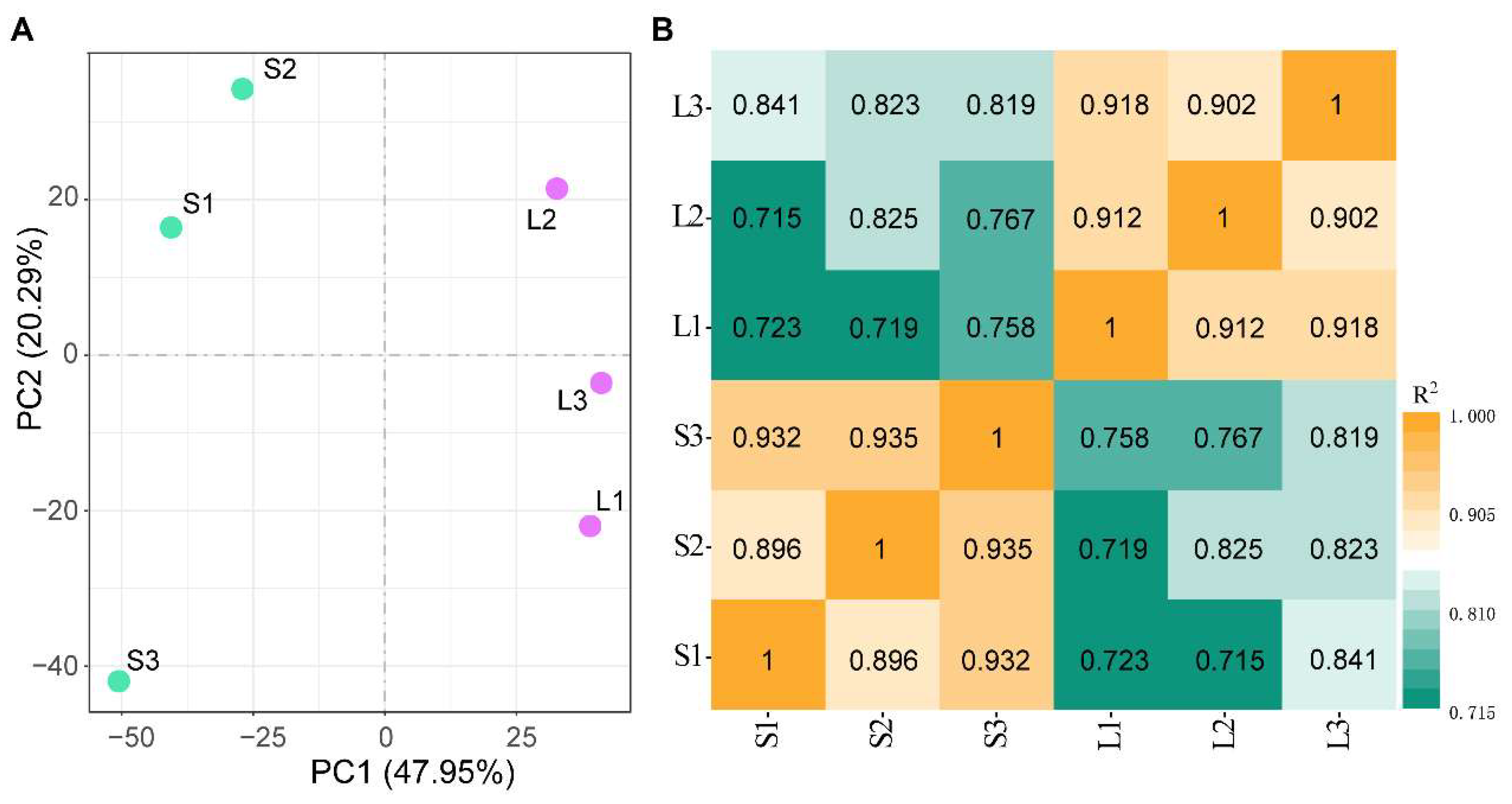
Figure 1. Principal component analysis (A) and heatmap (B) of transcriptomes of six samples based on Pearson’s correlation coefficient.
Fishes 08 00350 g001
Figure 2. GO functional annotation for differentially expressed genes. BP represents biological process, CC represents cellular components, and MF represents molecular function.
Fishes 08 00350 g002
Figure 3. The top 20 statistically significant KEGG classifications.
Fishes 08 00350 g003
Figure 4. The fold−change of differentially expressed genes determined by RNA−Seq and qRT−PCR. ICP: novel 358, EK: Ecdysteroid kinase, SCBP: Sarcoplasmic calcium−binding protein, beta chain, MHC: Myosin heavy chain, TC: Troponin C, TI: Troponin I, CAS: Crustacyanin−A2 subunit, Hsp70: Heat shock protein 70, IAP: Inhibitor of apoptosis protein, TRY: Trypsin−1. Each bar represents the mean ± S.D (n = 3). A significant difference between groups at p < 0.05 (n = 3, ANOVA) is indicated by different letters above the bars.
Fishes 08 00350 g004
Figure 5. Distribution of putative SNPs in M. japonicus.
Fishes 08 00350 g005
Figure 6. Multiple alignments of the deduced AA sequences of the cuticle protein (novel 358) with other cuticle proteins (A), structural domains (B), and three-dimensional prediction (C). The dotted line represents the signal peptide, and the black box represents low complexity.
Fishes 08 00350 g006
Figure 7. Phylogenetic analysis and protein domain analysis of cuticle protein, for Portunus trituberculatus (MPC28856.1), Eriocheir sinensis (XP_050700776.1), Cherax quadricarinatus (XP_053639595.1), Procambarus clarkii (BAM99303.1), Homarus americanus (XP_042218166.1), Fenneropenaeus chinensis (XP_047469304.1), Litopenaeus vannamei (XP_027239199.1), Penaeus monodon (XP_037780114.1), Aedes aegypti (EAT39943.1), Bombyx mori (NP_001166723.1), Eumeta japonica (GBP72130.1), Hyposmocoma kahamanoa (XP_026328697.1), Nilaparvata lugens (QCP68952.1), Homalodisca vitripennis (XP_046666636.1), Aphis craccivora (KAF0756676.1), Hirondellea gigas (LAC22872.1), and Hyalella azteca (XP_018010166.1).
Fishes 08 00350 g007
Table 1. Summary statistics for sequencing data.
SampleRaw ReadsClean ReadsClean Bases (G)Q20 (%)Q30 (%)GC (%)
S146,763,77245,912,3326.8998.0394.450.12
S248,942,93247,858,2727.1898.2694.8550.04
S345,487,75044,475,4086.6798.0394.5251.44
L148,220,79447,140,4427.0798.4895.4151.04
L247,360,73046,243,7666.9498.2894.8248.69
L344,954,98443,991,2666.698.4695.552.72
Table 2. Representative growth-related differentially expressed genes.
Gene_idGene_DescriptionLog2 (Fold Change)padj
Hic_asm_3.1371Cuticle protein AMP48.0050.026
Hic_asm_3.1791Chitin binding Peritrophin-A domain7.6750.020
Hic_asm_29.1989Insect cuticle protein5.6200.001
Hic_asm_3.3781Transmembrane protease serine 11D5.5330.045
Hic_asm_21.381Tubulin alpha-1 chain5.0780.004
Hic_asm_5.353Ecdysteroid kinase4.6280.001
Hic_asm_4.2410Myosin heavy chain C3.6300.002
Hic_asm_7.2362Sarcoplasmic calcium-binding protein4.3490.005
Hic_asm_28.2387Mitochondrial basic amino acids transporter3.9390.006
Hic_asm_37.790Troponin C2.7590.002
Hic_asm_16.1606Mitochondrial enolase superfamily member 12.6560.024
Hic_asm_27.1758phosphatidylinositol 4,5-bisphosphate phosphodiesterase2.6180.020
Hic_asm_13.2114Mitochondrial carnitine/acylcarnitine carrier protein2.0820.000
Hic_asm_12.2116Mitochondrial dicarboxylate carrier2.0610.048
Hic_asm_30.362Coactosin-like protein1.9650.025
Hic_asm_22.2436Carbonyl reductase (NADPH) 3−4.9780.002
Hic_asm_35.393Trypsin-1−3.5090.047
Hic_asm_12.273Inhibitor of apoptosis protein−3.0980.001
Hic_asm_3.2630Methyl farnesoate epoxidase−3.0640.002
Hic_asm_32.1003Alpha-amylase−2.8760.020
Hic_asm_26.1575Superoxide dismutase (Cu-Zn)−2.8050.024
Hic_asm_38.1026Actin-2, muscle-specific−2.5360.000
Hic_asm_14.203zinc-RING finger domain−2.5200.017
Hic_asm_8.3322Heat shock 70 kDa protein−2.4410.017
Hic_asm_16.604Crustacyanin-A2 subunit−2.4140.005
Hic_asm_1.2118Ubiquitin carboxyl-terminal hydrolase 22−2.3340.020
Hic_asm_25.2408Glutathione S-transferase D7−2.3940.006
Hic_asm_19.605Pyruvate kinase−1.9680.026
Hic_asm_36.203Superoxide dismutase (Mn), mitochondrial−1.6560.019
Hic_asm_17.2603Troponin I−1.5490.002
Disclaimer/Publisher’s Note: The statements, opinions and data contained in all publications are solely those of the individual author(s) and contributor(s) and not of MDPI and/or the editor(s). MDPI and/or the editor(s) disclaim responsibility for any injury to people or property resulting from any ideas, methods, instructions or products referred to in the content.