Article

The Effect of Cu2+ Exposure on the Nrf2 Signaling Pathway of Tilapia Hepatocyte, Base on Experiments In Vitro

1
Key Laboratory of Marine Biotechnology of Fujian Province, Institute of Oceanology, Fujian Agriculture and Forestry University, Fuzhou 350002, China
2
College of Marine Sciences, Fujian Agriculture and Forestry University, Fuzhou 350002, China
*
Author to whom correspondence should be addressed.
Fishes 2023, 8(6), 280; https://doi.org/10.3390/fishes8060280
Submission received: 25 April 2023 / Revised: 16 May 2023 / Accepted: 22 May 2023 / Published: 24 May 2023
(This article belongs to the Section Fish Pathology and Parasitology)

Abstract

:
Copper is a common component of industrial heavy metal waste and a major component of some fish medicines, which can cause oxidative stress and damage the health of farmed fish. The Nrf2 signaling pathway is an important pathway related to the oxidative stress on vertebrates. Exploring the effect of copper on the Nrf2 signaling pathway in fish hepatocytes would help improve the understanding of the molecular mechanism of antioxidants in fish hepatocytes and provide theoretical data for relevant toxicological research. Adult tilapia were cultured under properly controlled conditions for two weeks to adapt to laboratory culture conditions. Primary tilapia hepatocytes were obtained by cell culture. MTT (3-[4,5-dimethylthiazol-2-yl]-2,5 diphenyl tetrazolium bromide) assay was used to detect the effect of copper ions on the viability of tilapia hepatocytes. The lipid peroxidation level (MDA) and antioxidant ability of tilapia hepatocytes (SOD and CAT activity) were detected. Quantitative PCR (qPCR) was used to detect the differential expression of each gene (Nrf2, Keap1a, Keap1b, CuZnSOD, MnSOD, HO-1, and GSTA) in the Nrf2 signaling pathway. The results suggested that after treatment with 100 μM copper ions for 4 h, 8 h, and 24 h, the viability of hepatocytes significantly decreased (p < 0.05). LDH and MDA after 8 h and 24 h treatment were significantly higher than those in the control group (p < 0.05). CAT activity significantly decreased after 4 h (p < 0.05), and SOD activity significantly decreased after 8 h and 24 h (p < 0.05). The results of qPCR showed that the expression of MnSOD significantly increased after a treatment with copper ions for 4 h, and the expression of Nrf2, Keap1a, CuZnSOD, HO-1 as well as GSTA significantly increased after a treatment with copper ions for 8 h, compared with the control group (p < 0.05). After being treated with copper ions for 24 h, the expression of Nrf2 and CuZnSOD significantly increased compared with the control group (p < 0.05). There was no significant difference in the expression of Keap1b or CAT at each time point. In conclusion, with copper ions exposure, the viability of tilapia hepatocytes was reduced, causing lipid peroxidation, a reduction in the antioxidant capacity of cells, the activation of the Nrf2 signaling pathway, and the increase in the expression of most genes in this pathway, which are defensive responses of hepatocytes to oxidative stress caused by copper ions. This study can provide theoretical data for related toxicological research.
Key Contribution: The changes in viability, biochemical indexes and Nrf2 signaling pathway of tilapia hepatocytes under copper ion stress were elucidated.

Graphical Abstract

1. Introduction

Industrial wastewater and drug abuse have been endangering the health of aquaculture animals for years. Copper ions are not only common components of industrial heavy metal waste, but also the main components of some aquatic drugs [1]. Scholars have carried out a great deal of meaningful work on the toxicology of copper ions in aquatic animals, achieving many research results [2,3]. It is currently widely believed that copper ions and their complexes in water induce the formation of reactive oxygen species (ROS), which damage biological molecules such as lipids, proteins, and nucleic acids [4]. In the aquaculture industry, copper tends to accumulate in the liver of fish which leads to lipid peroxidation and eventually fatty liver [5]. ROS produced by heavy metals such as copper ions can be removed by the defensive antioxidant system of fish, which includes superoxide dismutase (SOD), catalase (CAT), glutathione (GSH), and so on [6]. How these antioxidant systems are activated in response to oxidative stress is the core concern of this study. By exploring the resistance of the molecular mechanism of fish liver cells to copper-ion-induced oxidative stress, theoretical data can be provided for the toxicology of heavy metals in fish.
The expression of many antioxidant enzyme genes is regulated through the upstream Nrf2 (nuclear factor erythroid-2-related factor 2) signaling pathway [7]. Nrf2 is a transcription factor related to the cellular defense system of vertebrates. The promoters of some important antioxidants and detoxification-related genes contain antioxidant response elements (AREs), to which Nrf2 can bind to promote the transcription of downstream genes. The common downstream genes of Nrf2 include SOD, glutathione sulfide transferase (GST), heme oxidase-1 (HO-1) and so on [7]. Nrf2 is usually bound to the cytoskeleton via Kelch-like ECH associated protein 1 (Keap1) protein dimer, and Keap1 can also lead the ubiquitination degradation of Nrf2. When cells are subjected to oxidative stress, Nrf2 will be dissociated from Keap1 protein and enter the nucleus, where it binds to the promoters of the above target genes to accelerate the transcription of antioxidant genes [7]. Among teleost, Nrf2 was first found in zebrafish [8], and this pathway is highly conserved across vertebrates [9]. Researches have shown that Nrf2 signaling pathway plays an important role in the resistance of fish to oxidative stress in vivo [10,11].
Tilapia (Oreochromis niloticus), the model organism of this study, is a kind of farmed fish with fast growth, strong resistance and strong fecundity, which is recommended by the FAO of the United Nations to reduce poverty and malnutrition [12]. Tilapia primary hepatocyte culture which previously established by the author [13] was used in this study. In this study, the viability and biochemical indexes of tilapia hepatocytes under copper ion exposure were studied in vitro, and the expression of genes in the Nrf2 signaling pathway was studied by qPCR, to explore the molecular mechanism of fish hepatocytes against the oxidative stress of heavy metals. This study can provide theoretical data for the mechanism of heavy metal toxicology in tilapia and the development of related hepatoprotective medicine.

2. Materials and Methods

2.1. Animals

Adult tilapia were provided by the Freshwater Fisheries Research Institute of Fujian, China. Tilapia (n = 7, average weight = 53.51 ± 6.10 g) were maintained in a 100 L glass tank for two weeks to adapt to laboratory culture conditions. The water temperature was maintained at 28 °C, and dissolved oxygen was higher than 6 mg/L. Tilapia were fed (4% body weight) twice daily.

2.2. Primary Culture of Tilapia Hepatocytes

The procedure of tilapia primary hepatocyte culture was consistent with that in the previous study [13]. Healthy adult tilapia were anesthetized with MS-222 and exsanguinated. After sterilizing the body surface with 75% alcohol, the livers were carefully excised onto a sterile watch glass and cut into tissue pieces of approximately 1 mm3, after which irrelevant tissues such as fat and blood vessels were removed. The tissues were washed repeatedly with Hank’s buffer until the blood color subsided. Liver tissues were digested with 0.25% trypsin solution (28 °C) for three times, 15 min each time, and hepatocytes were collected at each end point of digestion. The cell suspension was filtered through a 75 μm mesh and centrifuged twice at 50× g for 3 min. Cells were seeded at 1 × 10⁶ cells/mL in a 6-well or 96-well plate containing DMEM/F12 medium, 100 IU/mL penicillin, 100 mg/mL streptomycin and 10% fetal bovine serum, and was cultured in a CO2 incubator. The incubator temperature was set at 28 °C with 5% CO2.

2.3. The Effect of Copper Ions on the Hepatocyte Viability of Tilapia

Tilapia hepatocytes were cultured in 96-well plates. Cells were treated with medium containing copper ion sulfate of different concentrations (1 mM, 100 μM, 10 μM, 1 μM) for 24 h. Cell viability was determined by MTT assay. Concentrations of copper ions at which the cell viability was significantly reduced without killing most cells would be selected and used in the following experiments. Cells were treated with medium containing 100 μM (the concentration selected in this report) copper sulfate for 4 h, 8 h, and 24 h, and the viability was determined by MTT assay as well. Eight replicates of cell culture in each group were used for MTT assay.

2.4. MTT Assay for Cell Viability

The MTT cell viability detection was studied by predecessors [14]. Tilapia hepatocytes were cultured in 96-well plates and treated with copper sulfate culture solution. 20 μL of MTT solution (5 mg/mL) and 100 μL of culture solution were added to each well and incubated in an incubator under dark condition for 4 h. The culture medium was removed, and 200 μL of dimethyl sulfoxide (DMSO) was added to each well and shaken for 10 min until methylzan was completely dissolved. The OD value was detected based on a microplate reader (570 nm).

2.5. Biochemical Indices

Hepatocytes cultured in 6-well plates were stressed with 100 μM copper sulfate for different times, and the cell culture supernatant was used to detect lactate dehydrogenase (LDH) activity. The cells were washed with phosphoric acid buffer (PBS) (0.01 M, pH = 7.4). The adherent cells were scraped off with a cell scraper and 1 mL of PBS was added to make cell suspension. The cells were lysed in a homogenizer on the ice. The homogenate was used to detect the total protein, malondialdehyde (MDA), and the activity of SOD as well as CAT. Four replicates of each sample were evaluated. The above test kits were purchased from Jiancheng Institute of Biotechnology (Nanjing, China), and the test protocol followed the kit instructions.

2.6. qPCR

Tilapia hepatocytes were treated with copper ions (100 μM) and the culture medium was removed. 1 mL phosphoric acid buffer was added to each well to clean and remove cell debris, then 1 mL TRIzol reagent (Invitrogen, Waltham, MA, USA) was added and blown repeatedly until all cells fell off. Total RNA was extracted according to the procedure suggested in the TRIzol reagent instructions. After total RNA was detected by electrophoresis, the optical density (OD) value and nucleic acid concentration of samples were detected by spectrophotometry (NanoDrop™ 2000, Thermo Fisher Scientific, Waltham, MA, USA). The cDNA template of each sample was prepared by using the reverse transcription kit (Yeasen, Shanghai, China) with four biological replicates in each group.
A 10 μL reaction system was used for qPCR detection, which contained both upstream and downstream primers (Table 1) (10 μM) 0.25 μL, 5 μL SYBR Green Master Mix (Promega, Madison, WI, USA), and 4.5 μL cDNA template. The cycling conditions for PCR were set as follows: 1 min at 95 °C, followed by 40 cycles at 95 °C for 15 s and at 60 °C for 1 min. The comparative CT (2−ΔΔct) value method was used to calculate the relative expression level of all these genes [15].

2.7. Statistical Analysis

All significant differences in the data were identified using IBM SPSS Statistics 19 software (Chicago, IL, USA). The data were analyzed using a one-way ANOVA followed by Tukey’s HSD to determine the significant differences (p < 0.05).

3. Results

3.1. The Effect of Copper Ions on the Hepatocyte Activity of Tilapia

After treatment with 1 mM and 100 μM copper ions for 24 h, the hepatocyte viability of tilapias significantly decreased to 2.96% and 66.40%, respectively. Ten μM or 1 μM copper ion treatment had no significant effect on hepatocyte viability (Figure 1A). With 100 μM copper ions, the activity of tilapia hepatocytes was significantly reduced without killing most hepatocytes. Therefore, 100 μM was used as the stress concentration for subsequent experiments. The results of MTT showed that with 100 μM copper ions, the activity of tilapia hepatocytes could decrease for 4 h, 8 h, and 24 h. In addition, the longer the cells experienced stress, the greater the decrease in cell viability would be (Figure 1B).

3.2. The Effect of Copper Ions on the Biochemical Indices of Tilapia Hepatocytes

LDH activity in supernatant increased with the extension of copper ions treatment time, which was significantly higher than that in the control group after 8 h and 24 h of copper ions treatment (p < 0.05) (Figure 2A). The intracellular MDA also increased with the extension of copper ions treatment time, which was significantly higher than that of the control group after 8 h and 24 h of copper treatment (p < 0.05) (Figure 2B). After 4 h of copper treatment, the CAT activity decreased significantly (p < 0.05) to about half of that of the control group, which gradually recovered after 8 h and 24 h (Figure 2C). The SOD activity did not significantly decrease after copper ions treatment for 4 h, but it significantly decreased after copper ion treatment for 8 h (p < 0.05), and this low level of SOD activity was maintained for 24 h in the experiment (Figure 2D).

3.3. The Effect of Copper Ions on Genes Expression in Nrf2 Signaling Pathway of Tilapia Hepatocytes

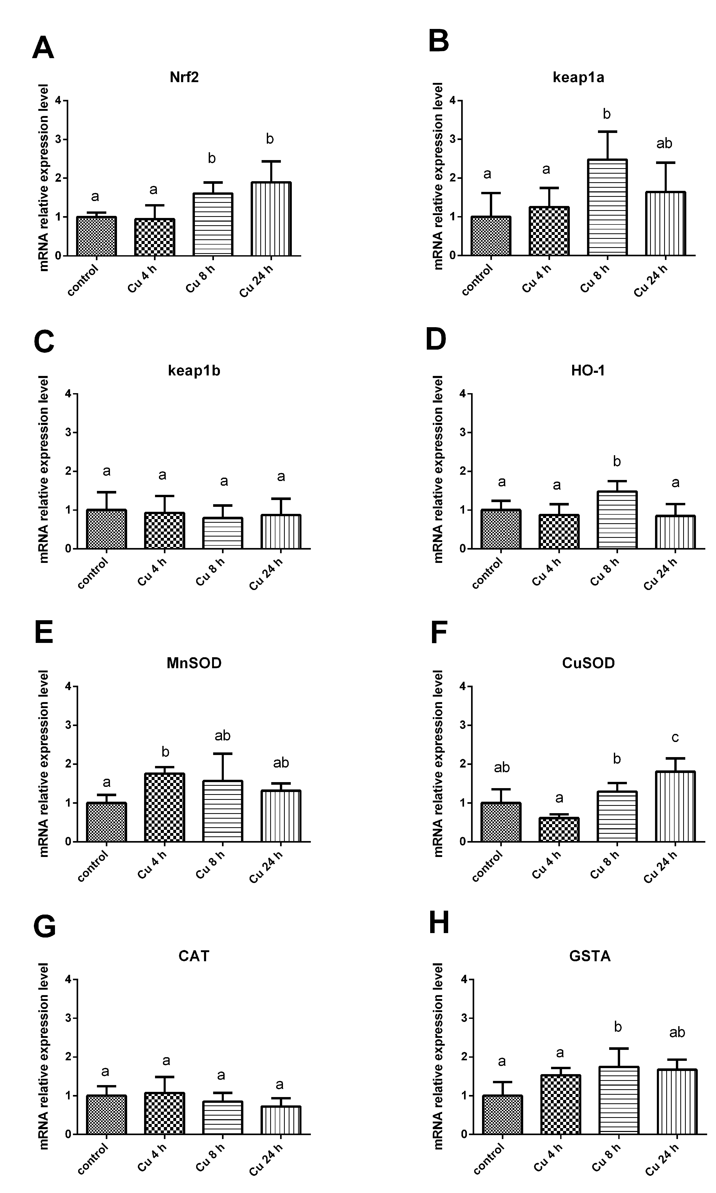
The expression of Nrf2 increased significantly (p < 0.05) after copper ion treatment for 8 h and maintained a high expression after 24 h, while copper ion stress in the first 4 h had little effect on Nrf2 expression (Figure 3A). The expression level of Keap1a after copper ion treatment for 8 h was significantly higher than that of the control group (p < 0.05), and the expression level of Keap1a decreased to the same level as that of the control group after 24 h (Figure 3B). Keap1b showed no significant difference at any time point after copper ion treatment (Figure 3C). The expression of HO-1 increased significantly (p < 0.05) after copper ion treatment for 8 h, and decreased to the same level as the control group after 24 h (Figure 3D). The expression of MnSOD significantly increased after copper ion treatment for 4 h (p < 0.05), and there was no significant difference at other time points (Figure 3E). The expression level of CuZnSOD significantly increased after 8 h (p < 0.05), and increased to a higher level at 24 h (Figure 3F). There was no significant difference in CAT gene expression among all groups (Figure 3G). The expression of glutathione thiotransferase (GSTA) increased significantly (p < 0.05) after copper ion treatment for 8 h, which had no significant difference at any other time points (Figure 3H).

4. Discussion

4.1. Copper Inhibited the Activity of Tilapia Hepatocytes and Caused Oxidative Stress

In this experiment, liver cell vitality was reduced to 2.96% with 1 mM copper ions treatment, resulting in a large number of cell deaths, which was not conducive to conduct subsequent experiments. 10 μM or 1 μM copper ions had no significant effect on hepatocyte viability. Therefore, the above concentrations of copper ions were not used in the following experiments. 100 μM copper can be used to significantly reduce cell viability to 66.40% without causing a large number of cell deaths, which provides suitable conditions for subsequent biochemical indicators and qPCR detection. Therefore, 100 μM is selected as the exposure concentration for subsequent experiments. Previous studies indicated that the half-inhibitory concentration (IC50) of copper on liver cell lines of grass carp after 48 h was 243.09 μM. When treated with 100 μM copper ions, the liver cell activity of grass carp was reduced to about 78% [16]. In a study of Epinephelus coioides, the liver cell viability was reduced to 40% with cultured medium containing 37.5 μM copper ions [17]. In this experiment, with 100 μM copper ions significantly reduced the liver cell viability of tilapia was significantly reduced to 66.40%. The resistance of tilapia hepatocytes to copper ions is similar to that of grass carp. The above results indicated that the hepatocyte tolerance to copper ions of freshwater fish (grass carp and tilapia) was stronger than that of marine fish (grouper). In addition, the authors found that the higher the density of cell culture was, the better the tolerance of the cells to copper ions would be. Therefore, cell density should be strictly controlled when conducting copper or other heavy metal exposure experiments, so as to ensure the accuracy and repeatability of the experiments.
The LDH activity of culture medium reflects cell integrity [18]. In this experiment, LDH of the cell culture medium treated with copper for 8 h and 24 h was significantly higher than that of the control group (p < 0.05). An increase in LDH activity was also observed in the supernatant of liver cells of grouper under the stress of copper ions [17], which was similar to our results. These results indicate that copper stress damages the cell membrane structure of fish, leading to cell rupture and LDH overflow. MDA is a stable metabolite of lipid peroxidation, which is considered as a reliable parameter reflecting the rise of ROS [18], and the rise of MDA is associated with oxidative damage. In this experiment, MDA increased with the extension of copper treatment time, which indicated that copper ions caused oxidative stress on tilapia hepatocytes. Heavy metals in water often cause oxidative stress in fish, and some researchers have also found that copper could cause the increase in ROS and the production of free radicals, meanwhile activating antioxidant defense systems [19]. Many antioxidant enzymes play an important role in protecting organisms from the toxic effects of ROS. SOD and CAT are antioxidant enzymes that respond most rapidly to oxidative stress, which are also important indicators of antioxidant capacity or oxidative damage [20]. In this study, copper ions also caused the decrease in CAT and SOD in different degrees, suggesting that copper inhibited the antioxidant capacity of liver cells while causing oxidative stress. This result is consistent with that of previous in vivo experiments [21]. This toxicological phenomenon of copper also occurs in other fish, such as Monopterus albus [20] and Mugil cephalus [22]. This indicates that with the oxidative stress caused by copper ions exposure, SOD and CAT can be consumed in the original antioxidant system of cells, ultimately leading to the decline of the liver antioxidant capacity.

4.2. Activate Nrf2 Signaling Pathway to Resist Oxidative Stress Caused by Copper Ions

In this study, the gene expression of Nrf2 signaling pathway in tilapia hepatocytes under copper ion stress was detected, and the results showed that Nrf2 expression increased significantly after copper stress for 8–24 h (Figure 3A). The results suggested that exposed copper ions could be used to improve the de novo synthesis of Nrf2 in liver cells. Some scholars believed that the activation of existing Nrf2 would help resist acute or early oxidative stress, and increase in de novo synthesis of Nrf2 could provide a lasting long-term effect, thus prolongating the continuous activation of related downstream genes [23,24]. Therefore, it can be inferred that during the first 4 h of copper ions treatment, the antioxidative stress mode of tilapia hepatocytes mainly depended on the existing Nrf2 separated from Keap1, which entered the nucleus to promote downstream gene transcription. After 8 h to 24 h, a large amount of Nrf2 in hepatocytes began to be synthesized and promote the transcription of downstream antioxidant enzymes. The promoting effect of copper ions on Nrf2 expression in fish has also been found in other fish species, such as Trachinotus ovatus [25], Megalobrama amblycephala [26], Cyprinus carpio [27], and Gobiocypris rarus [28].
In this study, the expressions of HO-1, CuZnSOD, MnSOD and, GSTA increased after copper ion treatment (Figure 3D–H), suggesting that copper ions could enhance the transcription of downstream genes by promoting Nrf2. In contrast to gene expression, SOD activity decreased after copper stress (Figure 2D), which might be due to the fact that a good deal of SOD was consumed in resistance to copper stress, whose synthetic rate of SOD was lower than the rate of depletion. In conclusion, the Nrf2 signaling pathway showed a tendency of high expression under the challenge of copper ions, and this high expression was a passive response of liver cells to copper stress. This response mechanism enabled liver cells to resist the stress of low concentrations of copper ions. When treated with 100 μM copper ions, cell vitality and antioxidant capacity were significantly decreased.
In this study, the expression of Keap1a also increased with the extension of copper treatment time. As a negative regulator of Nrf2, the increased expression of Keap1 seemed to be in contradiction with the upregulation of other antioxidant genes, which was due to the presence of ARE in the promoter of Keap1. Hence, Keap1 itself is also a downstream gene of Nrf2. There is an auto-regulatory feedback loop between Nrf2 and Keap1, which mediates their balance between them [29]. This phenomenon of Keap1 following Nrf2 expression also occurs in T. ovatus [25], Anguilla anguilla [30], grass carp [31] and so on. On the other hand, the expression level of Keap1b did not change with the challenge of copper ions in this study, and it was found in another study on carp that copper was more likely to affect the expression of Keap1a than that of Keap1b [27]. The reasons why copper has different effect on the heterodimer of Keap1 need further research.

5. Conclusions

The present study illustrated the mechanisms of copper ions toxicity to tilapia hepatocytes. In summary, exposure to copper ions increased cellular ROS, which could induce oxidative stress and lipid peroxidation. The biological membrane structure and anti-oxidant defense system were impaired, and the viability of tilapia hepatocytes was reduced. Copper ions promoted the synthesis of Nrf2. The activation of the Nrf2 signaling pathway enhanced the expression of downstream genes. The results suggest that exogenous ROS can induce the response of antioxidant system by regulating Nrf2 signaling pathway. Nrf2 could be adopted as a molecular target of hepatoprotective medicine in a future study.

Author Contributions

L.L.: conceptualization, methodology, validation, writing—original draft. R.W.: methodology. Z.Z.: original idea, writing—review and editing, Supervision. All authors contributed to preparing the final manuscript. All authors have read and agreed to the published version of the manuscript.

Funding

This study was supported by a Special Grant for a Leading Professor of Fujian Province (660160010). Funding data: 2016.

Institutional Review Board Statement

All of the study design and animal experiments were conducted in accordance with the guidelines of Fujian Agriculture and Forestry University’s Animal Care and Use Committee (Approval Code: PZCASFAFU23060, Approval Date: 9 February 2023).

Informed Consent Statement

Not applicable.

Data Availability Statement

The data that support the findings of this study are available on request from the corresponding author. The data are not publicly available due to privacy or ethical constraints.

Conflicts of Interest

The authors declare no conflict of interest.

References

  1. Gao, M.Q.; Cui, H.; Fang, W.H.; Hu, H.J.; Miao, L.; Jin, S.; Xie, J.S.; Ma, R.R. The effects of CuSO4 on Cryptocaryon irritans tomonts and its potential mechanism. Aquaculture 2022, 560, 738578. [Google Scholar] [CrossRef]
  2. Yang, Z.G.; Lian, W.; Waiho, K.; Zhu, L.L.; Chen, A.Q.; Cheng, Y.X.; Wang, Y.J. Effects of copper exposure on lipid metabolism and SREBP pathway in the Chinese mitten crab Eriocheir sinensis. Chemosphere 2022, 308, 136556. [Google Scholar] [CrossRef] [PubMed]
  3. Monier, M.N.; El-Naby, A.S.A.; Samir, F.; Abdel-Tawwab, M. Positive effects of dietary nanosized sodium butyrate on growth performance, immune, antioxidant indices, and resistance of Nile tilapia to waterborne copper toxicity. Aquac. Rep. 2022, 26, 101323. [Google Scholar] [CrossRef]
  4. Chen, S.J.; Liu, Y.T.; Xie, S.W.; Guo, Y.C.; Yang, H.R.; Wei, Y.R.; Xu, Q.; Ye, T.; Meng, B.S.; Huang, R.B.; et al. Role of myo-inositol supplementation against toxicity of excessive dietary copper in Pacific white shrimp Litopenaeus vannamei. Ecotoxicol. Environ. Saf. 2022, 241, 113712. [Google Scholar] [CrossRef]
  5. Liu, X.J.; Luo, Z.; Xiong, B.X.; Liu, X.; Zhao, Y.H.; Hu, G.F.; Lv, G.J. Effect of waterborne copper exposure on growth, hepatic enzymatic activities and histology in Synechogobius hasta. Ecotoxicol. Environ. Saf. 2010, 73, 1286–1291. [Google Scholar] [CrossRef]
  6. Zhang, L.; Yang, Z.; Yang, M.; Yang, F.; Wang, G.; Liu, D.; Li, X.; Yang, L.; Wang, Z. Copper-induced oxidative stress, transcriptome changes, intestinal microbiota, and histopathology of common carp (Cyprinus carpio). Ecotoxicol. Environ. Saf. 2022, 246, 114136. [Google Scholar] [CrossRef]
  7. Osburn, W.O.; Kensler, T.W. Nrf2 signaling: An adaptive response pathway for protection against environmental toxic insults. Mutat. Res. Rev. Mutat. Res. 2008, 659, 31–39. [Google Scholar] [CrossRef]
  8. Kobayashi, M.; Itoh, K.; Suzuki, T.; Osanai, H.; Nishikawa, K.; Katoh, Y.; Takagi, Y.; Yamamoto, M. Identification of the interactive interface and phylogenic conservation of the Nrf2-Keap1 system. Genes Cells 2002, 7, 807–820. [Google Scholar] [CrossRef]
  9. Kobayashi, M.; Yamamoto, M. Molecular mechanisms activating the Nrf2-Keap1 pathway of antioxidant gene regulation. Antioxid. Redox Signal. 2005, 7, 385–394. [Google Scholar] [CrossRef]
  10. Bao, S.A.; Nie, X.P.; Ou, R.K.; Wang, C.; Ku, P.J.; Li, K.B. Effects of diclofenac on the expression of Nrf2 and its downstream target genes in mosquito fish (Gambusia affinis). Aquat. Toxicol. 2017, 188, 43–53. [Google Scholar] [CrossRef]
  11. Wu, P.; Zhang, L.; Jiang, W.D.; Liu, Y.; Jiang, J.; Kuang, S.Y.; Li, S.W.; Tang, L.; Tang, W.E.; Zhou, X.Q.; et al. Dietary Vitamin A Improved the Flesh Quality of Grass Carp (Ctenopharyngodon idella) in Relation to the Enhanced Antioxidant Capacity through Nrf2/Keap 1a Signaling Pathway. Antioxidants 2022, 11, 148. [Google Scholar] [CrossRef] [PubMed]
  12. Yuan, Y.; Yuan, Y.M.; Dai, Y.Y.; Gong, Y.C. Economic profitability of tilapia farming in China. Aquac. Int. 2017, 25, 1253–1264. [Google Scholar] [CrossRef]
  13. Li, L.m.; Zhang, Z.p.; Huang, Y.f. Integrative transcriptome analysis and discovery of signaling pathways involved in the protective effects of curcumin against oxidative stress in tilapia hepatocytes. Aquat. Toxicol. 2020, 224, 105516. [Google Scholar] [CrossRef] [PubMed]
  14. Morozesk, M.; Franqui, L.S.; Mansano, A.S.; Martinez, D.S.T.; Fernandes, M.N. Interactions of oxidized multiwalled carbon nanotube with cadmium on zebrafish cell line: The influence of two co-exposure protocols on in vitro toxicity tests. Aquat. Toxicol. 2018, 200, 136–147. [Google Scholar] [CrossRef]
  15. Zhang, X.; Li, Y.T.; Sun, Y.L.; Guo, M.X.; Feng, J.J.; Wang, Y.L.; Zhang, Z.P. Regulatory effect of heat shock transcription factor-1 gene on heat shock proteins and its transcriptional regulation analysis in small abalone Haliotis diversicolor. BMC Mol. Cell Biol. 2020, 21, 83. [Google Scholar] [CrossRef] [PubMed]
  16. Zhu, Q.L.; Luo, Z.; Zhuo, M.Q.; Tan, X.Y.; Sun, L.D.; Zheng, J.L.; Chen, Q.L. In vitro exposure to copper influences lipid metabolism in hepatocytes from grass carp (Ctenopharyngodon idellus). Fish Physiol. Biochem. 2014, 40, 595–605. [Google Scholar] [CrossRef]
  17. Wang, T.; Chen, X.; Long, X.; Liu, Z.; Yan, S. Copper Nanoparticles and Copper Sulphate Induced Cytotoxicity in Hepatocyte Primary Cultures of Epinephelus coioides. PLoS ONE 2016, 11, e0149484. [Google Scholar] [CrossRef]
  18. Yin, G.J.; Cao, L.P.; Xu, P.; Jeney, G.; Nakao, M.; Lu, C.P. Hepatoprotective and antioxidant effects of Glycyrrhiza glabra extract against carbon tetrachloride (CCl4)-induced hepatocyte damage in common carp (Cyprinus carpio). Fish Physiol. Biochem. 2011, 37, 209–216. [Google Scholar] [CrossRef]
  19. Shaw, B.J.; Al-Bairuty, G.; Handy, R.D. Effects of waterborne copper nanoparticles and copper sulphate on rainbow trout, (Oncorhynchus mykiss): Physiology and accumulation. Aquat. Toxicol. 2012, 116–117, 90–101. [Google Scholar] [CrossRef]
  20. Liu, L.; Zhou, Q.B.; Lin, C.G.; He, L.; Wei, L.L. Histological alterations, oxidative stress, and inflammatory response in the liver of swamp eel (Monopterus albus) acutely exposed to copper. Fish Physiol. Biochem. 2021, 47, 1865–1878. [Google Scholar] [CrossRef]
  21. Wang, J.; Xiao, J.; Zhang, J.; Chen, H.; Li, D.; Li, L.; Cao, J.; Xie, L.; Luo, Y. Effects of dietary Cu and Zn on the accumulation, oxidative stress and the expressions of immune-related genes in the livers of Nile tilapia (Oreochromis niloticus). Fish Shellfish. Immunol. 2020, 100, 198–207. [Google Scholar] [CrossRef] [PubMed]
  22. Akbary, P.; Sartipi Yarahmadi, S.; Jahanbakhshi, A. Hematological, hepatic enzymes’ activity and oxidative stress responses of gray mullet (Mugil cephalus) after sub-acute exposure to copper oxide. Environ. Sci. Pollut. Res. Int. 2018, 25, 1800–1808. [Google Scholar] [CrossRef] [PubMed]
  23. Baird, L.; Dinkova Kostova, A.T. The cytoprotective role of the Keap1-Nrf2 pathway. Arch. Toxicol. 2011, 85, 241–272. [Google Scholar] [CrossRef] [PubMed]
  24. Kwak, M.K.; Itoh, K.; Yamamoto, M.; Kensler, T.W. Enhanced expression of the transcription factor Nrf2 by cancer chemopreventive agents: Role of antioxidant response element-like sequences in the nrf2 promoter. Mol. Cell. Biol. 2002, 22, 2883–2892. [Google Scholar] [CrossRef] [PubMed]
  25. Xie, J.; He, X.; Fang, H.; Liao, S.; Liu, Y.; Tian, L.; Niu, J. Identification of heme oxygenase-1 from golden pompano (Trachinotus ovatus) and response of Nrf2/HO-1 signaling pathway to copper-induced oxidative stress. Chemosphere 2020, 253, 126654. [Google Scholar] [CrossRef]
  26. Liang, H.L.; Ji, K.; Ge, X.P.; Mi, H.F.; Xi, B.W.; Ren, M.C. Effects of dietary copper on growth, antioxidant capacity and immune responses of juvenile blunt snout bream (Megalobrama amblycephala) as evidenced by pathological examination. Aquac. Rep. 2020, 17, 100296. [Google Scholar] [CrossRef]
  27. Jiang, W.D.; Liu, Y.; Hu, K.; Jiang, J.; Li, S.H.; Feng, L.; Zhou, X.Q. Copper exposure induces oxidative injury, disturbs the antioxidant system and changes the Nrf2/ARE (CuZnSOD) signaling in the fish brain: Protective effects of myo-inositol. Aquat. Toxicol. 2014, 155, 301–313. [Google Scholar] [CrossRef]
  28. Wu, L.; Yu, Q.G.; Zhang, G.; Wu, F.L.; Zhang, Y.Y.; Yuan, C.; Zhang, T.; Wang, Z.Z. Single and combined exposures of waterborne Cu and Cd induced oxidative stress responses and tissue injury in female rare minnow (Gobiocypris rarus). Comp. Biochem. Physiol. C-Toxicol. Pharmacol. 2019, 222, 90–99. [Google Scholar] [CrossRef]
  29. Lee, O.H.; Jain, A.K.; Papusha, V.; Jaiswal, A.K. An auto-regulatory loop between stress sensors INrf2 and Nrf2 controls their cellular abundance. J. Biol. Chem. 2007, 282, 36412–36420. [Google Scholar] [CrossRef]
  30. Giuliani, M.E.; Regoli, F. Identification of the Nrf2-Keap1 pathway in the European eel Anguilla anguilla: Role for a transcriptional regulation of antioxidant genes in aquatic organisms. Aquat. Toxicol. 2014, 150, 117–123. [Google Scholar] [CrossRef]
  31. Wang, B.; Feng, L.; Jiang, W.D.; Wu, P.; Kuang, S.Y.; Jiang, J.; Tang, L.; Tang, W.N.; Zhang, Y.A.; Liu, Y.; et al. Copper-induced tight junction mRNA expression changes, apoptosis and antioxidant responses via NF-κB, TOR and Nrf2 signaling molecules in the gills of fish: Preventive role of arginine. Aquat. Toxicol. 2015, 158, 125–137. [Google Scholar] [CrossRef] [PubMed]
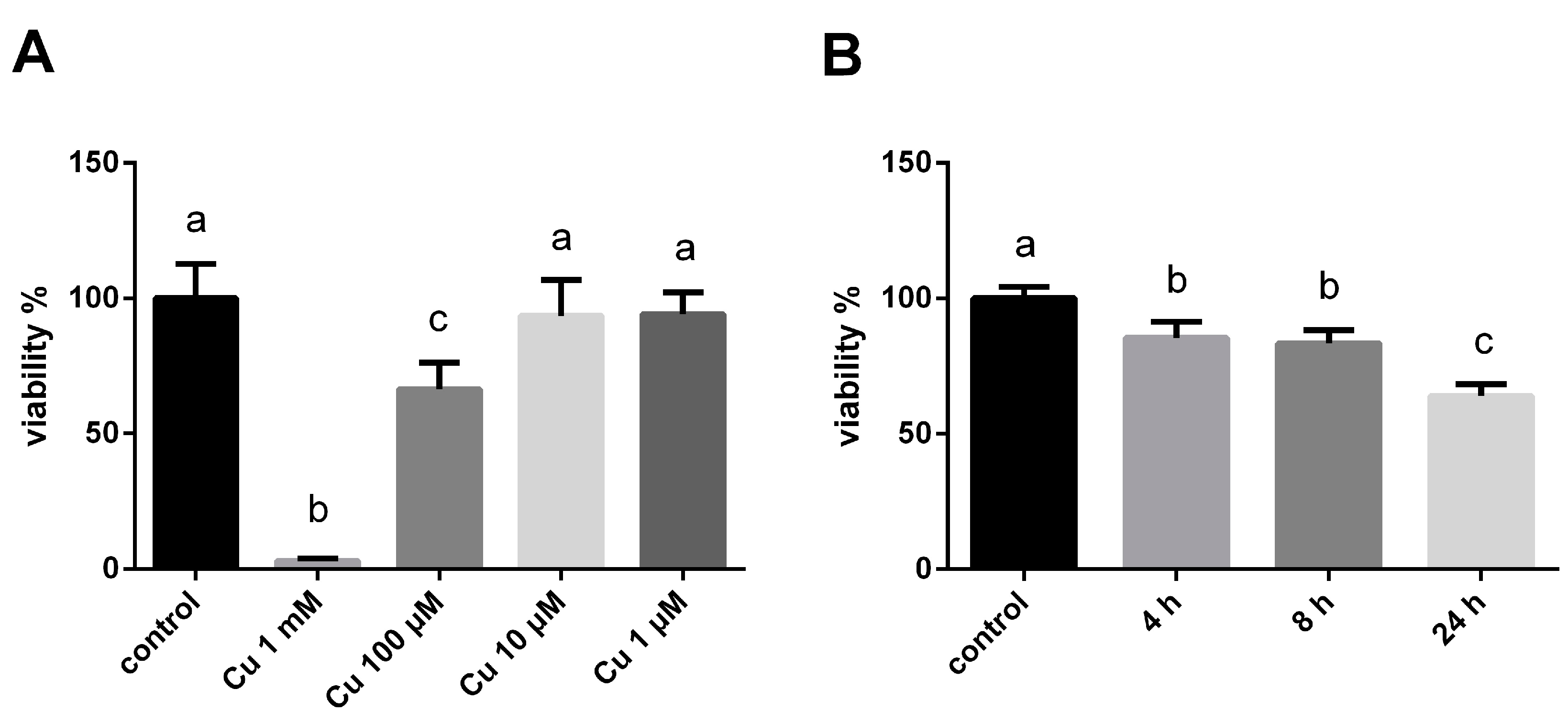
Figure 1. The effect of copper on the viability of tilapia hepatocytes (MTT assay). Data are shown as the means ± SDs. (A) Effects of different concentrations of copper ions on hepatocyte viability, (B) Effects of different time of 100 μM copper ions on hepatocyte viability. Significant differences (p < 0.05) among treatments were indicated by different letters.
Fishes 08 00280 g001
Figure 2. The effect of copper on biochemical indexes of tilapia hepatocytes. Data are shown as the means ± SDs. (A) LDH activity, (B) MDA, (C) CAT activity, (D) SOD activity. Significant differences (p < 0.05) among treatments were indicated by different letters.
Fishes 08 00280 g002
Figure 3. The effect of copper on the expression of genes in the Nrf2 signaling pathway of tilapia hepatocytes. Significant differences (p < 0.05) among treatments are indicated by different letters. (A): Nrf2, (B): keap1a, (C): keap1b, (D): HO-1, (E): MnSOD, (F): CuZnSOD, (G): CAT, (H): GSTA.
Fishes 08 00280 g003
Table 1. The primers used for qPCR.
GeneAcc. NO. (GenBank)PrimersPrimer Sequence (5′–3′)
Keap1aXM_003442916.5Keap1a-fCCCTCAACATCCCCAGGAAC
Keap1a-rGGATCCCACTTTTCCACCGT
Keap1bXM_003447926.4Keap1b-fCGGTGGAGCGCTATGATGTA
Keap1b-rGCCATCGTAACCTCCCAACA
HO-1XM_013272963.3HO-1-fGACAGGAACTCGGACCATCC
HO-1-rAGTGGCTGCAGGAATGACAA
MnSODXM_003449940.5MnSOD-fTCACAGCAAGCACCATGCTA
MnSOD-rATGTGGCCGCCTCCATTAAA
CuZnSODXM_003446807.5CuZnSOD-fGGAGACAACACAAACGGGTG
CuZnSOD-rTCTGCTGCAGTCACATTCCC
CATXM_003447521.5CAT-fGGACCATTATTCGAGCCATCAG
CAT-rAAACTGCAAGTGCTGCTGAAA
β-ACTINXM_003443127.5β-ACTIN-fTGCGGGATATCATTTGCCTGA
β-ACTIN-rGAATCCGGCCTTGCACATAC
Nrf2XM_003447296.3Nrf2-fCAGCCCAGAACTGCCGTAAA
Nrf2-rGCCAAAGACCTCCAGGTACA
GSTAXM_019350593.2GSTA-fGACAGACCCAGCATCAAAGC
GSTA-rCTCATCAGCTGACAAGCCAATC
Disclaimer/Publisher’s Note: The statements, opinions and data contained in all publications are solely those of the individual author(s) and contributor(s) and not of MDPI and/or the editor(s). MDPI and/or the editor(s) disclaim responsibility for any injury to people or property resulting from any ideas, methods, instructions or products referred to in the content.