Article

Glucocorticoid Receptor Mediates Cortisol Regulation of Glycogen Metabolism in Gills of the Euryhaline Tilapia (Oreochromis mossambicus)

1
Department of Life Sciences, National Chung Hsing University, Taichung City 402202, Taiwan
2
The Integrative Evolutionary Galliform Genomics (iEGG) and Animal Biotechnology Center, National Chung Hsing University, Taichung City 402202, Taiwan
3
Department of Biological Sciences and Technology, National University of Tainan, Tainan City 700301, Taiwan
*
Author to whom correspondence should be addressed.
Fishes 2023, 8(5), 267; https://doi.org/10.3390/fishes8050267
Submission received: 20 April 2023 / Revised: 15 May 2023 / Accepted: 16 May 2023 / Published: 18 May 2023
(This article belongs to the Section Physiology and Biochemistry)

Abstract

:
In this study, we investigated the effects of cortisol on the regulation of the glycogen metabolism biomarkers glycogen synthase (GS) and glycogen phosphorylase (GP) in the glycogen-rich cells of the gills of tilapia (Oreochromis mossambicus). In the gills of tilapia, GP, GS, and glycogen were immunocytochemically colocalized in a specific group of glycogen-rich cells adjacent to the gills’ main ionocytes and mitochondria-rich cells. Cortisol plays a vital role in the regulation of physiological functions in animals, including energy metabolism, respiration, immune response, and ion regulation. However, no studies have elucidated the mechanisms regulating cortisol and glycogen-rich cells in the gills. Therefore, we treated tilapia larvae with exogenous cortisol and a glucocorticoid receptor (GR) antagonist to investigate the regulatory mechanisms between cortisol and glycogen-rich cells in the gills. Our results showed that cortisol promoted the expression of gill glycogen phosphorylase isoform (GPGG) mRNA via GR, whereas the GS gene expression remained unaffected. We also found that GR mRNA was colocalized with some glycogen-rich cells in the gills, further confirming our hypothesis that cortisol directly acts on glycogen-rich cells in the gills of tilapia and regulates glycogen metabolism by promoting GPGG mRNA expression.
Key Contribution: Cortisol promoted the expression of gill glycogen phosphorylase isoform (GPGG) mRNA in tilapia via the glucocorticoid receptor, whereas the glycogen synthase (GS) gene expression remained unaffected.

1. Introduction

Glucose is the primary energy source for metabolism and is stored in the form of glycogen, a long-branching high-molecular-mass polysaccharide, in animal tissues [1]. Animals increase their blood glucose levels and regulate energy metabolism in response to stress. Glycogen metabolism is a source of energy for both vertebrates and invertebrates, particularly during periods of environmental fluctuation and stress responses [2,3,4,5]. The synthesis and degradation of liver glycogen are initiated by glycogen synthase (GS) and phosphorylase (GP), respectively. Glucose is mainly released via the GP-induced degradation of glycogen [1].
Corticosteroids are a group of hormones synthesized from cholesterol via a series of biochemical reactions. They are classified into glucocorticoids (GC) and mineralocorticoids (MC). Mammals mainly secrete corticosteroids from the adrenal cortex, while teleost fish secrete them from interrenal tissue [6]. Corticosteroids play vital roles in many physiological functions, including ion regulation, fluid balance, energy metabolism, respiration, and the immune response [7,8]. However, fish lack aldosterone synthase, which is required to produce a specific mineralocorticoid; therefore, cortisol is considered the major corticosteroid in fish [9,10]. In mammals, the effects of cortisol are mediated by two corticosteroid receptors: the glucocorticoid receptor (GR) and the mineralocorticoid receptor (MR) [11,12]. The type of physiological regulation that occurs depends on which receptor the cortisol binds to, which is determined by the complex formed (CS–GR or CS–MR) between the receptor and ligand (corticosteroids). This complex initiates transcription in the target cells and regulates various physiological processes [13], including ion regulation, fluid balance, energy metabolism, respiration, and the immune response.
Gills play a vital role in regulating ions in fish and serve as the primary organ for ionoregulation. During the early developmental stages, before the gills are fully developed, the skin serves as the main organ for ionoregulation [14,15]. Various studies have explored the significance of cortisol, along with the GR and/or MR, in fish osmoregulation. Cortisol, a crucial hormone in euryhaline fish, aids acclimation to seawater and freshwater ion regulation. Exogenous cortisol was found to regulate the activity of ion transporters, such as the Na+-K+-ATPase (NKA) and Na+-K+-2Cl cotransporter (NKCC), which are responsible for ion secretion by ionocytes. In addition, the expression of the genes encoding these ion transporters is regulated by cortisol [16,17,18].
Since gills are directly exposed to the external aquatic environment, they function in gas exchange, acid–base balance, and ion/osmotic regulation [18], and they directly resist drastic environmental changes, such as heavy metal pollution [5]. A series of studies have shown that these processes are highly correlated with cortisol and that acclimation to seawater environments can cause rapid increases in plasma cortisol levels [19,20,21,22,23,24,25]. Additionally, research has also shown that tilapia exposure to cadmium, a heavy metal, can increase the cortisol levels in the bloodstream [5]. These processes are energy-consuming [5,18,26]. Tseng et al. reported that a group of cells rich in glycogen deposits, called glycogen-rich cells, are surrounded by ionocytes [27,28]. These cells express a specific form of GP (gill glycogen phosphorylase isoform, GPGG). In this study, both the GP activity and protein expression in tilapia gills rapidly increased after transfer from freshwater to seawater. Simultaneously, the glycogen levels in the gills and liver were significantly depleted after transfer to seawater. These results suggest that changes in salinity promote the rapid supply of energy by the ionocytes adjacent to glycogen-rich cells, triggering osmotic regulation [27,28]. These results indicate that glycogenesis and glycogenolysis in the gills are involved in energy metabolism during acclimation to salinity changes [27,28], with cortisol potentially playing an important role. However, the detailed mechanisms remain unclear. No studies have elucidated the regulatory mechanisms of cortisol- and glycogen-rich cells in the gills during acclimation to salinity changes.
The Mozambique tilapia (Oreochromis mossambicus) is a euryhaline teleost species known for its strong salinity adaptation ability [29,30,31]. It can adapt to a wide range of salinities, from freshwater to seawater, and can even tolerate extremely low-ion water and high-salinity environments twice that of seawater. Owing to the well-defined types of ionocytes in freshwater and seawater, Mozambique tilapia is considered a suitable model for studying ionocyte regulation [32]. Additionally, this fish species is a maternal mouthbrooder, meaning the female carries fertilized eggs in its mouth until hatching, enabling researchers to obtain embryos for research purposes throughout the year. Previous studies have provided ample evidence regarding the cortisol levels in the plasma and glycogen-rich cell-related research in response to environmental stressors in tilapia, making it a suitable experimental species for this study [5,27,28].
In this study, we chose the euryhaline Mozambique tilapia as an animal model to investigate whether cortisol regulates GPGG or GS gene expression. We further designed experiments to determine whether cortisol affects gill glycogen metabolism and function through the GR, using the GR antagonist RU-486. Therefore, we treated tilapia larvae with exogenous cortisol to understand how cortisol acts on glycogen-rich cells through the GR by inhibiting GR function using antagonists.

2. Materials and Methods

2.1. Experimental Animals

Adult tilapia (Oreochromis mossambicus) weighing 30–60 g and measuring 9–15 cm in length were obtained from stocks maintained at the National University of Tainan, Taiwan. The fish were housed in a freshwater (local tap water; [Ca2+], 0.20 mM; [Mg2+], 0.16 mM; [Na+], 0.5 mM; [K+], 0.3 mM; [Cl], 0.45 mM) circulating system at 28 ± 0.5 °C, with a 14:10 h light:dark photoperiod. Tilapia larvae were obtained as follows: fertilized eggs were collected from the mouths of female tilapia during mouthbrooding and incubated in aerated freshwater. The fertilized eggs were used immediately upon hatching, with no feeding occurring during the incubation period. The adult fish and larvae were anesthetized using buffered 0.03% MS-222 (tricaine; Sigma-Aldrich, St. Louis, MO, USA) and dissected for subsequent experiments. The Institutional Animal Care and Use Group (Approval No. IACUG1050005) of the National University of Tainan reviewed and approved the animal use protocol. The 3R (reduce, replace, and refine) policy for animal handling followed the guidelines of the National Science and Technology Council of Taiwan.

2.2. Cortisol and Receptor Antagonist Treatment of Tilapia Larvae

The cortisol dosage was determined using previously established methods [33,34,35,36,37,38]. A cortisol (hydrocortisone, Sigma-Aldrich, Burlington, MA, USA) stock solution was prepared in dimethyl sulfoxide (DMSO) and then diluted to a final working solution (20 mg/L) in aerated tap water. Tilapia larvae were treated with cortisol media immediately after hatching. Newly hatched tilapia larvae were randomly divided into the control and drug treatment groups for the drug treatment experiments. The timing of the drug treatment was based on previous studies of glycogen metabolism in Mozambique tilapia [27,28]. After the experiments, three tilapia larvae were collected as a sample and five or six replicates (N = 5 or 6) were performed. Each experiment was performed with three replicates. The incubation media were refreshed daily to ensure consistent cortisol levels were maintained. The GR antagonists dosage were determined according to previous studies [38,39,40]. Tilapia larvae were grown in 10 µg/mL RU486 (GR antagonist, Sigma-Aldrich, Burlington, MA, USA) with 20 mg/L cortisol and then incubated in an incubator at 28 °C, and the medium was changed every day. In this study, higher dosages of cortisol and antagonists were employed as compared to some previous studies. Nonetheless, no significant mortality or aberrant behavior was noted. The used dosages of cortisol and antagonists had been proven to work in cultured gills and fish larvae in previous studies [33,35,36,37,38,38,39,40]. After the experiments, the tilapia larvae were anesthetized with buffered 0.03% MS-222 (Tricaine, Sigma-Aldrich, Burlington, MA, USA) for further analysis.

2.3. Total RNA Extraction

To extract the total RNA, the samples were homogenized in 1 ml of TRIzol reagent (Thermo Fisher Scientific, Waltham, MA, USA) and mixed with 0.2 ml of chloroform. The mixture was shaken thoroughly and centrifuged at 12,000× g at 4 °C for 45 min. An equal volume of isopropanol was added to the sample, and the mixture was then centrifuged at 12,000× g (4 °C, 30 min) to precipitate total RNA pellets. The pellets were washed twice with 70% alcohol via centrifugation at 12,000× g (4 °C, 30 min). The quantity and quality of the total RNA were assessed by the absorbance at 260 and 280 nm and by gel electrophoresis, and the samples were then stored at −20 °C until use.

2.4. Real-Time PCR

Next, we synthesized cDNA from the tilapia samples using the GoScript™ Reverse Transcription System (Promega, Madison, WI, USA) and poly(dT) primers. A StepOne Plus Real-Time PCR System (Applied Biosystems, Thermo Fisher Scientific, Waltham, MA, USA) was used to perform the real-time PCR in a final reaction volume of 20 μL, containing 10 μL of Fast SYBR™ Green Master Mix (Applied Biosystems, Thermo Fisher Scientific), 300 nM of the forward and reverse primers, 20–30 ng of cDNA, and nuclease-free water. The following primer sequences were used: GPGG forward (5′-CGAGCCCAGGGAAGCCATCGAA-3′) and reverse (5′-TGAAGGCTTAAACCAAACAGGAA-3′), a 105 bp fragment (accession no. DQ010415.1), GS forward (5′-TGGGATACTGCTCAGACTGTGA-3′) and reverse (5′-TGTCCTCCAGCATGTTGTGAGT-3′), a 187 bp fragment (accession no. EF565371.1), and β-actin forward (5′-CGGAATCCACGAAACCACCTA-3′) and reverse (5′-ATCTCCTGCATCCTGTCA-3′), a 135 bp fragment. The primers were obtained from previous studies [27,28]. β-actin was used as an internal control to normalize the mRNA expression, as described previously [28].

2.5. RNA Probe Synthesis

The RNA probe synthesis for in situ hybridization was performed as per previous studies [28,38,41]. Fragments of tilapia GR obtained via PCR were inserted into a pGEM-T easy vector (Promega). The primer set for GR included the forward (5′-TGATGGCAGGCATGAATCT-3′) and reverse (5′- =GACAAACTCGCTGCAAATC-3′) sequences, a 518 bp fragment (accession no. AB771724.1). For the in situ hybridization, RNA probes were synthesized using the DIG RNA Labeling Mix via in vitro transcription with T7 and M13 RNA polymerases (Promega). The labeled RNA probes were examined using RNA gels and dot-blot assays to confirm their quality and concentration.

2.6. In Situ Hybridization

The in situ hybridization was performed as per previous studies [41,42]. Briefly, the excised gills were fixed overnight with 4% paraformaldehyde in PBS. The fixed gills were washed with PBS and cryoprotected in 30% sucrose before being embedded in OCT compound embedding medium (Sakura, Tokyo, Japan) at −20 °C. Cross-sections of 10 μm were cut from the frozen tissue blocks using a CM 3050S rapid sectioning cryostat (Leica, Heidelberg, Germany) and mounted onto hydrophilic adhesion slides (PLATINUM PRO, MATSUNAMI, Osaka, Japan). Before the in situ hybridization, the slide-mounted gill sections were air-dried and rehydrated using a series of methanol and PBST (PBS with 0.1% Tween-20) mixtures.
The hybridization mix (HM) contained 50% formamide, 5 × saline sodium citrate (SSC), 9.2 mM citric acid, and 0.1% Tween-20. The slides were washed with PBST several times and then incubated in the pre-hybridization mix (HM+; HM with 500 ng/mL yeast tRNA and 50 μg/mL heparin) for two hours before being hybridized with the RNA probes (200 μL HM+ containing 30~50 ng DIG-labeled/fluorescein-labeled RNA probe) at 70 °C overnight.
Next, the hybridized slides were washed at 70 °C for 10 min in 100% HM (without tRNA and heparin), 10 min in 75% HM and 25% 2 × saline sodium citrate (SSC), 10 min in 50% HM and 50% 2 × SSC, 10 min in 25% HM and 75% 2 × SSC, 10 min in 2 × SSC, and finally, 30 min in 0.2 × SSC (this final step was repeated twice). The hybridized slides were then washed at room temperature for 10 min in 75% 0.2 × SSC and 25% PBST, 10 min in 50% 0.2 × SSC and 50% PBST, 10 min in 25% 0.2 × SSC and 75% PBST, and 10 min in PBST.
The washed slides were incubated for two hours in blocking buffer containing 5% sheep serum and 2 mg/mL bovine serum albumin (BSA) in PBST and then transferred into alkaline phosphatase-conjugated anti-DIG antibody (1:5000 dilution; Roche, Basel, Switzerland) and incubated overnight at 4 °C. Finally, the sections were washed with PBST six times for 15 min each and then transferred into alkaline Tris buffer containing 0.1 M Tris HCl (pH 9.5, 0.05 M MgCl2, 0.1 M NaCl, and 0.1% Tween 20). The tissues were stained with a mixture of 225 μg/mL NBT (Nitro Blue Tetrazolium) and 175 μg/mL BCIP (5-Bromo 4-Chloro 3-indolyl Phosphate) in alkaline Tris buffer. The labeling reaction was stopped with stop solution (containing 1 mM EDTA and 0.1% Tween 20 in 1 × PBS, pH 5.5) once the signal was strong enough for analysis.

2.7. Immunohistochemistry (IHC) of Gill Sections

After the in situ hybridization, the sections were sequentially washed at room temperature for 10 min each in 25% PBST, 75% methanol, 50% PBST, 50% methanol, 75% PBST, 25% methanol, and PBST. The sections were then incubated with 3% BSA in PBST for 2 h at room temperature. The samples were subsequently treated with anti-human glycogen synthase polyclonal antibody (diluted 1:200; Rockland Immunochemicals, Pottstown, PA, USA) and incubated overnight at 4 °C. Subsequently, the samples were washed six times with PBST for 10 min each and incubated with Alexa Fluor 488 goat anti-rabbit IgG antibodies (Thermo Fisher Scientific, diluted 1:400 with PBS) for 2 h at room temperature. The stained sections were mounted using Fluoromount-G mounting medium (Southern Biotech, Birmingham, AL, USA) and visualized using a Zeiss LSM 780 confocal microscope.

2.8. Statistical Analysis

Data were presented as the mean ± SD and were analyzed via a one-way analysis of variance (ANOVA) with Tukey’s comparisons (p < 0.05) and Student’s t test.

3. Results

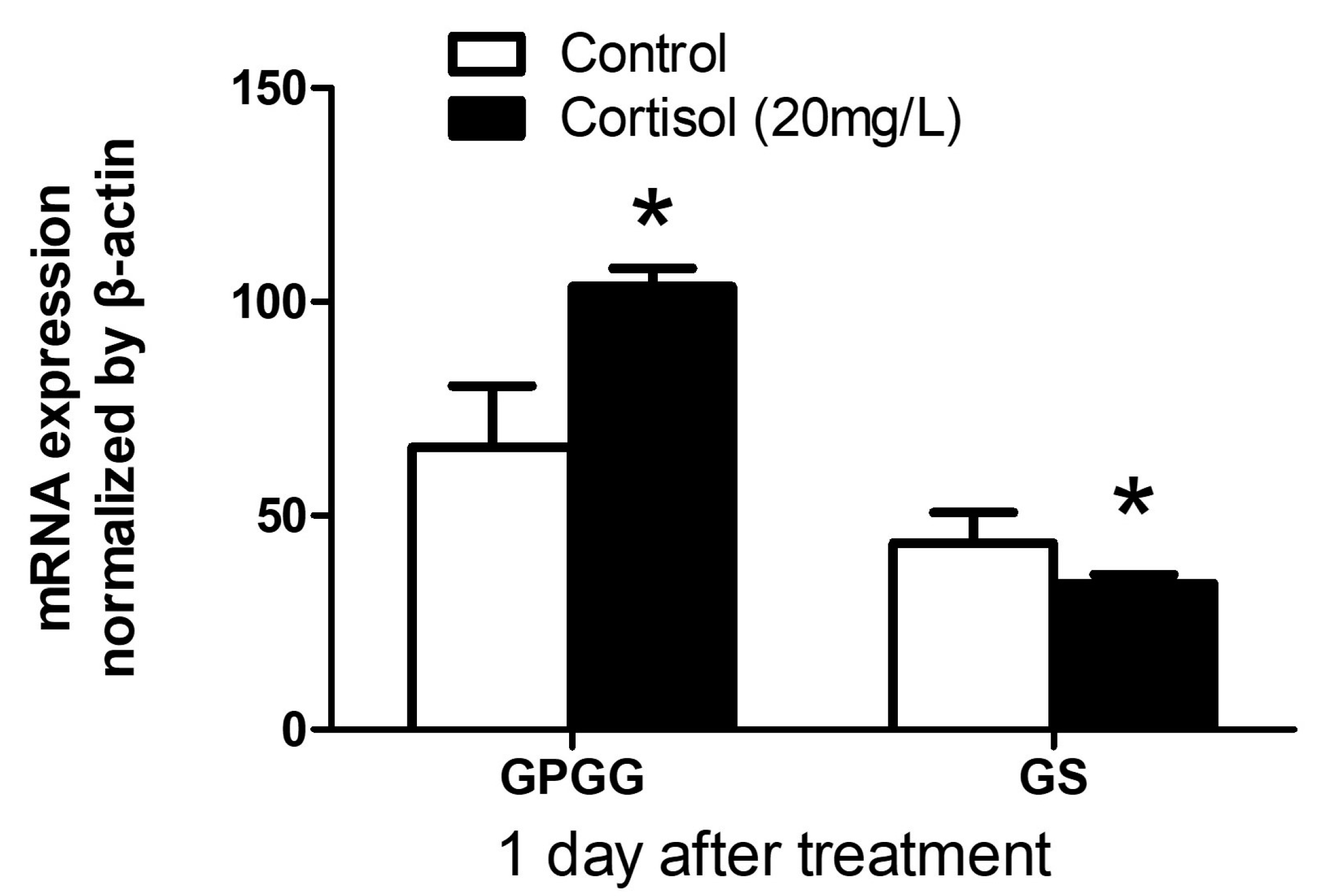
3.1. GPGG and GS mRNA Expression after Exogenous Cortisol Exposure

Tilapia larvae were treated with 20 mg/L cortisol to determine whether cortisol regulates glycogen metabolism [38]. Real-time PCR was performed to measure the GPGG and GS mRNA levels after cortisol treatment. Primers for GPGG and GS were designed based on previous studies [27,28]. The results showed that the expression of GPGG mRNA in the tilapia larvae was significantly upregulated after one day of exposure to cortisol (t = 6.171, df = 10, p < 0.0001), whereas the expression of GS was significantly suppressed (t = 3.161, df = 10, p = 0.0051) (Figure 1). This indicates that cortisol may be involved in glycogen metabolism in tilapia gills by promoting GPGG gene expression.

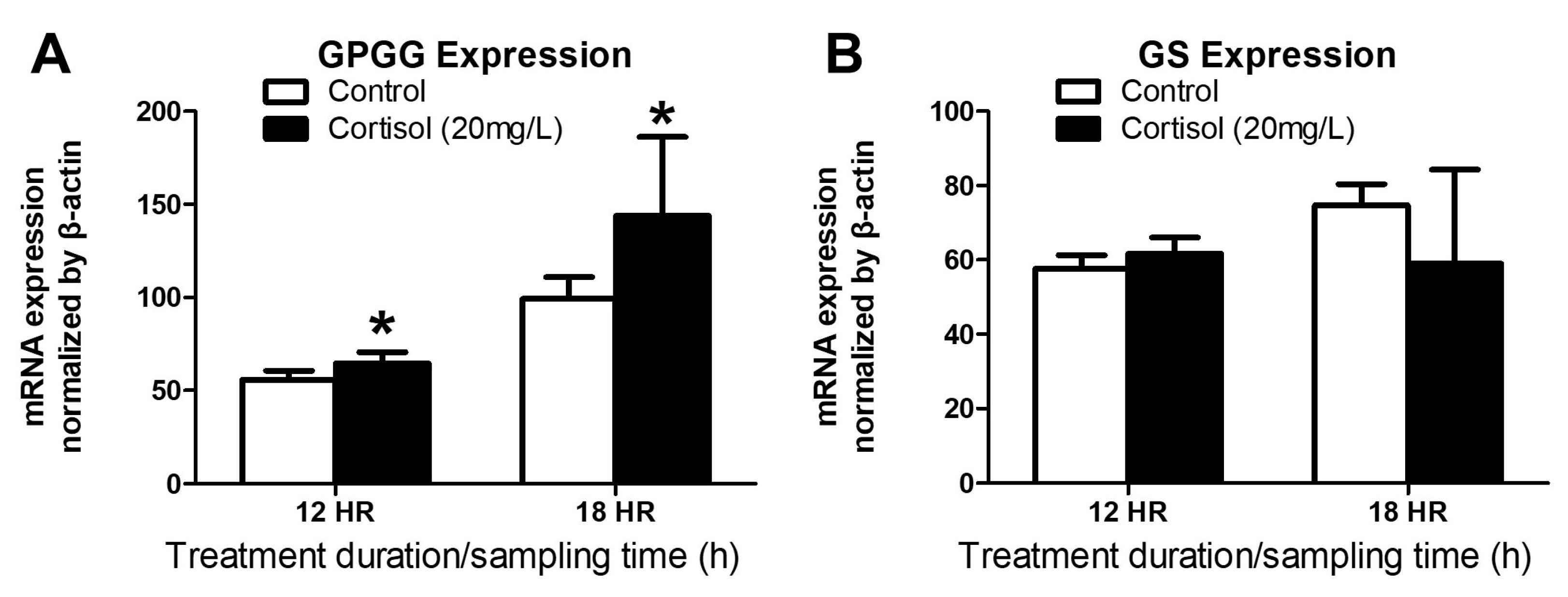
3.2. GPGG and GS mRNA Expression after Exogenous Cortisol Exposure from 12 to 18 h

A previous study has suggested that cortisol affects glycogen metabolism via GPGG and GS in tilapia larvae. However, according to Chang et al. [27], GP activity rapidly increased within 24 h in tilapia when faced with environmental stress from salinity. Therefore, we investigated whether cortisol alters the gene expression of GPGG and GS within a shorter timeframe. We collected tilapia larvae treated with 20 mg/L cortisol for 12 and 18 h as experimental samples and conducted real-time PCR to measure the mRNA levels of GPGG and GS after the cortisol treatment. The results showed that the mRNA expression levels of GPGG were significantly upregulated (12HR Group: t = 2.844, df = 10, p = 0.0087; 18HR Group: t = 2.487, df = 10, p = 0.0161) (Figure 2A), whereas there was no significant difference in the expression levels of GS (12HR Group: t = 1.093, df = 10, p = 0.15; 18HR Group: t = 1.48, df = 10, p = 0.0848) (Figure 2B). This result demonstrates that cortisol promotes GPGG gene expression from 12 h post-treatment but does not affect that of GS.

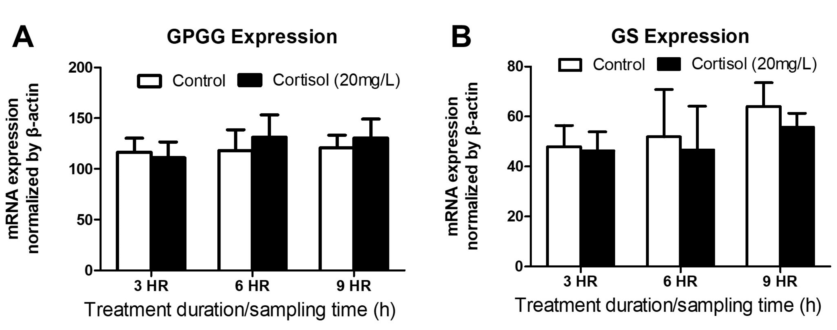
3.3. GPGG and GS mRNA Expression after Exogenous Cortisol Exposure from 3 to 9 h

The above results indicate that the gene expression of GPGG in the tilapia significantly increased starting from 12 h after treatment with exogenous cortisol, and this increase persisted until 24 h. However, according to previous studies, the plasma cortisol levels in tilapia rapidly increased within a very short time in response to environmental stressors, such as salinity or cadmium exposure [5,24,25,43]. Therefore, we investigated whether cortisol is involved in GPGG and GS gene expression within 12 h. To test this hypothesis, we collected tilapia larvae treated with 20 mg/L cortisol for 3 to 9 h as experimental samples and performed real-time PCR to measure the mRNA levels of GPGG and GS after the cortisol treatment. The results showed that there was no significant difference in the expression levels of GPGG (3HR Group: t = 0.6172, df = 10, p = 0.2755; 6HR Group: t = 0.7614, df = 10, p = 0.2320; 9HR Group: t = 1.035, df = 10, p = 0.1626) (Figure 3A) and GS (3HR Group: t =0.3483, df = 10, p = 0.3674; 6HR Group: t = 0.4969, df = 10, p = 0.3150; 9HR Group: t = 0.7236, df = 10, p = 0.2429) (Figure 3B) mRNA in the tilapia larvae exposed to cortisol for 3, 6, or 9 h.

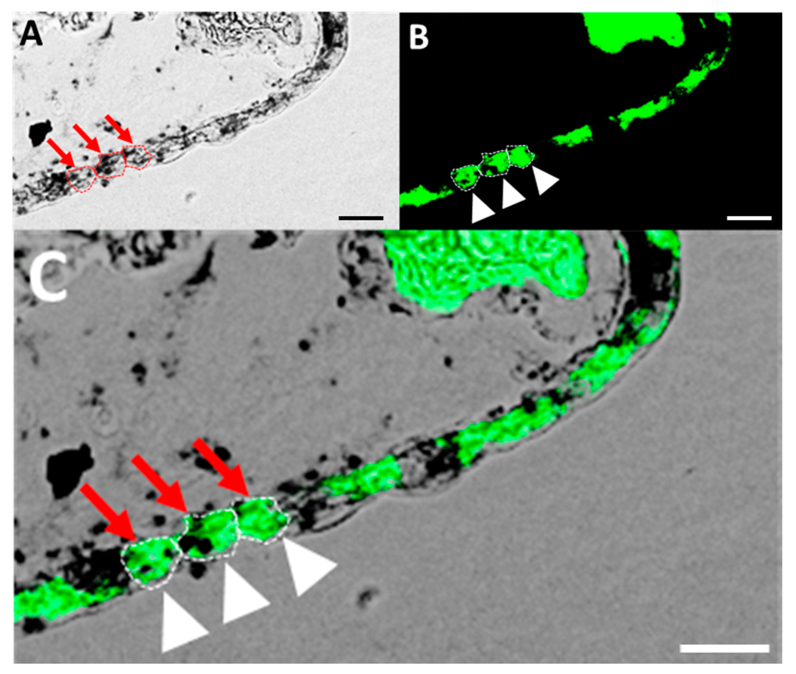
3.4. Localization of GR mRNA and Glycogen-Rich Cells in Adult Tilapia Gill Sections

Our results indicate that cortisol affects glycogen metabolism by promoting gene expression. However, there is no evidence to suggest that cortisol directly participates in glycogen metabolism in the gills of tilapia, and no studies have proposed a relationship between the GR and glycogen-rich cells. We speculated that cortisol may act directly on glycogen-rich cells. To test this hypothesis, we examined the expression of the GR and GS proteins in cryosections of adult tilapia gills. Double staining was performed to label the GR mRNA (black) using in situ hybridization (Figure 4A) and the GS protein (glycogen-rich cell marker, green) using immunostaining (Figure 4B). We found that the GR mRNA colocalized with glycogen-rich cells (Figure 4C). This result suggests that GR is expressed in glycogen-rich cells and that cortisol acts directly on these cells to regulate glycogen metabolism.

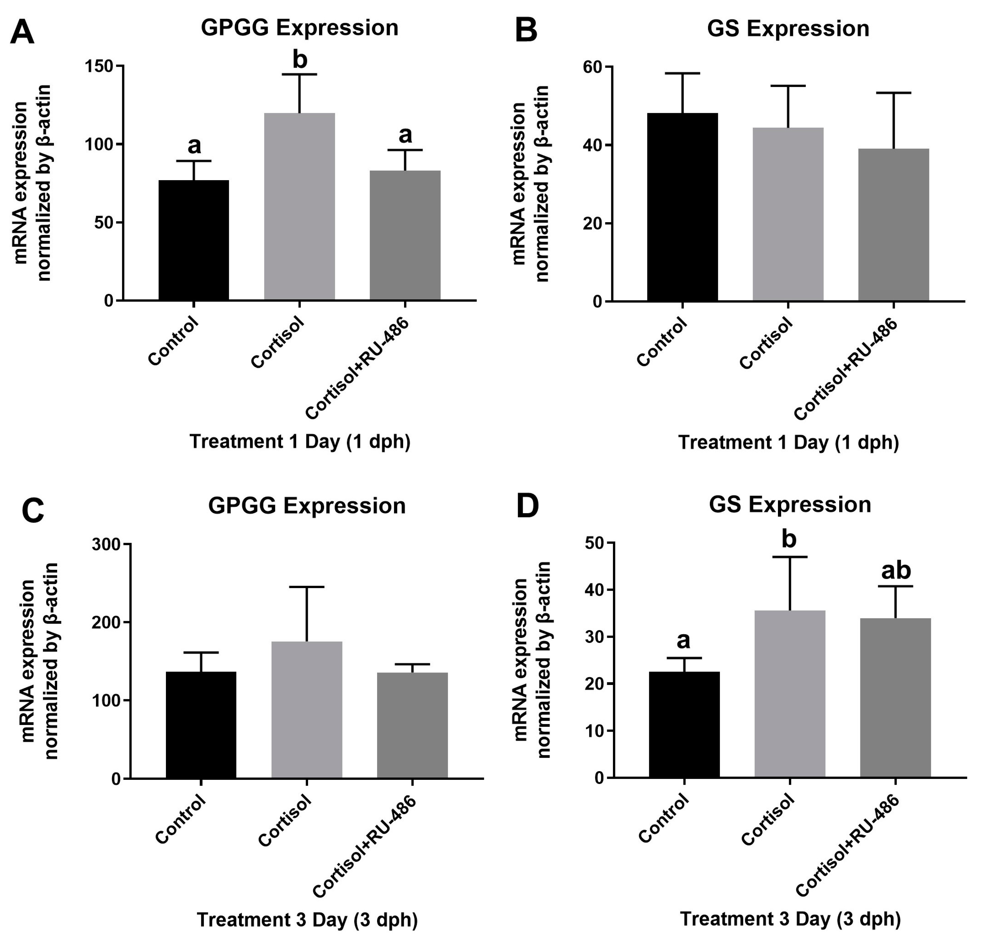
3.5. GPGG mRNA Expression after GR Antagonist Exposure

Based on information from the previous experiment, we hypothesized that cortisol may activate glycogen metabolism via GR signaling by promoting GPGG gene expression. To support this hypothesis, we conducted real-time PCR to measure the GPGG and GS mRNA levels after treatment with cortisol and a GR antagonist. Initially, we exposed 20 mg/L cortisol-treated tilapia larvae to RU486. After exposure for one or three days, the tilapia larvae were subjected to real-time PCR for the mRNA expression measurements. The results demonstrated that the GPGG mRNA expression levels were sharply upregulated in the tilapia larvae after cortisol exposure, and this upregulation was abolished after co-treatment with GR antagonists after one day of exposure (Figure 5A). This further confirms our hypothesis that cortisol acts directly on glycogen-rich cells in the gills of tilapia and regulates glycogen metabolism by promoting GPGG mRNA expression.

4. Discussion

We found that treatment with exogenous cortisol significantly increased the GPGG mRNA expression in larval tilapia. We used RNA probes specific to the tilapia GR gene to demonstrate that GR was present in glycogen-rich gill cells. We also provided pharmacological evidence that cortisol mediates glycogen metabolism by promoting GPGG mRNA expression through the GR. The GPGG expression increased significantly after 12 h of exogenous cortisol treatment and continued until 24 h of treatment. These results suggest that cortisol acts directly on glycogen-rich cells and regulates hepatic glucose metabolism by promoting GPGG gene expression. In contrast, cortisol did not appear to directly affect GS gene expression.
Glycogen metabolism is the primary pathway for energy acquisition in both vertebrates and invertebrates, particularly under environmental stress [3,4,44,45]. Teleosts require energy to cope with physiological regulation and environmental acclimation during stress responses. Under cadmium exposure, tilapia (Oreochromis mossambicus) used glycogen to produce energy to combat adversity in the gills [5]. When exposed to low-temperature environments, seawater-acclimated milkfish (Chanos chanos) used gill glycogen and lactate to meet the energy demands of aerobic ionocyte metabolism [46]. In a salinity challenge, glycogen utilization was observed in the gills of tilapia (Oreochromis mossambicus) and blue-spotted mudskippers (Boleophthalmus pectinirostris) to meet the energy demand for osmoregulation [27,28,47]. Glycogen is primarily stored in the skeletal muscles and liver of vertebrates as the primary carbohydrate reserve, providing tissues with an available source of glucose. Research has shown that this mechanism is similar to that in the gills. Glycogen-rich cells, a group of cells with glycogen deposits adjacent to the ionocytes, express a specific gill glycogen phosphorylase isoform (GPGG) [27,28]. The GP activity and protein expression in the gills of tilapia increased rapidly after transfer from freshwater to seawater, whereas the glycogen levels in the gills and liver decreased significantly after transfer to seawater. These results suggest that changes in salinity promote the rapid energy supply from the ionocytes adjacent to glycogen-rich cells, triggering an osmoregulatory mechanism [27,28].
Cortisol, the primary corticosteroid hormone, exerts its effects through the GR and/or MR in fish. Numerous studies have demonstrated that teleosts exhibit a rapid elevation in plasma cortisol levels in response to environmental stressors [5,19,20,21,22,23,24,25,43,48]. Prior investigations have revealed that cortisol is involved in the adaptive responses of freshwater and seawater fish, with the ionocytes in the skin and gills serving as crucial mediators of osmoregulation and ion regulation [17,18,43,49,50,51,52]. In previous studies conducted by Lin et al., cortisol was administered to tilapia larvae to examine the influx of Ca2+ and the expression of relevant ion transporters, such as the epithelial Ca2+ channel (ECaC), during embryonic development. These findings demonstrate that cortisol can elevate the Ca2+ levels in tilapia larvae by upregulating the expression of ECaC proteins [38]. Some studies have indicated that changes in the proportion and morphology of ionocytes during environmental acclimation in teleost fish are associated with cortisol-induced cell differentiation and proliferation [33,53,54,55,56,57,58,59,60,61,62,63,64,65]. However, these mechanisms require significant energy. To date, only a few studies have investigated the mechanisms of cortisol and glycogen in the liver and muscles of teleosts [25,48], and there is no direct evidence to suggest that the glycogen-rich cells in fish gills have a similar mechanism.
Herein, we treated tilapia larvae with exogenous cortisol and measured the gene expression of GPGG and GS using real-time PCR. The gene expression of GPGG increased significantly from 12 h post-treatment (Figure 2A) and continued until 24 h post-treatment (Figure 1). The cortisol treatment appeared to have inconsistent effects on GS gene expression, with little to no significant effects, which may be due to GPGG being a specific form expressed in the gills [28], while GS is widely distributed in fish tissues [27]. It is also possible that the GS gene expression in tilapia is not regulated by a single signaling pathway, and there may be other or multiple mechanisms regulating the expression of GS. To further confirm the experimental results mentioned above, we conducted in situ hybridization to determine the GR expression in the gill tissues of tilapia. We found that the GR mRNA colocalized with glycogen-rich cells (Figure 4C). We also provide pharmacological evidence to further validate our hypotheses. We exposed the cortisol-treated tilapia larvae to RU-486 (a GR antagonist) and discovered that RU-486 significantly blocked the cortisol-induced expression of GPGG mRNA, and this upregulation effect was abolished after co-treatment with RU-486 (a GR antagonist) after one day (Figure 5A) of exposure. These results suggest that cortisol regulates glycogen metabolism in the glycogen-rich cells in tilapia gills via the GR signaling pathway.
Ionocytes, which enclose the embryonic skin, are responsible for maintaining the osmoregulation of internal fluid homeostasis [66]. As development progresses, ionocytes emerge in functional organs, such as the gills of adult fish, and eventually become the primary regulators of osmotic pressure and ion regulation [14,51,52,66,67,68]. Therefore, the physiological mechanisms of the skin of tilapia larvae and the gills of adult tilapia are expected to be similar. In the present study, at least 72 h of cortisol treatment was required to significantly increase the number of ionocytes in the tilapia larvae. This finding reflects the fact that approximately 12 h of cortisol treatment is required to significantly increase the gene expression of GPGG. Although the plasma cortisol levels in tilapia increase rapidly under salinity challenge [25,43], it is possible that the increase in enzyme activity promotes glycogen degradation shortly before stimulating gene expression. After three days of cortisol treatment, the expression of the GS gene in the tilapia significantly increased, which may be a feedback effect due to liver glycogen depletion. Similar results have been reported in studies of muscle glycogen metabolism in rainbow trout (Oncorhynchus mykiss) [48], although the underlying mechanisms remain unclear.
The physiological response of fish to environmental stressors is a complex process, and energy regulation plays a critical role. In this study, we confirmed that cortisol, as a stress hormone, regulates glycogen metabolism in the glycogen-rich cells of fish gills via the GR. Our findings provide initial evidence of cortisol regulation of GPGG gene expression. However, cortisol may also participate in regulation through other aspects, such as protein expression, enzyme activity, and transcription or translation regulation of related genes. In order to gain a deeper understanding of this issue, it is necessary to explore it from multiple perspectives and employ more meticulous experimental designs. Transcriptomics has become a mature tool for physiological research, and it could be considered in the future to further explore the mechanism by which cortisol regulates gill energy utilization in fish.

5. Conclusions

Extensive research has shown that environmental stressors increase the plasma cortisol concentration in teleosts, and they also promote GP activity and gene expression to provide energy for glycogenolysis. However, there is no evidence that cortisol participates in glycogen metabolism in the glycogen-rich cells in fish gills. Here, we provide compelling molecular evidence that cortisol participates in the metabolism of glycogen-rich cells in the gills of tilapia. Exogenous cortisol treatment promotes GPGG gene expression. Co-treatment with exogenous RU-486 confirmed that cortisol participates in glycogenolysis in glycogen-rich cells via the GR. In slice experiments, we found that GR colocalized with glycogen-rich cells, supporting our previous experimental finding that cortisol regulates glycogen metabolism via the GR. In summary, our study provides the first evidence that cortisol participates in glycogen metabolism in the glycogen-rich cells of tilapia gills.

Author Contributions

Conceptualization, C.-Y.W. and D.-Y.T.; methodology, C.-Y.W., D.-Y.T. and T.-H.L.; software, C.-Y.W.; validation, C.-Y.W., D.-Y.T. and T.-H.L.; formal analysis, C.-Y.W.; resources, D.-Y.T. and T.-H.L.; writing—original draft preparation, C.-Y.W.; writing—review and editing, D.-Y.T.; supervision and funding acquisition, D.-Y.T. All authors have read and agreed to the published version of the manuscript.

Funding

This study was funded by the grants sponsored by the Ministry of Science and Technology MOST 107-2313-B-024-001 to D.-Y.T.

Institutional Review Board Statement

All the protocols and procedures involving tilapia were approved by the Institutional Animal Care and Use Group (Approval No. IACUG1050005, issue date 12 December 2016) at the National University of Tainan. The 3R (reduce, replace, and refine) policy for animal handling was followed, in accordance with the guidelines of the National Science and Technology Council, Taiwan.

Data Availability Statement

The data presented in this study are available on request from the corresponding author.

Acknowledgments

We thank Y.-H. Huang and T.-F. Kuo for the sample collection, Y.-C. Tseng for the antibody, and W.-F. Chen, H.-H. Huang, and F.-I. Lu for the technical assistance. We gratefully acknowledge the Instrument Development Center at National Cheng Kung University for allowing us to use the Zeiss LSM 780 confocal microscope.

Conflicts of Interest

The authors declare no conflict of interest. The funders had no role in the design of the study, in the collection, analysis, and interpretation of data, in the writing of the manuscript, or in the decision to publish the results.

References

  1. Roach, P.J.; Cheng, C.; Huang, D.; Lin, A.; Mu, J.; Skurat, A.V.; Wilson, W.; Zhai, L. Novel Aspects of the Regulation of Glycogen Storage. J. Basic Clin. Physiol. Pharmacol. 1998, 9, 139–152. [Google Scholar] [CrossRef] [PubMed]
  2. Bacca, H.; Huvet, A.; Fabioux, C.; Daniel, J.Y.; Delaporte, M.; Pouvreau, S.; Van Wormhoudt, A.; Moal, J. Molecular cloning and seasonal expression of oyster glycogen phosphorylase and glycogen synthase genes. Comp. Biochem. Physiol. Part B Biochem. Mol. Biol. 2005, 140, 635–646. [Google Scholar] [CrossRef] [PubMed]
  3. Karlsson, J. Some Features of Glycogen Metabolism in Human Skeletal Muscle. Bibl. Nutr. Dieta 1979, 27, 121–125. [Google Scholar]
  4. Oliveira, G.T.; Rossi, I.C.; Kucharski, L.C.; Da Silva, R.S.M. Hepatopancreas gluconeogenesis and glycogen content during fasting in crabs previously maintained on a high-protein or carbohydrate-rich diet. Comp. Biochem. Physiol. Part A Mol. Integr. Physiol. 2004, 137, 383–390. [Google Scholar] [CrossRef]
  5. Lin, Y.-S.; Tsai, S.-C.; Lin, H.-C.; Hsiao, C.-D.; Wu, S.M. Changes of glycogen metabolism in the gills and hepatic tissue of tilapia (Oreochromis mossambicus) during short-term Cd exposure. Comp. Biochem. Physiol. Part C Toxicol. Pharmacol. 2011, 154, 296–304. [Google Scholar] [CrossRef]
  6. Van Der Boon, J.; Van Den Thillart, G.E.E.J.M.; Addink, A.D.F. The effects of cortisol administration on intermediary metabolism in teleost fish. Comp. Biochem. Physiol. Part A Physiol. 1991, 100, 47–53. [Google Scholar] [CrossRef]
  7. Charmandari, E.; Tsigos, C.; Chrousos, G. Endocrinology of the Stress Response. Annu. Rev. Physiol. 2005, 67, 259–284. [Google Scholar] [CrossRef]
  8. McLaughlin, F.; Mackintosh, J.; Hayes, B.P.; McLaren, A.; Uings, I.J.; Salmon, P.; Humphreys, J.; Meldrum, E.; Farrow, S.N. Glucocorticoid-induced osteopenia in the mouse as assessed by histomorphometry, microcomputed tomography, and biochemical markers. Bone 2002, 30, 924–930. [Google Scholar] [CrossRef]
  9. Baker, M.E. Evolution of Glucocorticoid and Mineralocorticoid Responses: Go Fish. Endocrinology 2003, 144, 4223–4225. [Google Scholar] [CrossRef]
  10. Takahashi, H.; Sakamoto, T. The role of ‘mineralocorticoids’ in teleost fish: Relative importance of glucocorticoid signaling in the osmoregulation and ‘central’ actions of mineralocorticoid receptor. Gen. Comp. Endocrinol. 2013, 181, 223–228. [Google Scholar] [CrossRef]
  11. Carroll, S.M.; Ortlund, E.A.; Thornton, J.W. Mechanisms for the Evolution of a Derived Function in the Ancestral Glucocorticoid Receptor. PLoS Genet. 2011, 7, e1002117. [Google Scholar] [CrossRef]
  12. Bridgham, J.T.; Carroll, S.M.; Thornton, J.W. Evolution of Hormone-Receptor Complexity by Molecular Exploitation. Science 2006, 312, 97–101. [Google Scholar] [CrossRef] [PubMed]
  13. Gorissen, M.; Flik, G. Chapter 3—The Endocrinology of the Stress Response in Fish: An Adaptation-Physiological View. In Fish Physiology; Schreck, C.B., Tort, L., Farrell, A.P., Brauner, C.J., Eds.; Academic Press: Cambridge, MA, USA, 2016; Volume 35, pp. 75–111. [Google Scholar]
  14. Hwang, P.-P.; Lee, T.-H.; Lin, L.-Y. Ion regulation in fish gills: Recent progress in the cellular and molecular mechanisms. Am. J. Physiol. Regul. Integr. Comp. Physiol. 2011, 301, R28–R47. [Google Scholar] [CrossRef]
  15. Hwang, P.-P.; Tung, Y.-C.; Chang, M.-H. Effect of environmental calcium levels on calcium uptake in tilapia larvae Oreochromis mossambicus. Fish Physiol. Biochem. 1996, 15, 363–370. [Google Scholar] [CrossRef] [PubMed]
  16. McCormick, S.D.; Bradshaw, D. Hormonal control of salt and water balance in vertebrates. Gen. Comp. Endocrinol. 2006, 147, 3–8. [Google Scholar] [CrossRef] [PubMed]
  17. McCormick, S.D. Endocrine Control of Osmoregulation in Teleost Fish. Am. Zool. 2001, 41, 781–794. [Google Scholar] [CrossRef]
  18. Evans, D.H.; Piermarini, P.M.; Choe, K.P. The Multifunctional Fish Gill: Dominant Site of Gas Exchange, Osmoregulation, Acid-Base Regulation, and Excretion of Nitrogenous Waste. Physiol. Rev. 2005, 85, 97–177. [Google Scholar] [CrossRef]
  19. Redding, J.M.; Schreck, C.B. Influence of Ambient Salinity on Osmoregulation and Cortisol Concentration in Yearling Coho Salmon during Stress. Trans. Am. Fish. Soc. 1983, 112, 800–807. [Google Scholar] [CrossRef]
  20. Nichols, D.J.; Weisbart, M. Cortisol dynamics during seawater adaptation of Atlantic salmon Salmo salar. Am. J. Physiol. Regul. Integr. Comp. Physiol. 1985, 248, R651–R659. [Google Scholar] [CrossRef]
  21. Patiño, R.; Redding, J.M.; Schreck, C.B. Interrenal secretion of corticosteroids and plasma cortisol and cortisone concentrations after acute stress and during seawater acclimation in juvenile coho salmon (Oncorhynchus kisutch). Gen. Comp. Endocrinol. 1987, 68, 431–439. [Google Scholar] [CrossRef]
  22. Sheridan, M.A. Alterations in lipid metabolism accompanying smoltification and seawater adaptation of salmonid fish. Aquaculture 1989, 82, 191–203. [Google Scholar] [CrossRef]
  23. Franklin, C.E.; Forster, M.E.; Davison, W. Plasma cortisol and osmoregulatory changes in sockeye salmon transferred to sea water: Comparison between successful and unsuccessful adaptation. J. Fish Biol. 1992, 41, 113–122. [Google Scholar] [CrossRef]
  24. Dean, D.B.; Whitlow, Z.W.; Borski, R.J. Glucocorticoid receptor upregulation during seawater adaptation in a euryhaline teleost, the tilapia (Oreochromis mossambicus). Gen. Comp. Endocrinol. 2003, 132, 112–118. [Google Scholar] [CrossRef]
  25. Kammerer, B.D.; Cech, J.J.; Kültz, D. Rapid changes in plasma cortisol, osmolality, and respiration in response to salinity stress in tilapia (Oreochromis mossambicus). Comp. Biochem. Physiol. Part A Mol. Integr. Physiol. 2010, 157, 260–265. [Google Scholar] [CrossRef]
  26. Hirose, S.; Kaneko, T.; Naito, N.; Takei, Y. Molecular biology of major components of chloride cells. Comp. Biochem. Physiol. Part B Biochem. Mol. Biol. 2003, 136, 593–620. [Google Scholar] [CrossRef]
  27. Chang, J.C.-H.; Wu, S.-M.; Tseng, Y.-C.; Lee, Y.-C.; Baba, O.; Hwang, P.-P. Regulation of glycogen metabolism in gills and liver of the euryhaline tilapia (Oreochromis mossambicus) during acclimation to seawater. J. Exp. Biol. 2007, 210, 3494–3504. [Google Scholar] [CrossRef]
  28. Tseng, Y.-C.; Huang, C.-J.; Chang, J.C.-H.; Teng, W.-Y.; Baba, O.; Fann, M.-J.; Hwang, P.-P. Glycogen phosphorylase in glycogen-rich cells is involved in the energy supply for ion regulation in fish gill epithelia. Am. J. Physiol.-Regul. Integr. Comp. Physiol. 2007, 293, R482–R491. [Google Scholar] [CrossRef]
  29. Uchida, K.; Kaneko, T.; Miyazaki, H.; Hasegawa, S.; Hirano, T. Excellent Salinity Tolerance of Mozambique Tilapia: Elevated Chloride Cell Activity in the Branchial and Opercular Epithelia of the Fish Adapted to Concentrated Seawater. Zool. Sci. 2000, 17, 149–160. [Google Scholar] [CrossRef]
  30. Stickney, R.R. Tilapia Tolerance of Saline Waters: A Review. Progress. Fish-Cult. 1986, 48, 161–167. [Google Scholar] [CrossRef]
  31. Suresh, A.V.; Lin, C.K. Tilapia culture in saline waters: A review. Aquaculture 1992, 106, 201–226. [Google Scholar] [CrossRef]
  32. Hiroi, J.; Yasumasu, S.; McCormick, S.D.; Hwang, P.-P.; Kaneko, T. Evidence for an apical Na–Cl cotransporter involved in ion uptake in a teleost fish. J. Exp. Biol. 2008, 211, 2584–2599. [Google Scholar] [CrossRef] [PubMed]
  33. Cruz, S.A.; Chao, P.-L.; Hwang, P.-P. Cortisol promotes differentiation of epidermal ionocytes through Foxi3 transcription factors in zebrafish (Danio rerio). Comp. Biochem. Physiol. Part A Mol. Integr. Physiol. 2013, 164, 249–257. [Google Scholar] [CrossRef] [PubMed]
  34. Lin, C.-H.; Hu, H.-J.; Hwang, P.-P. Cortisol regulates sodium homeostasis by stimulating the transcription of sodium-chloride transporter (NCC) in zebrafish (Danio rerio). Mol. Cell. Endocrinol. 2016, 422, 93–102. [Google Scholar] [CrossRef]
  35. Lin, C.-H.; Shih, T.-H.; Liu, S.-T.; Hsu, H.-H.; Hwang, P.-P. Cortisol Regulates Acid Secretion of H+-ATPase-rich Ionocytes in Zebrafish (Danio rerio) Embryos. Front. Physiol. 2015, 6, 328. [Google Scholar] [CrossRef] [PubMed]
  36. Lin, C.-H.; Tsai, I.L.; Su, C.-H.; Tseng, D.-Y.; Hwang, P.-P. Reverse Effect of Mammalian Hypocalcemic Cortisol in Fish: Cortisol Stimulates Ca2+ Uptake via Glucocorticoid Receptor-Mediated Vitamin D3 Metabolism. PLoS ONE 2011, 6, e23689. [Google Scholar] [CrossRef]
  37. Lin, G.R.; Weng, C.F.; Wang, J.I.; Hwang, P.P. Effects of Cortisol on Ion Regulation in Developing Tilapia (Oreochromis mossambicus) Larvae on Seawater Adaptation. Physiol. Biochem. Zool. 1999, 72, 397–404. [Google Scholar] [CrossRef] [PubMed]
  38. Lin, C.-H.; Kuan, W.-C.; Liao, B.-K.; Deng, A.-N.; Tseng, D.-Y.; Hwang, P.-P. Environmental and cortisol-mediated control of Ca2+ uptake in tilapia (Oreochromis mossambicus). J. Comp. Physiol. B 2016, 186, 323–332. [Google Scholar] [CrossRef]
  39. Kiilerich, P.; Kristiansen, K.; Madsen, S.S. Cortisol regulation of ion transporter mRNA in Atlantic salmon gill and the effect of salinity on the signaling pathway. J. Endocrinol. 2007, 194, 417–427. [Google Scholar] [CrossRef]
  40. Kiilerich, P.; Tipsmark, C.K.; Borski, R.J.; Madsen, S.S. Differential effects of cortisol and 11-deoxycorticosterone on ion transport protein mRNA levels in gills of two euryhaline teleosts, Mozambique tilapia (Oreochromis mossambicus) and striped bass (Morone saxatilis). J. Endocrinol. 2011, 209, 115–126. [Google Scholar] [CrossRef]
  41. Hsiao, C.-D.; You, M.-S.; Guh, Y.-J.; Ma, M.; Jiang, Y.-J.; Hwang, P.-P. A Positive Regulatory Loop between foxi3a and foxi3b Is Essential for Specification and Differentiation of Zebrafish Epidermal Ionocytes. PLoS ONE 2007, 2, e302. [Google Scholar] [CrossRef]
  42. Han, T.-Y.; Wu, C.-Y.; Tsai, H.-C.; Cheng, Y.-P.; Chen, W.-F.; Lin, T.-C.; Wang, C.-Y.; Lee, J.-R.; Hwang, P.-P.; Lu, F.-I. Comparison of Calcium Balancing Strategies During Hypothermic Acclimation of Tilapia (Oreochromis mossambicus) and Goldfish (Carassius auratus). Front. Physiol. 2018, 9, 1224. [Google Scholar] [CrossRef] [PubMed]
  43. Hwang, P.-P.; Wu, S.-M. Role of Cortisol in Hypoosmoregulation in Larvae of the Tilapia (Oreochromis mossambicus). Gen. Comp. Endocrinol. 1993, 92, 318–324. [Google Scholar] [CrossRef]
  44. Li, M.; Wang, X.; Qi, C.; Li, E.; Du, Z.; Qin, J.G.; Chen, L. Metabolic response of Nile tilapia (Oreochromis niloticus) to acute and chronic hypoxia stress. Aquaculture 2018, 495, 187–195. [Google Scholar] [CrossRef]
  45. Li, S.; Sang, C.; Zhang, J.; Chen, N.; Li, Z.; Jin, P.; Huang, X. Effects of acute hyperglycemia stress on plasma glucose, glycogen content, and expressions of glycogen synthase and phosphorylase in hybrid grouper (Epinephelus fuscoguttatus ♀ × E. lanceolatus ♂). Fish Physiol. Biochem. 2018, 44, 1185–1196. [Google Scholar] [CrossRef] [PubMed]
  46. Chang, C.-H.; Liu, C.-J.; Lu, W.-J.; Wu, L.-Y.; Lai, K.-J.; Lin, Y.-T.; Lee, T.-H. Hypothermal Effects on Energy Supply for Ionocytes in Gills of Freshwater- and Seawater-Acclimated Milkfish, Chanos chanos. Front. Mar. Sci. 2022, 9, 880103. [Google Scholar] [CrossRef]
  47. Guo, T.; Yang, Y.; Meng, F.; Wang, S.; Xia, S.; Qian, Y.; Li, M.; Wang, R. Effects of low salinity on gill and liver glycogen metabolism of great blue-spotted mudskippers (Boleophthalmus pectinirostris). Comp. Biochem. Physiol. Part C Toxicol. Pharmacol. 2020, 230, 108709. [Google Scholar] [CrossRef] [PubMed]
  48. Milligan, C.L. A regulatory role for cortisol in muscle glycogen metabolism in rainbow trout Oncorhynchus mykiss Walbaum. J. Exp. Biol. 2003, 206, 3167–3173. [Google Scholar] [CrossRef]
  49. Hwang, P.-P.; Chou, M.-Y. Zebrafish as an animal model to study ion homeostasis. Pflügers Arch. Eur. J. Physiol. 2013, 465, 1233–1247. [Google Scholar] [CrossRef]
  50. Takei, Y.; Hwang, P.-P. Chapter 6—Homeostatic Responses to Osmotic Stress. In Fish Physiology; Schreck, C.B., Tort, L., Farrell, A.P., Brauner, C.J., Eds.; Academic Press: Cambridge, MA, USA, 2016; Volume 35, pp. 207–249. [Google Scholar]
  51. Guh, Y.-J.; Hwang, P.-P. Insights into molecular and cellular mechanisms of hormonal actions on fish ion regulation derived from the zebrafish model. Gen. Comp. Endocrinol. 2017, 251, 12–20. [Google Scholar] [CrossRef]
  52. Yan, J.-J.; Hwang, P.-P. Novel discoveries in acid-base regulation and osmoregulation: A review of selected hormonal actions in zebrafish and medaka. Gen. Comp. Endocrinol. 2019, 277, 20–29. [Google Scholar] [CrossRef]
  53. Perry, S.F.; Laurent, P. Adaptational Responses of Rainbow Trout to Lowered External NaCl Concentration: Contribution of the Branchial Chloride Cell. J. Exp. Biol. 1989, 147, 147–168. [Google Scholar] [CrossRef]
  54. McCormick, S.D. Cortisol directly stimulates differentiation of chloride cells in tilapia opercular membrane. Am. J. Physiol. Regul. Integr. Comp. Physiol. 1990, 259, R857–R863. [Google Scholar] [CrossRef]
  55. Uchida, K.; Kaneko, T.; Tagawa, M.; Hirano, T. Localization of Cortisol Receptor in Branchial Chloride Cells in Chum Salmon Fry. Gen. Comp. Endocrinol. 1998, 109, 175–185. [Google Scholar] [CrossRef] [PubMed]
  56. Hiroi, J.; Kaneko, T.; Tanaka, M. In vivo sequential changes in chloride cell morphology in the yolk-sac membrane of mozambique tilapia (Oreochromis mossambicus) embryos and larvae during seawater adaptation. J. Exp. Biol. 1999, 202, 3485–3495. [Google Scholar] [CrossRef] [PubMed]
  57. Wong, C.K.C.; Chan, D.K.O. Chloride cell subtypes in the gill epithelium of Japanese eel Anguilla japonica. Am. J. Physiol. Regul. Integr. Comp. Physiol. 1999, 277, R517–R522. [Google Scholar] [CrossRef] [PubMed]
  58. Sloman, K.A.; Desforges, P.R.; Gilmour, K.M. Evidence for a mineralocorticoid-like receptor linked to branchial chloride cell proliferation in freshwater rainbow trout. J. Exp. Biol. 2001, 204, 3953–3961. [Google Scholar] [CrossRef]
  59. Wong, C.K.; Chan, D.K. Effects of cortisol on chloride cells in the gill epithelium of Japanese eel, Anguilla japonica. J. Endocrinol. 2001, 168, 185–192. [Google Scholar] [CrossRef] [PubMed]
  60. Scott, G.R.; Keir, K.R.; Schulte, P.M. Effects of spironolactone and RU486 on gene expression and cell proliferation after freshwater transfer in the euryhaline killifish. J. Comp. Physiol. B 2005, 175, 499–510. [Google Scholar] [CrossRef]
  61. Shahsavarani, A.; Perry, S.F. Hormonal and environmental regulation of epithelial calcium channel in gill of rainbow trout (Oncorhynchus mykiss). Am. J. Physiol. Regul. Integr. Comp. Physiol. 2006, 291, R1490–R1498. [Google Scholar] [CrossRef]
  62. Cruz, S.A.; Lin, C.-H.; Chao, P.-L.; Hwang, P.-P. Glucocorticoid Receptor, but Not Mineralocorticoid Receptor, Mediates Cortisol Regulation of Epidermal Ionocyte Development and Ion Transport in Zebrafish (Danio rerio). PLoS ONE 2013, 8, e77997. [Google Scholar] [CrossRef] [PubMed]
  63. Trayer, V.; Hwang, P.-P.; Prunet, P.; Thermes, V. Assessment of the role of cortisol and corticosteroid receptors in epidermal ionocyte development in the medaka (Oryzias latipes) embryos. Gen. Comp. Endocrinol. 2013, 194, 152–161. [Google Scholar] [CrossRef] [PubMed]
  64. Madsen, S.S. The role of cortisol and growth hormone in seawater adaptation and development of hypoosmoregulatory mechanisms in sea trout parr (Salmo trutta trutta). Gen. Comp. Endocrinol. 1990, 79, 1–11. [Google Scholar] [CrossRef] [PubMed]
  65. Laurent, P.; Dunel-Erb, S.; Chevalier, C.; Lignon, J. Gill epithelial cells kinetics in a freshwater teleost, Oncorhynchus mykiss during adaptation to ion-poor water and hormonal treatments. Fish Physiol. Biochem. 1994, 13, 353–370. [Google Scholar] [CrossRef] [PubMed]
  66. Hwang, P.P.; Lee, T.H.; Weng, C.F.; Fang, M.J.; Cho, G.Y. Presence of Na-K-ATPase in Mitochondria-Rich Cells in the Yolk-Sac Epithelium of Larvae of the Teleost Oreochromis mossambicus. Physiol. Biochem. Zool. 1999, 72, 138–144. [Google Scholar] [CrossRef] [PubMed]
  67. Hwang, P.-P.; Perry, S.F. Chapter 8—Ionic and acid–base regulation. In Fish Physiology; Perry, S.F., Ekker, M., Farrell, A.P., Brauner, C.J., Eds.; Academic Press: Cambridge, MA, USA, 2010; Volume 29, pp. 311–344. [Google Scholar]
  68. Hwang, P.P. Salinity effects on development of chloride cells in the larvae of ayu (Plecoglossus altivelis). Mar. Biol. 1990, 107, 1–7. [Google Scholar] [CrossRef]
Figure 1. Effects of exogenous cortisol on the gill glycogen phosphorylase isoform (GPGG) and glycogen synthase (GS) mRNA expression in tilapia larvae. Tilapia (Oreochromis mossambicus) larvae were immersed in exogenous cortisol (20 mg/L) immediately after hatching for 1 day. Three tilapia larvae were collected as one sample, and six replicates were performed (N = 6). The mRNA expression was analyzed using real-time PCR, and the values were normalized to β-actin. * Indicates a significant difference (p < 0.05) using Student’s t-test. Values indicate the mean ± SD (N = 6).
Fishes 08 00267 g001
Figure 2. Effects of exogenous cortisol on the GPGG (A) and GS (B) mRNA expression in tilapia larvae from 12 to 18 h. Tilapia (Oreochromis mossambicus) larvae were immersed in exogenous cortisol (20 mg/L) immediately after hatching from 12 to 18 h. Three tilapia larvae were collected as one sample, and six replicates were performed (N = 6). The mRNA expression was analyzed using real-time PCR, and the values were normalized to β-actin. * Indicates a significant difference (p < 0.05) using Student’s t-test. Values indicate the mean ± SD (N = 6).
Fishes 08 00267 g002
Figure 3. Effects of exogenous cortisol on the GPGG (A) and GS (B) mRNA expression in tilapia larvae from 3 to 9 h. Tilapia (Oreochromis mossambicus) larvae were immersed in exogenous cortisol (20 mg/L) immediately after hatching from 3 to 9 h. Three tilapia larvae were collected as one sample, and six replicates were performed (N = 6). The mRNA expression was analyzed using real-time PCR, and the values were normalized to β-actin. Values indicate the mean ± SD (N = 6).
Fishes 08 00267 g003
Figure 4. In situ hybridization and immunohistochemistry results from the gill epithelial cryosections (AC). Double labeling for tilapia gill GR mRNA (A), and anti-GS protein antibody (glycogen-rich cell marker) (B) in the same section. Merged image (C) indicating that the GR (red arrows) was colocalized with GS (white arrows) in the glycogen-rich cells. Scale bar: 20 μm.
Fishes 08 00267 g004
Figure 5. Effects of exogenous cortisol and GR antagonists on the GPGG and GS mRNA expression in tilapia larvae. Tilapia (Oreochromis mossambicus) larvae were immersed in exogenous cortisol (20 mg/L) and GR antagonist (RU-486) (10 µg/mL) immediately after hatching. Three tilapia larvae were collected as one sample, and five replicates were performed (N = 5). The experimental samples were treated with the drug for 1 day post-hatching (1 dph) (A,B) and 3 days (3 dph) (C,D). The mRNA expression was analyzed using real-time PCR, and the values were normalized to β-actin). ab Indicate a significant difference (p < 0.05) using Tukey’s multiple comparison test following a one-way ANOVA. For the letters on each data bar, if no same letter can be found on two data bars, the difference between the two data is statistically significant (p < 0.05); otherwise, the difference between two compared data is not statically significant. Values indicate the mean ± SD (N = 5).
Fishes 08 00267 g005
Disclaimer/Publisher’s Note: The statements, opinions and data contained in all publications are solely those of the individual author(s) and contributor(s) and not of MDPI and/or the editor(s). MDPI and/or the editor(s) disclaim responsibility for any injury to people or property resulting from any ideas, methods, instructions or products referred to in the content.

