Article

Microencapsulation of Bacillus subtilis E20 Probiotic, a Promising Approach for the Enrichment of Intestinal Microbiome in White Shrimp, Penaeus vannamei

by
Ann-Chang Cheng
1,†,
Rolissa Ballantyne
2,†,
Shieh-Tsung Chiu
3 and
Chun-Hung Liu
3,*
1
Department and Graduate Institute of Aquaculture, National Kaohsiung University of Science and Technology, Kaohsiung 811213, Taiwan
2
Department of Tropical Agriculture and International Cooperation, National Pingtung University of Science and Technology, Pingtung 912301, Taiwan
3
Department of Aquaculture, National Pingtung University of Science and Technology, Pingtung 912301, Taiwan
*
Author to whom correspondence should be addressed.
These authors contributed equally to this work.
Fishes 2023, 8(5), 264; https://doi.org/10.3390/fishes8050264
Submission received: 16 April 2023 / Revised: 13 May 2023 / Accepted: 14 May 2023 / Published: 16 May 2023
(This article belongs to the Special Issue Nutrition and Immunity of Fish and Shellfish)

Abstract

:
Microencapsulation is an advanced technique used to improve the viability of probiotics and minimize sensitivity during processing, storage, and in the gastrointestinal environment. Two dietary treatments including a control and an encapsulated probiotic, Bacillus subtilis E20 (EP), were used to evaluate the efficacy in improving the intestinal microbiome of white shrimp, Penaeus vannamei, after a 60-feeding trial. The 16S rDNA next-generation sequencing (NGS) analysis indicated that shrimp fed the EP diet generated higher amplicon reads than shrimp fed the control diet. No significant differences were observed in the α-diversity index of the intestinal microbiota of shrimp that were fed the control and EP diet. At the phylum level, Proteobacteria was relatively abundant in the microbiota of shrimp fed both the control and EP diet. The treatment with EP increased the expression of Tenericutes, Bacteroidetes, and Firmicutes, more than the control. The PC analysis revealed that the EP diet altered the bacterial profile in shrimp’s intestines into forming different clusters. Unique genera such as Luteolibacter, Simkaniaceae, Haemophilus, Pirellulaceae, Filomicrobium, Sphingomonas, and Erysipelotrichaceae UCG-003 along with well-known probiotic genera Bacillus and Lactobacillus were found in the intestine of shrimp fed the EP diet. The PCA eigenvector plots indicated a higher abundance of Bacillus in shrimp fed with EP diet, but a higher abundance of Vibrio in shrimp fed with control diet. These results suggest that encapsulated B. subtilis E20 can be beneficial to shrimp microbiota.
Key Contribution: A. Encapsulation of probiotics enhanced cell viability and heightened the benefits after ingestion to further enrich the intestinal microbiome. B. Shrimp fed with encapsulated probiotic had lower Vibrio, which might benefit the prevention of disease in shrimp aquaculture.

1. Introduction

A healthy gut microbiome is essential in nutrient processing, energy balance, development, immune function, and providing resistance against pathogen colonization. Invertebrate animals, including crustaceans, lack gastric acid in their stomach, which makes it challenging for them to eliminate pathogens quickly since gastric acid, as the first line of defense, inactivates and inhibits foreign microorganisms from proliferating and reaching the intestine [1,2].
Penaeid shrimp, such as white shrimp Penaeus vannamei, are extensively produced for global consumption and equally studied due to their vulnerability to infectious diseases that continue to emerge. In the effort to reduce economic damage, probiotics have been incorporated into diets to restore microbial balance that supports gut barrier integrity. Several studies have reported the benefits of including probiotics in shrimp’s diet and modifying the bacterial profile of the shrimp intestine [3,4,5,6]. The supplementation of mixed-species probiotics also promotes growth, immunity, and the microbiota of white shrimp [7]. Administration of a probiotic, Bacillus amyloliquefaciens strain TOA5001, influenced the microbiota, which played a role in preventing acute hepatopancreatic necrosis disease (AHPND), Vibrio parahaemolyticus disease in white shrimp, and Marsupenaeus japonicus in kuruma shrimp [8]. Despite the benefits yielded, it is believed that the direct administration of live probiotics reduces cell viability, undermining the full potential of the probiotic. In particular, the sensitivity of probiotic bacteria to heat limits its application in the shrimp feed process, which often employs high temperatures [9]. Encapsulation techniques such as spray drying, freeze drying, and electrodynamics are deemed effective strategies to permit high viability and provide a high degree of protection against processing, storage, and gastrointestinal conditions [10]. These techniques control the release of probiotics in the intestine to exert modulatory effects on gut microbiota.
In different aquaculture production, encapsulated probiotics have been utilized for probiotic efficacy. Studies have reported that microencapsulation has the potential to prompt bivalve production, reduce production costs, improve human nutrition, and minimize environmental impacts [11,12]. Geotrichum candidum QAUGC01 in the encapsulated form demonstrated significant effects, as the growth performance, health status, and immunity of rohu Labeo rohita, Hamilton 1822, reared in a semi-intensive culture system were improved [13]. Encapsulated Lactobacillus plantarum isolated from fish gut tolerated pH 2 and pH 8 more efficiently, had higher cell survival, and showed better resistance to 50 °C for 1 h than unencapsulated cells. Thus, this makes it a suitable candidate for application in fish feed [14]. Under simulated conditions, alginate-coated gelatin microspheres encapsulated probiotic Bifidobacterium adolescentis 15703T [15] and Bifidobacterium login chitosan-coated alginate microcapsules using emulsification and internal gelation encapsulation [16] both produced a high number of surviving cells despite exposure to harsh environmental conditions. Our previous study also demonstrated an extended shelf-life and higher encapsulation survival of B. subtilis E20 when exposed to adverse conditions. In addition, shrimp fed with encapsulated B. subtilis E20 showed higher resistance to Vibrio infection at a dose of 107 CFU kg1 in comparison to a higher dose of an unencapsulated probiotic (109 CFU kg1), which was required to increase the protective capacity [17]. These findings suggest that an unknown factor influenced the shrimp’s ability to respond similarly. Therefore, further analysis was conducted to evaluate the bacterial composition in shrimp that were fed encapsulated probiotics and unencapsulated diets.
The application of next-generation sequencing (NGS) techniques for shrimp helps elucidate shrimp–bacteria interaction. To date, no studies have specifically addressed the effects of encapsulated probiotics on the composition, diversity, and function of microbiota in shrimp. This research analyzed the microbiota associated with the intestine of encapsulated probiotic-fed shrimp and unencapsulated-fed shrimp using next-generation sequencing (NGS) of 16S ribosomal RNA (16S rRNA).

2. Materials and Methods

2.1. Shrimp Husbandry and Culture Conditions

White shrimp were obtained from the Department of Aquaculture at the National Pingtung University of Science and Technology, in Pingtung, Taiwan. Before the study, shrimp at intermolt stage were acclimated for 7 days in 10 m3 cement tanks equipped with 5 tons of seawater at 20‰ salinity and air stones for aeration. The water temperature was maintained at 27 ± 1 °C. Dissolved oxygen (5.5~7.3 mg L−1), pH (7.7~8.3), and ammonia-N and nitrite-N (0.01–0.18 mg L−1 and 0–0.04 mg L−1) were kept within the acceptable range. A commercial diet produced by Chuen-Shin Feed Co., Ltd., Taiwan was fed to shrimp at 5% body weight daily. Excess feed and feces were siphoned after each feeding.

2.2. Probiotic Encapsulation

The probiotic B. subtilis E20 were encapsulated in alginate-chitosan bilayer microparticles. The procedure for encapsulation of microcapsule of B. subtilis E20 was described by Adilah et al. [15]. Briefly, B. subtilis E20 culture suspension and a sodium alginate solution were mixed for 5 min to obtain the final concentration of 109 CFU mL−1. Then, the chitosan solution was prepared using calcium chloride, canola oil, and glacial acid, then mixed thoroughly with a magnetic stirrer for 25 min to yield a gelling solution for the coating of alginate beads. The pH of the solution was adjusted to 5.7 using 1 M sodium hydroxide and autoclaved for 20 min at 120 °C before coating. The coating solution was transferred to a beaker and placed on an orbital shaker set at 100 rpm for 50 min. Subsequently, the mixture of B. subtilis E20 with alginate was coated with gelling solution containing chitosan. The resultant microcapsules coated by chitosan were filtered and washed twice with deionized water to remove excess chitosan, left to dry in a sterilized petri dish at 25 °C, then stored at 4 °C until use.

2.3. Experimental Population and Treatments

Two hundred juvenile shrimp (1.89 ± 0.06, mean ± SE) with all appendages in good condition were distributed into two cement tanks (6 × 2 × 1 m) with the same water parameters as mentioned before. Shrimp were allocated to two dietary treatments (n = 100 each), one being a control and the other being the encapsulated probiotic, B. subtilis E20. Experimental diets were prepared based on our previous study’s diet with the highest growth performance and improved health status [17]. The diet formulation using the encapsulated probiotic, B. subtilis E20 at 107 CFU kg−1 (EP7), and a basal control diet was prepared and fed to shrimp for 60 days (Table 1). The ingredients were combined, ground, and sieved through a 60-mesh screen. Distilled water was added and mixed to form a dough that was later pelleted using a ~2 mm pelletizer. Pellets were cut to ~2 mm and left to dry at room temperature until the moisture content was <10%. The experimental diets were stored in zip lock bags and at 4 °C until use. To avoid extreme variation and maintain microbial viability, fresh diets were prepared fortnightly and stored at 4 °C.

2.4. Intestinal Microbiota Analysis Using Next-Generation Sequencing (NGS)

For the intestinal microbial analysis, the shrimp were first euthanized on ice, then triplicates of whole intestines (each replicate contained the intestines pooled from three shrimp) were aseptically removed and kept on ice during sampling. The DNA extraction was performed using a FavorPrepTM Tissue Genomic DNA extraction Mini Kit (Favorgen Biotech, Pingtung, Taiwan) according to the protocol established by the manufacturer. The DNA concentration of each pool was analyzed by a NanoDrop spectrophotometer (Thermo Fisher Scientific Inc., Waltham, MA, USA) and only DNA samples with the optical density at 260/280 nm within the range of 1.8~2.0 were used for further analyses. For the identification of the microbial population, a SureCycler 8800 (Agilent Technologies, Santa Clara, CA, USA) was used to amplify the region of V3-V4 of the 16S rRNA gene with specific primers: the forward primer (S17): 5′-TCGTCGGCAGCGTCAGATGTGTATAAGAGACAGCCTACGGGNGGCWGCAG-3′ and reverse primer (A21): 5′-GTCTCGTGGGCTCGGAGATGTGTATAAGAGACAGGACTACHVGGGTATCTAATCC-3′ (Huang et al., 2022). The sequencing of the libraries was constructed in the Illumina MiSeq® platform (Illumina, San Diego, CA, USA), with a 2 × 300-bp paired-end configuration. Illumina paired-end reads with long reference sequences were aligned using Bowtie 2. Sequences with poor-quality reads and barcode-tagged primers were removed and trimmed, respectively. Overlapping pair-end reads of sequence were joined using FLASH. Mothur was used to filter potential chimeric sequences. The sequences were categorized into operational taxonomic units (OTU) using USEARCH (version 11) (https://www.drive5.com/usearch, accessed on 11 January 2023) at 95.12 to 97% similarity with the UPARSE algorithm. Effective reads and corresponding clean reads were in a range of 95.12~97.7%. The taxonomic levels and the clustering results were identified, and the data were analyzed using the principal component analysis (PCA) plot.

2.5. Biodiversity and Abundances of Intestinal Microbiota
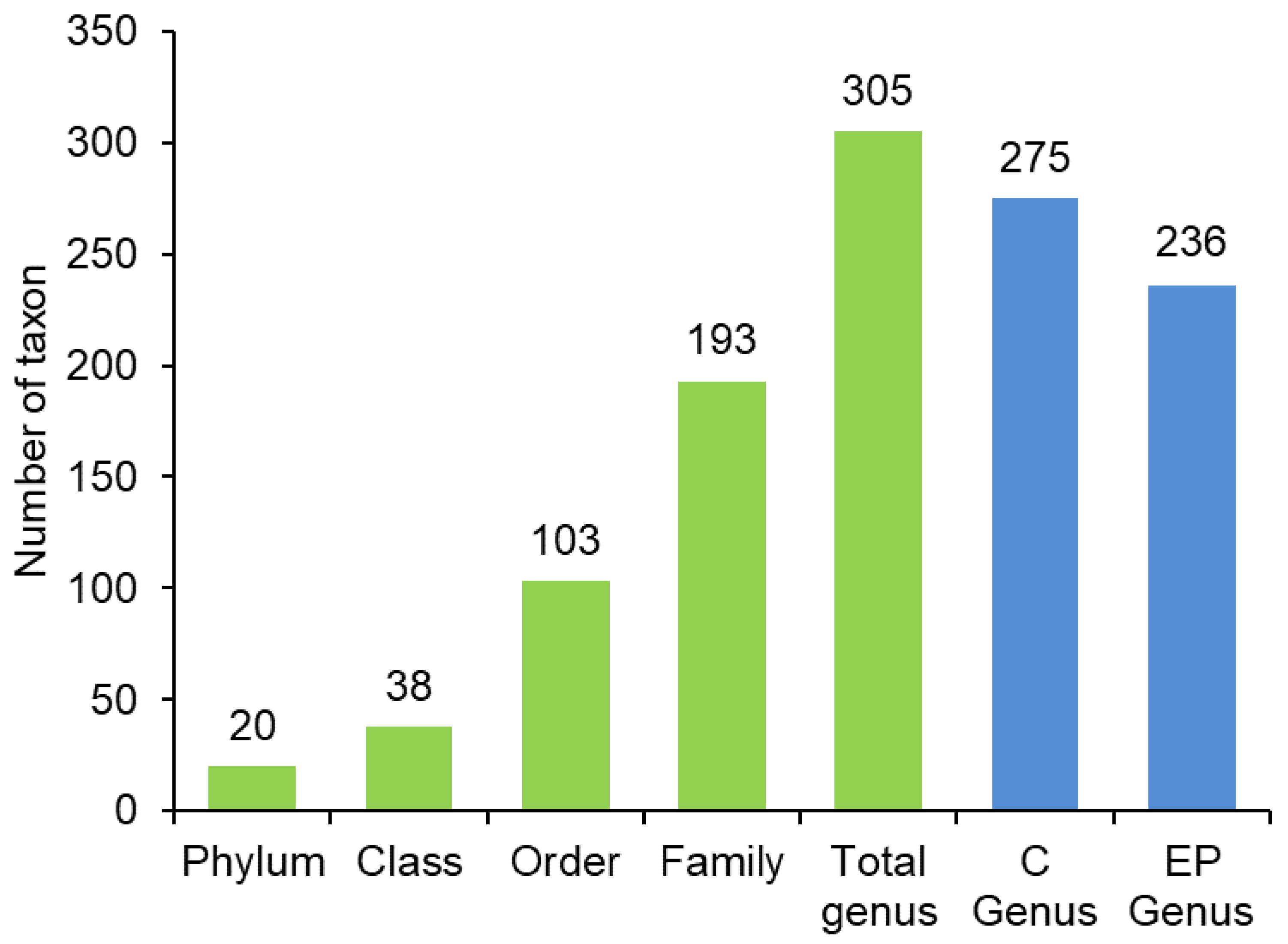
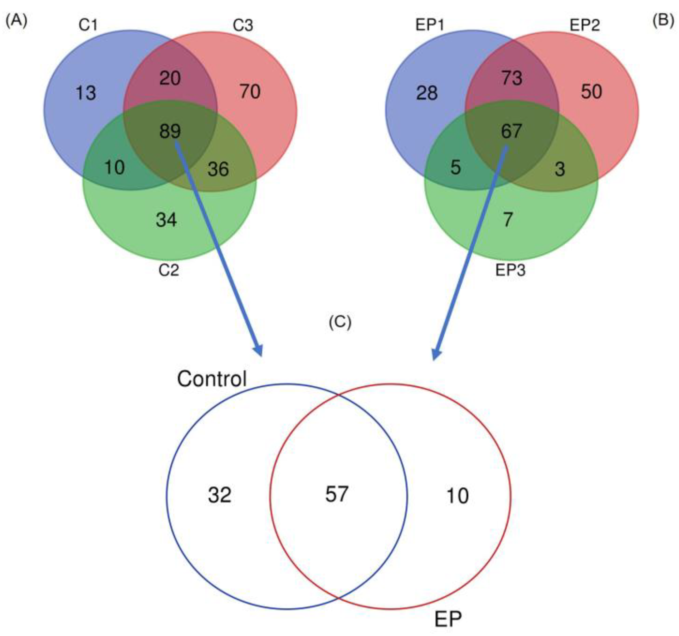
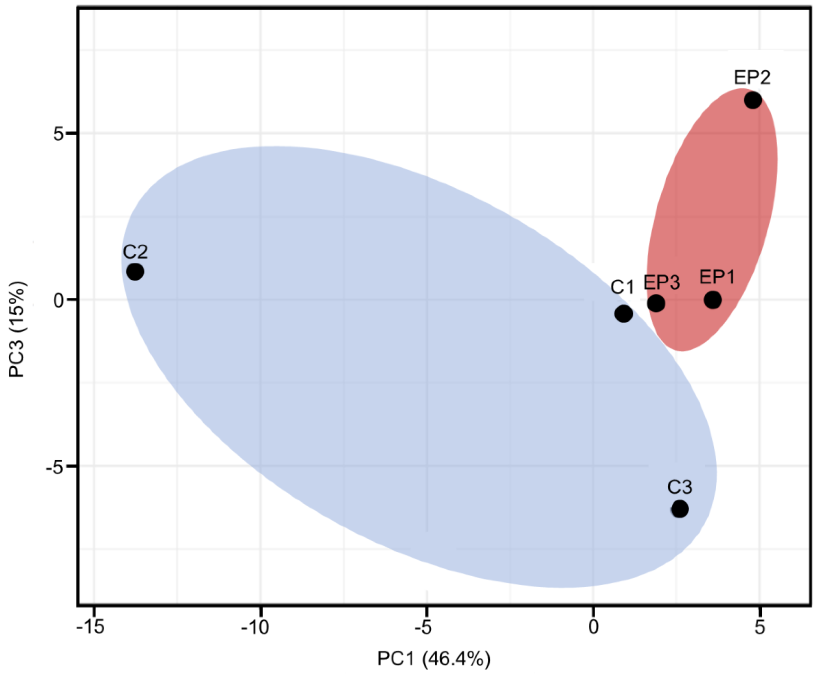
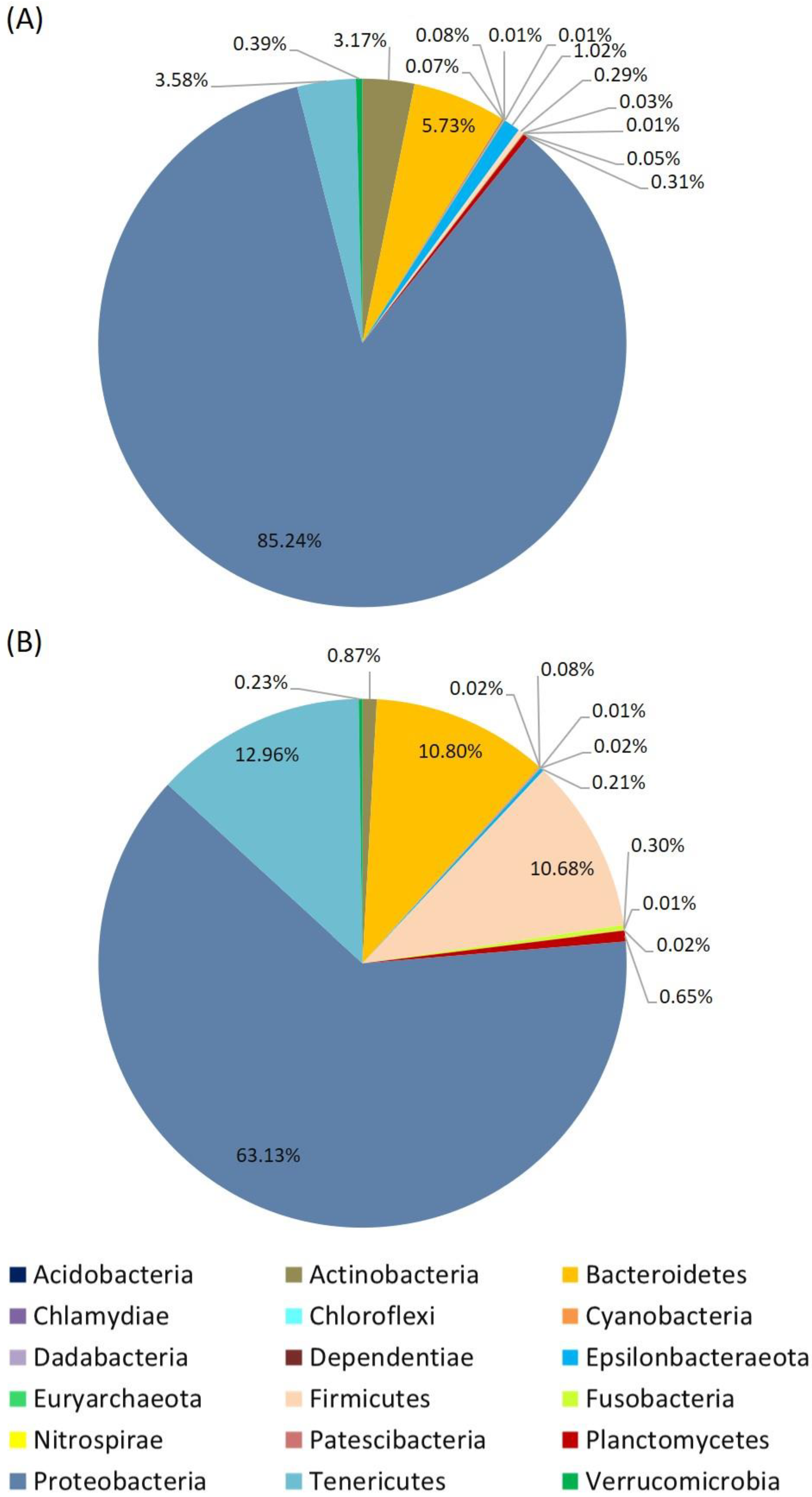
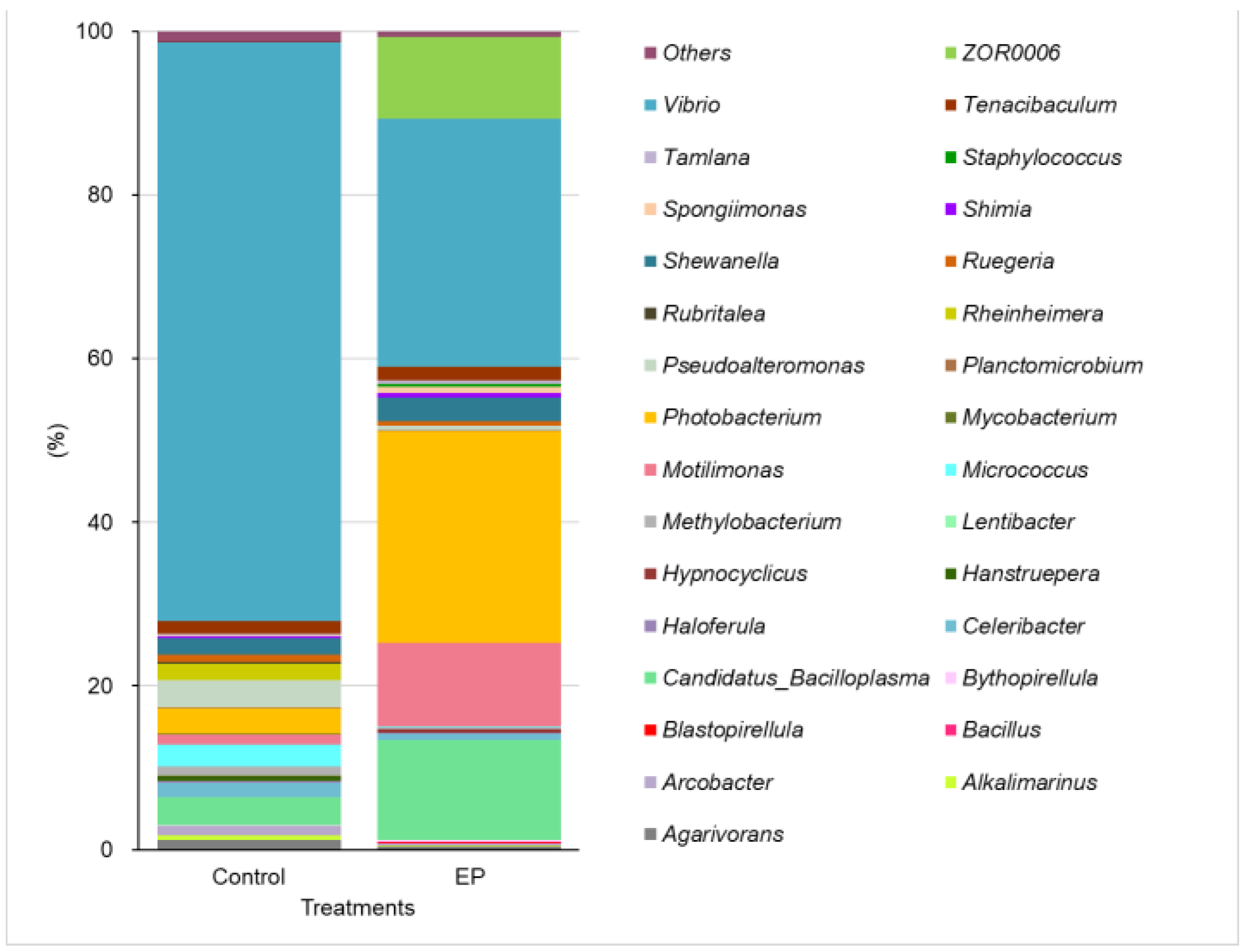
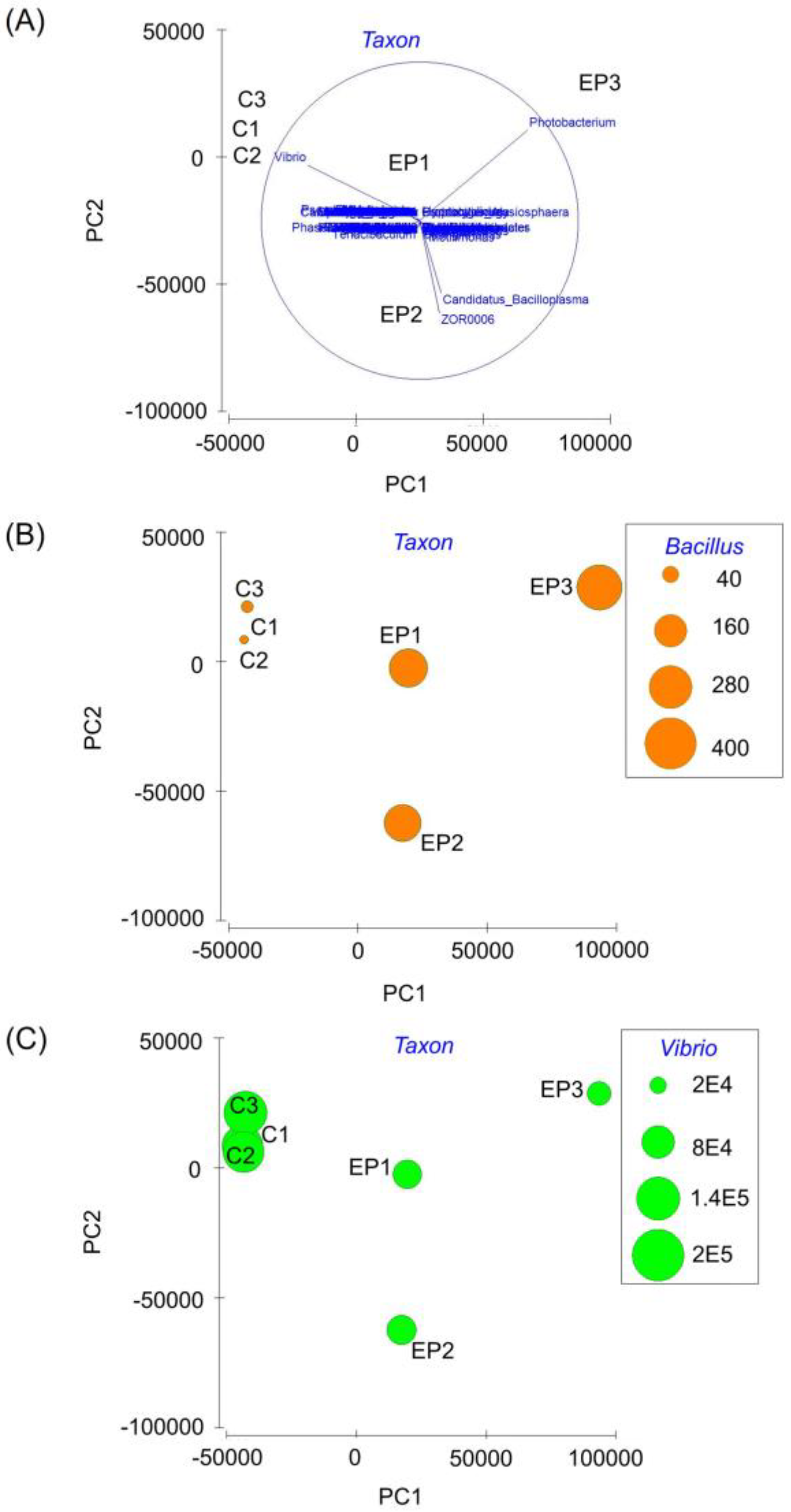
The genera of microbiota were determined by Pielou’s evenness (J), the Shannon diversity index, Margalef’s species richness (d), and the Simpson index using the alpha_diversity.py script on the website of QIIME (http://qiime.org/scripts/alpha_diversity.html, accessed on 11 January 2023). The PCA eigenvector plots and accumulated microbial dominance (%) plots were analyzed using the Plymouth Routines in Multivariate Ecological Research (PRIMER) version 6.1.5 (Clarke and Gorley, 2006).

3. Results

The 16S rDNA next-generation sequencing (NGS) analysis of the intestinal microbiome yielded 1,333,828 and 1,422,852 reads during the control and EP-feeding. After processing and filtering, the original sequences were grouped into 964 and 978 representative OTUs for the shrimp that were fed control and EP diets, respectively, at an identity cut-off of 97%. The intestinal microbiome of the shrimp showed substantial and distinct differentiation in each treatment. No significant difference was observed among the α-diversity indices for both control and EP-fed shrimp (Table 2).
The taxonomical analysis revealed that the majority of the bacterial genera were distributed among different families (193) with shrimp fed the control diet indicating a higher genus (275) than the shrimp fed the EP diet (236) (Figure 1). Among the intestinal samples in the control and EP group, the shared bacterial genera were 89 and 67, respectively (Figure 2A,B). However, between the two dietary treatments, 57 of the bacterial genera were similar (Figure 2C). The PCA plots compared the composition of microbiota in the two dietary groups. Upon PCA analysis, an obvious and regular variation was determined between the control and EP diet. The encapsulation of B. subtilis E20 (EP) modulated the bacterial profile in the shrimp’s intestines as different clusters were formed. In the dataset, the contribution of PC1 and PC3 was 46.4% and 15%, respectively (Figure 3).
At the phylum level, the relative abundance of bacterial groups in the intestinal microbiota of shrimp fed the control and EP diet was predominantly Proteobacteria at 85.24% and 63.13%, respectively. The EP diet was further influenced by the phylum Tenericutes (12.96%), Bacteroidetes (10.80%), and Firmicutes (10.68%), all of which were minimally expressed in the control group (Figure 4). In shrimp fed the control diet, the most abundant at a generic level were Vibrio (70.74%), compared to the EP group which had a lower abundance of 30.25%. In the following abundances, Photobacterium (25.83%), Candidatus Bacilloplasma (12.19%), Motilimonas (10.15%), and ZOR0006 (10.00%) were more highly expressed in the EP-fed shrimp than in the control group. Unique genus including Luteolibacter, Simkaniaceae, Haemophilus, Pirellulaceae, Filomicrobium, Sphingomonas, and Erysipelotrichaceae UCG-003 were discovered in EP-fed shrimp along with well-known probiotic genera Bacillus and Lactobacillus (Figure 5). The PCA eigenvector plots indicated that shrimp fed the EP diet had a significantly higher abundance of Bacillus, while Vibrio was mostly present in the control group of shrimp (Figure 6).

4. Discussion

The shrimp intestinal microbiome consists of several microbes and genes critical for health, metabolism, as well as disease pathogenesis. As shrimp are intimately connected to the aquatic environment, much of their intestinal microbes are influenced by the microbes present in the surrounding environment [19]. Consequently, culture systems that are either intensive or unfavorable adversely affect the microbial interaction between the shrimp and the environment, resulting in the proliferation of opportunistic pathogens that cause disease outbreaks [20]. Live probiotic bacteria, which are generally regarded as safe due to their immunomodulatory, antimicrobial, and antioxidant beneficial effects, are often incorporated into feeds as dietary supplements to maintain the microbial balance in shrimp gut [4,5,11]. However, the viability of probiotics is vastly affected by numerous factors, especially during production, storage, feeding, and passage through the gastrointestinal system. Thus, several techniques for microencapsulation have been attempted to preserve and protect the viability of probiotic cells [9,16,17,21]. While most of these studies focused on the immune response and growth performance as well as intestinal microbiota upon live probiotic administration without encapsulation, studies on the intestinal microbiome upon administering encapsulated probiotics is limited.
In this study, the B. subtilis E20 strain was encapsulated with alginate-chitosan to protect cell viability and determine the bacterial communities generated. Data from NGS analysis revealed a dominant presence of Proteobacteria in all shrimp microbiota. Similar results were obtained when B. subtilis E20-fermented soybean meal (FSBM) was provided to shrimp [22]. However, lower Proteobacteria was present in the intestines of shrimp fed the EP diet. Despite this, Tenericutes, Bacteroidetes, and Firmicutes were highly expressed in the EP-fed shrimp. These results suggest that microencapsulation of B. subtilis E20 (EP) can induce proliferation and diversification of bacteria in shrimp microbiota. Proteobacteria, Bacteroidetes, and Firmicutes are typically dominant bacteria associated with shrimp and other aquatic animals such as Nile tilapia Oreochromis niloticus, silver carp Hypophthalmichthys molitrix and bighead carp H. nobilis [23,24,25].
Generally, the health condition of shrimp and fish can be reflected by the relative abundance of Proteobacteria, which is a microbial sign of dysbiosis and disease in gut microbiota [26]. Tenericutes are free-living organisms affiliated with Bacilli, and exhibit metabolic and adaptivity flexibility commensal to the host [27,28]. Firmicutes helps to ferment carbon sources and control energy balance within the host [27,29]. Similarly, Bacteroidetes ferment plant-derived substrates in the intestines by producing short chain fatty acids (SCFAs) that allow the host to obtain excess energy [29]. It is known that SCFAs also play major roles in the homeostasis of immune cells in several organisms. Therefore, the interaction between Firmicutes and Bacteroidetes likely promoted more efficient fermentation of carbohydrates in the diet and increased the energy absorption in the intestine of shrimp fed with encapsulated B. subtilis E20. Furthermore, such interaction also explains the improved growth performance achieved in shrimp treated with microencapsulated probiotics (107−9 CFU g−1) in our previous study [17]. The results also agree with the recent research findings on juvenile Nile tilapia supplemented with microencapsulated probiotic additives containing Bacillus spp. (BACIL) or Saccharomyces cerevisiae (SACCH). The microbial profile showed a predominance of Firmicutes and Tenericutes in the intestinal microbiota of fish, reflecting better growth and immunity when compared to the control group [30].
In addition, the Vibrio species are among the dominant members of the white shrimp microbiota, and are considered the most important bacterial pathogens responsible for several diseases and mass mortalities [31]. Several studies have reported the importance of Vibrio during the different developmental stages of shrimp [31,32]. Findings revealed that the Vibrio population in the shrimp gut microbiota was higher during the nursery stage than in the adult stage, indicating that the microbiota in the latter stage is more diverse than in the nursery stage [33]. In most cases, Vibrio species are considered opportunistic pathogens that have detrimental effects on shrimp’s growth, metabolic activity, microbial balance, and immune response [19,34]. High expression in the gut is an indicator of disease in shrimp. In infected shrimp, V. parahaemolyticus increased intestinal permeability and impaired the ability to absorb the amino acids and glucose that are necessary for maintaining physiological activities.
The supplementation of probiotics has been proven to be a beneficial biocontrol agent for reducing Vibrio counts and preventing vibriosis [8,35,36,37]. The administration of marine bacterial microcapsule B. subtilis P2.24 reduced the total Vibrio count, total V. parahaemolyticus count in shrimp’s intestinal tract, and increased the intestinal microbiota diversity [36]. By analyzing the microbial community, it was found that the Vibrio count and abundance levels of Vibrio species were suppressed in the intestine of shrimp fed the encapsulated B. subtilis E20 compared to shrimp fed the control diet. A similar study reported that the inhibitory effect of Vibrio against Bacillus was attributed to the secretion of antibacterial peptides and competitive inhibition associated with probiotic B. subtilis UTM 126 [38].
Each bacterial genus hosts microorganisms with probiotic potential when present in the intestinal tract, helps to improve the physiological and metabolic functions of the host. In the present study, the Pirellula species, a non-pathogenic free-living bacterium in the aquatic environment was identified in shrimp that received the EP diet. Pirellulaceae are known as ammonia-oxidizing bacteria, are found in marine sponges Ircinia strobilina and Mycale laxissima [39], deep-sea octocoral Alcyonium grandiflorum [40], and contribute to nitrification as well as the removal of metabolic waste in the host microbiome. In tiger prawn Penaeus monodon, Pirellulales-like bacteria have been categorized as a commensal gut flora, as they proliferated under stressful conditions to reduce baculovirus infection in juvenile prawns [41]. These results may further explain the improved immune response and shrimp’s ability to resist V. alginolyticus when fed EP in our previous study [17].
In addition to Pirellulaceae, Erysipelotrichaceae and families within Firmicutes were also identified. Studies have shown that Erysipelotrichaceae increases energy absorption (cellular metabolism) in pigs [42]. However, the role of this bacteria in aquatic animals including shrimp remains unknown. Our NGS analysis specifically identified Erysipelotrichaceae UCG-003 as a unique genus in the EP-fed shrimp. Based on its characteristics, this strain is considered one of the main butyrate-producing bacteria when present in the microbiota, and is able to modulate bacterial diversity, playing a protective role [43]. Thus, the improved performance in shrimp fed EP could be attributed to the presence of Erysipelotrichaceae UCG-003, but further research is needed to confirm its functional properties in shrimp.
In a recent study, Luteolibacter in zebrafish was found to ameliorate the growth of Yersinia ruckeri, a salmon pathogen, by colonizing fish skin to repair the damaged tissues [44]. Similarly, regeneration of damaged skin microbiota was observed in Indian major carp, rohu Labeo rohita infected with Argulus siamensis due to an increase abundance of Luteolibacter [45]. Luteolibacter was also identified in the intestinal microbiota of shrimp that were fed with EP diet. Simkaniaceae has been found in several marine, coastal, and host-associated environments, including invertebrates. Though its role in shrimp microbiota has not yet been established, studies have reported that Simkaniaceae bacterium contains glutamate decarboxylase (GAD) enzymatic genes that catalyzes the conversion of glutamate into γ-aminobutyric acid (GABA) and carbon dioxide (CO2) [46]. The activation of the GAD enzyme allows host animals to tolerate acidic environments both externally and intracellularly. In this study, the presence of Simkaniaceae bacteria suggests that shrimp fed the EP diet would be able to tolerate stressful environments. GABA, which is usually associated with GAD, is an important neurotransmitter present at high concentrations in the brain and plays a key role in the metabolic pathways that regulate feed intake and nutrient utilization, behavior and immunity [47]. In addition, GABA is known to minimize the severity caused by environmental stressors and pathogenic organisms. Xie et al. [48] reported improved growth performance, antioxidative capacity and resistance against NH3 stress in L. vannamei fed GABA with low fishmeal diet. Given the importance of GAD and GABA in the host’s metabolic and physiological functions, Simkaniaceae can be considered a beneficial bacterium in the gut of shrimp when fed encapsulated B. subtilis E20. Other identified genera including Haemophilus and Filomicrobium were identified, however their roles are not known.
The microencapsulated probiotic also increased the abundance of Candidatus Bacilloplasma in shrimp. Candidatus Bacilloplasma are recognized as symbionts and can be used as potential taxonomic indicators for assessing the health status of shrimp. In previous studies, the detection of Candidatus Bacilloplasma showed commensal activities which inhibited the proliferation of Vibrio bacterial strains and infection [49]. The greater expression of Candidatus Bacilloplasma in this study suggests that the encapsulation of probiotics can preserve their viability to such an extent that it stimulated the growth of various beneficial bacteria that might be lost when the probiotic is unencapsulated. This is evident in the Cheng et al. [22] study that assessed the intestinal microbiota of white shrimp after feeding them B. subtilis E20-fermented soybean meal (FSBM), in which none of the unique genus and Candidatus Bacilloplasma were present in the microbiome. Probiotic genera Bacillus and Lactobacillus have been well documented in many studies, demonstrating immunomodulatory, growth, and metabolic enhancement. Pedicococcus pentosaceus also increased the intestinal counts of Bacillus sp., and Lactobacillus sp. in white shrimp [35].

5. Conclusions

The study concludes that microencapsulation of B. subtilis E20 can be helpful in tackling the sensitivity problems associated with probiotics during processing and application. Our results indicate that encapsulated B. subtilis E20 administration increased beneficial strains of bacteria such as Bacillus and reduced the harmful bacteria belonging to the Vibrio species. Thus, the encapsulation of B. subtilis E20 has the potential to modulate gut microbiota and control Vibrio species in shrimp.

Author Contributions

Conceptualization, A.-C.C. and C.-H.L.; data curation, C.-H.L.; funding acquisition, C.-H.L.; methodology, A.-C.C. and C.-H.L.; resources, S.-T.C. and C.-H.L.; supervision, C.-H.L.; writing—original draft, R.B. and C.-H.L.; writing—review and editing, C.-H.L. All authors have read and agreed to the published version of the manuscript.

Funding

This study was supported by a grant (110-2313-B-020-006-MY3 and 110-2637-B020-006-) from the National Science and Technology Council, ROC.

Institutional Review Board Statement

According to the Guideline for the Care and Use of Laboratory Animals, it is not required to have an “Affidavit of Approval of Animal Use Protocol” when using invertebrates, such as shrimp as experimental animals in Taiwan.

Data Availability Statement

The data presented in this study are available in the article.

Conflicts of Interest

The authors declare no conflict of interest.

References

  1. Tzuc, J.T.; Escalante, D.R.; Rojas Herrera, R.; Gaxiola Cortés, G.; Ortiz, M.L.A. Microbiota from Litopenaeus vannamei: Digestive tract microbial community of Pacific white shrimp (Litopenaeus vannamei). SpringerPlus 2014, 3, 280. [Google Scholar] [CrossRef] [PubMed]
  2. Dai, W.; Yu, W.; Zhang, J.; Zhu, J.; Tao, Z.; Xiong, J. The gut eukaryotic microbiota influences the growth performance among cohabitating shrimp. Appl. Microbiol. Biotechnol. 2017, 101, 6447–6457. [Google Scholar] [CrossRef] [PubMed]
  3. Vargas-Albores, F.; Porchas-Cornejo, M.A.; Martínez-Porchas, M.; Villalpando-Canchola, E.; Gollas-Galván, T.; Martínez-Córdova, L.R. Bacterial biota of shrimp intestine is significantly modified by the use of a probiotic mixture: A high throughput sequencing approach. Helgol. Mar. Res. 2017, 71, 5. [Google Scholar] [CrossRef]
  4. Ringø, E. Probiotics in shellfish aquaculture. Aquac. Fish. 2020, 5, 1–27. [Google Scholar] [CrossRef]
  5. Song, S.K.; Beck, B.R.; Kim, D.; Park, J.; Kim, J.; Kim, H.D.; Ringø, E. Prebiotics as immunostimulants in aquaculture: A review. Fish Shellfish Immunol. 2014, 40, 40–48. [Google Scholar] [CrossRef]
  6. Wang, A.; Ran, C.; Wang, Y.; Zhang, Z.; Ding, Q.; Yang, Y.; Olsen, R.E.; Ringø, E.; Bindelle, J.; Zhou, Z. Use of probiotics in aquaculture of China—A review of the past decade. Fish Shellfish Immunol. 2019, 86, 734–755. [Google Scholar] [CrossRef]
  7. Xie, J.J.; Liu, Q.Q.; Liao, S.; Fang, H.H.; Yin, P.; Xie, S.W.; Tian, L.X.; Liu, Y.J.; Niu, J. Effects of dietary mixed probiotics on growth, non-specific immunity, intestinal morphology and microbiota of juvenile Pacific white shrimp, Litopenaeus vannamei. Fish Shellfish Immunol. 2019, 90, 456–465. [Google Scholar] [CrossRef]
  8. Imaizumi, K.; Tinwongger, S.; Kondo, H.; Hirono, I. Analysis of microbiota in the stomach and midgut of two penaeid shrimps during probiotic feeding. Sci. Rep. 2021, 11, 9936. [Google Scholar]
  9. Jantarathin, S.; Borompichaichartkul, C.; Sanguandeekul, R. Microencapsulation of probiotic and prebiotic in alginate-chitosan capsules and its effect on viability under heat process in shrimp feeding. Mater. Today Proc. 2017, 4, 6166–6172. [Google Scholar] [CrossRef]
  10. Bauer-Estrada, K.; Sandoval-Cuellar, C.; Rojas-Muñoz, Y.; Quintanilla-Carvajal, M.X. The modulatory effect of encapsulated bioactives and probiotics on gut microbiota: Improving health status through functional food. Food Funct. 2022, 14, 32–55. [Google Scholar] [CrossRef]
  11. Willer, D.F.; Aldridge, D.C. Microencapsulated diets to improve growth and survivorship in juvenile European flat oysters (Ostrea edulis). Aquaculture 2019, 505, 256–262. [Google Scholar] [CrossRef]
  12. Willer, D.F.; Furse, S.; Aldridge, D.C. Microencapsulated algal feeds as a sustainable replacement diet for broodstock in commercial bivalve aquaculture. Sci. Rep. 2020, 10, 12577. [Google Scholar] [CrossRef]
  13. Amir, I.; Zuberi, A.; Kamran, M.; Imran, M.; Murtaza, M.U.H. Evaluation of commercial application of dietary encapsulated probiotic (Geotrichum candidum QAUGC01): Effect on growth and immunological indices of Rohu (Labeo rohita, Hamilton 1822) in semi-intensive culture system. Fish Shellfish Immunol. 2019, 95, 464–472. [Google Scholar] [CrossRef]
  14. Kumaree, K.K.; Akbar, A.; Anal, A.K. Bioencapsulation and application of Lactobacillus plantarum isolated from catfish gut as an antimicrobial agent and additive in fish feed pellets. Ann. Microbiol. 2015, 65, 1439–1445. [Google Scholar] [CrossRef]
  15. Annan, N.T.; Borza, A.D.; Hansen, L.T. Encapsulation in alginate-coated gelatin microspheres improves survival of the probiotic Bifidobacterium adolescentis 15703T during exposure to simulated gastro-intestinal conditions. Food Res. Int. 2008, 41, 184–193. [Google Scholar] [CrossRef]
  16. Ji, R.; Wu, J.; Zhang, J.; Wang, T.; Zhang, X.; Shao, L.; Chen, D.; Wang, J. Extending viability of Bifidobacterium longumin chitosan-coated alginate microcapsules using emulsification and internal gelation encapsulation technology. Front. Microbiol. 2019, 10, 1389. [Google Scholar] [CrossRef]
  17. Adilah, R.N.; Chiu, S.T.; Hu, S.Y.; Ballantyne, R.; Happy, N.; Cheng, A.C.; Liu, C.H. Improvement in the probiotic efficacy of Bacillus subtilis E20-stimulates growth and health status of white shrimp, Litopenaeus vannamei via encapsulation in alginate and coated with chitosan. Fish Shellfish Immunol. 2022, 125, 74–83. [Google Scholar] [CrossRef]
  18. Cheng, Z.J.; Hardy, R.W. Protein and lipid sources affect cholesterol concentrations of juvenile Pacific white shrimp, Litopenaeus vannamei (Boone). J. Anim. Sci. 2004, 82, 1136–1145. [Google Scholar] [CrossRef]
  19. Holt, C.C.; Bass, D.; Stentiford, G.D.; van der Giezen, M. Understanding the Role of the shrimp gut microbiome in health and disease. J. Invertebr. Pathol. 2021, 186, 107387. [Google Scholar] [CrossRef]
  20. Sun, F.; Wang, Y.; Wang, C.; Zhang, L.; Tu, K.; Zheng, Z. Insights into the intestinal microbiota of several aquatic organisms and association with the surrounding environment. Aquaculture 2019, 507, 196–202. [Google Scholar] [CrossRef]
  21. Riaz, Q.U.A.; Masud, T. Recent Trends and Applications of Encapsulating Materials for probiotic stability. Crit. Rev. Food Sci. Nutr. 2013, 53, 231–244. [Google Scholar] [CrossRef]
  22. Cheng, A.C.; Yeh, S.P.; Hu, S.Y.; Lin, H.L.; Liu, C.H. Intestinal microbiota of white shrimp, Litopenaeus vannamei, fed diets containing Bacillus subtilis E20-fermented soybean meal (FSBM) or an antimicrobial peptide derived from B. subtilis E20-FSBM. Aquac. Res. 2020, 51, 41–50. [Google Scholar] [CrossRef]
  23. Landsman, A.; St-Pierre, B.; Rosales-Leija, M.; Brown, M.; Gibbons, W. Impact of aquaculture practices on intestinal bacterial profiles of Pacific whiteleg shrimp Litopenaeus vannamei. Microorganisms 2019, 7, 93. [Google Scholar] [CrossRef]
  24. Li, J.; Ni, J.; Li, J.; Wang, C.; Li, X.; Wu, S.; Zhang, T.; Yu, Y.; Yan, Q. Comparative study on gastrointestinal microbiota of eight fish species with different feeding habits. J. Appl. Microbiol. 2014, 117, 1750–1760. [Google Scholar] [CrossRef]
  25. Kuang, T.; He, A.; Lin, Y.; Huang, X.; Liu, L.; Zhou, L. Comparative analysis of microbial communities associated with the gill, gut, and habitat of two filter-feeding fish. Aquac. Rep. 2020, 18, 100501. [Google Scholar] [CrossRef]
  26. Shin, N.R.; Whon, T.W.; Bae, J.W. Proteobacteria: Microbial signature of dysbiosis in gut microbiota. Trends Biotechnol. 2015, 33, 496–503. [Google Scholar] [CrossRef]
  27. Wang, Y.; Huang, J.M.; Zhou, Y.L.; Almeida, A.; Finn, R.D.; Danchin, A.; He, L.S. Phylogenomics of expanding uncultured environmental Tenericutes provides insights into their pathogenicity and evolutionary relationship with Bacilli. BMC Genom. 2020, 21, 408. [Google Scholar] [CrossRef]
  28. Huang, L.; Guo, H.; Chen, C.; Huang, X.; Chen, W.; Bao, F.; Liu, W.; Wang, S.; Zhang, D. The bacteria from large-sized bioflocs are more associated with the shrimp gut microbiota in culture system. Aquaculture 2020, 523, 735159. [Google Scholar] [CrossRef]
  29. Van Kessel, M.A.H.J.; Dutilh, B.E.; Neveling, K.; Kwint, M.P.; Veltman, J.A.; Flik, G.; Jetten, M.S.; Klaren, P.H.; den Camp, H.J.M.O. Pyrosequencing of 16S rRNA gene amplicons to study the microbiota in the gastrointestinal tract of carp (Cyprinus carpio L.). AMB Express 2011, 1, 41. [Google Scholar] [CrossRef]
  30. de Moraes, A.V.; Owatari, M.S.; da Silva, E.; de Oliveira Pereira, M.; Piola, M.; Ramos, C.; Farias, D.R.; Schleder, D.D.; Jesus, G.F.A.; Jatobá, A. Effects of microencapsulated probiotics-supplemented diet on growth, non-specific immunity, intestinal health and resistance of juvenile Nile tilapia challenged with Aeromonas hydrophila. Anim. Feed Sci. Technol. 2022, 287, 115286. [Google Scholar] [CrossRef]
  31. Garibay-Valdez, E.; Martínez-Córdova, L.R.; López-Torres, M.A.; Almendariz-Tapia, F.J.; Martínez-Porchas, M.; Calderón, K. The implication of metabolically active Vibrio spp. in the digestive tract of Litopenaeus vannamei for its post-larval development. Sci. Rep. 2020, 10, 11428. [Google Scholar] [CrossRef]
  32. Huang, Z.; Li, X.; Wang, L.; Shao, Z. Changes in the intestinal bacterial community during the growth of white shrimp, Litopenaeus vannamei. Aquac. Res. 2016, 47, 1737–1746. [Google Scholar] [CrossRef]
  33. Gainza, O.; Ramírez, C.; Ramos, A.S.; Romero, J. Intestinal microbiota of white shrimp Penaeus vannamei under intensive cultivation conditions in Ecuador. Microb. Ecol. 2018, 75, 562–568. [Google Scholar] [CrossRef]
  34. Jiao, L.F.; Dai, T.M.; Zhong, S.Q.; Jin, M.; Sun, P.; Zhou, Q.C. Vibrio parahaemolyticus infection impaired intestinal barrier function and nutrient absorption in Litopenaeus vannamei. Fish Shellfish Immunol. 2020, 99, 184–189. [Google Scholar] [CrossRef]
  35. Adel, M.; Yeganeh, S.; Dawood, M.A.O.; Safari, R.; Radhakrishnan, S. Effects of Pediococcus pentosaceus supplementation on growth performance, intestinal microflora and disease resistance of white shrimp, Litopenaeus vannamei. Aquac. Nutr. 2017, 23, 1401–1409. [Google Scholar] [CrossRef]
  36. Aribah, D.; Widanarni; Wahyudi, A.T. The effectiveness of marine bacterial microcapsules in controlling vibriosis disease caused by the infection of Vibrio parahaemolyticus in white shrimp Litopenaeus vannamei. Aquaculture 2022, 549, 737795. [Google Scholar] [CrossRef]
  37. Kongnum, K.; Hongpattarakere, T. Effect of Lactobacillus plantarum isolated from digestive tract of wild shrimp on growth and survival of white shrimp (Litopenaeus vannamei) challenged with Vibrio harveyi. Fish Shellfish Immunol. 2012, 32, 170–177. [Google Scholar] [CrossRef]
  38. Balcázar, J.L.; Rojas-Luna, T. Inhibitory activity of probiotic Bacillus subtilis UTM 126 against Vibrio Species confers protection against vibriosis in juvenile shrimp (Litopenaeus vannamei). Curr. Microbiol. 2007, 55, 409–412. [Google Scholar] [CrossRef]
  39. Mohamed, N.M.; Saito, K.; Tal, Y.; Hill, R.T. Diversity of aerobic and anaerobic ammonia-oxidizing bacteria in marine sponges. ISME J. 2009, 4, 38–48. [Google Scholar] [CrossRef]
  40. Lawler, S.N.; Kellogg, C.A.; France, S.C.; Clostio, R.W.; Brooke, S.D.; Ross, S.W. Coral-associated bacterial diversity is conserved across two deep-sea Anthothela Species. Front. Microbiol. 2016, 7, 458. [Google Scholar] [CrossRef]
  41. Fuerst, J.A.; Sambhi, S.K.; Paynter, J.L.; Hawkins, J.A.; Atherton, J.G. Isolation of a bacterium resembling Pirellula species from primary tissue culture of the giant tiger prawn (Penaeus monodon). Appl. Environ. Microbiol. 1991, 57, 3127–3134. [Google Scholar] [CrossRef] [PubMed]
  42. Wu, J.; Liu, M.; Zhou, M.; Wu, L.; Yang, H.; Huang, L.; Chen, C. Isolation and Genomic characterization of five novel strains of Erysipelotrichaceae from commercial pigs. BMC Microbiol. 2021, 21, 125. [Google Scholar] [CrossRef] [PubMed]
  43. Cornejo-Granados, F.; Lopez-Zavala, A.A.; Gallardo-Becerra, L.; Mendoza-Vargas, A.; Sánchez, F.; Vichido, R.; Brieba, L.G.; Viana, M.T.; Sotelo-Mundo, R.R.; Ochoa-Leyva, A. Microbiome of Pacific whiteleg shrimp reveals differential bacterial community composition between wild, aquaculture and AHPND/EMS Outbreak Conditions. Sci. Rep. 2017, 7, 11783. [Google Scholar] [CrossRef] [PubMed]
  44. Nakatani, H.; Hori, K.; Muiswinkel, V. Establishing a percutaneous infection model using zebrafish and a salmon pathogen. Biology 2021, 10, 166. [Google Scholar] [CrossRef]
  45. Mondal, H.K.; Maji, U.J.; Mohanty, S.; Sahoo, P.K.; Maiti, N.K. Alteration of gut microbiota composition and function of Indian major carp, Rohu (Labeo rohita) infected with Argulus siamensis. Microb. Pathog. 2022, 164, 105420. [Google Scholar] [CrossRef]
  46. Chen, Y.H.; Chiang, P.W.; Rogozin, D.Y.; Degermendzhy, A.G.; Chiu, H.H.; Tang, S.L. Salvaging high-quality genomes of microbial species from a meromictic lake using a hybrid sequencing approach. Commun. Biol. 2021, 4, 996. [Google Scholar] [CrossRef]
  47. Li, P.; Mai, K.; Trushenski, J.; Wu, G. New developments in fish amino acid nutrition: Towards functional and environmentally oriented aquafeeds. Amino Acids 2009, 37, 43–53. [Google Scholar] [CrossRef]
  48. Xie, S.W.; Li, Y.T.; Zhou, W.W.; Tian, L.X.; Li, Y.M.; Zeng, S.L.; Liu, Y.J. Effect of γ-aminobutyric acid supplementation on growth performance, endocrine hormone and stress tolerance of juvenile Pacific white shrimp, Litopenaeus vannamei, fed low fishmeal diet. Aquacult. Nutr. 2017, 23, 54–62. [Google Scholar] [CrossRef]
  49. Chen, W.Y.; Ng, T.H.; Wu, J.H.; Chen, J.W.; Wang, H.C. Microbiome dynamics in a shrimp grow-out pond with possible outbreak of acute hepatopancreatic necrosis disease. Sci. Rep. 2017, 7, 9395. [Google Scholar] [CrossRef]
Figure 1. Taxonomic identification of the intestinal microflora of white shrimp fed with the control (C) and the encapsulated-probiotic B. subtilis E20 (EP) diet.
Fishes 08 00264 g001
Figure 2. Venn diagram representation of shared and unique genera across the experimental feeding groups control (C) and encapsulated-probiotic B. subtilis E20 (EP) diet in the intestinal samples of white shrimp. The collective correlation between the bacterial genera within the control group (A) or within the embedded probiotic group (B) and between the control group and the embedded probiotic group (C).
Fishes 08 00264 g002
Figure 3. Principal composition of the intestinal bacterial communities in white shrimp at the generic level between the control (C) and the encapsulated-probiotic B. subtilis E20 (EP) diet. Score plot for PC1 (46.4%) vs. PC3 (15%) explained the variance.
Fishes 08 00264 g003
Figure 4. Relative abundance of phylum category of intestinal microbiota of shrimp fed the control diet (A) and the encapsulated-probiotic B. subtilis E20 (B) diet.
Fishes 08 00264 g004
Figure 5. The relative abundance of microbial genera in the intestinal tract of white shrimp fed the control diet and the encapsulated-probiotic B. subtilis E20 (EP) diet. Only data with the sequence number of OTUs > 0.1% are shown to represent a major proportion of the bacterial population.
Fishes 08 00264 g005
Figure 6. PCA eigenvector plots of intestinal microbial flora (A), Bacillus (B), and Vibrio (C) of white shrimp fed with the control diet (C1-3) and the encapsulated-probiotic B. subtilis E20 (EP1-3) diet.
Fishes 08 00264 g006
Table 1. The ingredients of the experimental diets.
IngredientsExperimental Diets (g kg⁻¹)
ControlEP
Fish meal410410
Soybean meal300300
Squid meal5050
Fish oil2929
α-Starch149.6149.6
Vitamin Premix *2020
Mineral premix *4040
E-probiotic (109 CFU g−1)00.01
α-Cellulose1.41.39
Probiotic level (CFU kg−1)02.5 × 10⁷
Proximate analysis
Moisture (%)7.2 ± 0.27.2 ± 0.3
Ash (%)14.1 ± 0.113.8 ± 0.2
Crude protein (%)40.1 ± 0.540.5 ± 0.2
Crude lipid (%)6.9 ± 0.66.5 ± 0.2
* Vitamin and mineral premix provided per kg of diet was according to [18].
Table 2. α-diversity index of the intestinal flora of white shrimp.
TreatmentsGenusMargalef’s Species Richness (d)Pielou’s Evenness (J’)Shannon IndexSimpson Index
Control27514.07 ± 2.390.32 ± 0.11.64 ± 0.520.55 ± 0.16
EP23612.12 ± 3.410.36 ± 0.051.79 ± 0.330.71 ± 0.08
Disclaimer/Publisher’s Note: The statements, opinions and data contained in all publications are solely those of the individual author(s) and contributor(s) and not of MDPI and/or the editor(s). MDPI and/or the editor(s) disclaim responsibility for any injury to people or property resulting from any ideas, methods, instructions or products referred to in the content.

