Article

Comparative Study on Ginger Powder and Ginger Extract Nanoparticles: Effects on Growth, Immune–Antioxidant Status, Tissue Histoarchitecture, and Resistance to Aeromonas hydrophila and Pseudomonas putida Infection in Oreochromis niloticus

by
Shaimaa A. A. Ahmed
1,
Abdelhakeem El-Murr
1,
Yasser Abd Elhakim
1,
Mohamed M. Metwally
2,
Amany Abd El Aziz Gharib
3,
Shimaa A. Amer
4,*,
Elsayed M. Younis
5,
Abdel-Wahab A. Abdel-Warith
5,
Simon J. Davies
6 and
Enas N. M. Khalil
3
1
Department of Aquatic Animal Medicine, Faculty of Veterinary Medicine, Zagazig University, Zagazig 44511, Egypt
2
Department of Pathology, Faculty of Veterinary Medicine, Zagazig University, Zagazig 44511, Egypt
3
Department of Hatchery and Fish Physiology, Central Laboratory for Aquaculture Research (CLAR), Agriculture Research Center, Sharkia 44662, Egypt
4
Department of Nutrition and Clinical Nutrition, Faculty of Veterinary Medicine, Zagazig University, Zagazig 44511, Egypt
5
Department of Zoology, College of Science, King Saud University, P.O. Box 2455, Riyadh 11451, Saudi Arabia
6
School of Science and Engineering, National University of Ireland Galway Republic of Ireland, H91 TK33 Galway, Ireland
*
Author to whom correspondence should be addressed.
Fishes 2023, 8(5), 259; https://doi.org/10.3390/fishes8050259
Submission received: 9 April 2023 / Revised: 9 May 2023 / Accepted: 10 May 2023 / Published: 13 May 2023

Abstract

:
A 10 week feeding trial was conducted to evaluate the potential effects of ginger powder (GP) and ginger extract nanoparticles (GNPs) on the growth parameters, digestive enzymes (lipase and amylase) activities, blood hematology, blood biochemical indices, immune indices (interleukin 10, immunoglobulin M, nitric oxide, and lysozymes), antioxidant activity, histological characteristics of kidney, spleen, liver, and intestine, and resistance to Aeromonas hydrophila or Pseudomonas putida infection in Nile tilapia, Oreochromis niloticus. Fish (n = 225, 27.01 ± 0.15 g) were stocked in 15 glass tanks (50 × 40 × 60 cm) and randomly allocated to five experimental treatments (TRTs) in triplicate (15 fish/replicate, 45 fish/TRT), consisting of five isocaloric–isonitrogenous diets. The treatments comprised the basal diet (1) without any additives (control group, CON), (2) with 0.5% GP (GP0.5), (3) with 1% GP (GP1), (4) with 0.5% GNPs (GNPs0.5), and (5) with 1% GNPs (GNPs1). Fish were manually fed to satiety three times a day (at 9 a.m., 12 p.m., and 2 p.m.). Fish were weighed at the start of the experiment, then the body weight, weight gain, feed intake, and feed conversion ratio were determined at the end of the experiment. At the end of the feeding period, 15 fish/TRT were intraperitoneally inoculated with two pathogenic bacterial strains (A. hydrophila or P. putida) in two separate challenge tests. Blood samples were collected from each TRT at two aliquots for hematological and biochemical analysis at the end of the feeding period. A significant improvement in fish growth was observed in GP and GNPs TRTs compared to the control group. There were no significant changes in the total amount of feed intake/fish in response to the experimental diets. Diets enriched with GNPs, particularly the GNPs1 TRT, resulted in a significant increase (p < 0.05) in digestive enzyme activity (lipase and amylase), serum growth hormone level, proteinogram, and immune indices (lysozyme, immunoglobulin M, interleukin 10, and nitric oxide). In addition, a significant increase in hepatic antioxidant enzymes (superoxide dismutase, reduced glutathione, and catalase) was observed in fish fed with GNPs-enriched diets. Survival percentages following bacterial challenge were higher in GNPs1, followed by GP1 and GNPs0.5 TRTs. Normal histomorphology was found in liver, kidney, and spleen tissues in all experimental TRTs. We conclude that GP and GNPs could be included in Nile tilapia diets for promoting fish growth, immunity, antioxidant status, and disease resistance without harming organ functions. In particular, the most effective treatment was GNPs1.
Key Contribution: Dietary addition of ginger powder and ginger extract nanoparticles at 1% are effective for improving growth, immune responses, and disease resistance to A. hydrophila and P. putida in O. niloticus.

1. Introduction

The widespread use of chemotherapeutic agents to control bacterial diseases of farmed fish is controversial because these agents may have adverse effects on non-target animals, the environment, and human health. Hence, an alternative strategy is necessary to manage bacterial infections and protect fish from such hazards. One such strategy involves reducing reliance on chemotherapeutic agents by using herbal remedies and natural extracts of medicinal plants [1,2]. Medicinal plants are excellent sources of antioxidants that can improve fish growth, immunity, and disease control [3,4,5].
Ginger (Zingiber officinale), a member of the Zingiberaceae family, is an edible plant, spice, and medicinal herb [6,7]. Ginger dry powder contains 3–6% lipid, 9% protein, 60–70% carbohydrates, 3–8% fiber, 2–6% proteases, 1–3% volatile compounds such as gingerol, zingiberol, shogaol, and zingiberene, as well as vitamins A, C, and B3 [8]. Gingerol is responsible for ginger’s intense aroma and taste, which increases food’s palatability and promotes digestive enzyme secretion [9,10]. Ginger contains various bioactive compounds, such as terpenes, oleoresin known as ginger oil, flavonoids, alkaloids, gingerol, paradols, shogaol, zingiberene, and zingerone, which have anti-inflammatory, anticancer, antidiabetic, anticlotting, antiarthritic, and antioxidative properties [11]. Ginger also has antifungal and antibacterial properties [12,13,14].
Nanotechnology is a rapidly evolving science of small bodies with increased reactivity and solubility and represents a new enabling technology suitable for biomedical, pharmaceutical, and food system applications [15]. It provides rapid disease detection and improved fish drug absorption. In aquaculture, it is used in fish biotechnology, genetics, and reproduction. Additionally, nanomaterials may inhibit and treat fish diseases [16,17]. Numerous studies have shown that ginger nanoparticles enhance antioxidant activity by raising antioxidant enzyme levels and reducing oxidative stress [18]. Ginger nanoparticles (GNPs) have been readily developed for large-scale manufacture. They are believed to be effective natural therapeutic products for preventing certain bacterial infections such as motile Aeromonas septicemia and Edwardsiellosis [17,19].
Nile tilapia (Oreochromis niloticus) is among the most important farmed freshwater fish species due to its nutritional value, rapid growth, and resistance to unfavorable environmental conditions [20,21]. Intensification of fish farming leads to outbreaks of bacterial diseases that continue to cause many deaths and significant economic losses [22]. Aeromonas and Pseudomonas species are among the bacteria identified as potential disease-causing agents in fish [23].
Aeromonas hydrophila is one of the most pathogenic bacteria related to diseases in freshwater fishes, especially for O. niloticus [24]. This bacterium becomes pathogenic during stressful conditions [25]. It produces disease signs such as extensive hemorrhages, pale gill color, caudal fin erosion, and ulceration, resulting in high mortality rates and significant economic losses for global aquaculture [26]. Pseudomonas spp. is another major aquaculture pathogen after Aeromonas spp. in terms of importance and economic impact [27]. Pseudomonas putida is a genetically related fluorescent pseudomonad from the Pseudomonadaceae family of the genus Pseudomonas. The most severe lesions associated with P. putida infection in fish are hemorrhagic areas on the skin and fins, exophthalmia (either unilateral or bilateral), ascites, and fin rot [28]. After P. putida infection, superficial or deep skin ulcerations were also detected. A post-mortem examination showed a slight enlargement of the liver, spleen, and kidney and engorgement of the intestine with a yellowish fluid [29,30].
The current study aimed to compare diets supplemented with conventional GP or GNPs by studying the effects of two doses, 0.5 and 1%, of those ginger derivatives on different growth, immune, and health aspects. The parameters evaluated were growth performance, digestive enzyme (lipase and amylase) activity, blood hematology, blood biochemical indices, immune indices (interleukin 10, immunoglobulin M, nitric oxide, and lysozymes), antioxidant activity, histological characteristics of kidney, spleen, liver, and intestine, and resistance to A. hydrophila or P. putida infection.

2. Materials and Methods

2.1. Preparation of Ginger Powder (GP)

Ginger (Zingiber officinale roscoe) bulbs were obtained from Giza Seeds & Herbs Company, Cairo, Egypt. The bulbs were cleaned, sun-dried, crushed, ground into a fine powder with an electric mixer, strained through a 0.25 mm sieve, and finally stored at room temperature in sterile glass containers for later use [31].

2.2. Preparation and Characterization of GNPs

GNPs were prepared by phase titration at the Nanomaterials Research and Synthesis Unit at Animal Health Research Institute. Firstly, the ginger powder was extracted with ethanol (95%) by Soxhlet apparatus (VELP SCIENTIFICA, Via Stazione 16–20865–Usmate Velate (MB)–Italy) until complete extraction. Then, ethanol was evaporated under reduced pressure using a rotary vacuum evaporator to obtain the extract. Five mL of ginger extract was mixed with 10 mL of Tween 80 and 85 mL of distilled deionized water for half an hour in a 1500 watt homogeneous blender. Finally, distilled water was slowly added to the mixed oil phase as described by Rao and McClements [32].
GNPs were characterized using high-resolution transmission electron microscopy (TEM, Tecnai G20, FEI, Netherlands) with an accelerating voltage of 200 kV, a Fourier transmittance Infrared FT/IR6100 Spectrometer (Malvern Instruments, Worcestershire, UK), and a Zetasizer Nano ZS instrument (Malvern Instruments, Worcestershire, UK).

2.3. Ethical Approval and Experimental Design

The experimental protocol was reviewed and approved by Zagazig University Institutional Animal Care and Use Committee (ZU-IACUC) (Approval number ZU-IACUC/2/F/6/2021).

2.4. Fish, Rearing Conditions, and Experimental Treatments

Fingerling Nile tilapia were transported from the Central Laboratory of Aquaculture Research, Abbasa, Egypt, checked according to CCoA [33] guidelines, and 15 fish were placed in each of 15 glass tanks (50 × 40 × 60 cm; 225 fish in total). The fish were initially held for 14 days, during which time they were fed the control diet (Table 1) and monitored for disease and mortality. If dead fish were found, they were replaced by healthy fish from a replacement tank set up at the beginning of the adaptation period. Then, individual fish weights were recorded (the average initial weight at the beginning of the experiment was 27.01 ± 0.15 g), and fish were randomly allocated to five triplicated treatments (45 fish/TRT, 15 fish/replicate), considering the tank a replicate. Fish were fed five isocaloric, isonitrogenous diets for ten weeks. The experimental TRTs consisted of the basal diet (BD) (1) without any additives (control TRT, CON), (2) with 0.5% (5 g Kg−1 diet) GP (GP0.5), (3) with 1% (10 g Kg−1 diet) GP (GP1), (4) with 0.5% (5 g Kg−1 diet) GNPs (GNPs0.5), and (5) with 1% (10 g Kg−1 diet) GNPs (GNPs1). The additives were mechanically mixed with other feed ingredients, pelleted, air-dried, and stored at 4 °C until use. Fish were manually fed to satiety (they stopped eating) thrice daily (at 9 am, 12 pm, and 2 pm). The basal diet was formulated according to NRC [34] and presented in Table 1. Feed ingredients and diets were chemically analyzed according to AOAC [35]. During the experimental period, water quality measurements were maintained at recommended levels [36]: the temperature at 27 ± 1 °C, nitrite at 0.04 ± 0.013 mg L–1, ammonia at 0.02 ± 0.002 mg L–1, and dissolved oxygen at 6.6 ± 0.5 mg L–1. Every other day, the water content of each tank was manually suctioned out and replaced with clean water. Throughout the experimental period, fish were observed for any clinical signs or mortality.

2.5. Growth Performance

The growth performance parameters considered were total weight gain (TWG)/fish, total feed intake (FI)/fish, feed conversion ratio (FCR), protein efficiency ratio (PER), and specific growth rate (SGR). They were calculated as described by Amer et al. [37] as follows:
TWG / fish g = FBW   g / tan k IBW   g / the   same   tank N
Where TWG is the total weight gain determined at the end of the experiment, FBW is the final body weight of fish in each tank determined at the end of the experiment, IBW is the initial body weight of fish in the same tank recorded at the beginning of the experiment, and N is the number of fish in the tank.
T o t a l   F I / f i s h   g = t o t a l   a m o u n t   o f   f e e d   i n t a k e   g / t a n k N  
Where FI is the feed intake determined at the end of the experiment.
FCR = total   FI   g TWG   g PER   = TWG   g protein   intake   g S G R   % / day = 100 × ln   FBW     ln   IBW time   in   days  
where ln is the natural logarithm.

2.6. Sampling

At the end of the experiment, fish (9 fish/TRT, 3 fish/tank) were randomly selected and anesthetized using benzocaine solution (100 mg/L) according to Neiffer and Stamper [38]. Then, blood samples were collected from the caudal blood vessels in two aliquots. One was collected using sterilized syringes without anticoagulant and centrifuged at 3000 rpm for 15 min to separate serum. The serum was kept in a refrigerator at –20 °C until it was used to analyze the immunological and biochemical parameters in triplicate. The second blood sample was collected using a 1 mL heparinized syringe and used for hematological assays in triplicate. Nine fish/TRT were randomly selected, euthanized, and dissected for tissue sampling. Liver and intestinal samples (n = 9 each) were collected and frozen at –20 °C until the activities of antioxidant enzymes and digestive enzymes were determined, respectively. Samples from the liver, intestine (anterior, posterior, and rectum), head kidney, and spleen (n = 9) were fixed in 10% neutral-buffered formalin for histological examination.

2.7. Intestinal Digestive Enzymes

The entire intestine (n = 9) was carefully weighed and homogenized in a tissue homogenizer with 10 volumes (v/w) of cold saline and placed in an ice bath. After that, the extract was centrifuged for 10 min at 1750× g. Lipase and amylase activities were measured in the extract according to the Izquierdo and Henderson [39] and Worthington [40] methods, respectively.

2.8. Blood Hematology

The following blood hematological parameters were estimated according to Feldman et al. [41] using an automatic cell counter (Hospitex Hema screen 18, Sesto Fiorentino, Italy): red blood cells count (RBCs), hemoglobin concentration (Hb), packed cell volume (PCV), mean corpuscular volume (MCV), mean corpuscular hemoglobin (MCH), mean corpuscular hemoglobin concentration (MCHC), and total and differential leukocyte count.

2.9. Liver Antioxidant Activity

Liver catalase (CAT) and superoxide dismutase (SOD) enzyme activities were determined colorimetrically using commercial kits (Bio diagnostic Co., Cairo, Egypt) as described by Aebi [42] and McCord and Fridovich [43], respectively. Reduced glutathione (GSH) was also assessed, according to Beutler et al. [44].

2.10. Serum Immune Indices

Serum lysozyme activity was evaluated spectrophotometrically, according to Ellis [45]. Nitric oxide (NO) content was determined colorimetrically, as described by Montgomery and Dymock [46]. Immunoglobulin M (IgM) and interleukin 10 (IL10) were measured using ELISA kits (MyBioSource Co., Cairo, Egypt, Cat. Nos. MBS282651 and MBS005953, respectively) following the manufacturer’s instructions.

2.11. Blood Biochemical Indices

Total protein (TP) and albumin (ALB) were assessed colorimetrically using BIOMED Diagnostic Egy. Chem. kits (Biomed Diagnostics, Cairo, Egypt), as described by Doumas et al. [47]. Serum globulin level was calculated as the difference between total protein values and albumin [48]. Growth hormone concentration (GH) was determined using a MyBioSource Co. ELISA kit (Cat. No. MBS266317) based on the instructions provided by the manufacturer. Serum alanine aminotransferase (ALT) and aspartate aminotransferase (AST) concentrations were measured using specialized kits (BIO. ADWIC, El Nasr Pharmaceutical Chemicals Company, Qaliubiya, Egypt) and a Spectronic 20 D spectrophotometer (Milton Roy Company, Ivyland, PA, USA) [49]. Serum creatinine and urea levels were determined as described by Fossati et al. [50] and Patton and Crouch [51], respectively.

2.12. Histological and Morphological Techniques

At the end of the experimental period, nine fish per TRT were randomly selected, euthanized by decapitation, and necropsied following standardized necropsy protocol [52]. Representative tissue specimens from the head kidney, liver, and spleens were collected, flushed with 0.9% normal saline, and fixed in 10% neutral-buffered formalin solution for 48 hrs. Post fixation, the specimens were dehydrated in an ascending series of ethanol, cleared in xylene, processed for paraffin impregnation and embedding, sectioned at 5 µm thickness, stained with hematoxylin and eosin [53], and examined microscopically, and any histological alterations were recorded. Additionally, the intestinal villus height (from the tip to the base), villus width [(villus basal width + villus apical width)/2], and villus surface area [(villus basal width + villus apical width)/2 × villus height] were calculated in 10 villi per fish (5 villi from the anterior intestine and 5 villi from the posterior portion per fish) according to Iji et al. [54] and Sun et al. [55]. All measurements were carried out using AmScope ToupView V3.7.13522 software, AmScope, and the results were expressed as percentages (means ± SD).

2.13. Bacterial Challenge

A. hydrophila and P. putida isolated from moribund O. niloticus at the Department of Aquatic Animal Medicine, Faculty of Veterinary Medicine, Zagazig University, were used for the bacterial challenge. Conventional biochemical tests were performed at the Microbiology and Immunology Department, National Research Centre (NRC), Dokki, Giza, Egypt, to identify both isolates. API® 20 kits (Oxoid, Hampshire, England, UK) were used for the biochemical profiling test and the VITEK 2-C15 automated system (BioMérieux, Craponne, France) was used for bacterial identification according to the manufacturer’s instructions [56,57]. The A. hydrophila and P. putida isolates were confirmed to be pathogenic for O. niloticus via the intraperitoneal (I/P) route of injection, with LD50 values (lethal dose, the dose which kills 50% of the injected fish) estimated to be 2.8 × 10⁷ CFU/mL and 1 × 10⁶ CFU/mL for A. hydrophila and P. putida, respectively.
After the end of the feeding trial, 15 fish from each experimental TRT (5 fish/tank) were randomly selected, held together in a new tank (one tank/TRT), and intraperitoneally injected with a sub-lethal dose of A. hydrophila. The inoculum consisted of a 0.1 mL suspension containing 1.5 × 10⁷ cells/mL, adjusted using McFarland standard tubes [37]. Another 15 randomly selected fish from each experimental TRT (5 fish/tank) were held together in a new tank (one tank/TRT) and intraperitoneally inoculated with 0.1 mL of a bacterial inoculum of P. putida containing 1 × 10⁵ cells/mL [29]. Challenged fish were observed daily for 15 days post-challenge and the survival rates, clinical signs, and post-mortem lesions were recorded [58]. During the post-challenge period, fish were fed experimental diets and kept in the same rearing conditions applied during the feeding period.

2.14. Statistical Analysis

One-way analysis of variance (ANOVA) was performed to statistically analyze the data using SPSS software (version 16.0, SPSS Inc., Chicago, IL, USA). Post hoc Tukey’s test was applied to compare differences between treatments for all indices, using a significance level < 0.05. Analyzed data were presented as means ± SE (standard error).

3. Results

3.1. Characterization of GNPs

The zeta potential of the ginger extract nanoemulsion is shown in Figure 1A. The surface charge of the ginger extract nanoemulsion obtained by the Zeta potential test was +ve 7.1 mV, Polarity (Automatic), Mobility 0.55 um/s/V/cm, Conductivity 181 uS/cm, Field Strength (Req/Act) 10/9.9 kV/mSOP, and Zeta Run Time 30 s.

3.2. TEM Analysis

A typical TEM micrograph of the ginger extract nanoemulsion is shown in Figure 1B. The ginger extract nanoemulsion has a nearly spherical shape, a smooth surface, and a size of about 32 nm.

3.3. Growth Performance Parameters

Significantly increased FBW and TWG values were observed in fish-fed GP- or GNPs-supplemented diets, following a descending order of GNPs1 > GP1 > GNPs0.5 > GP0.5 > CON (p < 0.01). FCR was lower (p = 0.01) and PER was higher (p = 0.01) in the GNPs1 group compared to the CON. Fish under GP1, GNPs0.5, and GNPs1 TRTs showed an increased SGR (p < 0.01) compared to the CON. There were no significant differences in the total FI among the experimental TRTs (p > 0.05) (Table 2).

3.4. Digestive Enzyme Activity

Amylase and lipase activities were increased in GP- or GNPs-supplemented TRTs compared to the CON. Treatments showed the following descending order in terms of digestive enzyme activity: GNPs1 > GP1 > GNPs0.5 > GP0.5 > CON (Figure 2A,B).

3.5. Blood Hematology

The fish fed supplemented diets presented a significant increase (p < 0.05) in RBCs, Hb, PCV, white blood cells (WBCs), lymphocytes, heterophils, and monocyte values (Table 3). Treatments showed the following descending order in terms of blood hematology parameters: GNPs1 > GP1 > GNPs0.5 > GP0.5 > CON (p < 0.05). On the other hand, a significant decrease in MCV, MCH, and MCHC was recorded in the GNPs1 compared to the CON (p < 0.05). The supplemented treatments had no significant effects on eosinophil and basophil counts compared to the CON (p > 0.05).

3.6. Antioxidant Activity and Immune Status

The hepatic antioxidant indices (SOD, CAT, and GSH) and immune indices (lysozyme activity, NO, IL10, and IgM levels) were significantly increased in the fish that were fed GP- or GNPs-supplemented diets compared to the CON, showing the following order: GNPs1 > GP1 > GNPs0.5 > GP0.5 > CON (p < 0.01) (Table 4).

3.7. Blood Biochemical Indices

Fish under treatments GNPs1 and GP1 showed higher serum TP, globulin, and GH levels and lower hepato-renal efficiency biomarkers (ALT, AST, urea, and creatinine) levels than the CON (p < 0.01). On the other hand, albumin levels and the albumin/globulin ratio were decreased in fish that were fed supplemented diets (p < 0.01) (Table 5).

3.8. Histological and Morphometric Investigation

Microscopic examination revealed normal histological pictures in the head kidneys of the control fish. Typically, it is a glomerular and almost exclusively hemopoietic tissue composed of erythropoietic, lymphopoietic, and granulopoietic blast cells located within connective tissue stroma and numerous discontinuous capillaries besides the melanomacrophage centers (MMCs) which contain varying amounts of yellow-pink, golden-brown, or black-brown pigments (Figure 3A). The head kidneys of the fish supplemented with 0.5% and 1% GP showed the same histological architecture as those of the control fish. Still, they were more cellular, particularly with the non-erythropoietic blast cells and MMCs (Figure 3B,C). No adverse histological changes were noticed in both groups. The specimens from the fish supplemented with 0.5% and 0.1% GNPs appeared densely cellular with the erythropoietic, lymphopoietic, and granulopoietic blast cells, besides hyperplasia of the MMCs associated with mild interstitial edema and notable hyperemia (Figure 3D,E). No dose-dependent significant morphological differences were noticed in the normal or nano forms of ginger.
Microscopic examination determined the histological pictures in the spleens of the control fish to be normal. Basically, it is hemopoietic tissue composed of diffuse white and red pulps and ellipsoids with an inconspicuous connective tissue framework (Figure 4A). The white pulp, consisting mainly of lymphoid cells, typically surrounds arterial vessels or forms small clusters in the splenic parenchyma. The red pulp consists mainly of erythroid cells and thrombocytes. The ellipsoids are thick-walled arterioles enclosed by MMCs. The spleens of the fish supplemented with 0.5% and 1% GP showed increased areas occupied with the white pulp and MMCs (Figure 4B,C) with no splenopathic alterations. The spleens of the fish supplemented with 0.5% and 0.1 % GNPs appeared highly cellular, particularly with the lymphopoietic blast cells, with notable hyperplasia of the MMCs associated with marked hyperemia (Figure 4D,E).
The microscopic examination of the livers of the control fish showed normal histological pictures where the liver was composed of branched two-hepatocyte-thick cords. These hepatocytes were polyhedral in shape, containing prominent spherical central nuclei with one nucleolus and vacuolated cytoplasm due to relatively large quantities of lipid and glycogen. The cords were separated with sinusoids lined by fenestrated endothelium, with an absence of Von Kupffer cells. The hepatic parenchyma also contained scattered clusters of pyramidal cells with basal nuclei and basophilic cytoplasm rich in eosinophilic zymogen granules. These clusters of cells represent the exocrine pancreas, present exclusively around the branches of the portal veins and with the hepatocytes from the hepatopancreas of O. niloticus (Figure 5A). The hepatic tissue sections of the fish supplemented with GP showed a notable decrease in the cytoplasmic vacuolations with a slight increase in the numbers of MMCs, besides the presence of a few mononuclear cells, particularly around the exocrine pancreas. These alterations were more pronounced in the fish supplemented with GNPs than those supplemented with GP, but no dose-dependent significant variations were noticed (Figure 5B,E).
The microscopic examination revealed normal histological pictures in the intestines of the control fish (Figure 6A). Supplementation with GP and GNPs induced no significant histological changes in the anterior intestine, posterior intestine, or rectum compared to the control fish (Figure 6B–E). The only difference was that the intestinal tissue sections of the fish supplemented with GP and GNPs showed slight hyperemic changes with increased numbers of the mucosal-associated lymphoid cells and wandering eosinophilic granular cells. Quantitative morphometric analysis for the intestinal histology among all groups is summarized in Table 6 and Table 7.

3.9. Survival Percentage and Behavioral, Clinical, and Post-Mortem Alterations following A. hydrophila and P. putida Challenges

Before the bacterial challenge, fish in all groups showed normal signs and behaviors throughout the feeding trial. However, after the challenge with A. hydrophila, fish fed the basal diet without supplements exhibited behavioral and clinical alterations, including off-food, slow movement, scale loss, severe fin rot, hemorrhages, eye cloudiness, and skin ulcerations. Post-mortem examinations of the same group revealed organ (liver, kidneys, and spleen) congestion, a moderately enlarged liver, and a substantially distended gall bladder. These symptoms and lesions were ameliorated in fish that received diets enriched with either GP or GNPs. The most relieved symptoms were shown by fish under the GNPs1 TRT. GNPs1-treated fish responded strongly to external stimuli, and their superficial lesions manifested as mild erythema on the pectoral fins. Furthermore, they exhibited the highest survival rate (80%) compared to fish under other treatments and in the control group (Table 8, Supplementary Figure S1).
Following the P. putida challenge, fish in the control group showed various clinical signs, including dark skin pigmentation, fin rot, ascites, loss of scales, and ulcerations on the dorsal surface. Post-mortem examination of freshly dead and/or sacrificed fish revealed an enlarged liver with a severely congested gall bladder. In addition, the kidney and spleen were also moderately enlarged and congested. On the other hand, fish fed on diets enriched with GP or GNPs showed ameliorated symptoms, and fish in the GNPs1 group showed the highest survival rates (86.66%) (Table 9, Supplementary Figure S2).

4. Discussion

Aquaculture has witnessed a significant increase in phytochemical agents (herbal components) used for various purposes, including growth promotion and disease control. These agents also help to reduce the utilization of hazardous antibiotics [59,60]. Furthermore, nanotechnology applications for aquaculture, although a relatively new approach, may, with improved technical innovation, provide tools to deal with most fish management and disease problems [61,62,63].
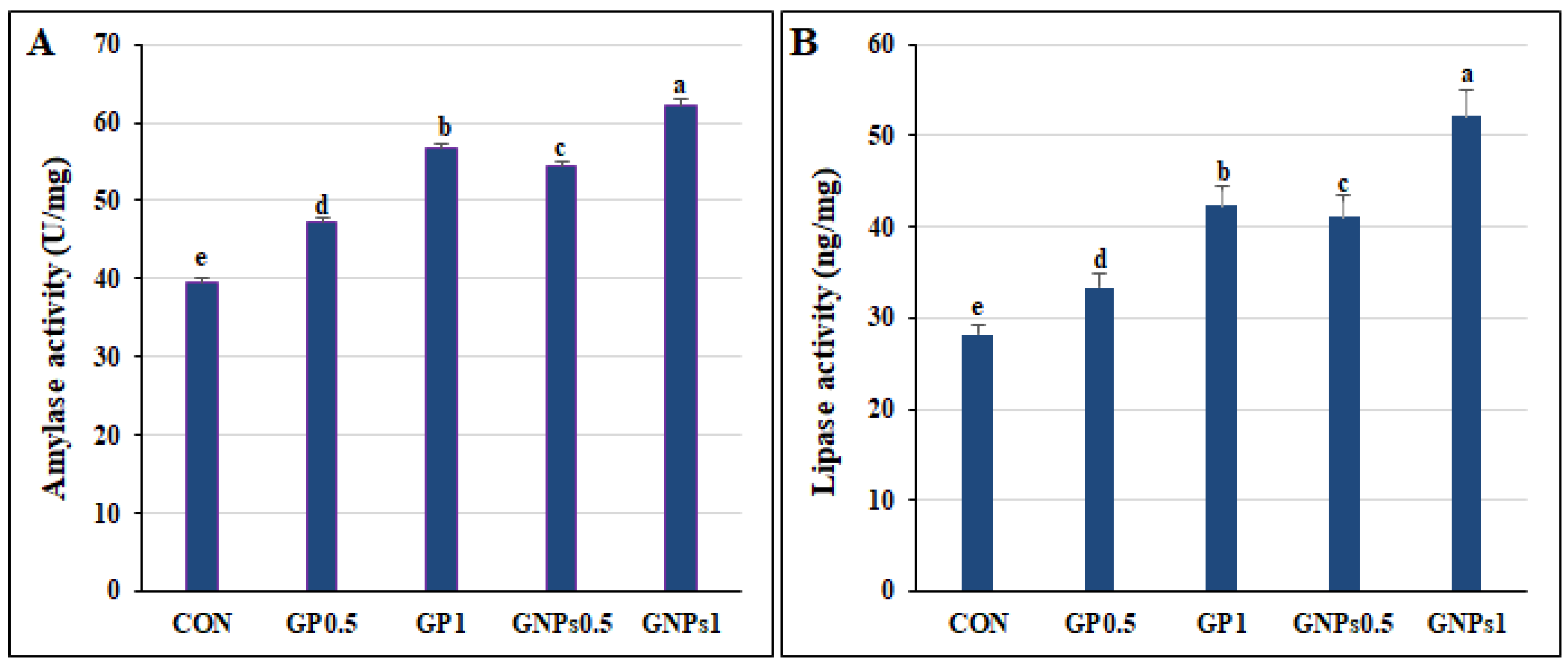
In the current study, diet enrichment with GP or GNPs improved fish growth, with the highest growth reported in the GNPs1 treatment. These results may indicate greater availability of nutrients for absorption, especially in GNPs TRTs. The growth-promoting effect of GP and GNPs can be attributed to their physicochemical properties and appetizer effect, which promote digestion, fat, and protein metabolism [12]. In addition, the increased growth hormone level and activity of digestive enzymes (amylase and lipase) reported in the supplemented TRTs would also promote fish growth. In addition to this, GP content of carbohydrates, minerals, vitamins, and other phytochemical compounds enhances animal growth and health [59]. Ginger rhizomes contain proteinase, which improves protein digestion and amino acid absorption [64]. Ginger also enhances the growth of microbial flora in the intestine [65]. The most significant growth was reported in the GNPs1 TRT, which can be explained by the enhanced bioavailability of nutrient nanoparticles and their high absorption, given their efficient entry into cells [66]. It was reported that growth rates of common carp (Cyprinus carpio) fingerlings were significantly increased after consuming diets containing ginger nanoparticles [17]. Moreover, Ude et al. [59] proposed that dietary supplementation with 1% ginger boosted the growth performance of African catfish (Clarias gariepinus). Accordingly, Korni and Khalil [17] hypothesized that adding GP and GNPs supplements to the diet of Cyprinus carpio fingerlings improved growth and FCR. Mohammadi et al. [67] reported improved weight gain, FCR, and RGR in common carp that were fed diets supplemented with ginger extract (0.1, 0.2, 0.4%), especially those that received 0.2% ginger extract. The improvement of fish growth by ginger has also been reported for Lates calcarifer fed 1% dried ginger plant for 15 days [12], Oreochromis niloticus fed 0.5% ginger essential oil for 55 days [68], and Labeo rohita fed 0.6–1% dried ginger plant for 60 days [69].
The present study showed increased total protein, growth hormone, and digestive enzyme activities in the supplemented groups. El-Sebai et al. [31] showed that ginger enhances protein and fat metabolism by increasing digestive enzyme secretion, improving digestion and absorption rates, and reducing pathogenic bacteria’s hazardous effects. Moreover, the phenolic compounds present in ginger, such as gingerols, paradols, and shogaols, enhance the secretion of pancreatic and intestinal lipase enzymes. This action facilitates fat digestion and absorption and stimulates protein synthesis [70]. Korni and Khalil [17] indicated improved growth and immunity by adding GP and GNPs to Cyprinus carpio fingerlings diets. Swain et al. [71] demonstrated that supplementing striped catfish (Pangasianodon hypophthalmus) diets with ginger promotes their growth performance.
Hematological parameters provide information on the animal’s health status [72]. In addition, leukocytes are essential for the innate immune system, and their count allows for the assessment of fish health and immunity [59]. The present study showed an improvement in the erythrogram and leukogram of fish fed diets enriched with GP or GNPs, particularly for GP1 and GNPs1 groups, indicating the immunostimulant properties of these ginger derivatives. Low levels of erythrocytes and hemoglobin are closely associated with impairment of the antioxidant system [73,74]. Hoseini et al. [75] established that exposure to ammonia causes oxidative stress by reducing the activities of antioxidant enzymes, causing anemia in Cyprinus carpio. Ginger is a powerful natural antioxidant [76] whose supplementation in the diet may protect RBCs from hemolysis by free radicals, thereby extending their lifespan. The increase in RBC count found in the current study is consistent with the reported antioxidant properties of GP and GNPs that protect RBC membranes from degradation [77]. Ginger is a source of phytochemical constituents such as phytate, oxalate, saponin, and tannin, as well as aromatic components which enhance the antioxidant and immune responses of fish [59]. Both GP and GNPs supplementation increases circulating hemoglobin levels in fish, consequently enhancing oxygen transport and improving health by stimulating growth and immune response [63,72]. In addition to this, the elevation of erythrocytes and hemoglobin could be due to the effect of ginger on hematopoiesis [78]. Positive effects of ginger supplementation on erythrocytes and hemoglobin have also been conveyed in earlier studies on Huso huso [79], Oncorhynchus mykiss [80], and Lates calcarifer [12]. The study by Mohammadi et al. [67] showed an increase in the erythrocyte and leucocyte count, as well as hematocrit and hemoglobin values, in common carp that received diets supplemented with ginger extract (0.1, 0.2, 0.4%). Udoh et al. [81] proved that high WBC counts, mainly due to increased lymphocytes and other phagocytes, enable fish to resist stressful conditions and infections. The improvement in hematological parameters can be explained by the presence of bioactive compounds in ginger, such as gingerols, zingerone, quercetin, paradols, shogaols, gingerenone-A, 6-dehydrogingerdione, β-bisabolene, zingiberene, α-farnesene, and β-sesquiphellandrene [6]. Furthermore, GNPs stimulate the production of anti-inflammatory cytokines while reducing that of pro-inflammatory cytokines [6]. GNPs are more stable than GP, preventing active component degradation and improving their effectiveness [82]. Korni and Khalil [17] indicated that ginger nanoparticles produced greater stimulation of Cyprinus carpio fingerlings’ immune response than ginger particles. Sharif Rohani et al. [83] stated that aloe vera extract nanoparticles promote and boost the immune response of Siberian sturgeon (Acipenser baerii).
Evidence demonstrated that ginger has strong antioxidant properties by boosting the action of several antioxidant enzymes and lowering reactive oxygen species (ROS) levels, which are responsible for the onset of many chronic diseases. It also reduces the number of free radicals, the precursors for lipid peroxidation [84]. This study revealed that fish fed diets supplemented with GP and GNPs presented a significant improvement in SOD, CAT, and GSH values, confirming the powerful antioxidant effect of the supplements. This effect can be attributed to the high content of polyphenolic compounds in ginger, including [6]-gingerol, paradols, shogaols, and diarylheptanoids, which have potent antioxidant properties [85]. GNPs are believed to have more powerful antioxidant properties than ginger extract. In particular, shogaols regulate numerous antioxidants and the detoxication of enzymes’ genetic expression [84,86]. The antioxidant activity of ginger have been reported by Ahmadifar et al. [9] in zebrafish, Sukumaran et al. [69] in Labeo rohita fingerlings, and Fazelan et al. [87] in Cyprinus carpio.
Lysozyme plays a crucial role in the immune system of fish, as it effectively lyses peptidoglycans present in bacterial cell walls. Furthermore, it stimulates phagocytosis and the complement system [88]. Nitric oxide has been shown to have powerful bactericidal properties [89]. The soluble IgM fraction has potent immunostimulatory activity [31]. IL-10, a cytokine associated with the adaptive immune system, protects tissues by decreasing the production of pro-inflammatory cytokines and eliminating excessive inflammation [70]. The present study has observed significant improvements in lysozyme, nitric oxide, IL-10, and IgM levels in the groups that received diets enriched with GP or GNPs. This finding indicates an enhancement of the fish immune system. El-Sebai et al. [31] showed that ginger supplementation in the O. niloticus diet boosted its immunity. Hence, the enhanced immune response may be attributed to phytochemical components (gingerol, paradols, and shogaols) and aromatic compounds present in ginger that stimulate antioxidant activity and immune functions in fish [6]. Mohammadi et al. [67] also reported the immunostimulant effect of ginger extract at 0.2 and 0.4% in Cyprinus carpio, indicated by increased lysozyme activity and immunoglobulin levels. Fazelan et al. [87] exhibited that ginger supplementation at 10 g kg−1 diet led to a significant increase in serum Ig in Cyprinus carpio exposed to high stocking-density stress, suggesting that dietary ginger may stimulate immune cells leading to enhanced immunity against harmful agents and stress.
Furthermore, our results indicated that GNPs have a more potent immunostimulatory effect than GP, which may be due to a smaller nanoparticle size and, therefore, a large exposed surface area, promoting greater solubility and bioavailability of active constituents [62,63]. Korni and Khalil [17] suggested that supplementing Cyprinus carpio fingerling diets with GNPs boosts their growth and immunity more than GP. This can be attributed to the nanoextraction of ginger, causing alterations in its characteristics. Zhang et al. [90] revealed that GNPs have a potent anti-inflammatory action by stimulating anti-inflammatory cytokines and reducing the concentrations of pro-inflammatory cytokines.
The present study showed that GP and GNPs supplementation increased resistance against A. hydrophila and P. putida infections. Ginger was reported to have potent antibacterial properties since it contains several bioactive substances such as gingerenone A, 6-shogaol, tannins, and saponins. These substances inhibit the growth of various Gram-positive and Gram-negative bacteria by suppressing bacterial biofilm formation and causing lysis of the bacterial cell wall [6]. Payung et al. [13] showed that dietary ginger supplementation promotes bacterial resistance of Nile tilapia to Aeromonas hydrophila. These findings matched those of Bakr et al. [86], who stated that ginger nanoparticles suppress the growth of a wide range of bacteria, including Salmonella typhimurium, Escherichia coli, Staphylococcus aureus, and Pseudomonas aeruginosa.
Enrichment of Nile tilapia diets with GP or GNPs improves liver function as indicated by reduced ALT and AST levels. The hepatoprotective effect of medicinal plants is primarily associated with their antioxidant activity, which prevents hepatic tissue damage [37]. This protective effect may be due to the bioactive ingredients in ginger, such as shogaols and gingerols, which act as free-radical scavengers preventing lipid peroxidation of hepatic cell membranes and, consequently, hepatic damage [85]. Abd-Elrhman et al. [84] stated that GNPs have a potent hepatoprotective effect against carbon tetrachloride (CCL4), which cause liver fibrosis in rats by decreasing the levels of ALT and AST enzymes. El-Sebai et al. [31] proved that supplementing the diet of Nile tilapia with ginger has a potent antioxidant effect and hepatoprotective function by preventing lipid peroxidation of cell membranes. Furthermore, dietary supplementation with GP or GNPs reduced serum urea and creatinine levels in the current study.
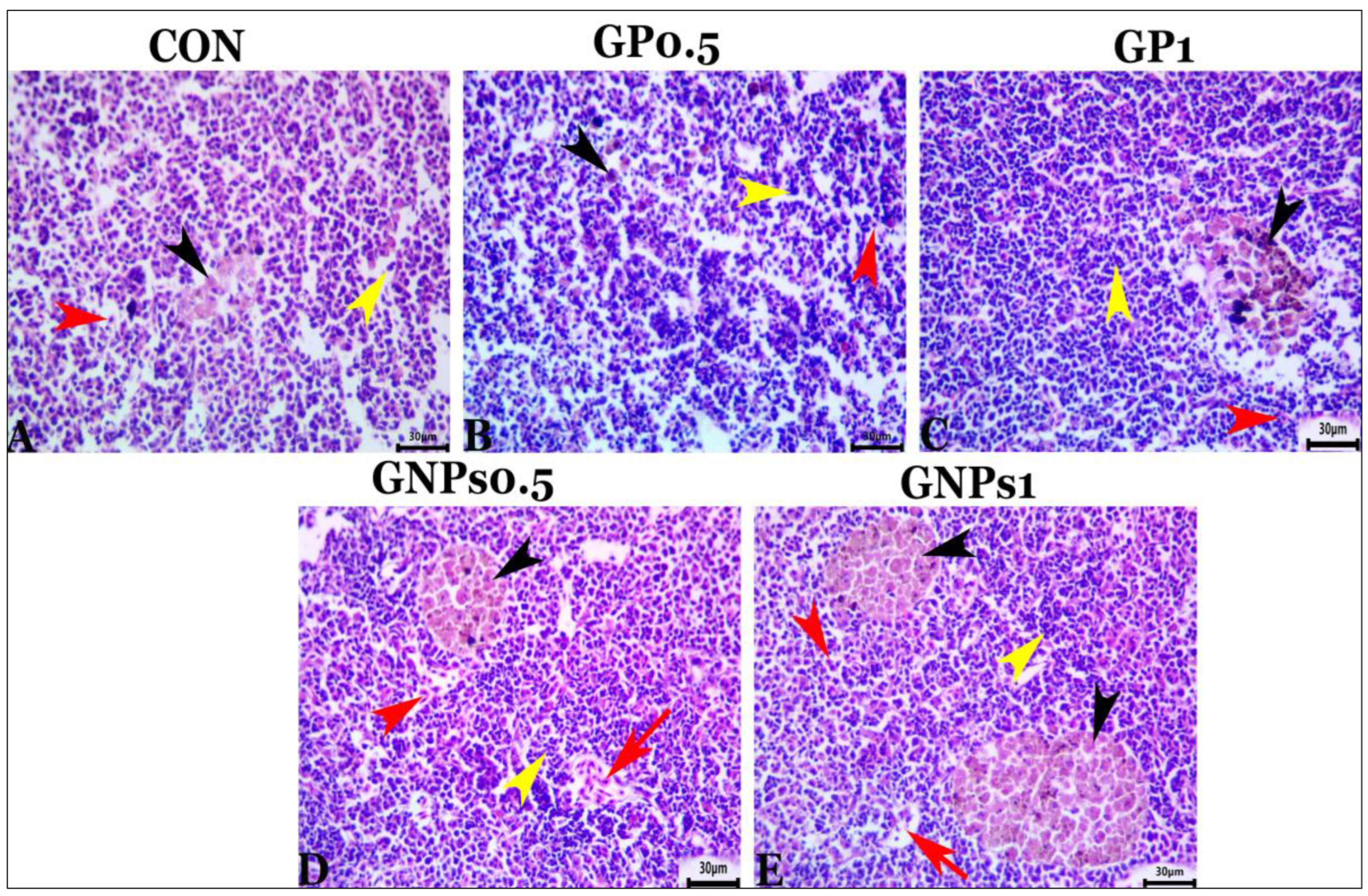
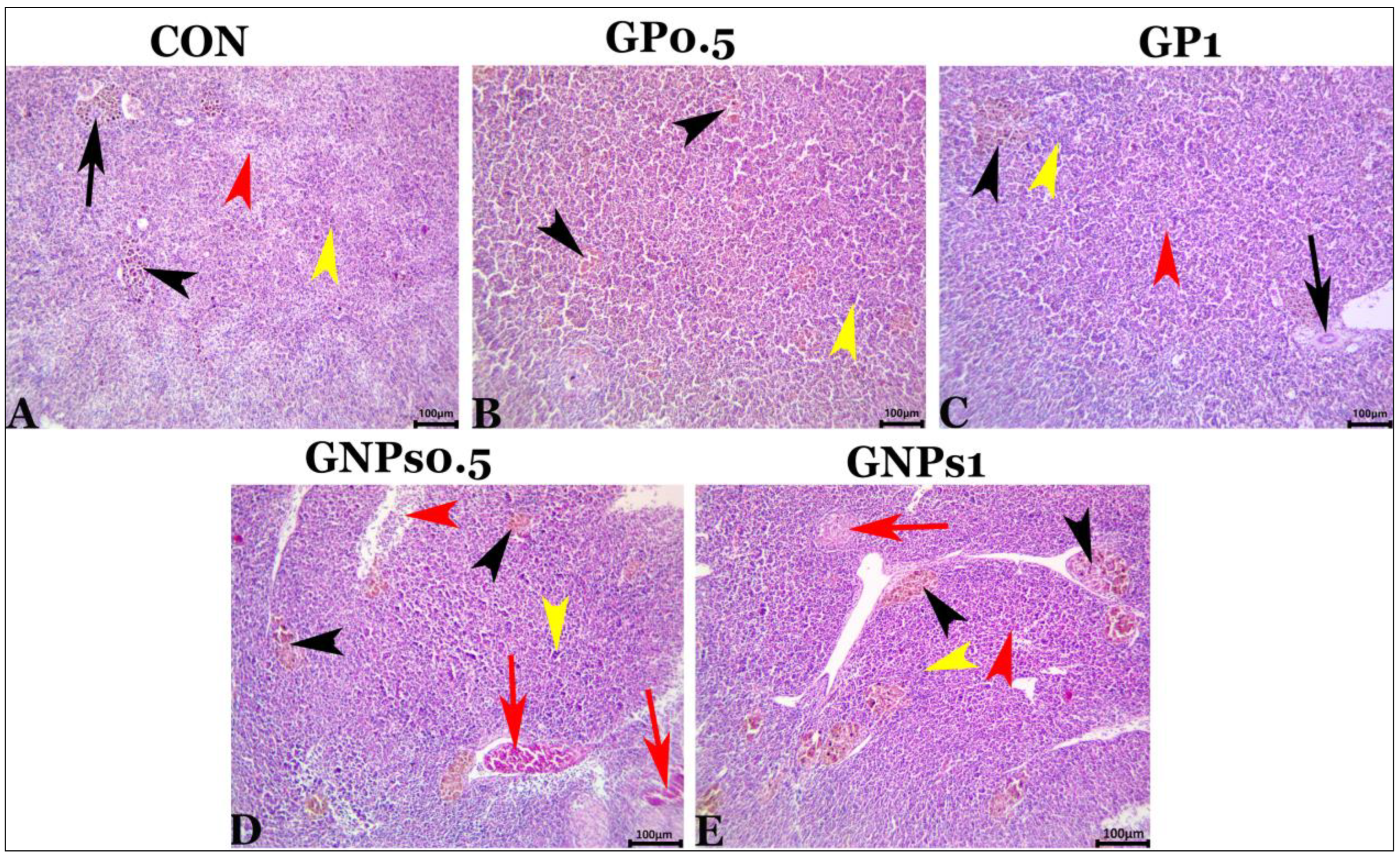
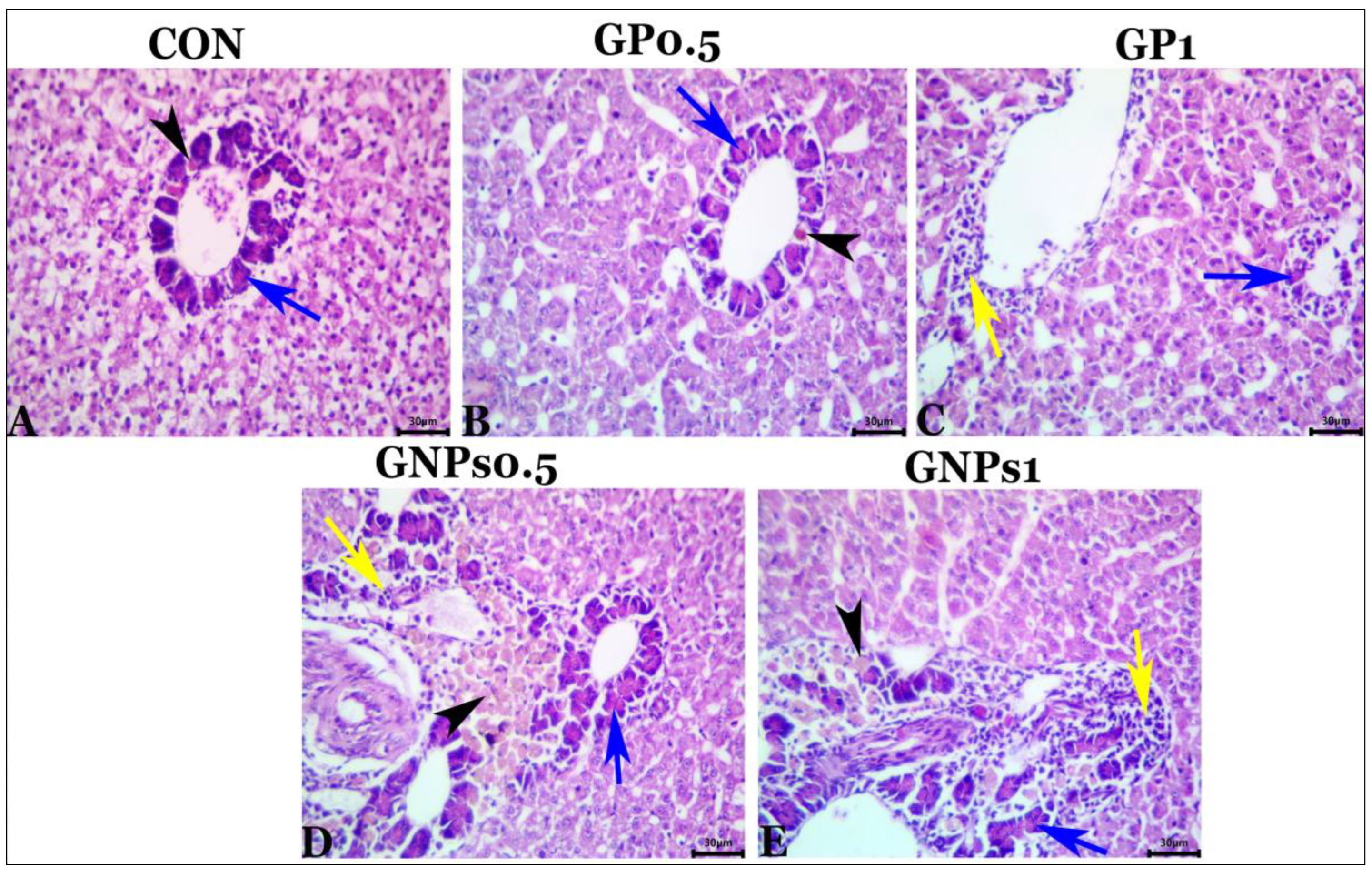
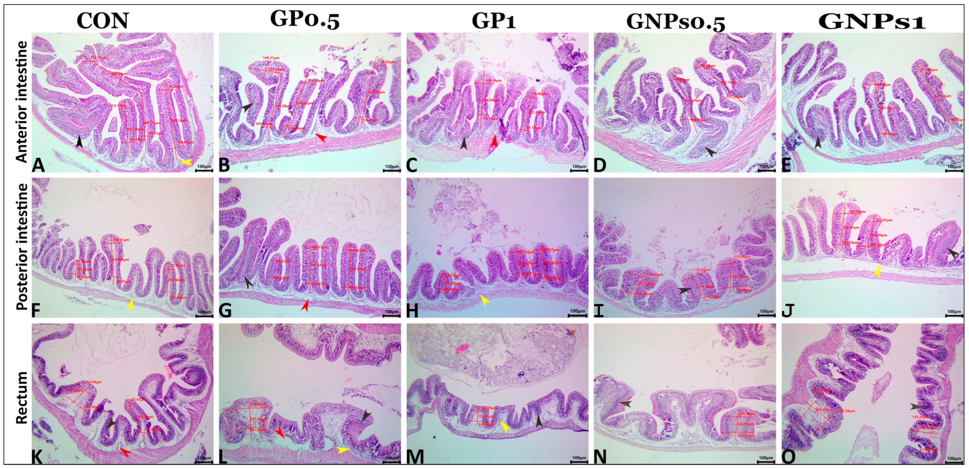
Regarding the histological examination of the spleen, liver, and kidney, fish fed the experimental diets showed the same typical histoarchitecture and morphology as the control group, indicating that dietary enrichment with GP or GNPs had no harmful effects on the internal organs of fish. These results confirmed the results of blood biochemistry (tests for liver and kidney function evaluation). It is well known that gut functions are crucial for nutrient digestion and absorption. Therefore, good and adequate growth depends mainly on the intestine’s condition. The villus of the digestive tract constitutes a vital sign of how effectively fish absorb nutrients [91]. Our results revealed that both ginger supplements, especially GNPs, enhance the mucosal-associated lymphoid tissue and the count of wandering eosinophilic granular cells. They also increase the values of all measured morphometric parameters (villus height, width, and surface area). The increase in villus height and width leads to a greater intestinal absorption area in fish fed the different experimental diets, consequently improving growth [92].

5. Conclusions

Based on the results obtained in the present study, we conclude that GP and GNPs could be effective dietary additives in Nile tilapia production at a supplementation level of 1%. Dietary supplementation with 1% GNPs or 1% GP increased fish growth by improving intestinal histomorphology and increasing digestive enzyme activity, serum growth hormone, and total protein without impairing organ function. Dietary enrichment with GNPs or GP at 1% improved blood hematology, immune status, and antioxidant activity, indicated by increased lysozyme and CAT activities and IgM, IL10, NO, SOD, and GSH levels. Furthermore, GP or GNPs supplementation at 1% increased the fish resistance to A. hydrophila and P. putida infections. We found that the most effective form and dose was the addition of 1% GNPs. Further studies are recommended to investigate the impact of GP and GNPs on protein absorption, amino acid transporter gene expression, and energy metabolism.

Supplementary Materials

The following are available online at https://www.mdpi.com/article/10.3390/fishes8050259/s1, Figure S1: The clinical picture of O. niloticus of the five different treatment groups, challenged with A. hydrophila: A- CON group, shows ocular opacity (arrow) and hemorrhagic areas on trunk and caudal peduncle regions (star). B- GP0.5 group shows severe fin rot of the dorsal fin (arrow), loss of scales, and hemorrhagic patches (star). C- GP1 group shows marked erythema of both pectoral fins (arrow). D- GNPs0.5 group shows severe erythema at the base of the pectoral fin (arrow). E- GNPs1 group shows normal appearance except for mild fin rot of the dorsal fin (arrow)., Figure S2: The clinical picture of O. niloticus of the five different treatment groups, challenged with P. putida: A- CON group shows the darkness of the caudal peduncle region with severe fin rot the caudal fin (arrow) and petechial hemorrhage at the operculum (star). B- GP0.5 group shows the darkness of the body, particularly at the caudal peduncle region, and severe fin rot of both the dorsal and the caudal fins (arrow). C- GP1 group shows the darkness of the whole body, erythema of the caudal fin with moderate fin rot, and mild hemorrhage at the lower jaw (arrow). D- GNPs0.5 group shows marked erythema of the caudal fin (arrow). E- GNPs1 group shows a normal clinical picture except for mild fin rot of the caudal fin (arrow).

Author Contributions

Conceptualization, S.A.A.A., A.E.-M., Y.A.E. and A.A.E.A.G.; methodology, E.N.M.K., S.A.A.A., A.E.-M., M.M.M., Y.A.E., A.A.E.A.G., S.A.A., E.M.Y., A.-W.A.A.-W. and S.J.D.; software, S.A.A.A., S.A.A., and E.N.M.K.; formal analysis, E.N.M.K., S.A.A., and S.A.A.A.; data curation, E.N.M.K., S.A.A. and S.A.A.A.; writing—original draft, E.N.M.K., S.A.A.A. and S.A.A.; writing—review and editing, E.N.M.K., S.A.A.A., S.A.A., A.E.-M., M.M.M., Y.A.E. and A.A.E.A.G.. All authors have read and agreed to the published version of the manuscript.

Funding

This work was supported by Zagazig University, Zagazig, Egypt.

Institutional Review Board Statement

The experiment procedures were approved by the Institutional Animal Care and Use Committee (ZU-IACUC) of Zagazig University, Egypt (Approval No. ZU-IACUC/2/F/6/2021).

Data Availability Statement

Data are contained in the article.

Acknowledgments

This work was supported by the Researchers Supporting Project (RSP2023R36), King Saud University, Riyadh, Saudi Arabia.

Conflicts of Interest

The authors declare that they have no conflict of interest.

References

  1. Vaseeharan, B.; Thaya, R. Medicinal plant derivatives as immunostimulants: An alternative to chemotherapeutics and antibiotics in aquaculture. Aquac. Int. 2014, 22, 1079–1091. [Google Scholar] [CrossRef]
  2. Li, M.; Wei, D.; Huang, S.; Huang, L.; Xu, F.; Yu, Q.; Liu, M.; Li, P. Medicinal herbs and phytochemicals to combat pathogens in aquaculture. Aquac. Int. 2022, 30, 1239–1259. [Google Scholar] [CrossRef]
  3. Amer, S.A.; El-Araby, D.A.; Tartor, H.; Farahat, M.; Goda, N.I.; Farag, M.F.; Fahmy, E.M.; Hassan, A.M.; Abo El-Maati, M.F.; Osman, A. Long-Term Feeding with Curcumin Affects the Growth, Antioxidant Capacity, Immune Status, Tissue Histoarchitecture, Immune Expression of Proinflammatory Cytokines, and Apoptosis Indicators in Nile Tilapia, Oreochromis niloticus. Antioxidants 2022, 11, 937. [Google Scholar] [CrossRef] [PubMed]
  4. Montaser, M.M.; El-Sharnouby, M.E.; El-Noubi, G.; El-Shaer, H.M.; Khalil, A.A.; Hassanin, M.; Amer, S.A.; El-Araby, D.A. Boswellia serrata resin extract in diets of Nile tilapia, Oreochromis niloticus: Effects on the growth, health, immune response, and disease resistance to Staphylococcus aureus. Animals 2021, 11, 446. [Google Scholar] [CrossRef] [PubMed]
  5. Al-Khalaifah, H.S.; Khalil, A.A.; Amer, S.A.; Shalaby, S.I.; Badr, H.A.; Farag, M.F.; Altohamy, D.E.; Abdel Rahman, A.N. Effects of dietary doum palm fruit powder on growth, antioxidant capacity, immune response, and disease resistance of African catfish, Clarias gariepinus (B.). Animals 2020, 10, 1407. [Google Scholar] [CrossRef] [PubMed]
  6. Mao, Q.-Q.; Xu, X.-Y.; Cao, S.-Y.; Gan, R.-Y.; Corke, H.; Beta, T.; Li, H.-B. Bioactive compounds and bioactivities of ginger (Zingiber officinale Roscoe). Foods 2019, 8, 185. [Google Scholar] [CrossRef]
  7. Thanikachalam, K.; Kasi, M.; Rathinam, X. Effect of garlic peel on growth, hematological parameters and disease resistance against Aeromonas hydrophila in African catfish Clarias gariepinus (Bloch) fingerlings. Asian Pac. J. Trop. Med. 2010, 3, 614–618. [Google Scholar] [CrossRef]
  8. Murray, M.T. The Healing Power of Herbs: The Enlightened Person’s Guide to the Wonders of Medicinal Plants; USA Prima Publisher: Rocklin, CA, USA, 1995. [Google Scholar]
  9. Ahmadifar, E.; Sheikhzadeh, N.; Roshanaei, K.; Dargahi, N.; Faggio, C. Can dietary ginger (Zingiber officinale) alter biochemical and immunological parameters and gene expression related to growth, immunity and antioxidant system in zebrafish (Danio rerio)? Aquaculture 2019, 507, 341–348. [Google Scholar] [CrossRef]
  10. Jesudoss, V.A.S.; Santiago, S.V.A.; Venkatachalam, K.; Subramanian, P. Zingerone (ginger extract): Antioxidant potential for efficacy in gastrointestinal and liver disease. In Gastrointestinal Tissue; Elsevier: Hoboken, NJ, USA, 2017; pp. 289–297. [Google Scholar]
  11. Maan, B.; Acharya, A. To evaluate the effect of ginger (Zigniber officinale) on body mass index an status of C-reactive protein in Diabetes Mellitus. Glob. J. Res. Anal. 2018, 7. Available online: https://www.worldwidejournals.com/global-journal-for-research-analysis-GJRA/article/to-evauate-the-effect-of-ginger-zingiber-officinale-on-body-mass-index-and-status-of-c-reactive-protein-in-diabetes-mellitus/ODI3Nw==/ (accessed on 20 April 2023).
  12. Talpur, A.D.; Ikhwanuddin, M.; Bolong, A.-M.A. Nutritional effects of ginger (Zingiber officinale Roscoe) on immune response of Asian sea bass, Lates calcarifer (Bloch) and disease resistance against Vibrio harveyi. Aquaculture 2013, 400, 46–52. [Google Scholar] [CrossRef]
  13. Payung, C.N.; Tumbol, R.A.; Manoppo, H. Dietary ginger (Zingiber officinale) enhance resistance of Nile tilapia (Oreochromis niloticus) against Aeromonas hydrophila. Aquac. Aquar. Conserv. Legis. 2017, 10, 962–968. [Google Scholar]
  14. Hussein, K.; Joo, J. Antifungal activity and chemical composition of ginger essential oil against ginseng pathogenic fungi. Curr. Res. Environ. Appl. Mycol. 2018, 8, 194–203. [Google Scholar] [CrossRef]
  15. Tatli Seven, P.; Seven, I.; Gul Baykalir, B.; Iflazoglu Mutlu, S.; Salem, A.Z. Nanotechnology and nano-propolis in animal production and health: An overview. Ital. J. Anim. Sci. 2018, 17, 921–930. [Google Scholar] [CrossRef]
  16. Rather, M.; Sharma, R.; Aklakur, M.; Ahmad, S.; Kumar, N.; Khan, M.; Ramya, V. Nanotechnology: A novel tool for aquaculture and fisheries development. A prospective mini-review. Fish. Aquac. J. 2011, 16, 3. [Google Scholar]
  17. Korni, F.; Khalil, F. Effect of ginger and its nanoparticles on growth performance, cognition capability, immunity and prevention of motile Aeromonas septicaemia in Cyprinus carpio fingerlings. Aquac. Nutr. 2017, 23, 1492–1499. [Google Scholar] [CrossRef]
  18. Abdu, S.B.; Abdu, F.; Khalil, W.K.B. Ginger nanoparticles modulate the apoptotic activity in male rats exposed to dioxin-induced cancer initiation. Int. J. Pharmacol. 2017, 13, 946–957. [Google Scholar] [CrossRef]
  19. Korni, F.M.; El-Ela, F.I.A.; Moawad, U.K.; Mahmoud, R.K.; Gadelhak, Y.M. Prevention of Edwardsiellosis in Clarias gariepinus using ginger and its nanoparticles with a reference to histopathological alterations. Aquaculture 2021, 539, 736603. [Google Scholar] [CrossRef]
  20. FAO. The State of World Fisheries and Aquaculture. Opportunities and Challenges. Food and Agriculture Organization of the United Nations. 2012. Available online: http://www.fao.org/3/i1820e/i1820e00.htm (accessed on 2 September 2020).
  21. Lim, C.; Webster, C. Nutrient requirements. In Tilapia: Biology, Culture and Nutrition; Food Products Press: New York, NY, USA, 2006; pp. 469–501. [Google Scholar]
  22. Chiew, I.K.M.; Salter, A.M.; Lim, Y.S. The significance of major viral and bacterial diseases in Malaysian aquaculture industry. Pertanika J. Trop. Agric. Sci. 2019, 42, 1023–1047. [Google Scholar]
  23. Ayoub, H.F.; Tohamy, E.Y.; Salama, H.M.; Mohamed, S.S. Isolation, Identification and Antimicrobial profile of Aeromonas spp., Pseudomonas spp. and Vibrio spp. from the Nile Tilapia, Oreochromis niloticus in fish farms. Egypt. J. Aquat. Biol. Fish. 2021, 25, 171–185. [Google Scholar]
  24. Kishk, D.; Moustafa, N.Y.; Kirrella, G.A. Prevalence and virulence characteristics of Aeromonas species isolated from fish farms in Egypt. Kafrelsheikh Vet. Med. J. 2020, 18, 5–8. [Google Scholar] [CrossRef]
  25. Swann, L.; White, M. Diagnosis and Treatment of “Aeromonas Hydrophila” Infection of Fish; Aquaculture Extension, Illinois-Indiana Sea Grant Program: Fact Sheet AS-461; Purdue University: West Lafayette, IN, USA, 1991; pp. 91–92. [Google Scholar]
  26. El-Son, M.; Abdelkhalek, N.; El-Ashram, A.; Zaki, V.H. Phenotypic and biochemical detection of Aeromonas hydrophila isolated from cultured Oreochromis niloticus during disease outbreaks. Int. J. Fish. Aquat. Stud. 2019, 7, 197–202. [Google Scholar]
  27. Austin, B.; Austin, D.A.; Austin, B.; Austin, D.A. Aeromonadaceae Representative (Aeromonas salmonicida). In Bacterial Fish Pathogens: Disease of Farmed and Wild Fish; Springer: Berlin/Heidelberg, Germany, 2016; pp. 215–321. [Google Scholar]
  28. Alzahrani, O.M.; Elumalai, P.; Nada, H.S.; Ahmed, S.A.; Zaglool, A.W.; Shawky, S.M.; Alkafafy, M.; Mahboub, H.H. Pseudomonas putida: Sensitivity to Various Antibiotics, Genetic Diversity, Virulence, and Role of Formic Acid to Modulate the Immune-Antioxidant Status of the Challenged Nile tilapia Compared to Carvacrol Oil. Fishes 2022, 8, 6. [Google Scholar] [CrossRef]
  29. Eissa, N.; El-Ghiet, E.; Shaheen, A.; Abbass, A. Characterization of Pseudomonas species isolated from tilapia “Oreochromis niloticus” in Qaroun and Wadi-El-Rayan lakes, Egypt. Glob. Vet. 2010, 5, 116–121. [Google Scholar]
  30. Li, M.; Mou, H.; Kong, Q.; Zhang, T.; Fu, X. Bacteriostatic effect of lipopeptides from Bacillus subtilis N-2 on Pseudomonas putida using soybean meal by solid-state fermentation. Mar. Life Sci. Technol. 2020, 2, 172–180. [Google Scholar] [CrossRef]
  31. El-Sebai, A.; El-Murr, A.E.-H.; Galal, A.A.; Abd El-Motaal, S.M. Effect of ginger dietary supplementation on growth performance, immune response and vaccine efficacy in Oreochromis niloticus challenged with Aeromonas hydrophila. Slov. Vet. Res. 2018, 55, 31–39. [Google Scholar]
  32. Rao, J.; McClements, D.J. Formation of flavor oil microemulsions, nanoemulsions and emulsions: Influence of composition and preparation method. J. Agric. Food Chem. 2011, 59, 5026–5035. [Google Scholar] [CrossRef]
  33. CCAC. Canadian Council on Animal Care Guidelines on: The Care and Use of Fish in Research, Teaching and Testing; CCAC: Ottawa, ON, Canada, 2005; p. 87. Available online: http://www.ccac.ca/Documents/Standards/Guidelines/Fish.pdf: (accessed on 18 July 2016).
  34. NRC. National Research Council. Nutrient Requirements of Fish and Shrimp; National Academies Press: Washington, DC, USA, 2011. [Google Scholar]
  35. AOAC. Official Methods of Analysis of AOAC International; AOAC: Rockville, MD, USA, 2000. [Google Scholar]
  36. APHA. WPCF (American Public Health Association, American Waterworks Association, Water Pollution Control Federation) (1992) Standard Methods for the Examination of Water and Wastewater. Stand. Methods Exam. Water Wastewater 1992, 17, 1193. [Google Scholar]
  37. Amer, S.A.; Ahmed, S.A.; Ibrahim, R.E.; Al-Gabri, N.A.; Osman, A.; Sitohy, M. Impact of partial substitution of fish meal by methylated soy protein isolates on the nutritional, immunological, and health aspects of Nile tilapia, Oreochromis niloticus fingerlings. Aquaculture 2020, 518, 734871. [Google Scholar] [CrossRef]
  38. Neiffer, D.L.; Stamper, M.A. Fish sedation, anesthesia, analgesia, and euthanasia: Considerations, methods, and types of drugs. ILAR J. 2009, 50, 343–360. [Google Scholar] [CrossRef]
  39. Izquierdo, M.; Henderson, R. The determination of lipase and phospholipase activities in gut contents of turbot (Scophthalmus maximus) by fluorescence-based assays. Fish Physiol. Biochem. 1998, 19, 153–162. [Google Scholar] [CrossRef]
  40. Worthington, V. Worthington Enzyme Manual: Enzymes and Related Biochemicals; Worthingthon Chemical: Lakewood, NJ, USA, 1993; p. 399. [Google Scholar]
  41. Feldman, B.; Zinkl, J.; Jain, N.C.; Schalm, O.W. Schalm’s Veterinary Hematology; Lippincott Williams & Wilkins: Philadelphia, PA, USA, 2000. [Google Scholar]
  42. Aebi, H. Catalase in vitro. In Methods in Enzymology; Elsevier: Amsterdam, The Netherlands, 1984; Volume 105, pp. 121–126. [Google Scholar]
  43. McCord, J.M.; Fridovich, I. Superoxide dismutase: An enzymic function for erythrocuprein (hemocuprein). J. Biol. Chem. 1969, 244, 6049–6055. [Google Scholar] [CrossRef] [PubMed]
  44. Beutler, E.; Duron, O.; Kelly, B.M. Improved method for the determination of blood glutathione. J. Lab. Clin. Med. 1963, 61, 882–888. [Google Scholar] [PubMed]
  45. Ellis, A.E. Lysozyme assays. Tech. Fish Immunol. 1990, 1, 101–103. [Google Scholar]
  46. Montgomery, H.; Dymock, J.F. Determination of nitrite in water. Analyst 1961, 86, 414–417. [Google Scholar]
  47. Doumas, B.T.; Bayse, D.D.; Carter, R.J.; Peters, T., Jr.; Schaffer, R. A candidate reference method for determination of total protein in serum. I. Development and validation. Clin. Chem. 1981, 27, 1642–1650. [Google Scholar] [CrossRef] [PubMed]
  48. Doumas, B.T.; Biggs, H.G.; Arends, R.L.; Pinto, P.V. Determination of serum albumin. In Standard Methods of Clinical Chemistry; Elsevier: Amsterdam, The Netherlands, 1972; Volume 7, pp. 175–188. [Google Scholar]
  49. Reitman, S.; Frankel, S. Colorimetric methods for aspartate and alanine aminotransferase. Am. J. Clin. Path 1957, 28, 55–60. [Google Scholar]
  50. Fossati, P.; Prencipe, L.; Berti, G. Enzymic creatinine assay: A new colorimetric method based on hydrogen peroxide measurement. Clin. Chem. 1983, 29, 1494–1496. [Google Scholar] [CrossRef]
  51. Patton, C.; Crouch, S. Determination of urea by Enzymatic colorimetric method. Anal. Chem. 1977, 49, 464–469. [Google Scholar] [CrossRef]
  52. Abbasi Ghadikolaei, H.; Kamali, A.; Soltani, M.; Sharifian, M. Effects of Zingiber officinale powder on growth parameters, survival rate and biochemical composition of body in juvenile common carp (Cyprinus carpio). Iran. J. Fish. Sci. 2017, 16, 67–85. [Google Scholar]
  53. Suvarna, K.S.; Layton, C.; Bancroft, J.D. Bancroft’s Theory and Practice of Histological Techniques E-Book; Elsevier Health Sciences: Amsterdam, The Netherlands, 2018. [Google Scholar]
  54. Iji, P.; Saki, A.; Tivey, D. Body and intestinal growth of broiler chicks on a commercial starter diet. 1. Intestinal weight and mucosal development. Br. Poult. Sci. 2001, 42, 505–513. [Google Scholar] [CrossRef]
  55. Sun, X.; McElroy, A.; Webb, K., Jr.; Sefton, A.; Novak, C. Broiler performance and intestinal alterations when fed drug-free diets. Poult. Sci. 2005, 84, 1294–1302. [Google Scholar] [CrossRef]
  56. Scheidegger, E.; Fracalanzza, S.; Teixeira, L.; Cardarelli-Leite, P. RFLP analysis of a PCR-amplified fragment of the 16S rRNA gene as a tool to identify Enterococcus strains. Memórias Do Inst. Oswaldo Cruz 2009, 104, 1003–1008. [Google Scholar] [CrossRef]
  57. Zhou, F.; Song, W.; Shao, Q.; Peng, X.; Xiao, J.; Hua, Y.; Owari, B.N.; Zhang, T.; Ng, W.K. Partial replacement of fish meal by fermented soybean meal in diets for black sea bream, Acanthopagrus schlegelii, juveniles. J. World Aquac. Soc. 2011, 42, 184–197. [Google Scholar] [CrossRef]
  58. Aly, S.M.; Ahmed, Y.A.-G.; Ghareeb, A.A.-A.; Mohamed, M.F. Studies on Bacillus subtilis and Lactobacillus acidophilus, as potential probiotics, on the immune response and resistance of Tilapia nilotica (Oreochromis niloticus) to challenge infections. Fish Shellfish Immunol. 2008, 25, 128–136. [Google Scholar] [CrossRef] [PubMed]
  59. Ude, E.; Iheanacho, S.C.; Ogbu, M.; Ayotunde, E.; Ogueji, E. Growth, Hematology and Immuno-Modulatory Potential of Ginger (Zingiber officinale) Supplemented Diets in Clarias gariepinus Juvenile (Burchell, 1822). Aquac. Stud. 2018, 18, 41–49. [Google Scholar]
  60. El-Araby, D.A.; Amer, S.A.; Attia, G.A.; Osman, A.; Fahmy, E.M.; Altohamy, D.E.; Alkafafy, M.; Elakkad, H.A.; Tolba, S.A. Dietary Spirulina platensis phycocyanin improves growth, tissue histoarchitecture, and immune responses, with modulating immunoexpression of CD3 and CD20 in Nile tilapia, Oreochromis niloticus. Aquaculture 2022, 546, 737413. [Google Scholar] [CrossRef]
  61. Fajardo, C.; Martinez-Rodriguez, G.; Blasco, J.; Mancera, J.M.; Thomas, B.; De Donato, M. Nanotechnology in aquaculture: Applications, perspectives and regulatory challenges. Aquac. Fish. 2022, 7, 185–200. [Google Scholar] [CrossRef]
  62. Ahmed, S.A.; Ibrahim, R.E.; Farroh, K.Y.; Moustafa, A.A.; Al-Gabri, N.A.; Alkafafy, M.; Amer, S.A. Chitosan vitamin E nanocomposite ameliorates the growth, redox, and immune status of Nile tilapia (Oreochromis niloticus) reared under different stocking densities. Aquaculture 2021, 541, 736804. [Google Scholar] [CrossRef]
  63. Ibrahim, R.E.; Amer, S.A.; Farroh, K.Y.; Al-Gabri, N.A.; Ahmed, A.I.; El-Araby, D.A.; Ahmed, S.A. The effects of chitosan-vitamin C nanocomposite supplementation on the growth performance, antioxidant status, immune response, and disease resistance of Nile tilapia (Oreochromis niloticus) fingerlings. Aquaculture 2021, 534, 736269. [Google Scholar] [CrossRef]
  64. Hashim, M.M.; Mingsheng, D.; Iqbal, M.F.; Xiaohong, C. Ginger rhizome as a potential source of milk coagulating cysteine protease. Phytochemistry 2011, 72, 458–464. [Google Scholar] [CrossRef]
  65. Ali, B.H.; Blunden, G.; Tanira, M.O.; Nemmar, A. Some phytochemical, pharmacological and toxicological properties of ginger (Zingiber officinale Roscoe): A review of recent research. Food Chem. Toxicol. 2008, 46, 409–420. [Google Scholar] [CrossRef] [PubMed]
  66. Ogunkalu, O.A. Utilization of nanotechnology in aquaculture and seafood sectors. Eurasian J. Food Sci. Technol. 2019, 3, 26–33. [Google Scholar]
  67. Mohammadi, G.; Rashidian, G.; Hoseinifar, S.H.; Naserabad, S.S.; Van Doan, H. Ginger (Zingiber officinale) extract affects growth performance, body composition, haematology, serum and mucosal immune parameters in common carp (Cyprinus carpio). Fish Shellfish Immunol. 2020, 99, 267–273. [Google Scholar] [CrossRef] [PubMed]
  68. Brum, A.; Pereira, S.A.; Owatari, M.S.; Chagas, E.C.; Chaves, F.C.M.; Mouriño, J.L.P.; Martins, M.L. Effect of dietary essential oils of clove basil and ginger on Nile tilapia (Oreochromis niloticus) following challenge with Streptococcus agalactiae. Aquaculture 2017, 468, 235–243. [Google Scholar] [CrossRef]
  69. Sukumaran, V.; Park, S.C.; Giri, S.S. Role of dietary ginger Zingiber officinale in improving growth performances and immune functions of Labeo rohita fingerlings. Fish Shellfish Immunol. 2016, 57, 362–370. [Google Scholar] [CrossRef] [PubMed]
  70. Kumar, R.M.; Rao, A.; Daggula, N.; Guguloth, G.; Das, B.Y.; Indhuri, A. Growth promoter effect of ginger, garlic and fenugreek on Pacific white leg shrimp (Litopenaeus vannamei). Int. J. Curr. Microbiol. Appl. Sci. 2019, 8, 2993–3001. [Google Scholar] [CrossRef]
  71. Swain, S.; Pawase, A.; Pai, R.; Tibile, R.; Indulkar, S.; Pawar, R. Effect of ginger (Zingiber officinale Roscoe) incorporated diet on growth performance of striped catfish, Pangasianodon hypophthalmus (Sauvage, 1878). J. Entomol. Zool. Stud. 2018, 6, 1094–1098. [Google Scholar]
  72. Ahmed, S.A.; Abd El-Rahman, G.I.; Behairy, A.; Beheiry, R.R.; Hendam, B.M.; Alsubaie, F.M.; Khalil, S.R. Influence of feeding quinoa (Chenopodium quinoa) seeds and prickly pear fruit (Opuntia ficus indica) peel on the immune response and resistance to Aeromonas sobria infection in Nile tilapia (Oreochromis niloticus). Animals 2020, 10, 2266. [Google Scholar] [CrossRef]
  73. Hoseini, S.M.; Mirghaed, A.T.; Iri, Y.; Ghelichpour, M. Effects of dietary cineole administration on growth performance, hematological and biochemical parameters of rainbow trout (Oncorhynchus mykiss). Aquaculture 2018, 495, 766–772. [Google Scholar] [CrossRef]
  74. Hajirezaee, S.; Mohammadi, G.; Naserabad, S.S. The protective effects of vitamin C on common carp (Cyprinus carpio) exposed to titanium oxide nanoparticles (TiO2-NPs). Aquaculture 2020, 518, 734734. [Google Scholar] [CrossRef]
  75. Hoseini, S.M.; Yousefi, M.; Hoseinifar, S.H.; Van Doan, H. Antioxidant, enzymatic and hematological responses of common carp (Cyprinus carpio) fed with myrcene-or menthol-supplemented diets and exposed to ambient ammonia. Aquaculture 2019, 506, 246–255. [Google Scholar] [CrossRef]
  76. Si, W.; Chen, Y.P.; Zhang, J.; Chen, Z.-Y.; Chung, H.Y. Antioxidant activities of ginger extract and its constituents toward lipids. Food Chem. 2018, 239, 1117–1125. [Google Scholar] [CrossRef] [PubMed]
  77. Al-Snafi, A.E. Chemical constituents, pharmacological effects and therapeutic importance of Hibiscus rosa-sinensis-A review. IOSR J. Pharm. 2018, 8, 101–119. [Google Scholar]
  78. Ferri-Lagneau, K.F.; Moshal, K.S.; Grimes, M.; Zahora, B.; Lv, L.; Sang, S.; Leung, T. Ginger stimulates hematopoiesis via Bmp pathway in zebrafish. PLoS ONE 2012, 7, e39327. [Google Scholar]
  79. Gholipour Kanani, H.; Nobahar, Z.; Kakoolaki, S.; Jafarian, H. Effect of ginger-and garlic-supplemented diet on growth performance, some hematological parameters and immune responses in juvenile Huso huso. Fish Physiol. Biochem. 2014, 40, 481–490. [Google Scholar] [CrossRef] [PubMed]
  80. Nya, E.J.; Austin, B. Use of dietary ginger, Zingiber officinale Roscoe, as an immunostimulant to control Aeromonas hydrophila infections in rainbow trout, Oncorhynchus mykiss (Walbaum). J. Fish Dis. 2009, 32, 971–977. [Google Scholar] [CrossRef]
  81. Udoh, J.P.; Emah, A.U.; George, I.E.; Philip, A.E. Growth performance and haematological response of Clarias gariepinus broodstock fed diets enriched with bitter leaf meal. Aquac. Aquar. Conserv. Legis. 2017, 10, 1281–1296. [Google Scholar]
  82. Luis, A.I.; Campos, E.V.; De Oliveira, J.L.; Guilger-Casagrande, M.; De Lima, R.; Castanha, R.F.; De Castro, V.L.; Fraceto, L.F. Zein nanoparticles impregnated with eugenol and garlic essential oils for treating fish pathogens. ACS Omega 2020, 5, 15557–15566. [Google Scholar] [CrossRef]
  83. Sharif Rohani, M.; Haghighi, M.; Bazari Moghaddam, S. Study on nanoparticles of Aloe vera extract on growth performance, survival rate and body composition in Siberian sturgeon (Acipenser baerii). Iran. J. Fish. Sci. 2017, 16, 457–468. [Google Scholar]
  84. Abd-Elrhman, S.Y.; Abd El-Fattah, H.M.; Morsy, G.M.; Elmasry, S. Effect of ginger nanoparticles on hepato-renal toxicity induced by carbon tetrachloride in rats. Annu. Res. Rev. Biol. 2020, 35, 36–55. [Google Scholar] [CrossRef]
  85. Ojarudi, M.; Moradi, A.; Hajihosseini, R.; Mazani, M.; Rezagholizadeh, L. Hepatoprotective and Antioxidant Activities of Combination of Cinnamomum zeylanicum and Zingiber officinale in CCl4-intoxicated Rats. J. Kerman Univ. Med. Sci. 2020, 27, 1–13. [Google Scholar]
  86. Bakr, A.F.; Abdelgayed, S.S.; El-Tawil, O.S.; Bakeer, A.M. Assessment of ginger extract and ginger nanoparticles protective activity against acetaminophen-induced hepatotoxicity and nephrotoxicity in rats. Pak. Vet. J. 2019, 39, 479–486. [Google Scholar] [CrossRef]
  87. Fazelan, Z.; Vatnikov, Y.A.; Kulikov, E.V.; Plushikov, V.G.; Yousefi, M. Effects of dietary ginger (Zingiber officinale) administration on growth performance and stress, immunological, and antioxidant responses of common carp (Cyprinus carpio) reared under high stocking density. Aquaculture 2020, 518, 734833. [Google Scholar] [CrossRef]
  88. Valipour, A.; Heidari, B.; Hadavi, M.; Yousefi, A. Changes in immune parameters (lysozyme, IgM, C3) in early life stages and broodstock of Siberian sturgeon, Acipenser Baerii. Fish. Aquat. Life 2018, 26, 21–30. [Google Scholar] [CrossRef]
  89. Giordano, D.; Verde, C.; Corti, P. Nitric Oxide Production and Regulation in the Teleost Cardiovascular System. Antioxidants 2022, 11, 957. [Google Scholar] [CrossRef] [PubMed]
  90. Zhang, M.; Viennois, E.; Prasad, M.; Zhang, Y.; Wang, L.; Zhang, Z.; Han, M.K.; Xiao, B.; Xu, C.; Srinivasan, S. Edible ginger-derived nanoparticles: A novel therapeutic approach for the prevention and treatment of inflammatory bowel disease and colitis-associated cancer. Biomaterials 2016, 101, 321–340. [Google Scholar] [CrossRef]
  91. Valladao, G.M.; Gallani, S.U.; Pala, G.; Jesus, R.B.; Kotzent, S.; Costa, J.C.; Silva, T.F.; Pilarski, F. Practical diets with essential oils of plants activate the complement system and alter the intestinal morphology of Nile tilapia. Aquac. Res. 2017, 48, 5640–5649. [Google Scholar] [CrossRef]
  92. Asghar, M.; Rahman, A.; Hayat, Z.; Rafique, M.; Badar, I.; Yar, M.; Ijaz, M. Exploration of Zingiber officinale effects on growth performance, immunity and gut morphology in broilers. Braz. J. Biol. 2021, 83, e250296. [Google Scholar] [CrossRef]
Figure 1. (A) Particle size distribution/Zeta potential of ginger extract nanoemulsion. (B) transmission electron microscopy (TEM) image of ginger extract nanoemulsion. Magnification: 40,000×.
Fishes 08 00259 g001
Figure 2. Amylase (A) and lipase (B) activities in fish fed the five experimental diets (n =3). Means not sharing a common superscript (a, b, c, d, e) differ significantly (Tukey’s test, p < 0.05).
Fishes 08 00259 g002
Figure 3. Representative photomicrographs of HE-stained head kidney sections (n= 9) showing normal histology in the CON (A), GP0.5 (B), and GP1 (C) groups. Dense cellularity with the erythropoietic, lymphopoietic, and granulopoietic blast cells, besides hyperplasia of the MMCs associated with mild interstitial edema and notable hyperemia, are seen in the GNPs0.5 (D) and GNPs1 (E) groups. Red arrowheads: erythropoietic blast cells. Yellow arrowheads: lymphopoietic blast cells. Back arrowheads: MMCs. Red arrows: hyperemic blood vessels. Scale bars = 30 microns (×40).
Fishes 08 00259 g003
Figure 4. Representative photomicrographs of HE-stained splenic tissue sections (n= 9) showing normal histology in the CON (A), GP0.5 (B), and GP1 (C) groups. Light cellularity, particularly with the lymphopoietic blast cells with notable hyperplasia of the MMCs associated with marked hyperemia, is seen in the GNPs0.5 (D) and GNPs1 (E) groups. Red arrowheads: erythropoietic blast cells. Yellow arrowheads: lymphopoietic blast cells. Black arrowheads: MMCs. Black arrows: ellipsoids. Red arrows: hyperemic blood vessels. Scale bars = 100 microns (×10).
Fishes 08 00259 g004
Figure 5. Representative photomicrographs of HE-stained hepatic tissue sections (n = 9) showing normal histology in the CON (A). Notable decrease in the cytoplasmic vacuolations with a slight increase in the numbers of MMCs; the presence of few numbers of mononuclear cells, particularly around the exocrine pancreas, are seen in the GP0.5 (B), GP1 (C), GNPs0.5 (D), and GNPs1 (E) groups. Black arrowheads: MMCs. Blue arrow: exocrine pancreatic cells. Yellow arrow: mononuclear cell infiltration. Scale bars = 30 microns (×40).
Fishes 08 00259 g005
Figure 6. Representative photomicrographs of HE-stained intestinal tissue sections (n = 9) showing the villus height and width, mucosal-associated lymphoid tissue (black arrowheads), wandering eosinophilic granular cells (yellow arrowheads), and blood capillaries (red arrowheads) in different groups. Scale bars = 100 microns (×10). CON group (A,F,K), GP0.5 group (B,G,L), GP1 group (C,H,M), GNPs0.5 group (D,I,N), and GNPs1 group (E,J,O).
Fishes 08 00259 g006
Table 1. Feed formulation and proximate composition (g kg−1 on a dry weight basis).
Ingredientsg kg−1
Soybean meal 49% CP240
Fish meal 70.7% CP200
Yellow corn230
Corn gluten 67% CP80
Wheat flour100
Wheat bran60
Fish oil30
Soy oil30
Premix 130
Chemical composition (g kg−1)
Crude protein358
Crude fiber34.78
Fat99.95
NFE 2441.6
Ash66.5
Lysine20.34
Methionine7.61
GE MJ/kg 320.65
1 Composition/kg: Vit. A 580,000 IU; Vit. D3 8600 IU; Vit. K3 142 mg; Vit. E 720 mg; Vit. C 0.1 mg; Vit. B1 58 mg; Vit. B2 34 mg; Vit. B6 34 mg; Vit. B12 58 mg; folic acid 86 mg; biotin 50 mg; pantothenic acid 8 mg; Zn methionine 3000 mg; Mn sulfate 65 mg; copper sulfate 3400 mg; iron sulfate 2000 mg; sodium selenite 25 mg; cobalt sulfate 572 mg; calcium iodide 25 mg; calcium carbonate as carrier up to 1 kg. 2 Nitrogen free extract (NFE), determined by difference = 100 − (protein % + fat % + crude fiber % + ash %). 3 Gross energy (GE) was calculated as 23.6 KJ/g protein, 39.5 KJ/g lipid, and 17.0 KJ/g NFE [34]. CP: crude protein.
Table 2. Growth performance parameters of Nile tilapia under different experimental TRTs.
ParametersCONGP0.5GP1 GNPs0.5 GNPs1p-Value
IBW/fish (g)27.11 ± 0.2027.35 ± 0.3426.62 ± 0.3826.66 ± 0.4027.32 ± 0.380.43
FBW/fish (g)80.87 ± 0.72 d84.72 ± 0.21 c91.15 ± 0.54 b89.67 ± 0.26 b95.02 ± 0.10 a<0.01
TWG/fish (g)53.76 ± 0.52 e57.38 ± 0.20 d64.53 ± 0.25 b63.01 ± 0.27 c67.70 ± 0.01 a<0.01
TFI/fish (g)101.84 ± 2.45103.22 ± 2.76107.88 ± 3.6106.94 ± 3.25105.88 ± 4.940.72
FCR1.89 ± 0.03 a1.79 ± 0.04 ab1.67 ± 0.05 ab1.69 ± 0.07 ab1.56 ± 0.04 b0.01
SGR (%)1.56 ± 0.01 b1.61 ± 0.01 b1.75 ± 0.01 a1.73 ± 0.01 a1.78 ± 0.01 a<0.01
PER1.63 ± 0.03 b1.72 ± 0.04 ab1.86 ± 0.06 ab1.83 ± 0.07 ab1.98 ± 0.03 a0.01
IBW, initial body weight; FBW, final body weight; TWG, total weight gain; TFI, total feed intake; SGR, specific growth rate; FCR, feed conversion ratio; PER, protein efficiency ratio. Values are expressed as mean ± SE. Values not sharing a common superscript (a, b, c, d, e) differ significantly (Tukey’s test, p < 0.05).
Table 3. Erythrogram and leukogram of Nile tilapia fed the experimental diets.
ParametersCONGP0.5GP1 GNPs0.5 GNPs1p-Value
Erythrogram
RBCs (106/µL)1.46 ± 0.01 e1.62 ± 0.01 d2.38 ± 0.02 b2.14 ± 0.02 c2.75 ± 0.02 a<0.01
Hb (g/dL)6.75 ± 0.01 e7.45 ± 0.03 d8.43 ± 0.07 b8.15 ± 0.02 c8.93 ± 0.02 a<0.01
PCV (%)24.47 ± 0.02 e25.78 ± 0.02 d30.80 ± 0.01 b30.17 ± 0.04 c31.57 ± 0.04 a<0.01
MCV (fL)167.98 ± 1.08 a158.83 ± 1.50 b129.41 ± 0.92 d141.02 ± 1.23 c114.66 ± 0.89 e<0.01
MCH (pg)46.36 ± 0.33 a45.90 ± 0.28 a35.43 ± 0.53 c38.11 ± 0.31 b32.43 ± 0.18 d<0.01
MCHC (%)27.60 ± 0.05 c28.90 ± 0.14 a27.37 ± 0.22 cd27.02 ± 0.03 d28.29 ± 0.07 b<0.01
Leukogram
WBCs (103/µL)7.23 ± 0.01 e8.12 ± 0.05 d9.16 ± 0.08 b8.93 ± 0.01 c10.32 ± 0.05 a<0.01
Lymphocytes (103/µL)4.35 ± 0.09 d5.13 ± 0.03 c5.63 ± 0.01 b5.53 ± 0.01 b6.21 ± 0.06 a<0.01
Heterophils (103/µL)2.33 ± 0.01 e2.64 ± 0.01 d3.57 ± 0.01 b3.08 ± 0.02 c3.82 ± 0.02 a<0.01
Monocytes (103/µL)0.52 ± 0.01 d0.58 ± 0.01 c0.64 ± 0.01 b0.62 ± 0.01 b72.0 ± 0.02 a<0.01
Eosinophils (103/µL)0.19 ± 0.01 a0.18 ± 0.01 ab0.17 ± 0.01 ab0.19 ± 0.01 a0.16 ± 0.01 b<0.01
Basophils (103/µL)0.05 ± 0.001 a0.05 ± 0.001 b0.05 ± 0.00 c0.05 ± 0.00 b0.05 ± 0.00 b<0.01
RBCs, red blood cells count; Hb, hemoglobin concentration; PCV, packed cell volume; MCV, mean corpuscular volume; MCH, mean corpuscular hemoglobin; MCHC, mean corpuscular hemoglobin concentration; WBCs, white blood cells count. Values are expressed as mean ± SE (n= 3). Values not sharing a common superscript (a, b, c, d, e) differ significantly (Tukey’s test, p < 0.05) (n= 3).
Table 4. Antioxidant activity and immune status of Nile tilapia under different experimental TRTs.
ParametersCONGP0.5GP1 GNPs0.5 GNPs1p-Value
Antioxidant indices
SOD (U/g)5.52 ± 0.17 c7.59 ± 0.18 b8.88 ± 0.03 a8.79 ± 0.01 a9.33 ± 0.07 a<0.01
CAT (U/G)14.73 ± 0.25 d19.41 ± 0.23 c21.66 ± 0.19 b20.82 ± 0.11 b23.90 ± 0.11 a<0.01
GSH (mmol/g)1.53 ± 0.01 e2.11 ± 0.03 d2.76 ± 0.01 b2.62 ± 0.01 c2.95 ± 0.01 a<0.01
Immune indices
Lysozyme (µg/mL)16.37 ± 0.52 d25.69 ± 0.71 c30.35 ± 0.43 b29.17 ± 0.36 b34.42 ± 0.35 a<0.01
Nitric oxide (μmol/L)34.88 ± 0.18 d39.36 ± 0.25 c44.78 ± 0.28 b44.19 ± 0.18 b49.93 ± 0.21 a<0.01
IL10 (pg/mL)361.30 ± 0.28 e411.54 ± 0.25 d632.60 ± 0.27 b587.26 ± 0.28 c704.32 ± 0.27 a<0.01
IgM (µg/mL)26.28 ± 0.20 e31.93 ± 0.10 d37.89 ± 0.09 b36.81 ± 0.15 c41.28 ± 0.05 a<0.01
SOD, superoxide dismutase; GSH, reduced glutathione; CAT, catalase; IgM, immunoglobulin M; IL10, interleukin 10; NO, nitric oxide. Values are expressed as mean ± SE (n= 3). Values not sharing a common superscript (a, b, c, d, e) differ significantly (Tukey’s test, p < 0.05).
Table 5. Blood biochemical indices of Nile tilapia under different experimental TRTs.
ParametersCONGP0.5GP1 GNPs0.5 GNPs1p-Value
Total Protein (g/dL)3.04 ± 0.02 e3.51 ± 0.01 d3.88 ± 0.01 b3.74 ± 0.01 c4.34 ± 0.01 a<0.01
Albumin (g/dL)1.71 ± 0.01 a1.64 ± 0.01 b1.52 ± 0.01 d1.55 ± 0.01 c1.32 ± 0.01 e<0.01
Globulin (g/dL)1.33 ± 0.02 e1.87 ± 0.01 d2.36 ± 0.01 b2.19 ± 0.01 c3.02 ± 0.01 a<0.01
A/G ratio1.28 ± 0.03 a0.88 ± 0.01 b0.65 ± 0.00 d0.71 ± 0.00 c0.44 ± 0.00 e<0.01
GH (pg/mL)80.4 ± 0.32 e93.1 ± 0.23 d112 ± 0.18 b108 ± 0.15 c121 ± 0.17 a<0.01
AST (U/L)22.7 ± 0.005 a21.2 ± 0.06 b20.8 ± 0.01 d21.01 ± 0.04 c19.1 ± 0.01 e<0.01
ALT (U/L)33.5 ± 0.08 a32.9 ± 0.04 b31.1 ± 0.03 d31.5 ± 0.02 c30.1 ± 0.02 e<0.01
Urea (mg/dL)14.7 ± 0.02 a13.9 ± 0.01 b13.5 ± 0.01 d13.6 ± 0.01 c13.2 ± 0.02 e<0.01
Creatinine (mg/dL)0.51 ± 0.005 a0.48 ± 0.003 b0.47 ± 0.002 bc0.46 ± 0.002 cd0.51 ± 0.003 d<0.01
A/G ratio, albumin/globulin ratio; GH, growth hormone; ALT, alanine aminotransferase; AST, aspartate aminotransferase. Values are expressed as mean ± SE (n= 3). Values not sharing a common superscript (a, b, c, d, e) differ significantly (Tukey’s test, p < 0.05).
Table 6. Effect of GP and GNPs supplementation on intestinal morphometric indices.
CONGP0.5GP1 GNPs0.5 GNPs1
Anterior intestine
Villus height (µm)319.50 ± 26.86323.10 ± 16.29350.00 ± 45.98293.00 ± 18.14341.20 ± 53.18
Villus width (µm)82.40 ± 2.90 ab79.80 ± 3.26 ab74.20 ± 3.73 b88.10 ± 2.54 a80.20 ± 5.58 ab
Villus surface area (µm)25,879 ± 168425,733 ± 155825,838 ± 344825,763 ± 170625,788 ± 2692
Posterior intestine
Villus height (µm) 193.60 ± 21.71183.50 ±16.88168.20 ± 10.93192.80 ± 26.75180.70 ± 19.84
Villus width (µm)96.60 ± 2.39 ab105.30 ± 8.18 ab111.00 ± 4.31 a93.20 ± 3.12 b99.10 ± 5.13 ab
Villus surface area (µm)18,666 ± 208218,622 ± 178618,473 ± 102318,455 ± 31118,409 ± 2971
Rectum
Villus height (µm)119.30 ± 20.95118.50 ±15.93114.10 ± 6.63110.40 ± 18.82121.80 ± 26.43
Villus width (µm)81.90 ± 3.8986.00 ± 3.9087.30 ± 5.8394.20 ± 5.1788.20 ± 6.39
Villus surface area (µm)9971 ± 20119776 ± 8779719 ± 5149650 ±7279544 ±1163
Values are expressed as mean ± SE (n = 9). Values in the same row with different superscripts (a, b) are significantly different (Tukey’s test, p < 0.05).
Table 7. Effect of GP and GNPs supplementation on intestinal histology.
LesionsCONGP0.5GP1 GNPs0.5 GNPs1
Vesicle formation0.00 ± 0.000.00 ± 0.000.00 ± 0.000.00 ± 0.000.00 ± 0.00
Epithelial desquamation0.00 ± 0.000.00 ± 0.000.00 ± 0.000.00 ± 0.000.00 ± 0.00
Necrosis of enterocytes0.00 ± 0.000.00 ± 0.000.00 ± 0.000.00 ± 0.000.00 ± 0.00
Goblet cell hyperplasia0.00 ± 0.00 b0.00 ± 0.00 b0.00 ± 0.00 b0.00 ± 0.00 b2.00 ± 1.33 a
Mucosal-associated lymphoid tissue hyperplasia2.00 ± 1.33 c2.00 ± 1.33 c3.00 ± 1.35 b2.00 ± 1.33 c 4.00 ± 1.45 a
Wandering eosinophilic granular cells2.00 ± 2.13 c2.00 ± 2.13 c3.00 ± 2.60 b2.00 ± 1.33 c4.00 ± 2.25 a
Hyperemia6.00 ± 2.67 a5.00 ± 1.67 ab5.00 ± 2.245.00 ± 1.67 ab0.00 ± 0.00 b
Hemorrhage0.00 ± 0.000.00 ± 0.000.00 ± 0.000.00 ± 0.000.00 ± 0.00
Edema0.00 ± 0.000.00 ± 0.000.00 ± 0.000.00 ± 0.000.00 ± 0.00
Leukocyte infiltration0.00 ± 0.000.00 ± 0.000.00 ± 0.000.00 ± 0.000.00 ± 0.00
Values are expressed as mean ± SE (n = 9). Values in the same row with different superscripts (a, b, c) are significantly different at p < 0.05.
Table 8. Survival percentage and behavioral and clinical signs following A. hydrophila challenge.
Items CONGP0.5GP1GNPs0.5GNPs1
No. of survived fish 6/159/1510/1511/1512/15
Survival % 40 60 66.6673.3380
Behavioral and clinical signs
Off foodNumber4/65/93/103/112/13
Score+++++++++
Slow swimming movementNumber4/63/93/102/111/13
Score+++++++++
Response to external stimuliNumber2/64/97/108/1111/13
Score++++++++++
Eye cloudinessNumber3/62/92/101/110/13
Score+++++
Loss of scalesNumber5/62/92/101/111/13
Score++++++++
Fin rotNumber5/65/94/103/112/13
Score+++++++++
HemorrhageNumber3/64/93/102/110/13
Score++++++
Skin ulcerationNumber3/61/91/100/110/13
Score++++
The score of symptoms was recorded as follows: (−) no; (+) weak; (++) moderate; (+++) severe.
Table 9. Survival percentage and behavioral and clinical signs following P. putida challenge.
Items CONGP0.5GP1GNPs0.5GNPs1
No. of survived fish 7/1510/1511/1512/1513/15
Survival % 46.6666.66 73.338086.66
Behavioral and clinical signs
Off foodNumber4/74/103/112/122/14
Score+++++++
Slow swimming movementNumber4/73/103/112/121/14
Score+++++++++
Response to external stimuliNumber2/75/108/1110/1214/14
Score++++++++++++
Dark skin pigmentationNumber3/74/103/112/121/14
Score++++++++
Fin rotNumber5/74/103/112/122/14
Score++++++++
Ascites Number2/71/101/110/120/14
Score+++
Skin ulcerationNumber3/71/101/110/120/14
Score++++
The score of symptoms was recorded as follows: (−) no; (+) weak; (++) moderate; (+++) severe.
Disclaimer/Publisher’s Note: The statements, opinions and data contained in all publications are solely those of the individual author(s) and contributor(s) and not of MDPI and/or the editor(s). MDPI and/or the editor(s) disclaim responsibility for any injury to people or property resulting from any ideas, methods, instructions or products referred to in the content.

