Next Article in Journal
The Environmental Niche of the Tuna Purse Seine Fleet in the Western and Central Pacific Ocean Based on Different Fisheries Data
Previous Article in Journal
Non-Dose-Dependent Relationship between Antipredator Behavior and Conspecific Alarm Substance in Zebrafish
 
 
Correction published on 16 May 2023, see Fishes 2023, 8(5), 263.
Article

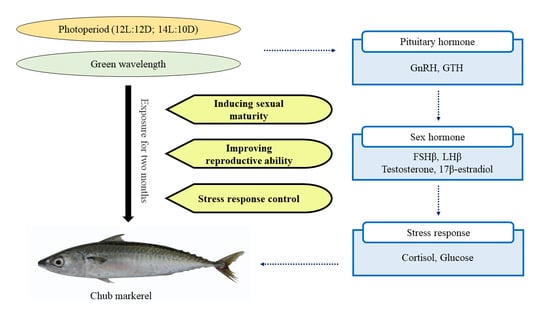
Physiological Effect of Extended Photoperiod and Green Wavelength on the Pituitary Hormone, Sex Hormone and Stress Response in Chub Mackerel, Scomber japonicus

1
Inland Fisheries Research Institute, National Institute of Fisheries Science, Geumsan 32762, Republic of Korea
2
Department of Aquatic Life and Medical Science, Sun Moon University, Asan 336-708, Republic of Korea
*
Author to whom correspondence should be addressed.
Fishes 2023, 8(2), 77; https://doi.org/10.3390/fishes8020077
Submission received: 30 December 2022 / Revised: 20 January 2023 / Accepted: 23 January 2023 / Published: 29 January 2023 / Corrected: 16 May 2023
(This article belongs to the Section Environment and Climate Change)

Abstract

:
Chub mackerel, Scomber japonicus, is heavily farmed and harvested due to its demand as a high-quality protein source rich in fatty acids. However, the effects of environmental cues on sexual maturation of the fish remain understudied. We aim to elucidate the effect of light manipulation on the hormones related to reproduction and on the stress response in the species. Mackerel were exposed to different photoperiods (12 h light:12 h dark or 14 h light:10 h dark) and light wavelengths (provided by white fluorescent bulbs or green LEDs). Total RNA extracted from the brain was assayed with quantitative polymerase chain reaction (a powerful technique for advancing functional genomics) and blood plasma was analyzed via immunoassay using ELISA kits. The mRNA expression of gene-encoding gonadotropin-releasing hormone, gonadotropin hormone, follicle-stimulating hormone, and luteinizing hormone were significantly increased through the use of an extended photoperiod and green wavelength, which also increased testosterone and 17β-estradiol plasma levels. Plasma levels of cortisol and glucose, which are indicators of a stress response, were significantly decreased through green LED exposure. Our results indicate that environmental light conditions affect the production of pituitary and sex hormones, and reduce the stress response in S. japonicus.

Graphical Abstract

1. Introduction

Environmental factors such as temperature and salinity (for freshwater/seawater adaptation) are critical in the timing of sexual maturation and spawning in fish [1,2,3]. Among such factors, light has a major influence on the physiology and behavior of fish, and the light cycle is essential for biological processes such as reproduction, migration and dispersal, as well as metabolic rate and body pigmentation [4,5,6,7,8]. Diurnal fish are most active during the day and less active during the night, while nocturnal fish are most active during the night and less active during the day [4]. The photoperiod and wavelength affect the timing of sexual maturation and spawning because they stimulate the release of melatonin from the pineal gland and retina, which in turn activates reproductive hormones [9,10]. Seasonal changes in photoperiod may have a major effect on the sexual maturity of fish by regulating gonadal maturation (which is associated with endogenous rhythms) and the synthesis/secretion of reproductive hormones [11].
The photoperiod and wavelength are critical environmental cues that initiate the breeding season in temperate fish species, and have a major impact on reproductive and aggressive behavior mediated by the hypothalamus–pituitary–gonadal axis [12]. Artificial lighting produces changes in endocrine hormones through the same mechanism, and the exact effect of varying photoperiod and wavelength depends on the fish species and developmental stage [7,13]. Photoperiod manipulation has been successfully used to promote the growth, development and survival of various juvenile fish, such as those of the gilthead seabream (Sparus aurata), European sea bass (Dicentrarchus labrax), and barramundi (Lates calcarifer) species; it has also been successfully used to increase the rate of sexual maturation and shorten spawning time of adult D. labrax, S. aurata, rainbow trout (Oncorhynchus mykiss), brown trout (Salmo trutta), and Atlantic salmon (Salmo salar)] [14]. A longer photoperiod promotes fry growth, because it presents more feeding opportunities [15]. Furthermore, it cues stimulation of the endocrine system, which prolongs the spawning period, thereby increasing the number of eggs produced; therefore, both the periodicity (in terms of a daily cycle that varies seasonally and with latitude) and quality (the wavelength, absorbed by water) of light should be considered in fish reproduction [16,17].
The pituitary gland produces hormones that play major roles in the endocrine regulation of growth, the metabolism, homeostasis, reproduction, and stress responses [18]. The anterior pituitary gland secretes gonadotropin-releasing hormone (GnRH), gonadotropin hormone (GTH), follicle-stimulating hormone (FSH), and luteinizing hormone (LH), which influence cell development via signaling molecules and transcription factors [19]. In fish, light stimulates the central nervous system to promote pituitary secretion, which also affects the development of fish gonadal cells, and fish ovarian development is closely connected to the light in the environment [20]. Sex steroid hormones such as testosterone and estrogen regulate digestion and food utilization, growth, reproduction, gut transport, osmoregulation, intermediary metabolism, behavior, and immunity in fish, as well as genes and proteins that are involved in tissue and skeletal growth, metabolism, gonadal development, neuroplasticity, and cognition [21,22]. Sex steroid hormones especially control various behavioral and neural processes including aggression, reproduction, memory, and learning [23]. Therefore, in fish, sex steroid hormones may be an important indicator for evaluating the effects of varying photoperiods and light wavelengths, which are closely linked with gonadal development and reproduction [24]. The fish retina absorbs light of various wavelengths that is transmitted to the brain via photoreceptors and results in physiological and behavioral reactions, and some wavelengths may cause physiological stress [25]. In fish, an appropriate level of light can have effects such as improved food intake, accelerated nutrient assimilation, proper growth, improved immune system, increased enzyme activity and crude protein/crude lipid accumulation, which acts as a stressor leading to mass mortality [26]. The cycle and wavelength of light are major factors that induce physiological changes as a direct stress to fish, and these stress-related physiological indicators such as cortisol and glucose are widely used to evaluate the effects of environmental stress [27].
Chub mackerel, Scomber japonicus, occurs worldwide, including the warm and temperate coastal waters of the Pacific, Indian, and Atlantic Oceans. It is one of the most heavily caught and farmed fish species due to its demand in Korea, Japan, China, and Russia as a high-quality protein source rich in omega-3 fatty acids [28]. Although widely used as an aquaculture species, insufficient studies have been conducted on the sexual maturation of S. japonicus. Light is the most influential factor on the sexual maturity of fish, which is essential for stable seedling production. In particular, in recent studies, green wavelengths increased the immunity of fish, reducing stress responses, and had a greater impact on growth than red and blue wavelengths [29,30]. Therefore, the aim of this study was to assess the effect of light manipulation on levels of pituitary hormones, sex hormones, and stress indicators in S. japonicus via exposure to extended photoperiods and the green wavelength.

2. Materials and Methods

2.1. Experimental Fish

The study was conducted using sea cage fish farms measuring 7 m × 7 m × 7 m in size because it is the most commonly used sea border farming facility size in Korea. Chub mackerel (n = 480; full length, 33.7 ± 3.5 cm; weight, 453.5 ± 10.4 g) were obtained from Geomun Island, Yeosu, Korea, and randomly divided into four cages of forty exposed to natural light from the outside. Each cage was exposed to either a 12 h light and 12 h dark (12L:12D) photoperiod or a 14 h light and 10 h dark (14L:10D) photoperiod over a two-month experimental period, using either green LEDs (530 nm) or a white fluorescent bulb (27 W) for lighting. These light sources were waterproofed and set in the center of their respective cages, and maintained at 150 cm under the water surface. The fish were provided commercial feed twice daily (at 09:00 and 17:00) and were reared under these conditions for two months.

2.2. Experimental Design and Sampling

Following the two months of rearing described in Section 2.1, the fish were sampled every month for the two subsequent months. We collected the brain, pituitary, and blood from fish in each group at 14:00 at the one- and two-month experimental periods (n = 40). Immediately after collection, the tissues were frozen in liquid nitrogen and stored at −80 °C until total RNA extraction was performed. Moreover, a blood sample was drawn from the caudal vasculature using a 1 mL syringe coated with heparin. After centrifugation (4 °C, 10,000× g, 10 min), the plasma was stored at −80 °C until analysis.

2.3. Pituitary Hormone

Quantitative polymerase chain reaction (qPCR) was conducted to determine the relative expression of GnRH and GTHα mRNA using total RNA extracted from the brain of chub mackerel. In general, this analysis technique was used because qPCR is used instead of semi-quantitative PCR as an analysis technique to quantify gene expression. The following qPCR primers were designed with reference to the known sequences of the chub mackerel (GenBank accession numbers: GnRH, HQ108195; GTHα, JF495131; and β-actin, GU731674): GnRH forward (5′-ACT GGT CCT ATG GAT GGC TAC-3′) and reverse (5′-TTC AGG AAG AGA CAC CAC ACC-3′) primers; GTHα forward (5′-ATC AAA CAT GGG CTG TGA GG-3′) and reverse (5′-TTT GAG TGG TGT CGG GTA GG-3′) primers; and β-actin forward (5′-ACC GGT ATT GTC ATG GAC TC-3′) and reverse (5′-TCA TGA GGT AGT CTG TGA GGT C-3′) primers.

2.4. Pituitary Gonadotropins

qPCR was conducted to determine the relative expression of FSHβ and LHβ mRNA using total RNA extracted from the brain of chub mackerel. The following qPCR primers were designed with reference to the known sequences of the chub mackerel (GenBank accession numbers: FSHβ, JF495132; LHβ, JF495133; and β-actin, GU731674): FSHβ forward (5′-TGT GAA GGA CAG TGT TAC CAC AGG G-3′) and reverse (5′-TCA TAG GTC CAG TCA CCG C-3′) primers; LHβ forward (5′-GAA ACA ACC ATC TGC AGC G-3′) and reverse (5′-AAA AGT CCC GAT ACG TGC AC-3′) primers; and β-actin forward (5′-ACC GGT ATT GTC ATG GAC TC-3′) and reverse (5′-TCA TGA GGT AGT CTG TGA GGT C-3′) primers.
PCR amplification was conducted using a Bio-Rad CFX96™ Real-time PCR Detection System (Bio-Rad, Hercules, CA, USA) and iQ™ SYBR Green Supermix (Bio-Rad) according to the manufacturer’s instructions. qPCR was performed as follows: one cycle of denaturation at 95 °C for 5 min, 35 cycles of denaturation at 95 °C for 20 s, and annealing at 55 °C for 20 s. The assay was performed in triplicate for each experimental group to confirm consistency. β-actin was used as the internal control, and all data are expressed relative to the corresponding β-actin-calculated ΔCt levels. The calibrated ΔCt value (ΔΔCt) for each sample and the internal control were calculated as ΔΔCt = 2^−(ΔCtsample − ΔCtinternal control).

2.5. Sex Hormone

The levels of plasma testosterone and 17β-estradiol were analyzed via immunoassay with an ELISA kit (testosterone, catalog no. MBS933475; 17β-estradiol, catalog no. MBS221037; Mybiosource, San Diego, CA, USA). An anti-antibody that was specific to the respective hormonal antibodies was pre-coated onto a microplate. Then, 50 μL of blood plasma was added per well, followed by 50 μL of horseradish peroxidase conjugate and 50 μL of antibody. The contents were mixed well and then incubated for 2 h at 37 °C. During the subsequent wash process, any remaining wash buffer was removed via aspiration or decantation. Next, 50 μL of substrate A and substrate B were added to each well, followed by incubation for 15 min at 37 °C in the dark. After incubation, 50 μL of stop solution was added to each well and the optical density of the wells was determined within 10 min, using a GloMax Explorer Multimode Microplate Reader (Promega, Madison, WI, USA) set to 450 nm.

2.6. Stress Hormone and Glucose

The levels of plasma cortisol were analyzed via immunoassay (catalog no. MBS007869; Mybiosource). An anti-antibody specific to the cortisol antibodies was pre-coated onto a microplate. The procedure followed for the remainder of the assay is identical to that described for the sex hormone analyses in Section 2.5. Levels of plasma glucose were measured with the dry multiplayer analytic slide method using a biochemistry auto-analyzer (FUJI DRI-CHEM 4000i; Fujifilm, Tokyo, Japan).

2.7. Statistical Analysis

Statistical analyses were performed using the SPSS/PC+ statistical package (SPSS Inc., Chicago, IL, USA). Significant differences between groups were identified using one-way ANOVA and Tukey’s post hoc test for multiple comparisons. The significance level was set at p < 0.05.

3. Results

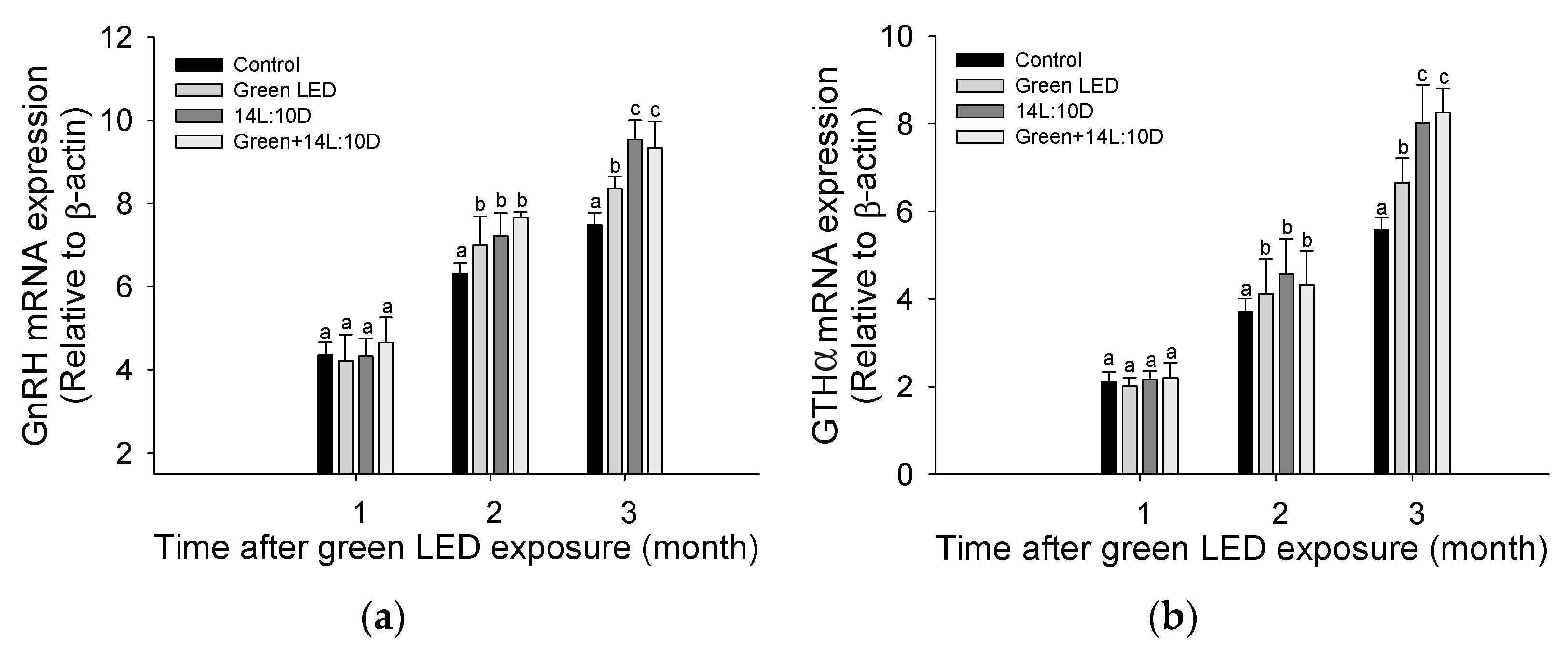
3.1. Pituitary Hormone

The expression of GnRH and GTHα mRNA in the brain of S. japonicus fish exposed to different photoperiods and light wavelengths is shown in Figure 1. The GnRH mRNA expression was significantly increased under green LED lighting and a 14L:10D photoperiod after 1 and 2 months of exposure, respectively. Fish exposed to a 14L:10D photoperiod for 2 months, under either a fluorescent bulb or green LEDs, displayed a significantly higher GnRH mRNA expression than fish under a 12L:12D cycle for that period. Similarly, GTHα mRNA expression was significantly increased with green LED and a 14L:10D photoperiod exposure at 1 and 2 months, respectively. Again, at the 2-month observation period, groups under a 14L:10D photoperiod indicated significantly higher GTHα mRNA expression than groups under a 12L:12D photoperiod.

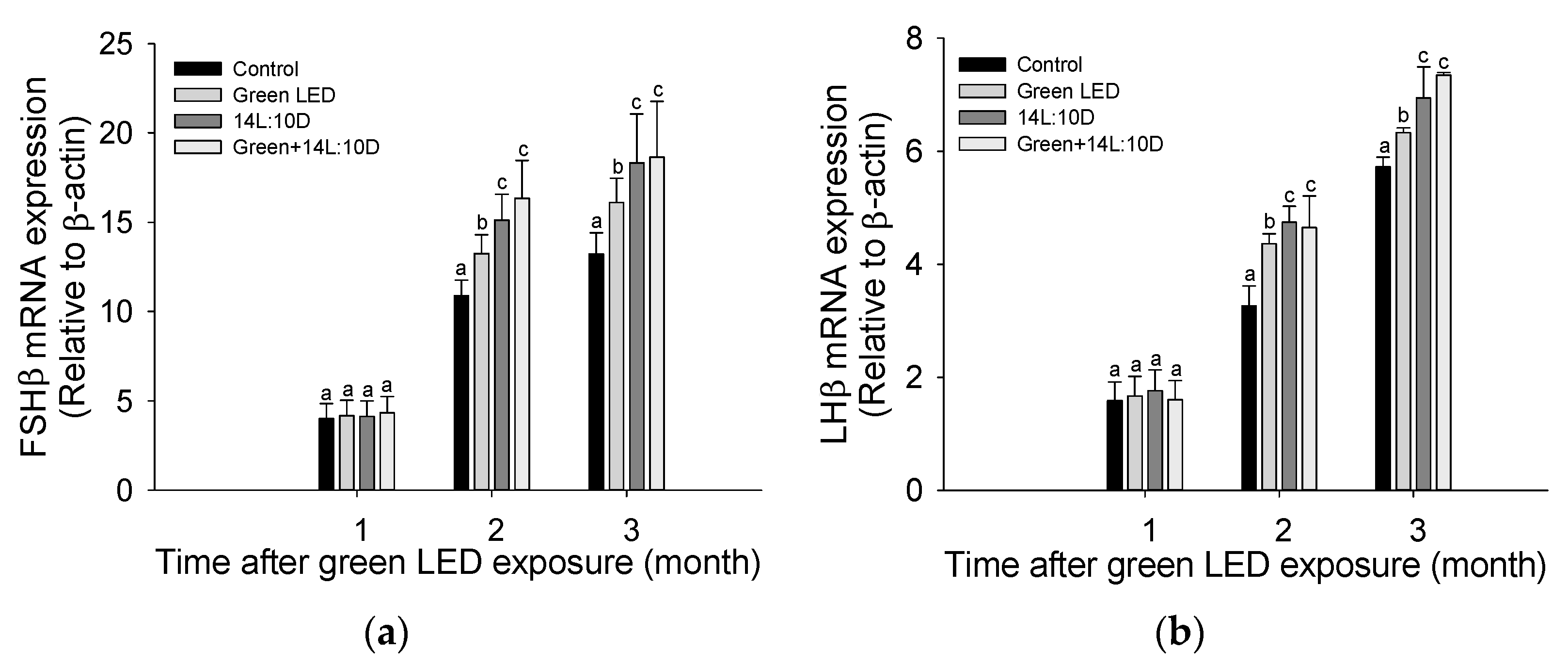
3.2. Pituitary Gonadotropins
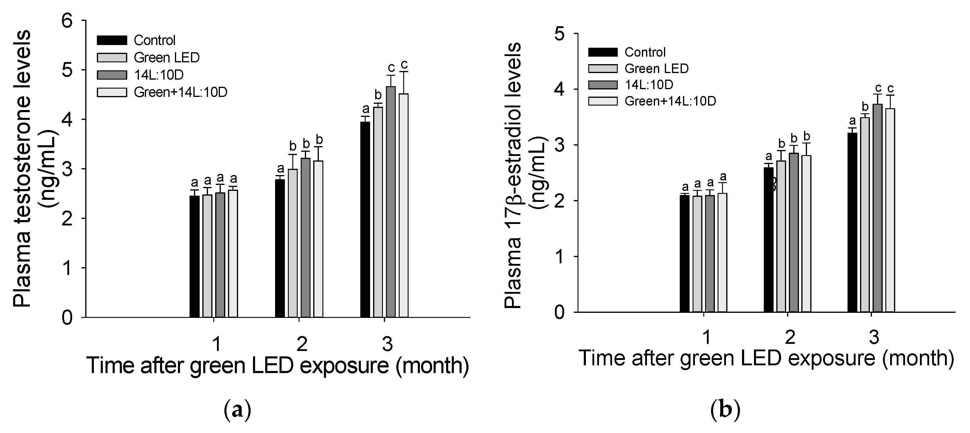
The mRNA expression of FSHβ and LHβ in the brain of S. japonicus fish exposed to different photoperiods and light wavelengths is depicted in Figure 2. The expression of both FSHβ and LHβ mRNA was significantly increased using green LED lighting and a lengthened 14L:10D photoperiod. The 14L:10D photoperiod produced a significantly higher expression of both FSHβ and LHβ mRNA than the 12L:12D photoperiod under either lighting, both at 1 and 2 months.

3.3. Sex Hormone

The levels of testosterone and 17β-estradiol sex hormones in the blood plasma of S. japonicus following exposure of the fish to different photoperiods and light wavelengths are presented in Figure 3. The plasma levels of both testosterone and 17β-estradiol were significantly increased under exposure to green LEDs and to a 14L:10D photoperiod of 1 and 2 months. Furthermore, the 14L:10D photoperiod produced significantly higher plasma levels of both sex hormones than a 12L:12D photoperiod after two months, regardless of light wavelength.

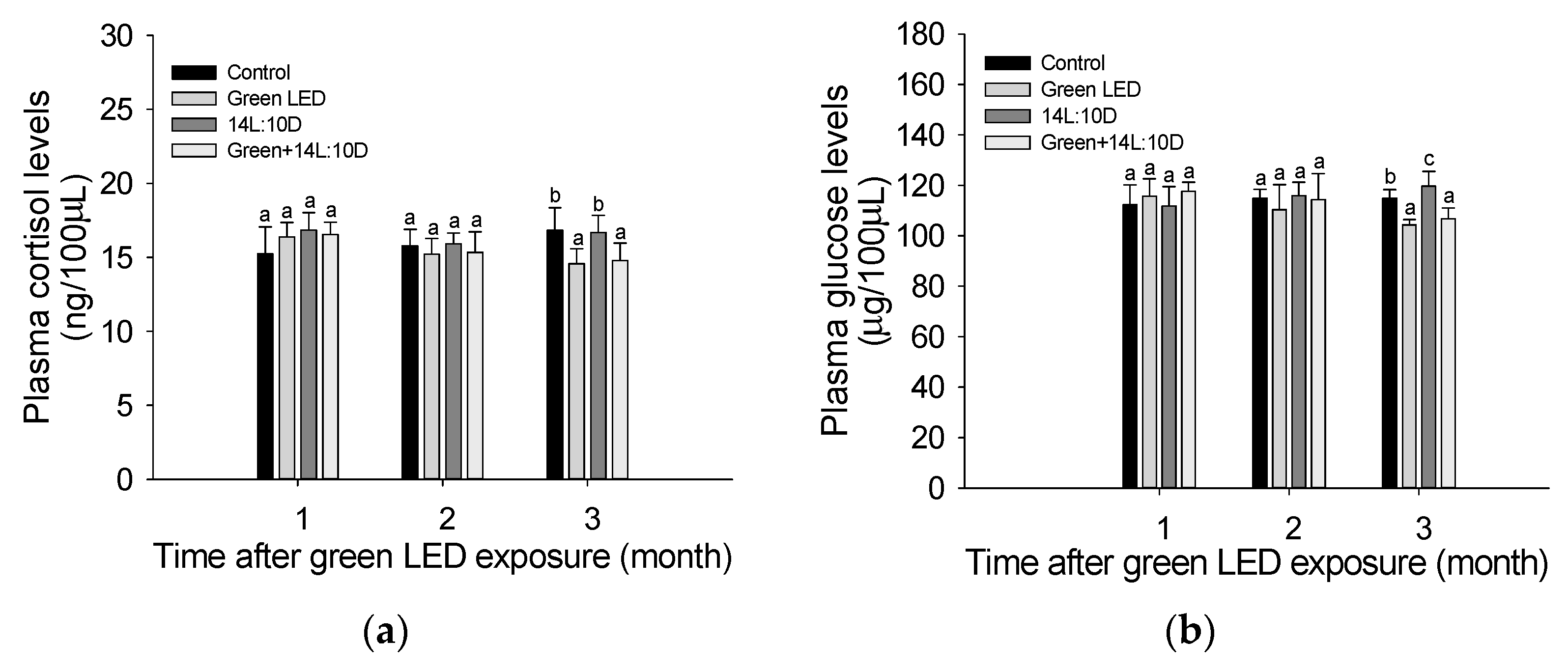
3.4. Stress Response

Figure 4 indicates the stress responses of S. japonicus as measured by cortisol and glucose levels in the plasma following exposure to different photoperiods and lighting wavelengths. Plasma levels of both cortisol and glucose were significantly decreased in the groups that received green LED lighting, regardless of photoperiod, compared to the control group at 12L:12D and the 14L:10D group, which received fluorescent lighting.

4. Discussion

Sexual development and maturation in fish are regulated by various sex hormones produced in the hypothalamus–pituitary–gonadal axis, including GnRH, GTH, steroid hormones, and other neurohormones [31,32]. GnRH is critical for the initiation and maintenance of reproductive function as well as sexual behavior in fish, and the release of GnRH is influenced by the interaction of excitatory and inhibitory signals in the hypothalamus [33]. Subsequently, GnRH transfers the information to the gonadotropic cells of the pituitary, thereby stimulating the synthesis and release of gonadotropic hormones such as FSH and LH. In this study, the GnRH mRNA expression of S. japonicus was significantly increased after exposure of the fish to green LEDs and a 14L:10D photoperiod; this increase was most pronounced for the 14L:10D photoperiod using a fluorescent bulb and green LEDs, which indicated that the photoperiod affected the experimental fish more than wavelength did. Ref. [34] suggested that the photoperiod promotes gonadal maturation in salmon species, reporting a significant increase in GnRH expression of masu salmon (Oncorhynchus masou) due to a lengthened photoperiod. Ref. [35] confirmed that the expression of GnRH mRNA in the hypothalamus of goldfish (Carassius auratus) was significantly higher in a long photoperiod (14L:10D) than in shorter photoperiods (12L:12D or 10L:14D). Similarly, ref. [36] reported that GnRH expression in the threespine stickleback (Gasterosteus aculeatus) significantly increased in a long photoperiod (16L:8D) compared to a short photoperiod (8L:16D), indicating sexual maturation through the photoperiod.
Furthermore, GnRH is a fundamental link in the brain–pituitary–gonad axis and regulates the production of two types of GTH (GTH-I, which is FSH-like, and GTH-II, which is LH-like) by the pituitary; together, these hormones have a major function in stimulating the maturation of gonads [37,38]. In this study, the GTH mRNA expression of S. japonicus was significantly increased by green LED exposure and a 14L:10D photoperiod. The increase was most pronounced in fish exposed to the 14L:10D photoperiod under fluorescent lighting and green LED. Ref. [39] suggested that a long photoperiod stimulates GTH synthesis and release in fish; they reported that the GTH of greater amberjack (Seriola dumerili) increased significantly under a long photoperiod (18L:6D) compared to other periods (natural or 8L:16D), which means that the long photoperiod can accelerate ovarian development. The stimulation of GnRH is closely related to a GTH increase and sexual maturation in various fish species, such as C. auratus, African catfish (Clarias gariepinus), S. aurata, eels (Anguilla anguilla), platyfish (Xiphophorus maculatus), and salmonids [40]. Ref. [36] reported that G. aculeatus remains immature in short photoperiods (8L:16D), but matures rapidly in long photoperiods (16L:8D). In this study, it was confirmed that a green LED wavelength could stimulate sexual maturation by inducing significant expression of GnRH and GTH mRNA in S. japonicus and that an increase in the photoperiod further increased GnRH and GTH expression.
FSH and LH are key hormones regulating fish reproduction; FSH is primarily responsible for gametogenesis initiation and gonadal growth, while LH regulates gonadal maturation, spermatogenesis, and ovulation [41]. The expression of FSH-β and LH-β in the pituitary gland is known to show a similar trend, but the cycle of synthesis and release of FSH and LH appears differentially: FSH increases in the early stages of yolk formation and spermatogenesis in fish, while LH increases in the final stage of maturation [42,43]. In this study, the FSHβ and LHβ mRNA expression in S. japonicus was significantly increased through green LED exposure and a longer photoperiod (14L:10D). This increase was most pronounced in fish exposed to a 14L:10D photoperiod under fluorescent lighting and in fish exposed to green LEDs, which suggests that both the photoperiod and green LED wavelength may play roles in inducing the production and release of sex hormones in S. japonicus. Ref. [39] showed that a long photoperiod (18L:6D) induced a significant increase in plasma FSH in previtellogenic S. dumerili compared to short photoperiods (8L:16D), suggesting that photoperiod cues are essential for inducing ovarian development in fish. Ref. [44] also reported that dark conditions (no light for 24 h) caused disturbances in ovarian growth in D. labrax, and that exposure to a long photoperiod significantly increased FSHβ expression. Ref. [45] argued that the photoperiod influences the expression of hormones such as FSHβ and LHβ, and they reported that a long photoperiod significantly increased LHβ levels in the cichlid fish, Cichlasoma dimerus.
As previously mentioned, GnRH promotes the synthesis of FSH and LH, which are transported to the gonads in the bloodstream where they bind to their respective receptors to induce the production of sex hormones [46]. Of the sex hormones, testosterone is not only essential for sperm production, but also acts as a precursor to estrogen biosynthesis; thus, it is equally important in the female reproductive process [47]. Estradiol induces yolk formation by affecting vitellogenin gene expression in the liver and increases the granulosa cells of ovarian follicles [46]. In this study, we found that an increase in photoperiod and light wavelength resulted in significant increases in testosterone and 17β-estradiol levels in S. japonicus, indicating increased production of these sex hormones. Ref. [48] reported that a lengthening photoperiod caused by seasonal changes from winter to early spring promotes ovulation and spermatogenesis in O. mykiss, and they argued that photoperiod was associated with increases in plasma 17β-estradiol and plasma testosterone in female salmon. Ref. [39] reported a significant increase in 17β-estradiol levels in S. dumerili in a long photoperiod (18L:6D) compared to a short photoperiod (8L:16D), which could be recognized as a signal to initiate ovarian development. Ref. [49] reported that the green LED wavelength induced sexual maturation in the yellowtail damselfish (Chrysiptera parasema) and consequently observed a large number of mature oocytes, indicating that green LEDs induced ovarian maturation. Similarly, our findings suggest that this approach may be valuable for inducing sexual maturation and improving the reproductive ability of S. japonicus.
Photoperiod manipulation in fish can affect stress responses [50]. Neuroendocrine responses to stressors (such as toxicity) in fish entail activation of the hypothalamus–pituitary–interrenal axis, which results in increased cortisol production; therefore, cortisol is widely used as a stress indicator in fish [51]. In this study, the plasma cortisol levels of S. japonicus were significantly decreased in fish exposed to green LED lights, compared to fish in the control group (12L:12D) and fish exposed to fluorescent lighting (14L:10D). This suggests that the wavelength of green LEDs imposed less stress on fish compared to that of fluorescent bulbs. Ref. [52] reported that a gradual decrease in photoperiod affected the plasma cortisol of pintado (Pseudoplatystoma corruscans), which indicates that the photoperiod may modulate the stress response in fish. Ref. [53] reported that the change in daily photoperiod induced a significant change in plasma cortisol in D. labrax. Ref. [50] reported a significant increase in plasma cortisol of juvenile great sturgeon (Huso huso), which indicates that extreme photoperiod changes can induce stress. Plasma glucose is closely linked to carbohydrates that induce metabolic stress in fish, and high blood glucose is a critical indicator for evaluating stress, being the result of glycolysis in liver tissue to provide energy during stress conditions [54]. Ref. [55] suggested that a change in photoperiod acts as a stress factor, reporting a significant increase in plasma glucose of common dentex (Dentex dentex) as a result thereof. On the other hand, refs. [56,57] reported that changes in photoperiods did not affect stress indicators in striped knifejaw (Oplegnathus fasciatus) and red sea bream (Pagrus major). Ref. [58] concluded that an artificial photoperiod does not cause an acute stress response, such as heightened plasma cortisol and glucose levels, in Nile tilapia (Oreochromis niloticus). In this study, the stress response of S. japonicus was not affected by a change in the photoperiod. However, a green LED wavelength showed a tendency to decrease stress indicators, which indicates that the photoperiod and green LEDs do not affect the stress of fish, and that breeding using green LEDs may rather reduce the stress of fish.

5. Conclusions

In conclusion, an increase in the amount of light and the wavelength of green LEDs induced significant increases in GnRH and GTH mRNA expression in S. japonicus, which encode the major pituitary hormones in fish. In addition, an increase in photoperiod and green LED lighting stimulated a significant expression of gonadotropic FSHβ and LHβ mRNA and an increase in testosterone and 17β-estradiol sex hormone levels in S. japonicus. The levels of two key stress indicators, cortisol and glucose, were significantly lower in fish exposed to the green LED wavelength compared to those exposed to fluorescent lighting. The results of this study indicated that a longer photoperiod and green LED wavelength both stimulated the production of key factors related to the sexual maturation and development of S. japonicus and lowered stress induction.

Author Contributions

Data curation, Y.J.C., S.G.N.R.P., A.-H.J., J.-H.K.; writing—original draft preparation, J.-H.K., S.G.N.R.P., A.-H.J.; writing—review and editing, J.-H.K.; supervision, Y.J.C., J.-H.K. All authors have read and agreed to the published version of the manuscript.

Funding

This research was supported by a grant from the National Institute of Fisheries Science [R2023021].

Institutional Review Board Statement

Ethics approval and consent to participate. The participants in this study were trained in animal protection, animal welfare, and animal experimentation by the National Institute of Fisheries Science. All experimental animals used in this study were maintained under a protocol approved by the Institutional Animal Care and Use Committee of the National Institute of Fisheries Science (NIFS-2022-30).

Acknowledgments

This research was supported by a grant from the National Institute of Fisheries Science [R2023021].

Conflicts of Interest

The authors declare no conflict of interest.

References

  1. Debes, P.V.; Piavchenko, N.; Ruokolainen, A.; Ovaskainen, O.; Moustakas-Verho, J.E.; Parre, N.; Primmer, C.R. Polygenic and major-locus contributions to sexual maturation timing in Atlantic salmon. Mol. Ecol. 2021, 30, 4505–4519. [Google Scholar] [CrossRef] [PubMed]
  2. Imsland, A.K.D.; Gunnarsson, S.; Thorarensen, H. Impact of environmental factors on the growth and maturation of farmed Arctic charr. Rev. Aquac. 2020, 12, 1689–1707. [Google Scholar] [CrossRef]
  3. Liu, Q.; Zheng, Y.; Fu, L.; Simco, B.A.; Goudie, C.A. Brood-stock management and natural spawning of American shad (Alosa sapidissima) in a recirculating aquaculture system. Aquaculture 2021, 532, 735952. [Google Scholar] [CrossRef]
  4. Veras, G.C.; Murgas, L.D.S.; Rosa, P.V.; Zangeronimo, M.G.; Ferreira, M.S.D.S.; Leon, J.A.S.D. Effect of photoperiod on locomotor activity, growth, feed efficiency and gonadal development of Nile tilapia. Rev. Bras. Zootec. 2013, 42, 844–849. [Google Scholar] [CrossRef]
  5. Czarnecka, M.; Kakareko, T.; Jermacz, Ł.; Pawlak, R.; Kobak, J. Combined effects of nocturnal exposure to artificial light and habitat complexity on fish foraging. Sci. Total Environ. 2019, 684, 14–22. [Google Scholar] [CrossRef]
  6. Ruchin, A.B. Environmental colour impact on the life of lower aquatic vertebrates: Development, growth, physiological and biochemical processes. Rev. Aquac. 2020, 12, 310–327. [Google Scholar] [CrossRef]
  7. Ruchin, A.B. Effect of illumination on fish and amphibian: Development, growth, physiological and biochemical processes. Rev. Aquac. 2021, 13, 567–600. [Google Scholar] [CrossRef]
  8. Schligler, J.; Cortese, D.; Beldade, R.; Swearer, S.E.; Mills, S.C. Long-term exposure to artificial light at night in the wild decreases survival and growth of a coral reef fish. Proc. Biol. Sci. 2021, 288, 20210454. [Google Scholar] [CrossRef]
  9. Akhtar, M.S.; Rajesh, M.; Kamalam, B.S.; Ciji, A. Effect of photoperiod and temperature on indicators of immunity and wellbeing of endangered golden mahseer (Tor putitora) broodstock. J. Therm. Biol. 2020, 93, 102694. [Google Scholar] [CrossRef]
  10. Baekelandt, S.; Milla, S.; Cornet, V.; Flamion, E.; Ledoré, Y.; Redivo, B.; Kestemont, P. Seasonal simulated photoperiods influence melatonin release and immune markers of pike perch Sander lucioperca. Sci. Rep. 2020, 10, 2650. [Google Scholar] [CrossRef]
  11. Garcia, I.D.; Plaul, S.E.; Torres, D.; Del Fresno, P.S.; Miranda, L.A.; Colautti, D.C. Effect of photoperiod on ovarian maturation in Cheirodon interruptus (Teleostei: Characidae). Braz. J. Biol. 2018, 79, 669–677. [Google Scholar] [CrossRef] [PubMed]
  12. Gonçalves-de-Freitas, E.; Carvalho, T.B.; Oliveira, R.F. Photoperiod modulation of aggressive behavior is independent of androgens in a tropical cichlid fish. Gen. Comp. Endocrinol. 2014, 207, 41–49. [Google Scholar] [CrossRef] [PubMed]
  13. O’Connor, J.J.; Fobert, E.K.; Besson, M.; Jacob, H.; Lecchini, D. Live fast, die young: Behavioural and physiological impacts of light pollution on a marine fish during larval recruitment. Mar. Pollut. Bull. 2019, 146, 908–914. [Google Scholar] [CrossRef] [PubMed]
  14. Kissil, G.W.; Lupatsch, I.; Elizur, A.; Zohar, Y. Long photoperiod delayed spawning and increased somatic growth in gilthead seabream (Sparus aurata). Aquaculture 2001, 200, 363–379. [Google Scholar] [CrossRef]
  15. Arambam, K.; Singh, S.K.; Biswas, P.; Patel, A.B.; Jena, A.K.; Pandey, P.K. Influence of light intensity and photoperiod on embryonic development, survival and growth of threatened catfish Ompok bimaculatus early larvae. J. Fish Biol. 2020, 97, 740–752. [Google Scholar] [CrossRef]
  16. Sánchez-Vázquez, F.J.; López-Olmeda, J.F.; Vera, L.M.; Migaud, H.; López-Patiño, M.A.; Míguez, J.M. Environmental cycles, melatonin, and circadian control of stress response in fish. Front. Endocrinol. 2019, 10, 279. [Google Scholar] [CrossRef] [PubMed]
  17. Lee, C.H.; Hur, S.W.; Kim, B.H.; Soyano, K.; Lee, Y.D. Induced maturation and fertilized egg production of the red-spotted grouper, Epinephelus akaara, using adaptive physiology of photoperiod and water temperature. Aquac. Res. 2020, 51, 2084–2090. [Google Scholar] [CrossRef]
  18. Murugananthkumar, R.; Sudhakumari, C.C. Understanding the impact of stress on teleostean reproduction. Aquac Fish. 2022, 7, 553–561. [Google Scholar] [CrossRef]
  19. Giordano, M. Genetic causes of isolated and combined pituitary hormone deficiency. Best Pract. Res. Clin. Endocrinol. Metab. 2016, 30, 679–691. [Google Scholar] [CrossRef] [PubMed]
  20. Qiang, J.; He, J.; Zhu, J.H.; Tao, Y.F.; Bao, J.W.; Yan, Y.; Zhu, X. Optimal combination of temperature and photoperiod for sex steroid hormone secretion and egg development of Oreochromis niloticus as determined by response surface methodology. J. Therm. Biol. 2021, 97, 102889. [Google Scholar] [CrossRef]
  21. Duarte-Guterman, P.; Navarro-Martín, L.; Trudeau, V.L. Mechanisms of crosstalk between endocrine systems: Regulation of sex steroid hormone synthesis and action by thyroid hormones. Gen. Comp. Endocrinol. 2014, 203, 69–85. [Google Scholar] [CrossRef] [PubMed]
  22. Sudo, R.; Yada, T. Anguillid Eels as a Model Species for Understanding Endocrinological Influences on the Onset of Spawning Migration of Fishes. Biology 2022, 11, 934. [Google Scholar] [CrossRef] [PubMed]
  23. Ogawa, S.; Pfaff, D.W.; Parhar, I.S. Fish as a model in social neuroscience: Conservation and diversity in the social brain network. Biol. Rev. 2021, 96, 999–1020. [Google Scholar] [CrossRef] [PubMed]
  24. Shahjahan, M.; Al-Emran, M.; Islam, S.M.; Baten, S.A.; Rashid, H.; Haque, M.M. Prolonged photoperiod inhibits growth and reproductive functions of rohu Labeo rohita. Aquac. Rep. 2020, 16, 100272. [Google Scholar] [CrossRef]
  25. Song, J.A.; Choi, C.Y. Effects of blue light spectra on retinal stress and damage in goldfish (Carassius auratus). Fish Physiol. Biochem. 2019, 45, 391–400. [Google Scholar] [CrossRef]
  26. Aragón-Flores, E.A.; Martínez-Cárdenas, L.; Hernández-González, C.; Barba-Quintero, G.; Zavala-Leal, O.I.; Ruiz-Velazco, J.M.; Juárez-López, P. Effect of light intensity and photoperiod on growth and survival of the Mexican cichlid, Cichlasoma beani in culture conditions. Lat. Am. J. Aquat. Res. 2017, 45, 293–301. [Google Scholar] [CrossRef]
  27. Dara, M.; Dioguardi, M.; Vazzana, M.; Vazzana, I.; Accardi, D.; Carbonara, P.; Cammarata, M. Effects of Social Hierarchy Establishment on Stress Response and Cell Phagocytosis in Gilt-Head Sea Bream (Sparus aurata). Fishes 2022, 7, 75. [Google Scholar] [CrossRef]
  28. Chen, J.; Wang, S.Z.; Chen, J.Y.; Chen, D.Z.; Deng, S.G.; Xu, B. Effect of cold plasma on maintaining the quality of chub mackerel (Scomber japonicus): Biochemical and sensory attributes. J. Food Sci. 2019, 99, 39–46. [Google Scholar] [CrossRef]
  29. Choi, C.Y.; Choi, J.Y.; Choi, Y.J.; Yoo, J.H. Physiological effects of various light spectra on oxidative stress by starvation in olive flounder, Paralichthys olivaceus. Mol. Cell Toxicol. 2018, 14, 399–408. [Google Scholar] [CrossRef]
  30. Takahashi, A.; Kasagi, S.; Murakami, N.; Furufuji, S.; Kikuchi, S.; Mizusawa, K.; Andoh, T. Effects of different green light intensities on the growth performance and endocrine properties of barfin flounder Verasper moseri. Gen. Comp. Endocrinol. 2018, 257, 203–210. [Google Scholar] [CrossRef]
  31. Lee, Y.H.; Du, J.L.; Yen, F.P.; Lee, C.Y.; Dufour, S.; Huang, J.D.; Chang, C.F. Regulation of plasma gonadotropin II secretion by sex steroids, aromatase inhibitors, and antiestrogens in the protandrous black porgy, Acanthopagrus schlegeli Bleeker. Comp. Biochem. Physiol. B Biochem. Mol. Biol. 2001, 129, 399–406. [Google Scholar] [CrossRef]
  32. Moussavi, M.; Wlasichuk, M.; Chang, J.P.; Habibi, H.R. Seasonal effect of GnIH on gonadotrope functions in the pituitary of goldfish. Mol. Cell. Endocrinol. 2012, 350, 53–60. [Google Scholar] [CrossRef]
  33. Ogawa, S.; Parhar, I.S. Single-cell gene profiling reveals social status-dependent modulation of nuclear hormone receptors in GnRH neurons in a male cichlid fish. Int. J. Mol. Sci. 2020, 21, 2724. [Google Scholar] [CrossRef]
  34. Fukaya, K.; Amano, M.; Ueda, H. Diurnal changes in salmon GnRH secretion in the brain of masu salmon (Oncorhynchus masou). Gen. Comp. Endocrinol. 2013, 192, 77–80. [Google Scholar] [CrossRef]
  35. Shin, H.S.; Song, J.A.; Choi, J.Y.; Kim, N.N.; Choi, Y.J.; Sung, S.N.; Choi, C.Y. Effects of various photoperiods on Kisspeptin and reproductive hormones in the goldfish, Carassius auratus. Anim. Cells Syst. 2014, 18, 109–118. [Google Scholar] [CrossRef]
  36. Shao, Y.T.; Roufidou, C.; Chung, P.C.; Borg, B. Changes in kisspeptin, GnRH, and gonadotropin mRNA levels in male Threespine stickleback (Gasterosteus aculeatus) during photoperiod-induced sexual maturation. Evol. Ecol. Res. 2019, 20, 317–329. [Google Scholar]
  37. Klausen, C.; Chang, J.P.; Habibi, H.R. The effect of gonadotropin-releasing hormone on growth hormone and gonadotropin subunit gene expression in the pituitary of goldfish, Carassius auratus. Comp. Biochem. Physiol. B Biochem. Mol. Biol. 2001, 129, 511–516. [Google Scholar] [CrossRef]
  38. Molés, G.; Hausken, K.; Carrillo, M.; Zanuy, S.; Levavi-Sivan, B.; Gómez, A. Generation and use of recombinant gonadotropins in fish. Gen. Comp. Endocrinol. 2020, 299, 113555. [Google Scholar] [CrossRef]
  39. Nyuji, M.; Hamada, K.; Kazeto, Y.; Mekuchi, M.; Gen, K.; Soyano, K.; Okuzawa, K. Photoperiodic regulation of plasma gonadotropin levels in previtellogenic greater amberjack (Seriola dumerili). Gen. Comp. Endocrinol. 2018, 269, 149–155. [Google Scholar] [CrossRef]
  40. Shao, Y.T.; Tseng, Y.C.; Chang, C.H.; Yan, H.Y.; Hwang, P.P.; Borg, B. GnRH mRNA levels in male three-spined sticklebacks, Gasterosteus aculeatus, under different reproductive conditions. Comp. Biochem. Physiol. A Mol. Integr. Physiol. 2015, 180, 6–17. [Google Scholar] [CrossRef]
  41. Mateos, J.; Mananos, E.; Carrillo, M.; Zanuy, S. Regulation of follicle-stimulating hormone (FSH) and luteinizing hormone (LH) gene expression by gonadotropin-releasing hormone (GnRH) and sexual steroids in the Mediterranean Sea bass. Comp. Biochem. Physiol. B Biochem. Mol. Biol. 2002, 132, 75–86. [Google Scholar] [CrossRef] [PubMed]
  42. Dickey, J.T.; Swanson, P. Effects of salmon gonadotropin-releasing hormone on follicle stimulating hormone secretion and subunit gene expression in coho salmon (Oncorhynchus kisutch). Gen. Comp. Endocrinol. 2000, 118, 436–449. [Google Scholar] [CrossRef] [PubMed]
  43. Trudeau, V.L. Neuroendocrine control of reproduction in teleost fish: Concepts and controversies. Annu. Rev. Anim. Biosci. 2022, 10, 107–130. [Google Scholar] [CrossRef]
  44. Bayarri, M.J.; Rodrıguez, L.; Zanuy, S.; Madrid, J.A.; Sanchez-Vazquez, F.J.; Kagawa, H.; Carrillo, M. Effect of photoperiod manipulation on the daily rhythms of melatonin and reproductive hormones in caged European sea bass (Dicentrarchus labrax). Gen. Comp. Endocrinol. 2004, 136, 72–81. [Google Scholar] [CrossRef] [PubMed]
  45. Fiszbein, A.; Cánepa, M.; Vázquez, G.R.; Maggese, C.; Pandolfi, M. Photoperiodic modulation of reproductive physiology and behaviour in the cichlid fish Cichlasoma dimerus. Physiol. Behav. 2010, 99, 425–432. [Google Scholar] [CrossRef]
  46. Wang, J.; Wang, W.; Li, J.; Luo, Z.; Li, Z.; Chai, M.; Liu, S. Effects of sex hormone rescue on gametogenesis in allotriploid crucian carp. Aquaculture 2022, 561, 738645. [Google Scholar] [CrossRef]
  47. Gabilondo, A.R.; Pérez, L.H.; Rodríguez, R.M. Hormonal and neuroendocrine control of reproductive function in teleost fish. Bionatura 2022, 6, 2122. [Google Scholar] [CrossRef]
  48. Taranger, G.L.; Haux, C.; Stefansson, S.O.; Björnsson, B.T.; Walther, B.T.; Hansen, T. Abrupt changes in photoperiod affect age at maturity, timing of ovulation and plasma testosterone and oestradiol-17β profiles in Atlantic salmon, Salmo salar. Aquaculture 1998, 162, 85–98. [Google Scholar] [CrossRef]
  49. Shin, H.S.; Kim, N.N.; Choi, Y.J.; Habibi, H.R.; Kim, J.W.; Choi, C.Y. Light-emitting diode spectral sensitivity relationship with reproductive parameters and ovarian maturation in yellowtail damselfish, Chrysiptera parasema. J. Photochem. Photobiol. B 2013, 127, 108–113. [Google Scholar] [CrossRef] [PubMed]
  50. Bani, A.; Tabarsa, M.; Falahatkar, B.; Banan, A. Effects of different photoperiods on growth, stress and haematological parameters in juvenile great sturgeon Huso huso. Aquac. Res. 2009, 40, 1899–1907. [Google Scholar] [CrossRef]
  51. Faught, E.; Vijayan, M.M. Maternal stress and fish reproduction: The role of cortisol revisited. Fish Fish. 2018, 19, 1016–1030. [Google Scholar] [CrossRef]
  52. Fagundes, M.; Urbinati, E.C. Stress in pintado (Pseudoplatystoma corruscans) during farming procedures. Aquaculture 2008, 276, 112–119. [Google Scholar] [CrossRef]
  53. Cerdá-Reverter, J.M.; Zanuy, S.; Carrillo, M.; Madrid, J.A. Time-course studies on plasma glucose, insulin, and cortisol in sea bass (Dicentrarchus labrax) held under different photoperiodic regimes. Physiol. Behav. 1998, 64, 245–250. [Google Scholar] [CrossRef]
  54. De Araújo, F.C.T.; Ribeiro, R.P.; Campos, E.C.; Todesco, H.; Tsujii, K.M.; Mantovani, L.S.C.; de Oliveira, C.A.L. Could serum glucose be a selection criterion in Nile tilapia breeding programs? Aquaculture 2022, 548, 737573. [Google Scholar] [CrossRef]
  55. Pavlidis, M.; Greenwood, L.; Paalavuo, M.; Mölsä, H.; Laitinen, J.T. The effect of photoperiod on diel rhythms in serum melatonin, cortisol, glucose, and electrolytes in the common dentex, Dentex dentex. Gen. Comp. Endocrinol. 1999, 113, 240–250. [Google Scholar] [CrossRef] [PubMed]
  56. Biswas, A.K.; Seoka, M.; Ueno, K.; Yong, A.S.; Biswas, B.K.; Kim, Y.S.; Kumai, H. Growth performance and physiological responses in striped knifejaw, Oplegnathus fasciatus, held under different photoperiods. Aquaculture 2008, 279, 42–46. [Google Scholar] [CrossRef]
  57. Biswas, A.; Seoka, M.; Inagaki, H.; Takii, K. Reproduction, growth and stress response in adult red sea bream, Pagrus major (Temminck & Schlegel) exposed to different photoperiods at spawning season. Aquac. Res. 2010, 41, 519–527. [Google Scholar] [CrossRef]
  58. Biswas, A.K.; Maita, M.; Yoshizaki, G.; Takeuchi, T. Physiological responses in Nile tilapia exposed to different photoperiod regimes. J. Fish Biol. 2004, 65, 811–821. [Google Scholar] [CrossRef]
Figure 1. Chub mackerel, Scomber japonicus, were exposed to a 12 h light and 12 h dark photoperiod, either under fluorescent lighting (Control) or under green LEDs (Green LED); mackerel were also exposed to an extended 14 h light and 10 h dark photoperiod under fluorescent lighting (14L:10D) or green LEDs (Green+14L:10D). Total RNA was extracted from the brains of fish (n = 40; full length, 33.7 ± 3.5 cm; weight, 453.5 ± 10.4 g per group) and subjected to quantitative polymerase chain reaction to assess the expression of (a) GnRH mRNA and (b) GTHα mRNA at onset of the experiment, after 1 month, and after 2 months. Vertical bars denote standard errors. Values with different superscripts (a, b, or c) within a given observation period indicate significant differences between groups (p < 0.05) as determined using Tukey’s multiple range test.
Fishes 08 00077 g001
Figure 2. Chub mackerel, Scomber japonicus, were exposed to a 12 h light and 12 h dark photoperiod, either under fluorescent lighting (Control) or under green LEDs (Green LED); mackerel were also exposed to an extended 14 h light and 10 h dark photoperiod under fluorescent lighting (14L:10D) or green LEDs (Green+14L:10D). Total RNA were extracted from the brains of fish (n = 40; full length, 33.7 ± 3.5 cm; weight, 453.5 ± 10.4 g per group) and subjected to quantitative polymerase chain reaction to assess the expression of (a) FSHβ mRNA and (b) LHβ mRNA at onset of the experiment, after 1 month, and after 2 months. Vertical bars denote standard errors. Values with different superscripts (a, b, or c) within a given observation period indicate significant differences between groups (p < 0.05) as determined using Tukey’s multiple range test.
Fishes 08 00077 g002
Figure 3. Chub mackerel, Scomber japonicus, were exposed to a 12 h light and 12 h dark photoperiod, either under fluorescent lighting (Control) or under green LEDs (Green LED); mackerel were also exposed to an extended 14 h light and 10 h dark photoperiod under fluorescent lighting (14L:10D) or green LEDs (Green+14L:10D). Blood was drawn from the caudal veins of fish (n = 40; full length, 33.7 ± 3.5 cm; weight, 453.5 ± 10.4 g per group) and subjected to immunoassay via ELISA kits to measure plasma levels of (a) testosterone and (b) 17β-estradiol at onset of the experiment, after 1 month, and after 2 months. Vertical bars denote standard errors. Values with different superscripts (a, b, or c) within a given observation period indicate significant differences between groups (p < 0.05) as determined using Tukey’s multiple range test.
Fishes 08 00077 g003
Figure 4. Chub mackerel, Scomber japonicus, were exposed to a 12 h light and 12 h dark photoperiod, either under fluorescent lighting (Control) or under green LEDs (Green LED); mackerel were also exposed to an extended 14 h light and 10 h dark photoperiod under fluorescent lighting (14L:10D) or green LEDs (Green+14L:10D). Blood was drawn from the caudal veins of fish (n = 40; full length, 33.7 ± 3.5 cm; weight, 453.5 ± 10.4 g per group) and subjected to immunoassay via ELISA kits to measure plasma levels of (a) cortisol and (b) glucose at onset of the experiment, after 1 month, and after 2 months. Vertical bars denote standard errors. Values with different superscripts (a, b, or c) within a given observation period indicate significant differences between groups (p < 0.05) as determined using Tukey’s multiple range test.
Fishes 08 00077 g004
Disclaimer/Publisher’s Note: The statements, opinions and data contained in all publications are solely those of the individual author(s) and contributor(s) and not of MDPI and/or the editor(s). MDPI and/or the editor(s) disclaim responsibility for any injury to people or property resulting from any ideas, methods, instructions or products referred to in the content.

