Brief Report

Characterization of Red Sea Bream (Pagrus major) Interferon Regulatory Factor 5 and 6 Genes and Their Expression in Response to RSIV Infection

1
Department of Marine Biology and Aquaculture, College of Marine Science, Gyeongsang National University, 2, Tongyeonghaean-ro, Tongyeong 53064, Republic of Korea
2
East Sea Fisheries Research Institute, National Institute of Fisheries Science, Gangneung-si 25435, Republic of Korea
*
Author to whom correspondence should be addressed.
These authors contributed equally to this work.
Fishes 2023, 8(2), 114; https://doi.org/10.3390/fishes8020114
Submission received: 25 January 2023 / Revised: 14 February 2023 / Accepted: 15 February 2023 / Published: 16 February 2023

Abstract

:
Interferon regulatory factors (IRFs) play crucial roles in antiviral processes, such as in the transcriptional induction of interferon (IFN) and IFN-stimulated genes (ISGs). In this study, the genes encoding IRF5 and IRF6 were identified in Pagrus major, and their expression in various organs after pathogen infection was analyzed. In the coding sequences of P. major (Pm)IRF5 and PmIRF6, the DNA binding domain, IRF association domain, and viral-activated domain were found to be highly conserved. Phylogenetic analysis revealed that PmIRF5 and PmIRF6 were most closely related to IRF5 and IRF6 of large yellow croakers. The mRNAs for PmIRF5 and PmIRF6 were constitutively expressed in all organs analyzed but were highly expressed in the liver and gills. As a result of an infection with red sea bream iridovirus, a major pathogen of red sea bream, PmIRF5 and PmIRF6 expression was significantly upregulated in the spleen and kidney. On the basis of these results, it can be concluded that IRF5 and IRF6 expression play an influential role in the immune system of red sea bream infected with viruses.

1. Introduction

Interferon regulatory factors (IRFs) are essential regulators involved in the transcriptional induction of interferon (IFN) and IFN-stimulated genes (ISGs) [1]. In addition, IRFs are critical components of innate and adaptive immunity, playing significant roles in antiviral defense, immune response, cell growth regulation, and apoptosis [2,3]. Currently, 11 IRF families (IRF1–11) have been identified in vertebrates, with IRF10 in birds and IRF11 in fish [1,3,4,5,6]. The N-terminus of IRF contains a DNA binding domain (DBD) of approximately 120 amino acids (aa), containing 5–6 conserved tryptophan repeats forming a helix-turn-helix motif [7]. It recognizes and binds IFN-stimulated response elements (ISREs) to regulate the expression of several immune-related genes [8,9,10]. IRFs contain an IRF association domain (IAD) at their non-conserved C-terminus, which mediates the interaction of IRFs with other transcription factors to form transcriptional complexes [7,11,12]. IRFs are classified into three groups based on differences in the C-terminal region. The IRF family members are activators (IRF1, 3, 5, 7, 9, and 10), repressors (IRF2 and 8), and multifunctional factors that inhibit and activate gene transcription (IRF2, 4, 5, and 7) [4,6,13,14].
Mammalian IRF5 regulates the expression of IFN-α and IFN-β to participate in antiviral responses, activate inflammatory factors, and suppress tumors [15,16,17]. It has been shown that IRF5 knockout mice show high susceptibility to viral infections and are involved in the TLR-MyD88 signaling pathway for gene induction of proinflammatory cytokines, such as interleukin (IL)-6, IL-12, and TNFα [16,18,19]. Mammalian IRF6 is associated with the formation of connective tissue, and mutations in the IRF6 gene can cause Van der Woude syndrome (VWS) and popliteal pterygium syndrome (PPS); however, it has been reported that it is not related to IFN expression [20,21,22]. The overexpression of fish IRF6 significantly upregulated IFN promoter activity and activated the transcription of ISG15, RIG-I, and MAVS [23]. In addition, IRF6 can be phosphorylated by MyD88 and TBK1 and is an IFN-positive regulator, in contrast to mammalian IRF6 [23]. Nevertheless, reports of them in teleost fish are extremely rare, except for zebrafish (Danio rerio), half-smooth tongue sole (Cynoglossus semilaevis), mandarin fish (Siniperca chuatsi), Atlantic cod (Gadus morhua), and blunt snout bream (Megalobrama amblycephala) [23,24,25,26,27,28]. Identifying IRF5 and IRF6 in fish models and studying their functional characteristics will assist in predicting the prognosis of related diseases. This will enable us to understand their roles in the immune system.
Red sea bream iridovirus (RSIV) is a DNA virus that causes mortality in more than 30 species of farmed fish. The first report on RSIV was made in 1990 on red sea bream (Pagrus major) cultured in Japan [29,30]. Since it was first reported in 1998 in aquaculture rock bream (Oplegnathus fasciatus), RSIV has led to significant economic losses in the Republic of Korea yearly [31]. To reduce the damage caused by RSIV, basic research, disease prevention, and control measures must be prepared. Although these efforts have continued over time, there is still limited information regarding the immune system in particular. Therefore, in this study, the first nucleotide sequences of IRF5 and IRF6 identified in red sea bream were obtained, and their characteristics were confirmed. In addition, by confirming the expression patterns of P. major (Pm)IRF5 and PmIRF6 mRNA after RSIV exposure, we intend to provide new insights into IRF5 and IRF6 in the red sea bream immune system.

2. Materials and Methods

2.1. Experimental Fish

Red sea bream were obtained from a net pen farm in Tongyeong (Gyeongsangnam-do, Republic of Korea) and used in the experiment. To ensure that the fish were not infected with pathogens, five randomly selected fish were tested in the laboratory for bacterial, parasitic, and viral diseases. The first step was to observe the external symptoms of the fish with a microscope and with the naked eye. This was to ensure that they were not infected with parasites. During the procedure, the spleen and kidney were removed, and tissue cuts were smeared on the brain and heart infusion agar medium to determine if bacteria were present. RSIV infection was confirmed using DNA extraction from the spleen and PCR using primers recommended by WOAH and qPCR in the references. The purpose of this was to confirm the virus was not infected [30,32]. Red sea bream (total length: 12.5 ± 1.6 cm, weight: 52.1 ± 4.6 g) were acclimatized in a 1600 L tank for 2 weeks before the experiment and filtered. In addition, UV-treated seawater was continuously flowing. During the acclimatization period, the water temperature was 22 ± 1 °C, and commercial feed was fed twice daily.

2.2. Virus

In August 2019, the spleen of RSIV-infected rock bream was collected, and the tissue sample was stored at −80 °C [33]. WOAH provided a sequencing method through PCR that confirmed the presence of RSIV [30]. RSIV was classified as RSIV genotype Ⅱ (accession number: AY532608) by phylogenetic analysis of major capsid protein gene sequences [34].
Before the infection experiment, RSIV-infected tissue was homogenized in phosphate-buffered saline (PBS) and centrifuged at 10,000× g for 20 min at 4 °C. After filtering the supernatant containing the virus with a 0.45 μm syringe filter, it was inoculated into a P. major fin (PMF) cell line [35]. The PMF cell line was provided by the National Fishery Products Quality Management Service (Busan, Republic of Korea) and was cultured at 25 °C in L-15 medium (Gibco) and supplemented with 10% fetal bovine serum (FBS) (Gibco), 1% antibiotic-antimycotic (A-A; 100 U/mL penicillin, 100 μg/mL streptomycin, and 25 μg/mL amphotericin B, Gibco). PMF cells were then inoculated with filtered RSIV. After 4 h of infection, the medium was replaced with an L-15 medium containing 10% FBS and 1% A-A and cultured for 7 d. The cell supernatant containing the virus was used as an inoculum for subsequent experiments to observe the expression patterns of PmIRF5 and PmIRF6 mRNA in RSIV-challenged red sea bream.

2.3. Sequence and Phylogenetic Analysis of PmIRF5 and PmIRF6 Genes

The coding sequences (CDSs) of PmIRF5 and PmIRF6 were obtained through the RNA-seq method of next-generation sequencing (NGS) analysis using RNA extracted from the RSIV-stimulated spleen of red sea bream. Sanger sequencing was performed to verify the complete length sequence of the CDS, and PCR was conducted with the primer set shown in Table 1. IRF5 and IRF6 nucleotide sequences and deduced amino acid sequence analysis were performed with the GENETYX program version 8.0 (Genetyx Corporation, Tokyo, Japan) and the Basic Local Alignment Search Tool (BLAST) algorithm of the National Center for Biotechnology Information (NCBI). Molecular weight and isoelectric point (pI) were predicted using the Expert Protein Analysis System ProtParam tool (ExPASy) (http://web.expasy.org/protparam/ (accessed on 1 July 2020)). To identify specific domains and motifs of PmIRF5 and PmIRF6, the Simple Modular Architecture Research Tool (SMART) (http://smart.embl-heidelberg.de/ (accessed on 2 October 2020)) and SignalP version 6.0 software (https://services.healthtech.dtu.dk/service.php?SignalP-6.0 (accessed on 7 November 2022)) were analyzed. Multiple sequence alignments to amino acid sequences from different fish species were analyzed with Clustal Omega (https://www.ebi.ac.uk/Tools/msa/clustalo/ (accessed on 1 February 2022)). The phylogenetic analysis was conducted using the neighbor-joining (NJ) method of the MEGA (Molecular Evolutionary Genetics Analysis) version X program, and bootstraps were repeated 2000 times.

2.4. Viral Challenge, Nucleic Acid Extraction, and cDNA Synthesis

To confirm the distribution of the mRNA expressions of PmIRF5 and PmIRF6 from healthy red sea bream, head and trunk kidneys, skin, stomach, gills, heart, liver, spleen, eyes, brain, and intestines of three fish were aseptically collected and stored at −80 °C. To observe the mRNA expression patterns of PmIRF5 and PmIRF6 in various organs of red sea bream after being infected with RSIV, 100 μL of RSIV inoculum (1 × 10⁷ RSIV copies/fish) was artificially injected intraperitoneally in a 500 L water tank at 25 °C. At 0 (control, no infection), 1, 3, 6, 12, 24, and 36 h and 3, 5, and 7 d after virus inoculation, the gills, spleen, liver, and trunk kidneys were aseptically removed from three fish. Total RNA extraction from collected tissues was performed using the easy-spin Total RNA Extraction Kit (iNtRON Biotechnology, Seongnam, Republic of Korea) according to the manufacturer’s manual. cDNA synthesis was performed using the PrimeScript 1st strand cDNA Synthesis Kit (Takara, Kusatsu, Japan). Further, 8 μL of total RNA, 1 μL of the random hexamer, and 1 μL of dNTP mixture were heated at 65 °C for 5 min and then cooled on ice for 5 min. Then, 4 μL of 5× PrimeScript buffer, 0.5 μL of RNase Inhibitor (40 U/μL), 1 μL of PrimeScript RTase (200 U/μL), and 4.5 μL of RNase-free water were added and mixed carefully. A reaction mixture of 20 μL was heated at 30 °C for 10 min and 42 °C for 1 h. Subsequently, the reaction was heated at 95 °C for 5 min to inactivate the enzyme, and the synthesized cDNA was stored at −20 °C pending use. Genomic DNA extraction was performed using the AccuPrep Genomic DNA Extraction Kit (Bioneer, Daejeon, Republic of Korea) according to the manufacturer’s manual. All experimental protocols followed the guidelines of the Institutional Animal Care and Use Committee of Gyeongsang National University (approval number: GNU-220427-E0041).

2.5. Quantitative PCR Analysis

An RSIV copy number determination was performed according to a previously reported method [32]. The final volume of the reaction mixture was 25 μL and consisted of 12.5 μL of the HS Prime qPCR Premix with UDG (2×) (Genetbio, Daejeon, Republic of Korea), 900 nM of each primer, 250 nM of the probe, and 5 μL of DNA. There were 45 cycles of 5 s at 95 °C followed by 10 s at 60 °C, with one cycle lasting 1 min at 95 °C.
A reverse transcription quantitative PCR (RT–qPCR) was performed to measure the mRNA expression levels for PmIRF5 and PmIRF6 using the SYBR green method. The RT-qPCR reaction mixture consisted of 12.5 μL TB Green premix Ex Taq (Takara), 400 nM of each primer, and 1 μL cDNA in a final volume of 25 μL. The RT-qPCR reaction conditions were initial denaturation at 95 °C for 10 min, followed by denaturation at 95 °C for 20 s, and annealing at 60 °C for 1 min for a total of 45 cycles. Final dissociation was performed at 95 °C for 15 s, then at 60 °C for 30 s, and lastly at 95 °C for 15 s. Melt curve analysis was performed at the end of the 45 amplification cycles to test for the presence of the unique PCR products.
The relative expression levels of PmIRF5 and PmIRF6 mRNA were compared with the threshold cycle (Ct) of the mRNA of the elongation factor 1 alpha gene (EF-1α; GenBank accession No. AY190693), known as a red sea bream housekeeping gene, and quantified using the 2−ΔΔCt method, (ΔΔCt = 2^ − [ΔCtsample − ΔCtinternal control]) [36]. The experiment was performed in triplicate. The PCR reaction was analyzed using Thermal Cycler Dice Real-Time System III (Takara), and the sequences of primer sets and probes used are shown in Table 1.

2.6. Statistical Analysis

The expression analysis of PmIRF5 and PmIRF6 in organs during RSIV infection was assessed using a one-way analysis of variance (ANOVA), followed by Tukey’s multiple comparison tests (* p <0.05 and ** p <0.01) and compared to controls (0 h). The statistical analysis was performed using GraphPad Prism 9.5.

3. Results

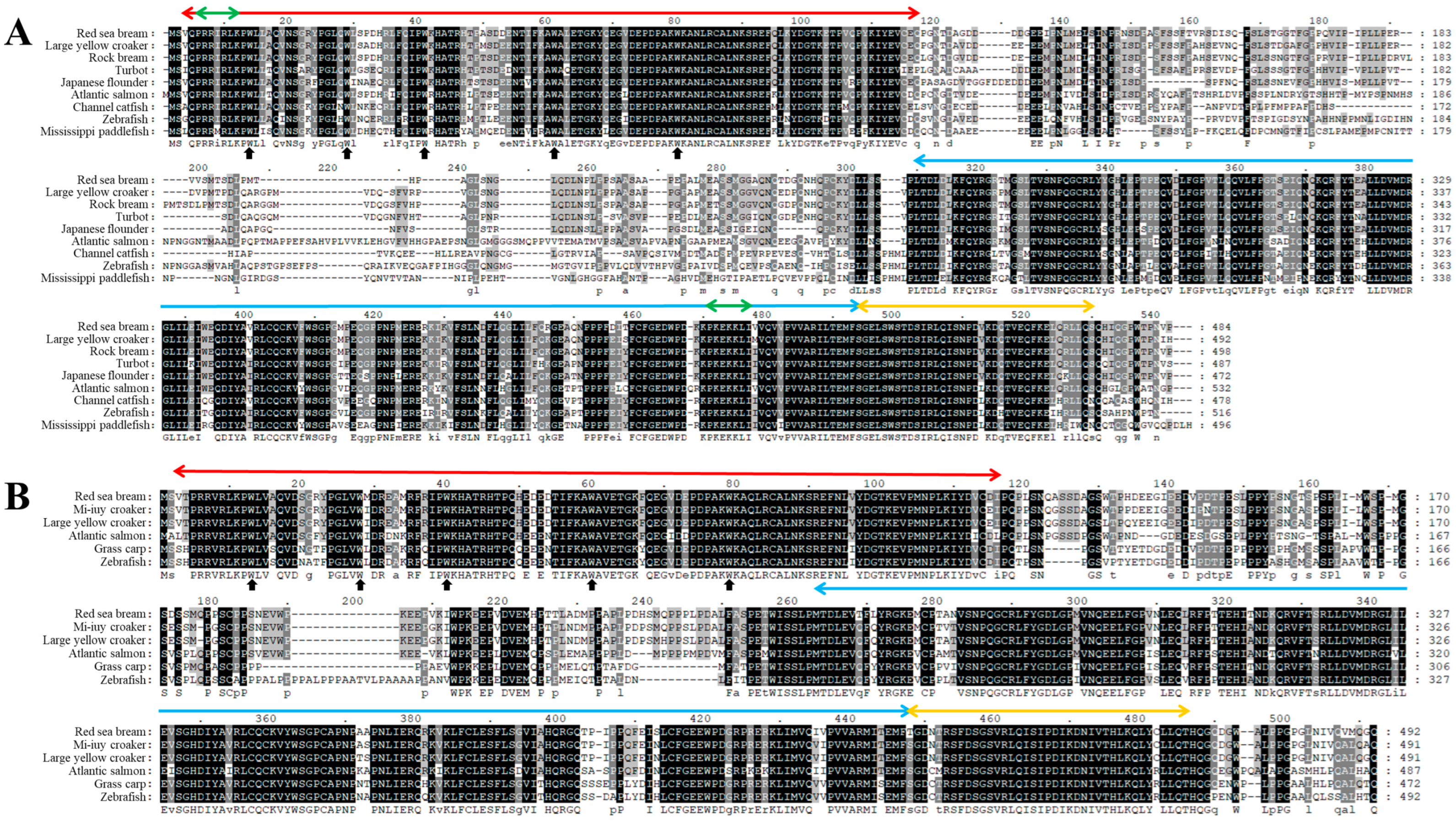
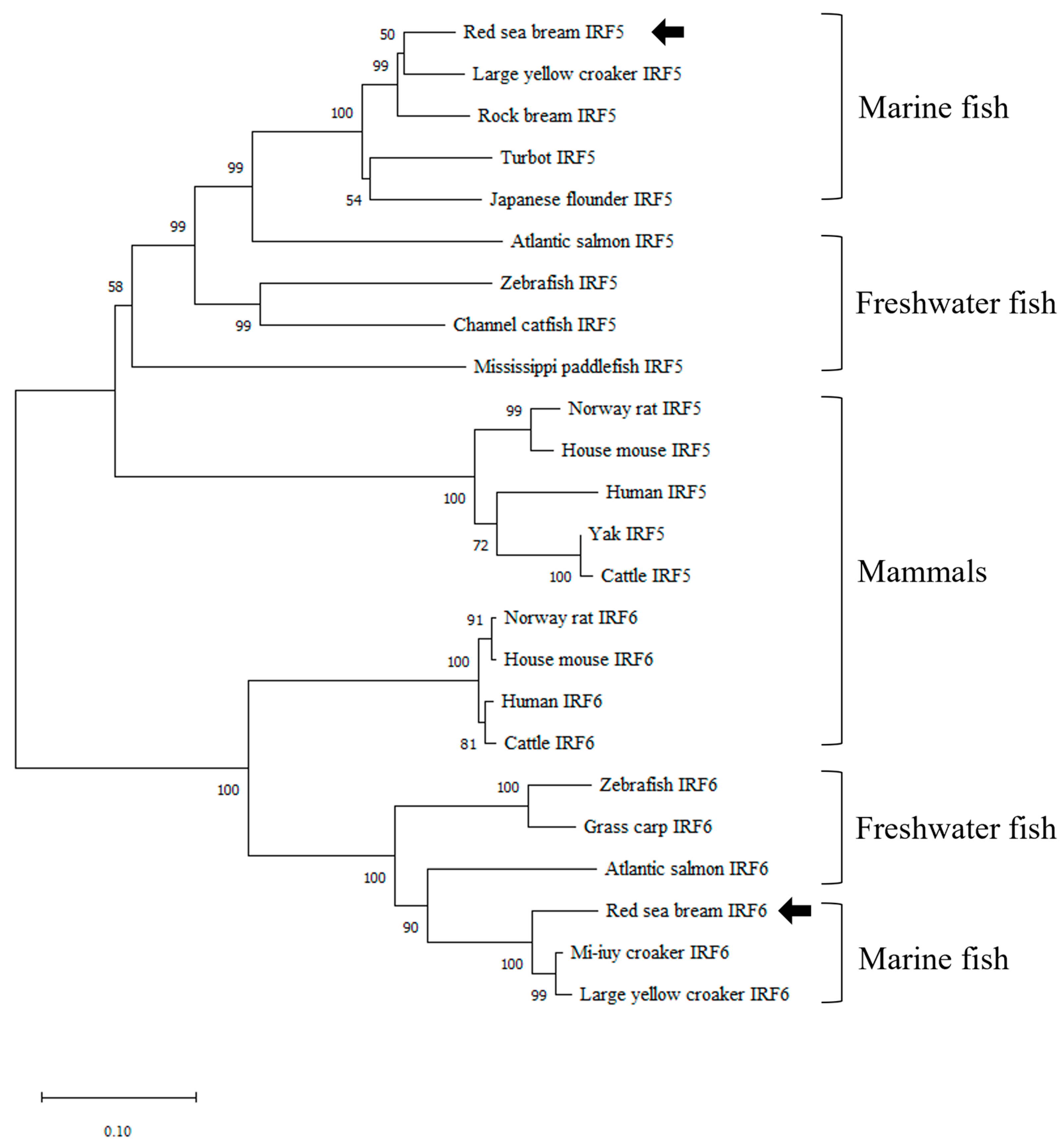
3.1. Identification and Characterization of PmIRF5 and PmIRF6 Sequence

The CDSs of PmIRF5 and PmIRF6 are 1455 bp (GenBank accession No. OK340058) and 1479 bp (GenBank accession No. OK340059), respectively (Figure S1). The CDS of PmIRF5 encodes a mature peptide of 484 aa with a calculated molecular weight of 54.5 kDa and an isoelectric point of 5.11 (Figure S1A). The CDS of PmIRF6 encodes a 492 aa peptide with a molecular weight of 55.5 kDa and an isoelectric point of 5.12 (Figure S1B). PmIRF5 was confirmed to have a DBD (3−116 aa) with five tryptophan (W) residues at the N-terminus, IAD (253−436 aa), a viral activated domain (VAD; 436−472 aa) at the C-terminus, and two nuclear localization signals (NLSs; 5−11 aa, 413−419 aa) (Figure S1A). In addition, PmIRF6 contained a DBD (3−116 aa) with five W residues at the N-terminus, IAD (246−430 aa), and VAD (430−468 aa) at the C-terminus (Figure S1B). As a result of the multiple sequence alignment, PmIRF5 had the highest similarity with large yellow croaker IRF5 (89.84%), followed by rock bream IRF5 (88.8%), turbot IRF5 (82.62%), Japanese flounder IRF5 (80.16%), and Atlantic salmon IRF5 (77.35%). There was a comparatively low level of identity with channel catfish IRF5 (68.11%), zebrafish IRF5 (63.39%), and Mississippi paddlefish IRF5 (61.2%) (Figure 1A). In PmIRF6, mi-iuy croaker IRF6 (93.5%) showed the highest similarity, followed by large yellow croaker IRF6 (93.29%), Atlantic salmon IRF6 (78.59%), grass carp IRF6 (76.11%), and zebrafish IRF6 (71.63%) (Figure 1B). As a result of phylogenetic analysis using the deduced amino acid sequences, PmIRF5 and PmIRF6 were clustered into corresponding subgroups and showed the closest relationship to marine fish. In addition, IRF5 and IRF6 in marine fish, freshwater fish, and mammals each formed distinct clusters (Figure 2).

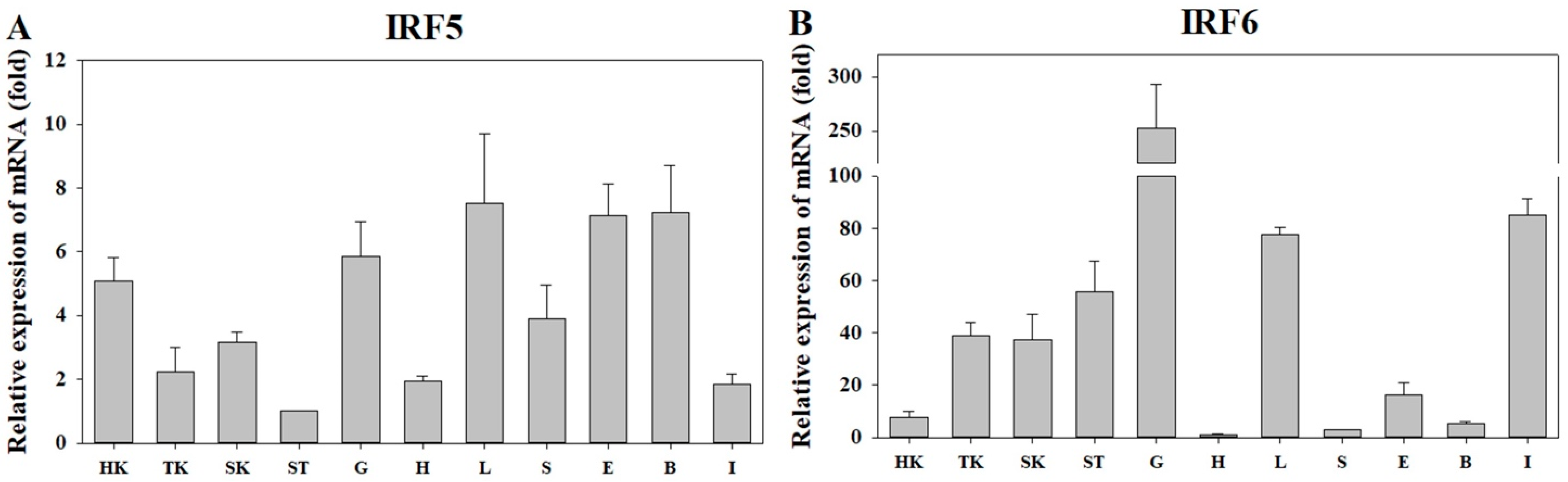
3.2. Expression of PmIRF5 and PmIRF6 mRNA in Various Organs

As a result of analyzing the distribution of PmIRF5 and PmIRF6 mRNA in various organs of healthy red sea bream, they were ubiquitously expressed in all 11 organs (head and trunk kidneys, skin, stomach, gills, heart, liver, spleen, eyes, brain, and intestine) (Figure 3). In comparison to the stomach where PmIRF5 mRNA was least expressed, the liver demonstrated the highest expression (7.52-fold), followed by the brain (7.24-fold) and eyes (7.12-fold). The expression level was relatively low in the intestine (1.83-fold), heart (1.93-fold), and trunk kidneys (2.21-fold) (Figure 3A). As compared to the heart, PmIRF6 mRNA expression was highest in the gills (252.75-fold), and moderately high levels were found in the intestine (85.0-fold) and liver (77.56-fold). A relatively low level of expression was observed in the spleen (2.81-fold), the brain (5.39-fold), and the head kidneys (7.68-fold) (Figure 3B).

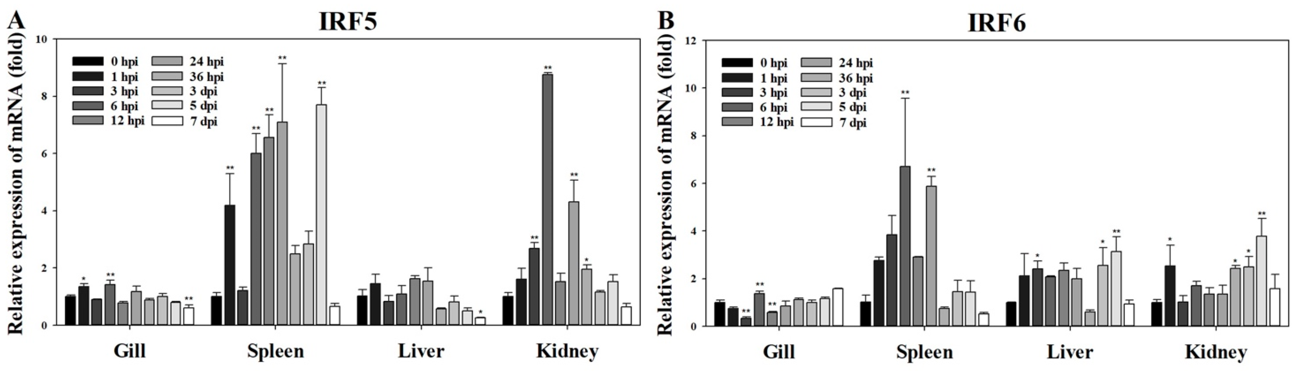
3.3. Expression of PmIRF5 and PmIRF6 mRNA after RSIV Challenge

The mRNA expression levels of PmIRF5 and PmIRF6 in the gill, spleen, liver, and kidney at 0, 1, 3, 6, 12, 24, and 36 hpi (hours post-infection) and 3, 5, and 7 dpi (days post-infection) after RSIV infection were determined using RT-qPCR. The expression of PmIRF5 mRNA was mostly upregulated in all tested organs, and limited downregulated was observed (Figure 4A). The expression was significantly upregulated at 1 and 6 hpi in the gill and 1, 6, 12, 24 hpi, and 5 dpi in the spleen. In the kidney, the expression value was significantly increased at 3, 6, 24, and 36 hpi. In contrast, significant downregulation was confirmed in the gills and liver at 7 dpi. The highest expression level was observed at 6 hpi in the kidney (5.73-fold) (Figure 4A). As a result of RSIV exposure, PmIRF6 mRNA exposure was mostly upregulated in all organs tested, and some organs showed a decrease in expression. The expression value in the gills decreased significantly at 3 hpi, upregulated at 6 hpi, and downregulated again at 12 hpi. In addition, there was significant upregulation of expression in the spleen at 6 and 24 hpi. Furthermore, a significant increase in expression was observed at 3 hpi and from 3 to 5 dpi in the liver, and at 1 hpi and from 36 hpi to 5 dpi in the kidney (Figure 4B).

4. Discussion

The IRF family of transcription factors plays an essential role in regulating type I IFNs and ISGs [8]. The IRF5 and IRF6 genes have been identified in several vertebrates. Despite this, there is no information on the identification of IRF5 and IRF6 in red sea bream and their expression patterns following infection with RSIV, a severe viral disease. In this study, we identified the full-length CDSs of PmIRF5 and PmIRF6. As with other fish species, IRF5 and IRF6 encode conserved W residues, DBD, IAD, and VAD. The DBDs of PmIRF5 and PmIRF6 contain five well-conserved W pentad-repeats that form a helix-turn-helix structure that binds to the ISRE/IRF regulatory element (IRF-E) consensus in target promoters via contacts [37,38], as do other vertebrates with IRF5 and IRF6. The five W residues of both PmIRF5 and PmIRF6 were located at 13, 28, 40, 60, and 79 aa, which compares to those of IRF5 in zebrafish [39], grass carp [40], and turbot [41] and those of IRF6 in common carp [42] and zebrafish [24]. Among these, three W residues have been reported to be involved in binding to DNA through hydrogen bonding by recognizing the “GAAA” sequence [38]. A further feature of the C-terminus of PmIRF5 and PmIRF6 is the presence of IAD1, which is similar to those of IRF39. In the IRF family, IADs consist of IAD1 (IRF39) and IAD2 (IRF1 and 2), which are structurally different [3,43]. IADs can initiate the transcription of target genes by forming transcriptional complexes with other IRFs or transcriptional co-regulators [44,45]. The VAD contains conserved serine residues in other vertebrate IRFs that are phosphorylation sites during viral infection. These functions are similar to those of the serine-rich domains (SRDs) of IRF3 and IRF7 [46,47]. The VAD domain is greatly involved in the transcriptional activity of IRF7 in response to viral infection, with its deletion resulting in transcriptionally inactive IRF7 [48]. Additionally, the deletion of only the C-terminal SRD results in no virus-induced transcriptional activity, suggesting that the VAD domain alone is not transcriptionally active and requires cooperation with the SRD to play its important role in the antiviral response. Similarly to other IRF5s, the predicted PmIRF5 protein contains two NLSs at its N- and C-termini, and these NLSs are vital for IRF nuclear translocation and maintenance in virus-infected cells [2]. NLSs were identified in IRF1, 3, 4, 5, 8, and 9, of which only IRF5 contained two NLSs, with the others containing one in the N-terminal domain [2]. Previous studies reported that the N-terminal NLS and C-terminal NLS of IRF5 are involved in nuclear translocation, with the N-terminal NLS playing a more significant role [2]. Overall, these generally conserved amino acid sequences and structural features of PmIRF5 and PmIRF6 suggest that their activation and action patterns in the immune response to viral infection have remained relatively unchanged.
Phylogenetic analysis indicated that all IRF5 and IRF6 family members were divided into marine fish, freshwater fish, and mammalian groups. In addition, PmIRF5 and PmIRF6 were more closely related to large yellow croaker IRF5 and 6, including IRF5 and 6 of marine fish. These results are consistent with the observed evolutionary relationship between the various tibia species at the genomic and structural level of the IRF5 and 6 genes.
PmIRF5 and PmIRF6 mRNAs were ubiquitously expressed in all organs examined. In particular, PmIRF5 was expressed at high levels in the liver, brain, eyes, and gills, and high levels of PmIRF6 were found in the gills, intestines, and liver. Similarly, rock bream IRF5 was highly expressed in the liver [49], and common carp IRF5 was highly expressed in the gills and brain [50]. It was found that IRF6 was highly expressed in the gills, liver, and intestines of large yellow croakers, which is consistent with our findings [51]. The high expression of IRF5 and IRF6 in the gills and intestine, and mucosal-associated lymphoid tissues with lymphocytes, suggests that they may play a significant role in the activity of the mucosal immune system in fish.
IRF5 and IRF6 mRNA expression was significantly upregulated in RSIV-infected red sea bream, particularly in the spleen and kidneys which are important target organs of the virus [30,32] and may, therefore, require higher levels of immunity than other organs. In contrast, IRF5 and IRF6 mRNAs were highly expressed in the gills of healthy red sea bream. This may be explained by the constantly exposure of the teleost fish to potential pathogens present in the aquatic environment [52]. In addition, the relatively low immunity in the gills of the infected fish may have been due to the delivery of RSIV antigen via the peritoneal cavity. Further studies on the effect of the infection method on the expression patterns of antiviral genes in different organs are required. Turbot significantly stimulated the expression of IRF5 in the spleen on 1 dpi and the kidney on 2 dpi after infection with turbot reddish body iridovirus (TRBIV) [41]. After infection with rock bream iridovirus (RBIV), IRF5 expression in the head kidney was greatest at 12 hpi and decreased until 48 hpi [49]. Nevertheless, in a time course study of grass carp infected with grass carp hemorrhagic reovirus (GCRV), high upregulation of IRF5 (>300-fold) was observed in the head kidney at 6 d after infection [40]. Among Japanese flounders infected with the Lymphocystis disease virus (LCDV), significant upregulation was observed in the muscle at 3 dpi [53]. Considering that IRF5 mediates the antiviral response in a virus-specific manner, further research on the expression pattern of IRF5 according to the host-virus relationship is necessary. Atlantic cod showed no significant changes in IRF6 expression in response to poly(I:C) and lipopolysaccharide stimulation [27]. While these results are consistent with the understanding that IRF6 plays an instrumental role in epithelial cell differentiation, a previous study showed evidence of an upregulation of IRF6 following poly(I:C) stimulation [23]. Currently, limited information is available on the expression profile of IRF6 in fish following exposure to pathogens. During our study, we found that IRF6 levels were upregulated after RSIV stimulation in red sea bream and that distinct changes were observed in the spleen in particular. However, information regarding the functional characterization of IRF6 by pathogen infection in fish is still limited, and further research is required. IRF5 and IRF6 are known to stimulate the expression of IFN-related genes in virus-infected cells [15,16,17,23]. Our results showed that the expression of PmIRF5 and PmIRF6 mRNA was low in all organs at 7 dpi. Red sea bream infected with RSIV showed onset of death at 6 dpi and 100% cumulative mortality at 8 dpi. Therefore, at this time, RSIV infection may have disrupted host cell function, or apoptosis may have disrupted the transcription of immune genes.
In summary, we identified and characterized the CDSs of the IRF5 and IRF6 genes in red sea bream. PmIRF5 and PmIRF6 appeared to be constitutively expressed in several organs, such as the liver and gills of red sea bream. Red sea bream infected with RSIV showed upregulation of PmIRF5 and PmIRF6 in the spleen and kidney at an early stage of infection. Functional studies of IRF5 and IRF6 on viral diseases in teleost fish are still limited. Furthermore, developing an understanding of pathogen-derived diseases and the immune system will be essential for the effective control and treatment of disease.

5. Conclusions

We identified the genetic sequences of PmIRF5 and PmIRF6 in red sea bream and characterized their corresponding aa sequences and conserved domains. Analysis of the Expression profiles of PmIRF5 and PmIRF6 mRNAs revealed that they are constitutively expressed and that, post-infection, they significantly increase in the spleen and kidneys, which are major targets of RSIV.

Supplementary Materials

The following supporting information can be downloaded at: https://www.mdpi.com/article/10.3390/fishes8020114/s1, Figure S1: (A) PmIRF5 and (B) PmIRF6 nucleotide sequence and translated amino acid sequence. The red box at the N-terminus indicates the DNA binding domain (DBD), and five conserved tryptophan residues are marked with arrows. The blue box at the C-terminus indicates the IRF association domain (IAD), and the viral-activated domain (VAD) is shown in the yellow box. The nuclear localization signals (NLS) are shown in green boxes.

Author Contributions

Conceptualization, K.-H.K., M.-S.J. and C.-I.P.; methodology, K.-H.K. and M.-S.J.; formal analysis, K.-H.K., G.K., W.-S.W., M.-Y.S. and H.-J.S.; investigation, K.-H.K.; resources, C.-I.P.; writing—original draft preparation, K.-H.K.; writing—review and editing, C.-I.P.; supervision, C.-I.P.; project administration, C.-I.P.; funding acquisition, C.-I.P. All authors have read and agreed to the published version of the manuscript.

Funding

This research was funded by the Ministry of Oceans and Fisheries, Republic of Korea (R2023024).

Institutional Review Board Statement

All experimental protocols followed the guidelines of the Institutional Animal Care and Use Committee of the Gyeongsang National University (approval number: GNU-220427-E0041).

Data Availability Statement

Not applicable.

Acknowledgments

This research was supported by the National Institute of Fisheries Science, Ministry of Oceans and Fisheries, Republic of Korea (R2023024).

Conflicts of Interest

The authors declare no conflict of interest.

References

  1. Honda, K.; Taniguchi, T. IRFs: Master regulators of signaling by toll-like receptors and cytosolic pattern-recognition receptors. Nat. Rev. Immunol. 2006, 6, 644–658. [Google Scholar] [CrossRef] [PubMed]
  2. Barnes, B.J.; Kellum, M.J.; Field, A.E.; Pitha, P.M. Multiple regulatory domains of IRF-5 control activation, cellular localization, and induction of chemokines that mediate recruitment of T lymphocytes. Mol. Cell. Biol. 2002, 22, 5721–5740. [Google Scholar] [CrossRef] [Green Version]
  3. Yanai, H.; Negishi, H.; Taniguchi, T. The IRF family of transcription factors: Inception, impact and implications in oncogenesis. Oncoimmunology 2012, 1, 1376–1386. [Google Scholar] [CrossRef] [Green Version]
  4. Nehyba, J.; Hrdlicková, R.; Burnside, J.; Bose, H.R., Jr. A novel interferon regulatory factor (IRF), IRF-10, has a unique role in immune defense and is induced by the v-Rel oncoprotein. Mol. Cell. Biol. 2002, 22, 3942–3957. [Google Scholar] [CrossRef] [PubMed] [Green Version]
  5. Huang, W.S.; Zhu, M.H.; Chen, S.; Wang, Z.X.; Liang, Y.; Huang, B.; Nie, P. Molecular cloning and expression analysis of a fish specific interferon regulatory factor, IRF11, in orange spotted grouper, Epinephelus coioides. Fish Shellfish Immunol. 2017, 60, 368–379. [Google Scholar] [CrossRef]
  6. Huang, B.; Qi, Z.T.; Xu, Z.; Nie, P. Global characterization of interferon regulatory factor (IRF) genes in vertebrates: Glimpse of the diversification in evolution. BMC Immunol. 2010, 11, 22. [Google Scholar] [CrossRef] [PubMed] [Green Version]
  7. Tamura, T.; Yanai, H.; Savitsky, D.; Taniguchi, T. The IRF family transcription factors in immunity and oncogenesis. Annu. Rev. Immunol. 2008, 26, 535–584. [Google Scholar] [CrossRef]
  8. Taniguchi, T.; Ogasawara, K.; Takaoka, A.; Tanaka, N. IRF family of transcription factors as regulators of host defense. Annu. Rev. Immunol. 2001, 19, 623–655. [Google Scholar] [CrossRef]
  9. Veals, S.A.; Schindler, C.; Leonard, D.; Fu, X.Y.; Aebersold, R.; Darnell, J.E.; Levy, D.E. Subunit of an alpha-interferon-responsive transcription factor is related to interferon regulatory factor and Myb families of DNA-binding proteins. Mol. Cell. Biol. 1992, 12, 3315–3324. [Google Scholar] [CrossRef]
  10. Darnell, J.E., Jr.; Kerr, I.M.; Stark, G.R. Jak-STAT pathways and transcriptional activation in response to IFNs and other extracellular signaling proteins. Science 1994, 264, 1415–1421. [Google Scholar] [CrossRef] [Green Version]
  11. Panne, D.; Maniatis, T.; Harrison, S.C. An atomic model of the interferon-beta enhanceosome. Cell 2007, 129, 1111–1123. [Google Scholar] [CrossRef] [PubMed] [Green Version]
  12. Chen, W.; Royer, W.E., Jr. Structural insights into interferon regulatory factor activation. Cell. Signal. 2010, 22, 883–887. [Google Scholar] [CrossRef] [PubMed] [Green Version]
  13. Antonczyk, A.; Krist, B.; Sajek, M.; Michalska, A.; Piaszyk-Borychowska, A.; Plens-Galaska, M.; Wesoly, J.; Bluyssen, H.A.R. Direct inhibition of IRF-dependent transcriptional regulatory mechanisms associated with disease. Front. Immunol. 2019, 10, 1176. [Google Scholar] [CrossRef] [PubMed] [Green Version]
  14. Hu, G.; Chen, X.; Gong, Q.; Liu, Q.; Zhang, S.; Dong, X. Structural and expression studies of interferon regulatory factor 8 in Japanese flounder, Paralichthys olivaceus. Fish Shellfish Immunol. 2013, 35, 1016–1024. [Google Scholar] [CrossRef]
  15. Schoenemeyer, A.; Barnes, B.J.; Mancl, M.E.; Latz, E.; Goutagny, N.; Pitha, P.M.; Fitzgerald, K.A.; Golenbock, D.T. The interferon regulatory factor, IRF5, is a central mediator of toll-like receptor 7 signaling. J. Biol. Chem. 2005, 280, 17005–17012. [Google Scholar] [CrossRef] [Green Version]
  16. Krogsgaard, M.; Li, Q.J.; Sumen, C.; Huppa, J.B.; Huse, M.; Davis, M.M. Agonist/endogenous peptide-MHC heterodimers drive T cell activation and sensitivity. Nature 2005, 434, 238–243. [Google Scholar] [CrossRef]
  17. Hu, G.; Mancl, M.E.; Barnes, B.J. Signaling through IFN regulatory factor-5 sensitizes p53-deficient tumors to DNA damage-induced apoptosis and cell death. Cancer Res. 2005, 65, 7403–7412. [Google Scholar] [CrossRef] [Green Version]
  18. Takaoka, A.; Yanai, H.; Kondo, S.; Duncan, G.; Negishi, H.; Mizutani, T.; Kano, S.; Honda, K.; Ohba, Y.; Mak, T.W.; et al. Integral role of IRF-5 in the gene induction programme activated by toll-like receptors. Nature 2005, 434, 243–249. [Google Scholar] [CrossRef]
  19. Paun, A.; Reinert, J.T.; Jiang, Z.; Medin, C.; Balkhi, M.Y.; Fitzgerald, K.A.; Pitha, P.M. Functional characterization of murine interferon regulatory factor 5 (IRF-5) and its role in the innate antiviral response. J. Biol. Chem. 2008, 283, 14295–14308. [Google Scholar] [CrossRef] [Green Version]
  20. Ingraham, C.R.; Kinoshita, A.; Kondo, S.; Yang, B.; Sajan, S.; Trout, K.J.; Malik, M.I.; Dunnwald, M.; Goudy, S.L.; Lovett, M.; et al. Abnormal skin, limb and craniofacial morphogenesis in mice deficient for interferon regulatory factor 6 (Irf6). Nat. Genet. 2006, 38, 1335–1340. [Google Scholar] [CrossRef] [Green Version]
  21. Richardson, R.J.; Dixon, J.; Malhotra, S.; Hardman, M.J.; Knowles, L.; Boot-Handford, R.P.; Shore, P.; Whitmarsh, A.; Dixon, M.J. Irf6 is a key determinant of the keratinocyte proliferation-differentiation switch. Nat. Genet. 2006, 38, 1329–1334. [Google Scholar] [CrossRef] [PubMed]
  22. Leslie, E.J.; Standley, J.; Compton, J.; Bale, S.; Schutte, B.C.; Murray, J.C. Comparative analysis of IRF6 variants in families with Van der Woude syndrome and popliteal pterygium syndrome using public whole-exome databases. Genet. Med. 2013, 15, 338–344. [Google Scholar] [CrossRef] [PubMed] [Green Version]
  23. Li, S.; Lu, L.F.; Wang, Z.X.; Chen, D.D.; Zhang, Y.A. Fish IRF6 is a positive regulator of IFN expression and involved in both of the MyD88 and TBK1 pathways. Fish Shellfish Immunol. 2016, 57, 262–268. [Google Scholar] [CrossRef] [PubMed]
  24. Ben, J.; Jabs, E.W.; Chong, S.S. Genomic, cDNA and embryonic expression analysis of zebrafish IRF6, the gene mutated in the human oral clefting disorders van der Woude and popliteal pterygium syndromes. Gene Expr. Patterns 2005, 5, 629–638. [Google Scholar] [CrossRef]
  25. Zhang, J.; Li, Y.X.; Hu, Y.H. Molecular characterization and expression analysis of eleven interferon regulatory factors in half-smooth tongue sole, Cynoglossus semilaevis. Fish Shellfish Immunol. 2015, 44, 272–282. [Google Scholar] [CrossRef]
  26. Laghari, Z.A.; Li, L.; Chen, S.N.; Huo, H.J.; Huang, B.; Zhou, Y.; Nie, P. Composition and transcription of all interferon regulatory factors (IRFs), IRF1–11 in a perciform fish, the mandarin fish, Siniperca chuatsi. Dev. Comp. Immunol. 2018, 81, 127–140. [Google Scholar] [CrossRef]
  27. Inkpen, S.M.; Solbakken, M.H.; Jentoft, S.; Eslamloo, K.; Rise, M.L. Full characterization and transcript expression profiling of the interferon regulatory factor (IRF) gene family in Atlantic cod (Gadus morhua). Dev. Comp. Immunol. 2019, 98, 166–180. [Google Scholar] [CrossRef]
  28. Zhan, F.B.; Jakovlić, I.; Wang, W.M. Identification, characterization and expression in response to Aeromonas hydrophila challenge of five interferon regulatory factors in Megalobrama amblycephala. Fish Shellfish Immunol. 2019, 86, 204–212. [Google Scholar] [CrossRef]
  29. Inouye, K.; Yamano, K.; Maeno, Y.; Nakajima, K.; Matsuoka, M.; Wada, Y.; Sorimachi, M. Iridovirus infection of cultured red sea bream, Pagrus major. Fish Pathol. 1992, 27, 19–27. [Google Scholar] [CrossRef]
  30. WOAH (World Organisation for Animal Health). Red Sea bream iridoviral disease. In Manual of Diagnostic Tests for Aquatic Animals; WOAH: Paris, France, 2012; Available online: https://www.woah.org/fileadmin/Home/eng/Health_standards/aahm/current/2.3.07_RSIVD.pdf (accessed on 29 March 2022).
  31. Sohn, S.G.; Choi, D.L.; Do, J.W.; Hwang, J.Y.; Park, J.W. Mass mortalities of cultured striped beakperch, Oplegnathus fasciatus by iridoviral infection. Fish Pathol. 2000, 13, 121–127. [Google Scholar]
  32. Kim, K.H.; Choi, K.M.; Kang, G.; Woo, W.S.; Sohn, M.Y.; Son, H.J.; Yun, D.; Kim, D.H.; Park, C.I. Development and validation of a quantitative polymerase chain reaction assay for the detection of red sea bream Iridovirus. Fishes 2022, 7, 236. [Google Scholar] [CrossRef]
  33. Kim, K.H.; Choi, K.M.; Joo, M.S.; Kang, G.; Woo, W.S.; Sohn, M.Y.; Son, H.J.; Kwon, M.G.; Kim, J.O.; Kim, D.H.; et al. Red sea bream Iridovirus (RSIV) kinetics in rock bream (Oplegnathus fasciatus) at various fish-rearing seawater temperatures. Animals 2022, 12, 1978. [Google Scholar] [CrossRef]
  34. Kim, K.I.; Hwang, S.D.; Cho, M.Y.; Jung, S.H.; Kim, Y.C.; Jeong, H.D. A natural infection by the red sea bream Iridovirus-type Megalocytivirus in the golden mandarin fish Siniperca scherzeri. J. Fish Dis. 2018, 41, 1229–1233. [Google Scholar] [CrossRef] [PubMed]
  35. Kwon, W.J.; Yoon, M.J.; Jin, J.W.; Kim, K.I.; Kim, Y.C.; Hong, S.; Jeong, H.D. Development and characterization of megalocytivirus persistently-infected cell cultures for high yield of virus. Tissue and Cell 2020, 66, 101387. [Google Scholar] [CrossRef]
  36. Livak, K.J.; Schmittgen, T.D. Analysis of relative gene expression data using real-time quantitative PCR and the 2(-Delta Delta C(T)) Method. Methods 2001, 25, 402–408. [Google Scholar] [CrossRef]
  37. Au, W.C.; Moore, P.A.; Lowther, W.; Juang, Y.T.; Pitha, P.M. Identification of a member of the interferon regulatory factor family that binds to the interferon-stimulated response element and activates expression of interferon-induced genes. Proc. Natl. Acad. Sci. USA 1995, 92, 11657–11661. [Google Scholar] [CrossRef] [PubMed] [Green Version]
  38. Escalante, C.R.; Nistal-Villán, E.; Shen, L.; García-Sastre, A.; Aggarwal, A.K. Structure of IRF-3 bound to the PRDIII-I regulatory element of the human interferon-beta enhancer. Mol. Cell 2007, 26, 703–716. [Google Scholar] [CrossRef]
  39. Xiang, Z.; Dong, C.; Qi, L.; Chen, W.; Huang, L.; Li, Z.; Xia, Q.; Liu, D.; Huang, M.; Weng, S.; et al. Characteristics of the interferon regulatory factor pairs zfIRF5/7 and their stimulation expression by ISKNV Infection in zebrafish (Danio rerio). Dev. Comp. Immunol. 2010, 34, 1263–1273. [Google Scholar] [CrossRef]
  40. Xu, Q.Q.; Chang, M.X.; Xiao, F.S.; Huang, B.; Nie, P. The gene and virus-induced expression of IRF-5 in grass carp Ctenopharyngodon idella. Vet. Immunol. Immunopathol. 2010, 134, 269–278. [Google Scholar] [CrossRef]
  41. Xia, J.; Hu, G.B.; Dong, X.Z.; Liu, Q.M.; Zhang, S.C. Molecular characterization and expression analysis of interferon regulatory factor 5 (IRF-5) in turbot, Scophthalmus maximus. Fish Shellfish Immunol. 2012, 32, 211–218. [Google Scholar] [CrossRef]
  42. Liang, Y.; Liu, R.; Zhang, J.; Chen, Y.; Shan, S.; Zhu, Y.; Yang, G.; Li, H. Negative regulation of interferon regulatory factor 6 (IRF6) in interferon and NF-κB signalling pathways of common carp (Cyprinus carpio L.). BMC Vet. Res. 2022, 18, 433. [Google Scholar] [CrossRef] [PubMed]
  43. Meraro, D.; Hashmueli, S.; Koren, B.; Azriel, A.; Oumard, A.; Kirchhoff, S.; Hauser, H.; Nagulapalli, S.; Atchison, M.L.; Levi, B.Z. Protein-protein and DNA-protein interactions affect the activity of lymphoid-specific IFN regulatory factors. J. Immunol. 1999, 163, 6468–6478. [Google Scholar] [CrossRef]
  44. Yoneyama, M.; Suhara, W.; Fukuhara, Y.; Fukuda, M.; Nishida, E.; Fujita, T. Direct triggering of the type I interferon system by virus infection: Activation of a transcription factor complex containing IRF-3 and CBP/p300. EMBO J. 1998, 17, 1087–1095. [Google Scholar] [CrossRef] [Green Version]
  45. Yang, H.; Ma, G.; Lin, C.H.; Orr, M.; Wathelet, M.G. Mechanism for transcriptional synergy between interferon regulatory factor (IRF)-3 and IRF-7 in activation of the interferon-beta gene promoter. Eur. J. Biochem. 2004, 271, 3693–3703. [Google Scholar] [CrossRef] [PubMed]
  46. Marié, I.; Smith, E.; Prakash, A.; Levy, D.E. Phosphorylation-induced dimerization of interferon regulatory factor 7 unmasks DNA binding and a bipartite transactivation domain. Mol. Cell. Biol. 2000, 20, 8803–8814. [Google Scholar] [CrossRef] [Green Version]
  47. Holland, J.W.; Bird, S.; Williamson, B.; Woudstra, C.; Mustafa, A.; Wang, T.; Zou, J.; Blaney, S.C.; Collet, B.; Secombes, C.J. Molecular characterization of IRF3 and IRF7 in rainbow trout, Oncorhynchus mykiss: Functional analysis and transcriptional modulation. Mol. Immunol. 2008, 46, 269–285. [Google Scholar] [CrossRef]
  48. Lin, R.; Mamane, Y.; Hiscott, J. Multiple regulatory domains control IRF-7 activity in response to virus infection. J. Biol. Chem. 2000, 275, 34320–34327. [Google Scholar] [CrossRef] [PubMed] [Green Version]
  49. Wickramaarachchi, W.D.; Wan, Q.; Lim, B.S.; Jung, H.B.; De Zoysa, M.; Park, M.A.; Lee, J.; Whang, I. Genomic characterization of interferon regulatory factor 5 from rock bream (Oplegnathus fasciatus) and its role in antiviral defense. Fish Shellfish Immunol. 2014, 37, 256–267. [Google Scholar] [CrossRef]
  50. Zhu, Y.; Qi, C.; Shan, S.; Zhang, F.; Li, H.; An, L.; Yang, G. Characterization of common carp (Cyprinus carpio L.) interferon regulatory factor 5 (IRF5) and its expression in response to viral and bacterial challenges. BMC Vet. Res. 2016, 12, 127. [Google Scholar] [CrossRef] [Green Version]
  51. Guan, Y.; Chen, X.; Luo, T.; Ao, J.; Ai, C.; Chen, X. Molecular characterization of the interferon regulatory factor (IRF) family and functional analysis of IRF11 in the large yellow croaker (Larimichthys crocea). Fish Shellfish Immunol. 2020, 107, 218–229. [Google Scholar] [CrossRef]
  52. Yu, Y.; Wang, Q.; Huang, Z.; Ding, L.; Xu, Z. Immunoglobulins, Mucosal Immunity and Vaccination in Teleost Fish. Front. Immunol. 2020, 11, 567941. [Google Scholar] [CrossRef] [PubMed]
  53. Hu, G.B.; Lou, H.M.; Dong, X.Z.; Liu, Q.M.; Zhang, S.C. Characteristics of the interferon regulatory factor 5 (IRF5) and its expression in response to LCDV and poly I:C challenges in Japanese flounder, Paralichthys olivaceus. Dev. Comp. Immunol. 2012, 38, 377–382. [Google Scholar] [CrossRef] [PubMed]
Figure 1. Multiple sequence alignment analysis of the (A) IRF5- and (B) IRF6-derived amino acid sequences in various fish species. Red double-headed arrows at the N-termini indicate the DNA binding domain (DBD), with five conserved tryptophan residues indicated by black arrows. The blue double-headed arrows at the C-termini indicate the IRF association domain (IAD), while the viral-activated domain (VAD) is indicated by the yellow double-headed arrows. The nuclear localization signals (NLSs) are indicated by green double-headed arrows.
Fishes 08 00114 g001
Figure 2. A phylogenetic tree of P. major interferon regulatory factor (PmIRF5), PmIRF6, and other known IRF5 and IRF6 homologs based on the neighbor-joining (NJ) method. The scale bar indicates a branch length of 0.10. Numbers are bootstrap percentiles from 2000 replicates. This analysis is based on the following sequence data: Red sea bream IRF5 (UIR15468), Large yellow croaker IRF5 (QPZ85366), Rock bream IRF5 (AFZ93894), Turbot IRF5 (AEG76960), Japanese flounder IRF5 (AEY55357), Atlantic salmon IRF5 (NP_001133324), Channel catfish IRF5 (AHH37262), Zebrafish IRF5 (NP_001314746), Mississippi paddlefish IRF5 (AEW27153), Norway rat IRF5 (NP_001100056), House mouse IRF5 (EDL13770), Human IRF5 (EAL24108), Yak IRF5 (ELR49399), Cattle IRF5 (NP_001030542), Norway rat IRF6 (NP_001102329), House mouse IRF6 (NP_058547), Human IRF6 (AAH14852), Cattle IRF6 (NP_001070402), Grass carp IRF6 (AMT92196), Zebrafish IRF6 (NP_956892), Atlantic salmon IRF6 (XP_014022332), Red sea bream IRF6 (UIR15469), Large yellow croaker IRF6 (QPZ85367), and Mi-iuy croaker IRF6 (AHB59739).
Fishes 08 00114 g002
Figure 3. Distribution of (A) IRF5 and (B) IRF6 mRNA expression related to EF-1α expression in red sea bream organs (exhibited relative to the organ with the lowest mRNA expression level). The 11 organs are the head kidneys (HK), trunk kidneys (TK), skin (SK), stomach (ST), gills (G), liver (L), heart (H), spleen (S), eyes (E), brain (B), and intestine (I). Results exhibit the mean ± SD of triplicate (n = 3).
Fishes 08 00114 g003
Figure 4. Expression pattern of (A) IRF5 and (B) IRF6 mRNA in the gill, spleen, liver, and kidney of red sea bream infected with RSIV. The PmIRF5 and PmIRF6 transcription levels were compared to that of the EF-1α using RT-qPCR. The data are shown as the means ± SD of triplicate for each organ (n = 3). Asterisks indicate differences (* p < 0.05, ** p < 0.01) compared with the control (0 h).
Fishes 08 00114 g004
Table 1. Primer sequences used in this study.
Primer NameSequence of Primer (5′−3′)Usage
PmIRF5-F1ATGAGCGTGCAGCCTCGGAAmplification for reaffirmation of full-length CDS
PmIRF5-R1TGTCCCTCCGGTGGACAGAC
PmIRF5-F2AGGGACATTTGGACCTCCTC
PmIRF5-R2TGTAGAACCGCTGCTTCTGG
PmIRF5-F3GCGGTTCTACACTGAGGCCC
PmIRF5-R3TCAGGGGACATTGGGGGTCC
PmIRF6-F1ATGTCAGTCACCCCTCGGC
PmIRF6-R1CTCTTTGGGCCAGACCTCGT
PmIRF6-F2TCAGACTCCTCCATGCAGCC
PmIRF6-R2TGGGCTATCACCCCACTGAG
PmIRF6-F3TTTTCTCAGTGGGGTGATAG
PmIRF6-R3TCACTGTCCCTGCATAAC
qPCR-PmIRF5-FACCTGTTTGGACCTGTCACCRT-qPCR amplification
qPCR-PmIRF5-RAGCAGGGCCTCAGTGTAGAA
qPCR-PmIRF6-FCTCTGCCAGTGCAAGGTGTA
qPCR-PmIRF6-RGGCTATCACCCCACTGAGAA
qPCR-PmEF-1α-FACGTGTCCGTCAAGGAAATC
qPCR-PmEF-1α-RTGATGACCTGAGCGTTGAAG
qPCR-RSIV-Meg 1041-FCCACCAGATGGGAGTAGACRSIV copy number determination [32]
qPCR-RSIV-Meg 1139-RGGTTGATATTGCCCATGTCCA
qPCR-RSIV-Meg 1079-P[FAM]CCTACTA[i-EBQ]CTTTGCGCCCAGCATG[phosphate]
Disclaimer/Publisher’s Note: The statements, opinions and data contained in all publications are solely those of the individual author(s) and contributor(s) and not of MDPI and/or the editor(s). MDPI and/or the editor(s) disclaim responsibility for any injury to people or property resulting from any ideas, methods, instructions or products referred to in the content.

