Article

Cloning and Expression of Sox2 and Sox9 in Embryonic and Gonadal Development of Lutraria sieboldii

1
Guangxi Key Laboratory of Beibu Gulf Marine Biodiversity Conservation, Beibu Gulf University, Qinzhou 535011, China
2
Guangxi Institute of Oceanology, Beihai 536000, China
3
Guangxi Key Laboratory of Veterinary Biotechnology, Guangxi Veterinary Research Institute, Nanning 530001, China
*
Authors to whom correspondence should be addressed.
These authors contributed equally to this work.
Fishes 2022, 7(6), 392; https://doi.org/10.3390/fishes7060392
Submission received: 26 November 2022 / Revised: 10 December 2022 / Accepted: 13 December 2022 / Published: 16 December 2022
(This article belongs to the Section Genetics and Biotechnology)

Abstract

:
The Sox family plays essential roles as transcription factors in vertebrates; however, little is known about the Sox family in Lutraria sieboldii. L. sieboldii are pleasant to eat with a short growth cycle and have become one of the best bottom-seeded enrichment species in Guang Xi. In this study, Sox2 (named LsSox2) and Sox9 (named LsSox9) from L. sieboldii were cloned, and their expression patterns were analyzed. The length of the LsSox2 gene coding sequence was 1011 bp, encoding 336 amino acids, and LsSox9 was 1449 bp, encoding 482 amino acids. LsSox2 had its highest expression levels in the ovary, which were 356 times those in testis, whereas LsSox9 presented higher expression in testis, which was 6 times more highly expressed than in the ovary. LsSox2 exhibited the highest expression during the morula stage, which was 20 times that of the D-shaped larvae or zygote. LsSox9 exhibited two expression peaks, one at the four-cell stage and the other at the trochophore stage, while the lowest expression was in the zygote. LsSox9 was 73 times more highly expressed in the four-cell stage than in the zygote stage. During gonadal development, LsSox2 presented the highest expression in the mature ovary, which was 756 times more highly expressed than in mature testis. LsSox9 presented higher expression in testis at the emission stage which was 6 times more highly expressed than in the ovary. These results indicate that LsSox2 and LsSox9 may play important roles in embryonic and gonadal development.

1. Introduction

The sex determining region Y-box (Sox) gene family is involved in sex differentiation and embryonic development. Its main features are a conserved High Mobility Group (HMG) motif that binds specifically to DNA sequences and has more than 50% sequence identity in the same family [1]. The Sox gene family consists of more than 20 members that mediate DNA binding through the HMG domain [2], which has been divided into eight groups (sex determining region Y-box A to sex determining region Y-box H, SoxA–SoxH) [3] that play essential roles in development, cell division, and differentiation [4].
Sex determining region Y (SRY) is the only member in the SoxA group [5]. Although SRY plays a regulatory role in sex determination, little information is available on the sequences surrounding the HMG structure. The SoxB group is subdivided into SoxB1 and SoxB2. The SoxB1 group includes Sox1, Sox2, and Sox3, while Sox14 and Sox21 are part of the SoxB2 subgroup. SoxB1 members play major roles in nervous system development and architecture, while SoxB2 has a C-terminal trans-inhibitory domain but not a trans-activating domain [6]. SoxE contains Sox8, Sox9, and Sox10, which are similar in structure and contain a unique dimerization domain in the HMG structure [7]. These 20 Sox genes are currently found only in animals, including invertebrates, but not in plants.
The Sox transcription factor is a fundamental molecule that plays a key role in almost all stages of embryonic development and differentiation of many cell types [4]. A study of the Sox gene family in cyprinid fish revealed that the Sox gene family is expressed at different levels and duplicated in cyprinid fish with different ploidy after a whole-genome duplication even [8]. Studies on metamorphosis in the African clawed toad have shown that many members of the Sox family play important roles in the formation and proliferation of intestinal stem cells and play an active role in the intestinal epithelium [9]. Sox2, a member of the SoxB1 subfamily [10], has also been implicated in intestinal development [11]. Early studies showed that, like other members of the SoxB1 subfamily, Sox2 promotes neural cell differentiation during vertebrate embryonic development, stem cell maintenance [12], and is required for maintenance of central nervous system stem cells [12,13]. Lc-Sox2 is zygotically expressed in Larimichthys crocea embryos, with high levels starting at the gastrulation stage [14]. Sox2 is not maternally inherited in Japanese flounder, and the transcripts are present from the high blastula stage onwards, with the highest level at the mid-gastrula stage [14].
Sox9, a member of the SoxE subfamily, is an important transcription factor in the development of many tissues, particularly in sex determination and chondrogenesis. The Sox9 expression pattern changes throughout the reproductive cycle of male Astyanax altiparanae and is significantly downregulated after spermatogenesis [15]. The Sox9 gene is conserved in mammals and birds, and its structure and function are preserved in teleost fish. Two Sox9 genes, Sox9a and Sox9b have been identified in Paralichthys olivaceus, and quantitative real-time polymerase chain reaction (qRT-PCR) indicated that PoSox9a and PoSox9b were expressed at higher levels in testis than in ovaries of adults [14,16]. On the other hand, Sox9a is preferentially expressed in the brain and ovary of Oryzias latipes, while Sox9b is expressed more in the testis than in the ovary [17].
Mollusks are the second largest group in the animal kingdom after arthropods; however, the phylogenetic characterization of the Sox2 gene family is very rare in mollusks, particularly in Lutraria sieboldii which are pleasant to eat, have a short growth cycle, and have become one of the best bottom-seeded enrichment species in Guang Xi, with an annual output of about 76,000 tons (4.5–13.68 billion Yuan). Here, we report a study of the Sox2 and Sox9 genes in the L. sieboldii. The qPCR results suggest that these transcription factors have evolved potentially functions during embryogenesis.

2. Materials and Methods

2.1. Tissue and Embryo Collection

L. sieboldii were purchased from a culture farm in BeiHai, Guangxi Province, China. Tissues, such as the gills, mantle, pipe, visceral mass, digestive gland, gonads, muscle, and genital duct, were collected from three individuals. RNA was extracted with the RNAiso Plus reagent (Takara, Dalian, China). The RNA was used for cloning and tissue expression analyses of the LsSox2 and LsSox9 genes.
Jiao et al. reported that embryonic development of L. sieboldii can be divided into cleavage, blastocyst, gastrulation, dandruff larvae, and disc larvae stages [18]. In our study, embryos were collected from adult females and males after artificial insemination. Furthermore, 46 L. sieboldii gonads were collected during gonadal development and paraffin sections of the gonads were prepared. Gonadal development in L. sieboldii peaks from October to April of the following year, three samples were collected at each development stage [19].

2.2. Gene Isolation and Cloning

RNA was isolated and cDNA was synthesized following a protocol previously established in our laboratory [20]. The coding sequences (CDSs) of the LsSox2 and LsSox9 genes were cloned by RT-PCR. Two pairs of primers were designed for PCR amplification and qRT-PCR of LsSox2 and LsSox9 based on the results of L. sieboldii transcriptome data previously generated in our laboratory. PCR, sequencing, and sequence assembly were performed following a protocol previously established in our laboratory [20], and sequences were submitted to NCBI under Accession numbers OM243913 and OM243914.

2.3. Bioinformatics Analysis

Bioinformatics analysis was performed following a protocol previously established in our laboratory [21]. The online websites (http://www.ncbi.nlm.nih.gov (accessed on 6 June 2022) and http://www.expasy.org (accessed on 6 June 2022)) were used to analyze the nucleotide sequences, deduced amino acid sequences, and open reading frames. The BLAST tool (http://www.ncbi.nlm.nih.gov, accessed on 6 June 2022) was used to compare the LsSox2 and LsSox9 sequences of the homologs with other CDSs. Signal peptides were predicted at http://www.cbs.dtu.dk/services/signalp/ (accessed on 6 June 2022). Protein functional loci were detected by Prosite, Motif Scan, and Protter. The cloned LsSox2 and LsSox9 sequences were aligned using DNAMAN software, and phylogenetic analysis of bootstrap resampling (with 1000 pseudocopies) was performed with MEGA10 using the neighbor-joining method to construct the evolutionary tree [22].

2.4. Tissue Distribution and LsSox2 and LsSox9 Expression Profiles in Adults and Embryos

The expression profiles of the two genes in L. sieboldii were determined using the CFX96TM Real-time System (Bio-Rad Laboratories, Hercules, CA, USA), as described previously [23]. The gene-specific primers (LsSox2 and LsSox9) are shown in Table 1. Each treatment was carried out in triplicate with a reaction volume of 20 µL, and independently repeated three times.

2.5. Statistical Analysis

The fold-change in the relative mRNA expression of the target genes was determined using the 2−ΔΔct method. The results were analyzed using one-way analysis, and mean comparisons of the treatments were determined using Duncan’s method with SPSS statistical 19 software (IBM Corp., Armonk, NY, USA). The significant difference standard was set as p < 0.05, and the highly significant difference was set as p < 0.01.

3. Results and Analysis

3.1. Cloning and Sequencing Analysis of the LsSox2 and LsSox9 Genes

The specific bands of the LsSox2 and LsSox9 genes were 1574 and 1504 bp via PCR amplification, respectively (Figure 1A,B).

3.2. Phylogenetic Analysis

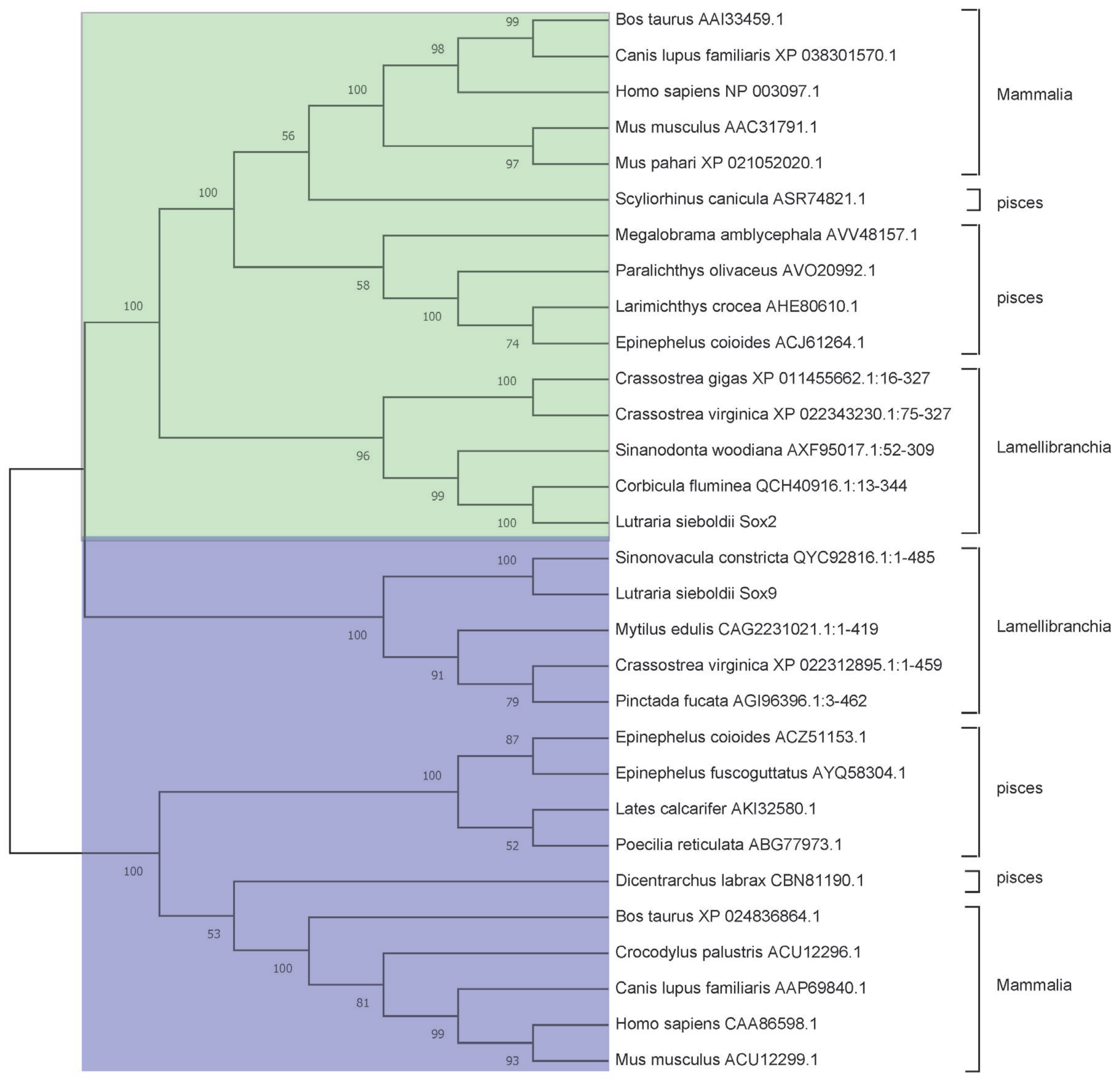
The homology analysis by Blast2GO showed that the LsSox2 (OM243913) CDS was 85.00% homologous for Corbicula fluminea, 71.33% for Sinanodonta woodiana and 62.39% for Crassortrea gigas. The homology of the LsSox9 (OM243914) CDS was 88.17% for C. fluminea and 45.60% for Mytilus edulis. The amino acid sequences of the Sox2 and Sox9 proteins were used to construct a phylogenetic tree among L. sieboldii with 30 other Sox genes, including 10 in Lamellibranchia, 10 in Mammalia, and 10 in Pisces (Figure 2). The phylogenetic analysis showed that the L. sieboldii Sox2 and Sox9 genes were clustered with their respective counterparts from C. fluminea and Sinonovacula constricta. The most striking result to emerge from the tree is that Sox2 and Sox9 were conserved in the Sox gene family, and our phylogenetic tree provided strong evidence for correctly naming the L. sieboldii Sox genes.

3.3. Bioinformatics Analysis

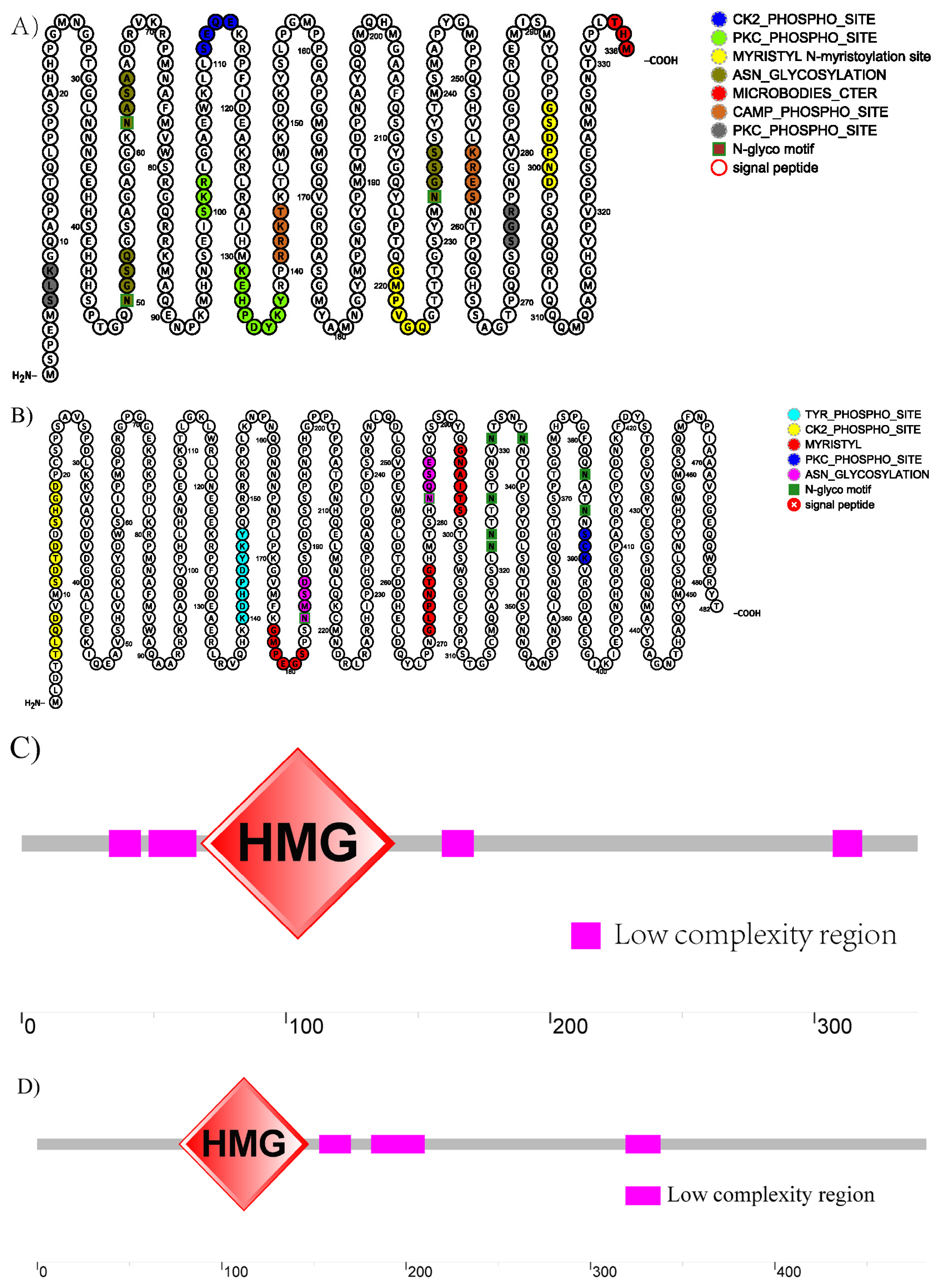
The CDSs of the LsSox2 and LsSox9 genes were 1011 and 1449 bp, encoding 336 and 482 amino acids, respectively. LsSox2 was predicted to contain cAMP- and cGMP-dependent protein kinase phosphorylation sites, N-glycosylation sites, and other sites (Figure 3A), while LsSox9 was predicted to contain casein kinase II phosphorylation sites, N-myristoylation sites, and other sites (Figure 3B). The SMART program predicted that LsSox2 has an HMG domain at residue sites 68–141 aa (Figure 3C), while LsSox9 has HMG domains at residue sites 77–147 aa (Figure 3D).
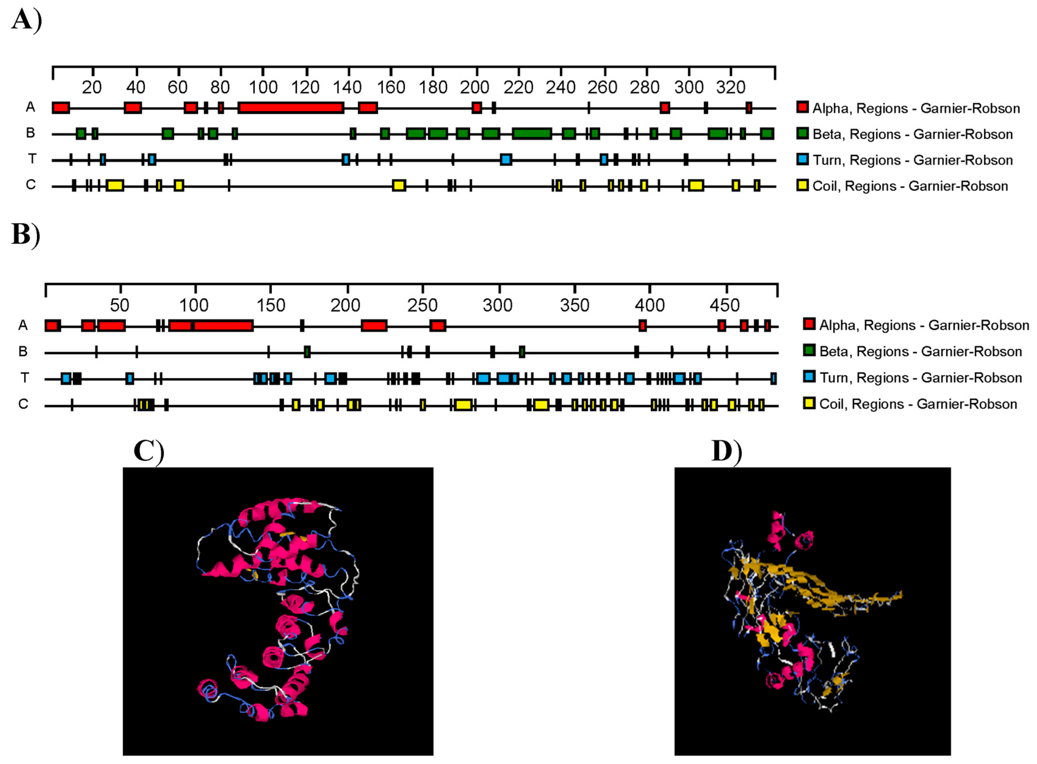
The formula, theoretical Mw, and pI of LsSox2 were C1557H2437N479O487S30, 36.62 kDa, and 9.67, respectively. No signal peptide was detected, but it was predicted to be nuclear with a score of 9.0. The formula, theoretical Mw, and pI of LsSox9 were C2301H3547N695O745S27, 53.73 kDa, and 6.53, respectively. It had no signal peptide and was predicted to be nuclear with a score of 7.2 and plasma membrane with a score of 1.7. LsSox2 was predicted to have a total of 13 α-helices, 24 β-folds, 23 T-turns, and 26 irregular coils (Figure 4A), while LsSox9 had a total of 15 α-helices, 13 β-folds, 43 T-turns, and 41 irregular coils (Figure 4B). The tertiary structures of LsSox2 and LsSox9 are shown in Figure 4C,D.

3.4. Tissue Distribution of LsSox2 and LsSox9 mRNA

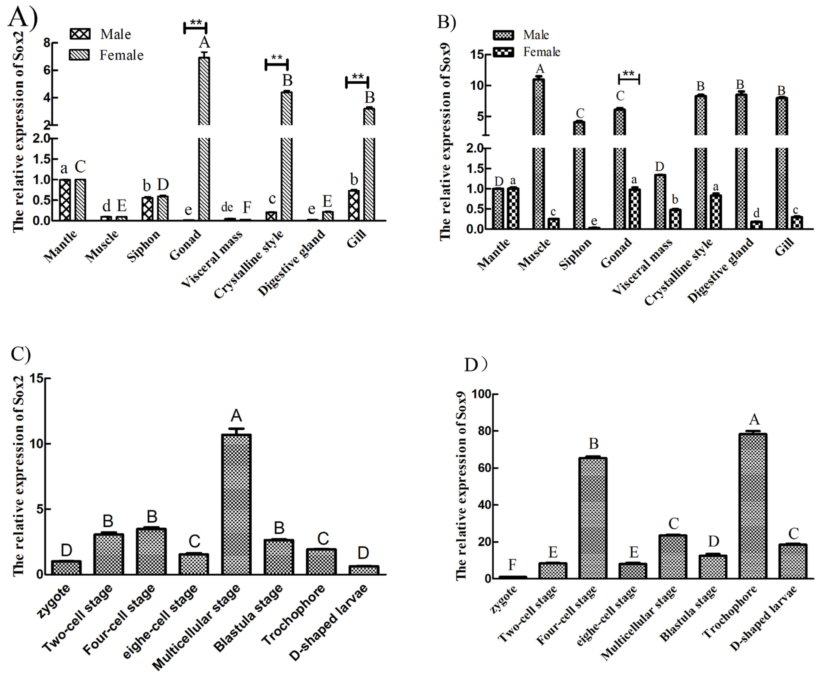
The quantitative real time-polymerase chain reaction (qRT-PCR) results suggested that LsSox2 was abundantly expressed in the ovary of females, moderately expressed in the gill, mantle, pipe, and genital duct, and weakly expressed in muscle, the visceral mass, and the digestive gland, LsSox2 was 250 times more highly expressed in the ovary than the visceral mass. In males, LsSox2 was abundantly expressed in the mantle, moderately expressed in the gill and pipe, and weakly expressed in muscle, the visceral mass, the digestive gland, and the testis, LsSox2 was 51 times more highly expressed in the mantle than in the testis. LsSox2 was more highly expressed in female tissues than in males (Figure 5A).
In males, LsSox9 was abundantly expressed in the muscle, pipe, gill, testis, digestive gland, and genital duct and moderately expressed in the mantle and visceral mass. LsSox9 was six times more highly expressed in testis than in the mantle. In females, LsSox9 was abundantly expressed in the mantle, ovary, and genital duct; moderately expressed in the visceral mass, muscle, and gill, and weakly expressed in the pipe and digestive gland. LsSox9 was 32 times more highly expressed in the ovary than in the pipe. LsSox9 was more highly expressed in male tissues than females (Figure 5B).
We next investigated Sox gene expression during embryogenesis by collecting cells from L. sieboldii zygotes after artificial insemination, including dividing, blastocyst stage, and D-shaped larvae. During embryogenesis, LsSox2 mRNA initially increased, and then decreased, exhibiting the highest expression at the multicellular stage (morula stage) and the lowest expression in the zygote and D-shaped larvae. The former was 20 times that of the D-shaped larvae (Figure 5C). During embryogenesis, LsSox9 expression exhibited two peaks. One peak occurred at the four-cell stage and the other peak occurred at the trochophore stage. The lowest expression was observed during the early stages of embryonic development (zygote stage), and it was 73 times more highly expressed at the four-cell stage than in the zygote stage (Figure 5D).

3.5. Gene Expression Analysis of L. sieboldii Sox Genes in Gonads

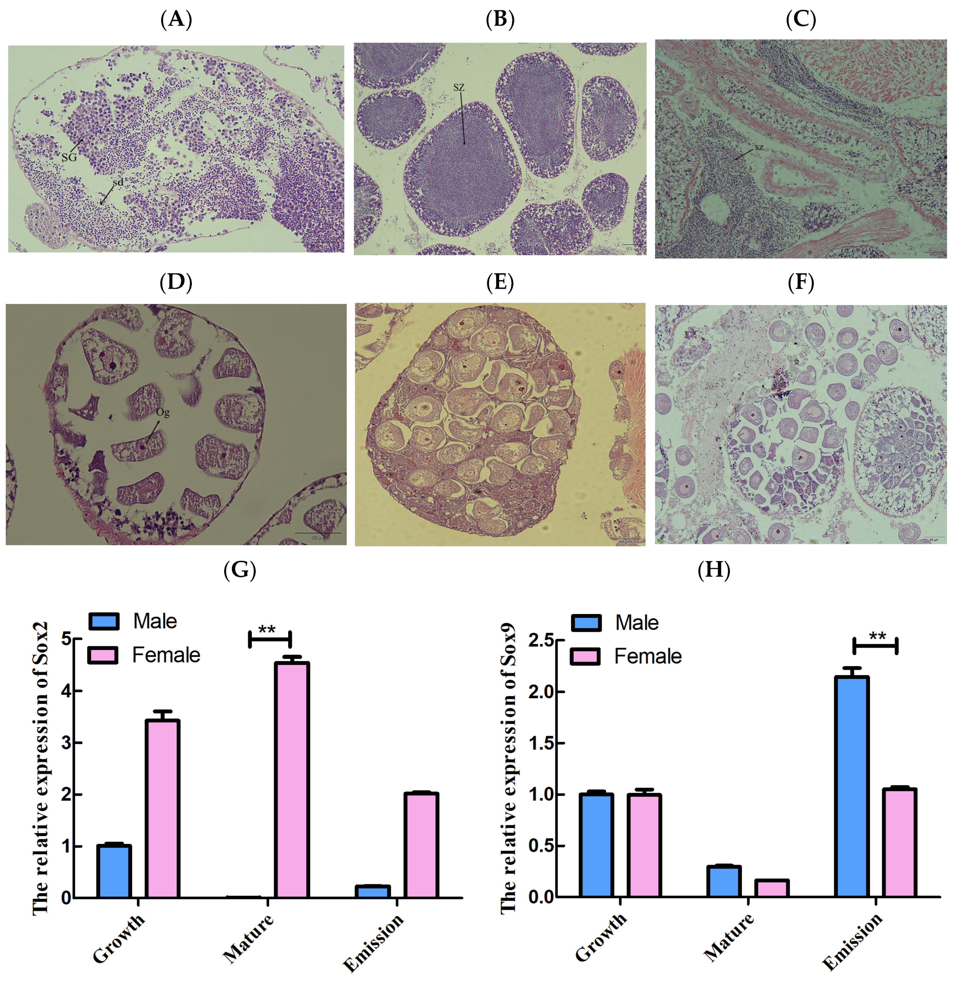
L. sieboldii gonads were collected and paraffin sections were prepared to monitor gonadal development according to previous studies. In the growth stage, the germ cells of males presented in cysts, and some were in advanced stages of spermatogenesis (Figure 6A). Mature sperm filled the vesicles during the maturation stage, indicating that these animals were sexually mature (Figure 6B). Mature sperm were expelled during the emission stage (Figure 6C). Primary oocytes were recognized in females during the growth stage (Figure 6D). Then, the entire follicular cavity was filled with mature eggs and oocytes during the late growth stage, and the irregularly shaped mature eggs were squeezed together in the cavity (Figure 6E). The follicles developed cavities of different sizes during the emission stage, but there were still immature oocytes remaining on the follicle wall (Figure 6F).
Notably, our results show that LsSox2 was significantly higher in the ovary than in the testis and reached its highest expression during gonadal maturation (p < 0.05) (Figure 6G). LsSox2 was 756 times more highly expressed in the ovary than in the testis during this stage (Figure 6G). Similarly, the LsSox9 gene was significantly more expressed in males than in females and was twice as high during the emission period (Figure 6H), suggesting that Sox9 plays a unique role during gonadal development in L. sieboldii.

4. Discussion

Mollusks are the second largest group in the animal kingdom after arthropods; however, expression analysis and functional characterization of the Sox gene family are very rare in mollusks. In the present study, the length of the LsSox2 gene CDS was 1011 bp, encoding 336 amino acids. It contained cAMP- and cGMP-dependent protein kinase phosphorylation sites, N-glycosylation sites, and an HMG domain, which are typical structural features of the Sox2 family [16]. The HMG domain of LsSox2 was highly similar to that of Anodonta woodiana [16] and Larimichthys crocea [24]. LsSox9 was 1449 bp, encoding 482 amino acids, and containing casein kinase II phosphorylation sites, N-myristoylation sites, and an HMG domain. Alignment of the LsSox9 protein sequence with sequences from other species indicated that the LsSox9 HMG box was highly conserved across taxa, consistent with earlier reports [19]. The highly conserved HMG domain of LsSox2 and LsSox9 with other species, assumed that the HMG box domain plays an important role in the recognition of specific target DNA elements, and suggested that the two genes likely functions as an important player in the gonadal tissue of L. sieboldii [25].
As reported in previous studies, many Sox genes have been associated with gonadal development [26,27], and are widely expressed during different stages of embryogenesis [28]. Herein, we report that Sox2 was highly expressed in the gonads and gills of normal L. sieboldii tissues, which was consistent with a previous study that the A. woodiana Sox2 gene is highly expressed in the gonads and gills [16]. While LsSox9 was more highly expressed in tissues of males than females, and it was six-fold more highly expressed in testis than in ovary, which was consistent with previous study that Sox9 was mainly expressed in the gonad, eyestalk, and cerebral ganglion of Scylla paramamosain [17], or with the highest expression levels in the brain and testis of Cyprinus carpio [18,19,29].
LsSox2 gene was more highly expressed during maturation of the L. sieboldii gonads, particularly during oocyte maturation, when it was significantly more expressed than in sperm. This finding is consistent with a study on Paralichthys olivaceus [14] but contrasted with one on A. woodiana [16]. Sox9 is a ‘hub’ gene of gonadal development, regulated positively in males and negatively in females [30]. LsSox9 was more highly expressed in tissues of males than females. LsSox9 was initially expressed at the same level during gonadal development in both sexes, but then declined sharply at the onset of ovarian differentiation and was expressed at the average level during spermatogonial emission stage. We suspect that this occurred due to the large number of sperm released to fill the spermathecae, making LsSox9 expression significantly higher in males than in females during the emission phase [31,32]. Similarly, the massive division and differentiation of oocytes at maturity results in much higher expression of Sox2 in the ovary than in testis. The differential expression patterns of LsSox2 and LsSox9 genes in the ovary and testis suggest that they may be involvement in the regulation of sex determination of L. sieboldii.
Previous studies have shown that the SoxB1 genome is critical for early embryogenesis, particularly as Sox2 promotes neural cell differentiation [15]. Okuda et al. provided evidence that the SoxB1 gene is highly redundant in early zebrafish embryos and that their encoded proteins are functionally interchangeable during early embryonic development by knocking out Sox2/3/19a/19b [33]. In the present study, LsSox2 exhibited the highest expression during the L. sieboldii morula stage and its lowest expression in D-shaped larvae. The results of these experiments indicate that Sox2 is a key transcriptional regulator in the oocyte-to-embryo transition and show that Sox2 expression was associated with the number of cell divisions, suggesting that Sox2 also plays a role in the embryonic development of L. sieboldii, which is consistent with previous studies [14]. For example, White et al. showed that Sox2-DNA binding predicts the fate of mammalian cells as early as the four-cell stage [34]. Pan et al. reported that Sox2, which is localized in the nucleus, is first expressed during the two-cell stage and that its expression dramatically increases between the morula and blastocyst stages, indicates that asymmetric distribution of Sox2 before the morula stage is essential for lineage segregation [35,36]. Sox9 gene, acts as a potent transcriptional regulator, involved in sex determination, gonad development and embryo development [29]. During embryo development, Spsox9 was highly expressed in 5 pairs of appendages, 7 pairs of appendages, and eyepigment formation stage [17]. Herein, LsSox9 gene exhibited two expression peaks, one at the four-cell stage and the other at the trochophore stage, it had been reported that during embryonic development, MnSoxE1 was mainly expressed in the gastrula stage, implicating its involvement in tissue cell differentiation and formation [37].

5. Conclusions

The Sox gene family plays a unique role in development. In this study, gene cloning and expression analyses were used to characterize Sox2 and Sox9 in L. sieboldii. The LsSox2 and LsSox9 expression patterns in normal tissues during gonadal development and embryogenesis suggest that the Sox2 and Sox9 genes play unique roles in L. sieboldii growth and development, and maybe sex-determining genes.

Author Contributions

P.Z. and J.Y. designed this project. M.L., Z.X. and Y.Z. cultured the experimental Lutraria sieboldii. M.L., Z.X., H.P. and J.Z. collected samples, extracted RNA and qRT-PCR. Y.X., J.L., M.L. and S.Q. performed date analysis and drafted the manuscript. Z.S., P.W. and M.L. participated in the sequence alignment and performed the statistical analysis. S.F.D., P.Z. and J.Y. revised the manuscript. P.Z. and J.Y. conceived the study, participated in its design and coordination. All authors have read and agreed to the published version of the manuscript.

Funding

This research was funded by the Guangxi innovation driven development special funds (Grant no. AA19254008-3), Guangxi Natural Science Foundation (Grant No. 2019GXNSFBA245081), Marine Science Program for Guangxi First-Class Discipline, Beibu Gulf University (Grant nos. DRB005, DTB001) and College Students’ innovation training program (202211607104, S202111607045 and Beike 202082030).

Institutional Review Board Statement

This article does not contain any studies with human participants. All animals used in this study were purchased from market, and the experimental protocols adhered to the the Guidelines of Animal Care by the Animal Management and Ethics Committee of BeiBu Gulf University.

Informed Consent Statement

Not applicable.

Data Availability Statement

The datasets used and/or analysed during the current study are available from the corresponding author on reasonable request.

Conflicts of Interest

The authors declare no conflict of interest.

References

  1. Grimm, D.; Bauer, J.; Wise, P.; Krüger, M.; Simonsen, U.; Wehland, M.; Infanger, M.; Corydon, T.J. The role of SOX family members in solid tumours and metastasis. Semin. Cancer Biol. 2020, 67, 122–153. [Google Scholar] [CrossRef]
  2. She, Z.-Y.; Yang, W.-X. SOX family transcription factors involved in diverse cellular events during development. Eur. J. Cell Biol. 2015, 94, 547–563. [Google Scholar] [CrossRef]
  3. Schepers, G.E.; Teasdale, R.D.; Koopman, P. Twenty Pairs of Sox: Extent, Homology, and Nomenclature of the Mouse and Human Sox Transcription Factor Gene Families. Dev. Cell 2002, 3, 167–170. [Google Scholar] [CrossRef] [Green Version]
  4. Hou, L.; Srivastava, Y.; Jauch, R. Molecular basis for the genome engagement by Sox proteins. Semin. Cell Dev. Biol. 2017, 63, 2–12. [Google Scholar] [CrossRef] [Green Version]
  5. Kashimada, K.; Koopman, P. Sry: The master switch in mammalian sex determination. Development 2010, 137, 3921–3930. [Google Scholar] [CrossRef] [Green Version]
  6. Miyagi, S.; Kato, H.; Okuda, A. Role of SoxB1 transcription factors in development. Cell. Mol. Life Sci. 2009, 66, 3675. [Google Scholar] [CrossRef]
  7. Bernard, P.; Tang, P.; Liu, S.; Dewing, P.; Harley, V.R.; Vilain, E. Dimerization of SOX9 is required for chondrogenesis, but not for sex determination. Hum. Mol. Genet. 2003, 12, 1755–1765. [Google Scholar] [CrossRef] [Green Version]
  8. Huang, X.; Wu, C.; Gong, K.; Chen, Q.; Gu, Q.; Qin, H.; Zhao, C.; Yu, T.; Yang, L.; Fu, W.; et al. Sox Gene Family Revealed Genetic Variations in Autotetraploid Carassius auratus. Front. Genet. 2020, 11, 804–804. [Google Scholar] [CrossRef]
  9. Fu, L.; Shi, Y.-B. The Sox transcriptional factors: Functions during intestinal development in vertebrates. Semin. Cell Dev. Biol. 2017, 63, 58–67. [Google Scholar] [CrossRef] [Green Version]
  10. Bowles, J.; Schepers, G.; Fau-Koopman, P.; Koopman, P. Phylogeny of the SOX family of developmental transcription factors based on sequence and structural indicators. Dev. Biol. 2000, 227, 239–255. [Google Scholar] [CrossRef]
  11. Kuzmichev, A.N.; Kim Sk Fau-D’Alessio, A.C.; D’Alessio Ac Fau-Chenoweth, J.G.; Chenoweth Jg Fau-Wittko, I.M.; Wittko Im Fau-Campanati, L.; Campanati, L.; Fau-McKay, R.D.; McKay, R.D. Sox2 acts through Sox21 to regulate transcription in pluripotent and differentiated cells. Curr. Biol. 2012, 22, 1705–1710. [Google Scholar] [CrossRef] [PubMed] [Green Version]
  12. Archer, T.C.; Jin, J.; Casey, E.S. Interaction of Sox1, Sox2, Sox3 and Oct4 during primary neurogenesis. Dev. Biol. 2011, 350, 429–440. [Google Scholar] [CrossRef] [PubMed] [Green Version]
  13. Hoffmann, S.A.; Hos, D.; Fau-Küspert, M.; Küspert, M.; Fau-Lang, R.A.; Lang Ra Fau-Lovell-Badge, R.; Lovell-Badge, R.; Fau-Wegner, M.; Wegner, M.; Fau-Reiprich, S.; et al. Stem cell factor Sox2 and its close relative Sox3 have differentiation functions in oligodendrocytes. Development 2014, 141, 39–50. [Google Scholar] [CrossRef] [PubMed] [Green Version]
  14. Gao, J.; Wang, Z.; Shao, K.; Fan, L.; Yang, L.; Song, H.; Liu, M.; Wang, Z.; Wang, X.; Zhang, Q. Identification and characterization of a Sox2 homolog in the Japanese flounder Paralichthys olivaceus. Gene 2014, 544, 165–176. [Google Scholar] [CrossRef]
  15. Cui, C.-P.; Zhang, Y.; Wang, C.; Yuan, F.; Li, H.; Yao, Y.; Chen, Y.; Li, C.; Wei, W.; Liu, C.H.; et al. Dynamic ubiquitylation of Sox2 regulates proteostasis and governs neural progenitor cell differentiation. Nat. Commun. 2018, 9, 4648. [Google Scholar] [CrossRef] [Green Version]
  16. Xia, X.; Guan, C.; Chen, J.; Qiu, M.; Qi, J.; Wei, M.; Wang, X.; Zhang, K.; Lu, S.; Zhang, L.; et al. Molecular characterization of AwSox2 from bivalve Anodonta woodiana: Elucidating its player in the immune response. Innate Immun. 2020, 26, 381–397. [Google Scholar] [CrossRef] [Green Version]
  17. Wan, H.; Liao, J.; Zhang, Z.; Zeng, X.; Liang, K.; Wang, Y. Molecular cloning, characterization, and expression analysis of a sex-biased transcriptional factor sox9 gene of mud crab Scylla paramamosain. Gene 2021, 774, 145423. [Google Scholar] [CrossRef]
  18. Takamatsu, N.; Kanda, H.; Ito, M.; Yamashita, A.; Yamashita, S.; Shiba, T. Rainbow trout SOX9: cDNA cloning, gene structure and expression. Gene 1997, 202, 167–170. [Google Scholar] [CrossRef]
  19. Du, Q.Y.; Wang, F.Y.; Hua, H.Y.; Chang, Z.J. Cloning and study of adult-tissue-specific expression of Sox9 in Cyprinus carpio. J. Genet. 2007, 86, 85–91. [Google Scholar] [CrossRef]
  20. Liao, Y.; Liu, K.; Ren, T.; Zhang, Z.; Ma, Z.; Dan, S.F.; Lan, Z.; Lu, M.; Fang, H.; Zhang, Y.; et al. The characterization, expression and activity analysis of three superoxide dismutases in Eriocheir hepuensis under azadirachtin stress. Fish. Shellfish. Immunol. 2021, 117, 228–239. [Google Scholar] [CrossRef]
  21. Lu, M.; Yang, J.; Wang, Z.; Song, J.; Hu, Y.; Wang, P.; Zhang, H.; Xu, Y.; Zhu, P. Cloning and expression of the ChGstα and ChGstκ genes in the gills of Crassostrea hongkongensis under nanoparticulate and ionic Zn stress. Comp. Biochem. Physiology. Toxicol. Pharmacol. CBP 2021, 244, 109007. [Google Scholar] [CrossRef] [PubMed]
  22. Kumar, S.; Stecher, G.; Li, M.; Knyaz, C.; Tamura, K. MEGA X: Molecular Evolutionary Genetics Analysis across Computing Platforms. Mol. Biol. Evol. 2018, 35, 1547–1549. [Google Scholar] [CrossRef] [PubMed]
  23. Xu, Y.; Pan, H.; Lu, M.; Liu, Q.; Shafique, L.; Peng, J.; Ahmed, T.; Wang, R.; Zhang, H.; Wang, Q.; et al. Analysis of transcripts and splice isoforms in red claw crayfish (Cherax quadricarinatus) using single-molecule long-read sequencing. Aquaculture 2021, 541, 736828. [Google Scholar] [CrossRef]
  24. Jiang, Y.-H.; Han, K.-H.; Wang, S.-H.; Chen, Y.; Wang, Y.-L.; Zhang, Z.-P. Identification and expression of transcription factor sox2 in large yellow croaker Larimichthys crocea. Theriogenology 2018, 120, 123–137. [Google Scholar] [CrossRef]
  25. Kurtz, S.; Lucas-Hahn, A.; Schlegelberger, B.; Göhring, G.; Niemann, H.; Mettenleiter, T.C.; Petersen, B. Knockout of the HMG domain of the porcine SRY gene causes sex reversal in gene-edited pigs. Proc. Natl. Acad. Sci. USA 2021, 118, e2008743118. [Google Scholar] [CrossRef]
  26. Jeng, S.R.; Wu, G.C.; Yueh, W.S.; Kuo, S.F.; Dufour, S.; Chang, C.F. Gonadal development and expression of sex-specific genes during sex differentiation in the Japanese eel. Gen. Comp. Endocrinol. 2018, 257, 74–85. [Google Scholar] [CrossRef]
  27. Kanda, H.; Kojima, M.; Miyamoto, N.; Ito, M.; Takamatsu, N.; Yamashita, S.; Shiba, T. Rainbow trout Sox24, a novel member of the Sox family, is a transcriptional regulator during oogenesis. Gene 1998, 211, 251–257. [Google Scholar] [CrossRef]
  28. Kamachi, Y.; Uchikawa, M.; Kondoh, H. Pairing SOX off: With partners in the regulation of embryonic development. Trends Genet. TIG 2000, 16, 182–187. [Google Scholar] [CrossRef]
  29. Adolfi, M.C.; Carreira, A.C.; Jesus, L.W.; Bogerd, J.; Funes, R.M.; Schartl, M.; Sogayar, M.C.; Borella, M.I. Molecular cloning and expression analysis of dmrt1 and sox9 during gonad development and male reproductive cycle in the lambari fish, Astyanax altiparanae. Reprod. Biol. Endocrinol. RBE 2015, 13, 2. [Google Scholar] [CrossRef] [Green Version]
  30. Vining, B.; Ming, Z.; Bagheri-Fam, S.; Harley, V. Diverse Regulation but Conserved Function: SOX9 in Vertebrate Sex Determination. Genes 2021, 12, 486. [Google Scholar] [CrossRef]
  31. Raghuveer, K.; Senthilkumaran, B. Isolation of sox9 duplicates in catfish: Localization, differential expression pattern during gonadal development and recrudescence, and hCG-induced up-regulation of sox9 in testicular slices. Reproduction 2010, 140, 477–487. [Google Scholar] [CrossRef] [PubMed] [Green Version]
  32. Degani, G. Expression of SOX3 and SOX9 Genes in Gonads of Blue Gourami. Adv. Biol. Chem. 2014, 4, 2162–2183. [Google Scholar] [CrossRef]
  33. Okuda, Y.; Ogura, E.; Kondoh, H.; Kamachi, Y. B1 SOX coordinate cell specification with patterning and morphogenesis in the early zebrafish embryo. PLoS Genet. 2010, 6, e1000936. [Google Scholar] [CrossRef] [Green Version]
  34. White, M.D.; Angiolini, J.F.; Alvarez, Y.D.; Kaur, G.; Zhao, Z.W.; Mocskos, E.; Bruno, L.; Bissiere, S.; Levi, V.; Plachta, N. Long-Lived Binding of Sox2 to DNA Predicts Cell Fate in the Four-Cell Mouse Embryo. Cell 2016, 165, 75–87. [Google Scholar] [CrossRef] [PubMed] [Green Version]
  35. Pan, H.; Schultz, R.M. Sox2 modulates reprogramming of gene expression in two-cell mouse embryos. Biol. Reprod. 2011, 85, 409–416. [Google Scholar] [CrossRef] [Green Version]
  36. Lee, M.; Choi, K.H.; Oh, J.N.; Kim, S.H.; Lee, D.K.; Choe, G.C.; Jeong, J.; Lee, C.K. SOX2 plays a crucial role in cell proliferation and lineage segregation during porcine pre-implantation embryo development. Cell Prolif. 2021, 54, e13097. [Google Scholar] [CrossRef]
  37. Hu, Y.; Jin, S.; Fu, H.; Qiao, H.; Zhang, W.; Jiang, S.; Gong, Y.; Xiong, Y.; Wu, Y. Functional analysis of a SoxE gene in the oriental freshwater prawn, Macrobrachium nipponense by molecular cloning, expression pattern analysis, and in situ hybridization (de Haan, 1849). 3 Biotech. 2019, 10, 10. [Google Scholar] [CrossRef]
Figure 1. Cloning of the LsSox2 and LsSox9 genes of Lutraria sieboldii. (A) Cloning of the LsSox2 gene, M: DL10000 DNA marker, 1 LsSox2: amplification product. (B) Cloning of the LsSox9 gene, M: DL10000 DNA marker, 1: water, 2: LsSox9 amplification product.
Fishes 07 00392 g001
Figure 2. Phylogenetic analyses of the (green) LsSox2 and (purple) LsSox9 proteins.
Fishes 07 00392 g002
Figure 3. Sequence analysis of LsSox2 and LsSox9. (A) LsSox2 motif sites. (B) LsSox2 protein domain structures. (C) LsSox9 motif sites. (D) LsSox9 protein domain structures.
Fishes 07 00392 g003
Figure 4. Tertiary structures of (A) LsSox2 and (B) LsSox9; secondary structures of (C) LsSox2 and (D) LsSox9.
Fishes 07 00392 g004
Figure 5. Relative expression of LsSox2 and LsSox9 in tissues of Lutraria sieboldii. (A) LsSox2 and (B) LsSox9. Expression of LsSox2 and LsSox9 at different stages of development. (C) LsSox2 and (D) LsSox9. Each bar represented mean ± SEM (n = 3), asterisks above the bars indicate the results of Duncan’s test at ** p < 0.01; Different letters indicate a significant difference.
Fishes 07 00392 g005
Figure 6. Paraffin sections of the Lutraria sieboldii gonad at different stages of development. (A) Growth stage, (B) maturation stage, and (C) emission stage of male L. sieboldii. (D) Growth stage, (E) maturation stage, and (F) emission stage of female L. sieboldii. (G) LsSox2 and (H) LsSox9. Expression of LsSox2 and LsSox9 at different stages of gonadal development in L. sieboldii. Each bar represented mean ± SEM (n = 3), asterisks above the bars indicate the results of Duncan’s test at ** p < 0.01. sd: spermatids; SG: spermatogonia; sz: spermatozoa. Og: oogonia.
Fishes 07 00392 g006
Table 1. Primer sequences.
PrimersSequence (5′-3′)Amplified Length (bp)Tm
(°C)
Note
LsSox2F1CAAGCACTGTTTTTGAGAGG157456CDS
cloning
LsSox2R1TTCATTTCACGATTTAACGG
LsSox2dlF1TTTACCCGTTAAATCGTGAAAT9259qRT-PCR
LsSox2dlR1TGAAAGTTGAGAATCGCCTCT
LsSox9F1AGCGAACTTGTTTGGACATA150457CDS
cloning
LsSox9R1ACCGTTTCTCAGCGTCTAAT
LsSox9dlF1AAAGTAAGAGACGACGCAG12358qRT-PCR
LsSox9dlR1GTAACGAGACACAGAGGGG
LsEf1dlF1CTGTCTTAGATTGTCACACTGCC10465qRT-PCR
LsEf1dlR1CGTCTTAGGGTTGTCTTCCAA
Publisher’s Note: MDPI stays neutral with regard to jurisdictional claims in published maps and institutional affiliations.