Article

Differences in Intestinal Microbial Composition between Red Claw Crayfish (Cherax quadricarinatus) and Red Swamp Crayfish (Procambarus clarkii) Cultured in Pond

1
State Key Laboratory for Managing Biotic and Chemical Threats to the Quality and Safety of Agro-Products, Institute of Hydrobiology, Zhejiang Academy of Agricultural Sciences, Hangzhou 310004, China
2
School of Fishery, Zhejiang Ocean University, Zhoushan 316022, China
3
Faculty of Life Sciences, Huzhou University, Huzhou 313000, China
4
Key Laboratory of Freshwater Aquatic Genetic Resources, Shanghai Ocean University, Ministry of Agriculture, Shanghai 201306, China
*
Authors to whom correspondence should be addressed.
These authors contributed equally to this work.
Fishes 2022, 7(5), 241; https://doi.org/10.3390/fishes7050241
Submission received: 16 August 2022 / Revised: 7 September 2022 / Accepted: 13 September 2022 / Published: 14 September 2022

Abstract

:
Intestinal microbiota communities participate in several metabolic processes in the host, and are highly correlated to digestion, nutrition, growth, and immunity. However, the intestinal microbiota of aquatic invertebrates is poorly understood, especially in freshwater crayfish. In this study, the intestinal microbiota of two important freshwater economic aquaculture species, the invasive species, the red swamp crayfish (Procambarus clarkii, Pc), and the introduced species, the red claw crayfish (Cherax quadricarinatus, Cq), were investigated. The results showed that the community richness and diversity of Pc were higher than those of Cq, which might be one of the reasons that Pc have stronger environmental adaptability than Cq. Five core phyla were identified in the Pc group, including Proteobacteria (26.92%), Tenericutes (25.73%), Bacteroidetes (25.12%), Firmicutes (14.03%), and RsaHF231 (8.02%), and three phyla were detected in the Cq group, including Tenericutes (67.35%), Proteobacteria (25.98%), and Firmicutes (4.69%). In Pc and Cq groups, Proteobacteria exhibited significant differential abundance between males and females. In addition, Vibrio coralliilyticus were found particularly in the intestine of Cq. This study provides information on intestinal microbiota differences of Pc and Cq, contributing to the development of new dietary formulations and providing significance to future aquaculture.

1. Introduction

Freshwater crayfish play key roles in the natural ecosystems and are important components of aquaculture [1]. China is one of the countries with the least distribution of freshwater crayfish; only three native species of Cambaroides exist and all of them are distributed in northeast area [1,2]. Due to the severe damage to the habitat caused by water pollution, their population has decreased significantly to endangerment over the past 20 years [3]; thus, none of them have become the breeding species. Procambarus clarkii (Pc) invaded China from Japan in the 1920s and has become the only economically important farmed freshwater crayfish species in China [4] that benefits from its strong adaptability to the environment. Cherax quadricarinatus (Cq) was introduced into China from Australia and has been cultured in many places in China as a promising aquaculture species. As a tropical species, Cq grows best in warm water [5]. Long growth period and feeding habits are the major concerns of successful aquaculture of Cq. The intestinal microbiota affects several metabolic processes in the host and reflects the diet of the host and their adaption to the living environment [6,7]; it is also highly associated with the health of the host [8,9,10]. For example, long-term starvation in Cherax cainii would cause significant decreases in microbial density and diversity and the core microbiota to be replaced by Vibrio [11]. Exposure to cadmium caused changes in the composition and decreased the community diversity of intestinal microbiota of Rana chensinensis [12]. The intestinal microbiota can boost or depress host immune system; in Litopenaeus vannamei, the total bacterial count in white spot syndrome virus-infected individuals is higher than that in uninfected individuals [13]. Recent research has demonstrated that bacterial microbiomes can have pivotal roles in invasion ecology. In signal crayfish, the intestinal microbial communities may affect and be affected by range expansion [14]. A survey of Cambarus sciotensis showed that the microbial diversity and composition of carapace and gills are determined by interactions within the local environment and host microsites [15].
Compared with mammals, the intestinal microbiota of aquatic invertebrates is poorly understood [5]. With the increase in global aquaculture, the influence of the intestinal microbial community on culture species has received increasing attention. Investigation of the diversity of intestinal microbiota composition and understanding of the microbiota’s functions in digestion and immune systems of aquaculture species could contribute to establishing healthy culture strategies. In this study, Illumina PE250 high-throughput sequencing was used to investigate the bacterial communities of the intestinal microbes of introduced Cq and invasive Pc. The results will help understand the dietary differences between two widely cultured crayfish and provide more information about the nutritional requirements, health, and environment adaption, which will be beneficial for the development of new dietary formulations, including microbial agents.

2. Materials and Methods

2.1. Sample Collection

Healthy Pc and Cq crayfish with body weights of 32.55 ± 3.25 g and 54.89 ± 11.52 g were obtained from a farming pond on 10 September 2021 in Jiaxing, Zhejiang Province, China. The crayfish were cultured for about four months and were fed twice a day with commercial diets (Tech-Bank Food Co, Ltd., Nanjing, China), the rearing density was 7.5 larva per meters squared of water. A total of 20 male and female Pc and Cq individuals were randomly selected for sampling. After fasting for 24 h, the crayfish were euthanized by a cold shock. The complete intestinal tissues were immediately sampled using sterile equipment, flash-frozen in liquid nitrogen, and stored at −80 °C until DNA extraction.

2.2. Genomic DNA (gDNA) Extraction, Polymerase Chain Reaction (PCR) Amplification, and Sequencing

The gDNA of each sample was extracted using the E.Z.N.A.® Soil DNA Kit (Omega Bio-tek, Norcross, GA, USA) according to the manufacturer’s instructions. The quality and quantity of DNA were assessed by agarose gel electrophoresis and a NanoDrop 2000 spectrophotometer (Bio-Rad Laboratories Inc., Hercules, CA, USA). The 16S rRNA genes were amplified using the primer pair (341F: CCTAYGGGRBGCASCAG, 806R: GGACTACNNGGGTATCTAAT) with the barcode. PCR was performed (95 °C for 5 min, followed by 27 cycles at 95 °C for 30 s, 55 °C for 30 s, and 72 °C for 45 s and a final extension at 72 °C for 10 min) in a 20 μL reaction mixture containing 4 μL of 5 × FastPfu Buffer, 2 μL of 2.5 mM deoxynucleotide triphosphates (dNTPs), 0.8 μL of each primer (5 μM), 0.4 μL of FastPfu Polymerase, and 10 ng of template DNA. The PCR products were detected by 2% agarose gel electrophoresis, and the samples with target electrophoresis bands were chosen for further experiments. PCR products with different barcodes were mixed in equal quantities and purified with a GeneJET Gel Extraction Kit (Thermo Fisher Scientific, Waltham, MA, USA). Sequencing libraries were generated using an NEB Next®Ultra™DNA Library Prep Kit for Illumina (NEB, Ipswich, MA, USA) following the manufacturer’s recommendations, and index codes were added. The library quality was assessed on the Qubit@ 2.0 Fluorometer (Thermo Fisher Scientific) and Agilent Bioanalyzer 2100 system. Finally, the library was sequenced on an Illumina MiSeq platform, and 250–300 bp paired-end reads were generated.

2.3. Data Analysis

Paired-end reads from the original DNA fragments were merged using FLASH [16] and assigned to each sample according to the unique barcodes. The sequences were analyzed using UPARSE-OTU and UPARSE-OTUref algorithms in the UPARSE software package [17]. In-house Perl scripts were used to analyze alpha (within samples) and beta (among samples) diversities. Sequences with ≥97% similarity were assigned to the same operational taxonomic units (OTUs). A representative sequence for each OTU was picked, and the RDP Classifier was used to annotate taxonomic information for each representative sequence. To compute alpha diversity, the OTU table was rarified, and three metrics were calculated. Chao1 estimated the species abundance, and Observed Species estimated the amount of unique OTUs found in each sample and the Shannon index. Rarefaction curves were generated based on these three metrics’ indication of the diversity estimated by the Shannon index.

2.4. Phylogenic Distance and Community Distribution

A graphical representation of the relative abundance of bacterial diversity from phylum to species was visualized using the Krona chart. Cluster analysis was preceded by principal component analysis (PCA) to reduce the dimension of the original variables using the QIIME software package. QIIME calculated both weighted and unweighted UniFrac distances, which were phylogenetic measures of beta diversity. An unweighted UniFrac distance was used for principal coordinate analysis (PCoA) and unweighted pair-group method with arithmetic mean (UPGMA) clustering.

2.5. Statistical Analysis

To confirm differences in the abundances of individual taxa between the two groups, Metastats software was utilized. Linear discriminant analysis effect size (LEfSe) (http://huttenhower.sph.harvard.edu/galaxy, accessed on 2 September 2022) was used to analyze biomarkers within different groups. To identify differences in microbial communities between the two groups, analysis of similarities (ANOSIM) (Vegan-package in R) and the multi-response permutation procedure (MRPP) (Vegan-package in R) were performed based on the Bray–Curtis dissimilarity distance matrices.

3. Results

3.1. OTU Analysis and Alpha Diversity

A total of 472,373 and 481,012 high-quality sequences were obtained from the intestine content of Pc and Cq, respectively, with an average of 48,728 reads per sample. All sequences were clustered into OTUs according to the same similarity values of 97%, and the rarefaction curves of all samples and the species accumulation curves tended to reach a saturation plateau (Figure 1A,B), indicating that the sequencing depth and sampling amount were sufficient to cover the majority of the microbial community. In addition, the average OTU number obtained from each individual in Cq and Pc groups were 237 ± 118 and 347 ± 87. In total, 531 OTUs were unique in the Cq group, 447 OTUs were unique in the Pc group, and 523 OTUs were shared (Figure 1C). To understand the species classification information, all OTUs were annotated at different taxonomic levels, and 26 taxa were identified at the phylum level.
To investigate the community richness and diversity of Pc and Cq groups, the alpha diversity index, as well as Chao (community richness indexes), Simpson, and Shannon (diversity indexes) indexes, were calculated. The indexes of diversity at a genetic distance of 3% are presented in Table 1. The completeness of sequencing was estimated by the Good’s coverage, ranging from 99.75 to 99.82%. The community richness index (Chao and ACE) and Shannon index of samples in the Pc group were significantly higher than those of the Cq group (p < 0.01), and the Simpson index of samples in the Pc group was lower than that of the Cq group, revealing that the intestinal microbial community richness and variety in the Pc group were higher than those in the Cq group.

3.2. Beta Diversity

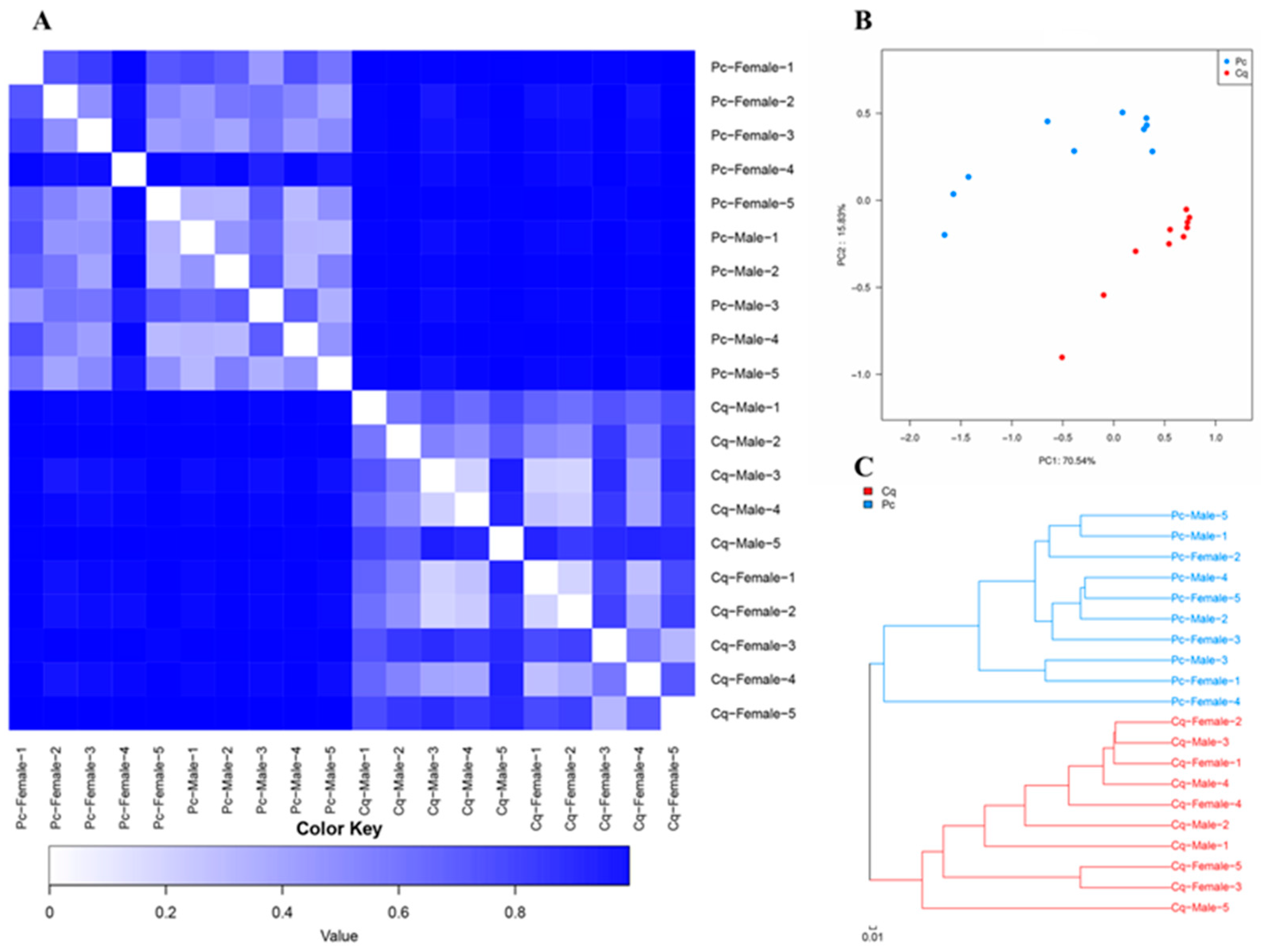
Bray–Curtis was used to compare the distance between samples in pairs, and a heatmap was used to visualize the results. From the heatmap (Figure 2A), the distances of individuals within the Pc group were significantly lower than those within the Cq group, which showed higher diversity. The similarity and difference of microbial community compositions of all samples were analyzed by PCoA, with PC1 = 70.54% and PC2 = 15.83% (Figure 2B). The Pc and Cq groups could be divided into two clusters. The hierarchical clustering showed that the Pc group gathered in one branch, and the Cq group gathered in another branch, but the males and females in each group did not show an obvious clustering relationship (Figure 2C).

3.3. Taxonomic Composition

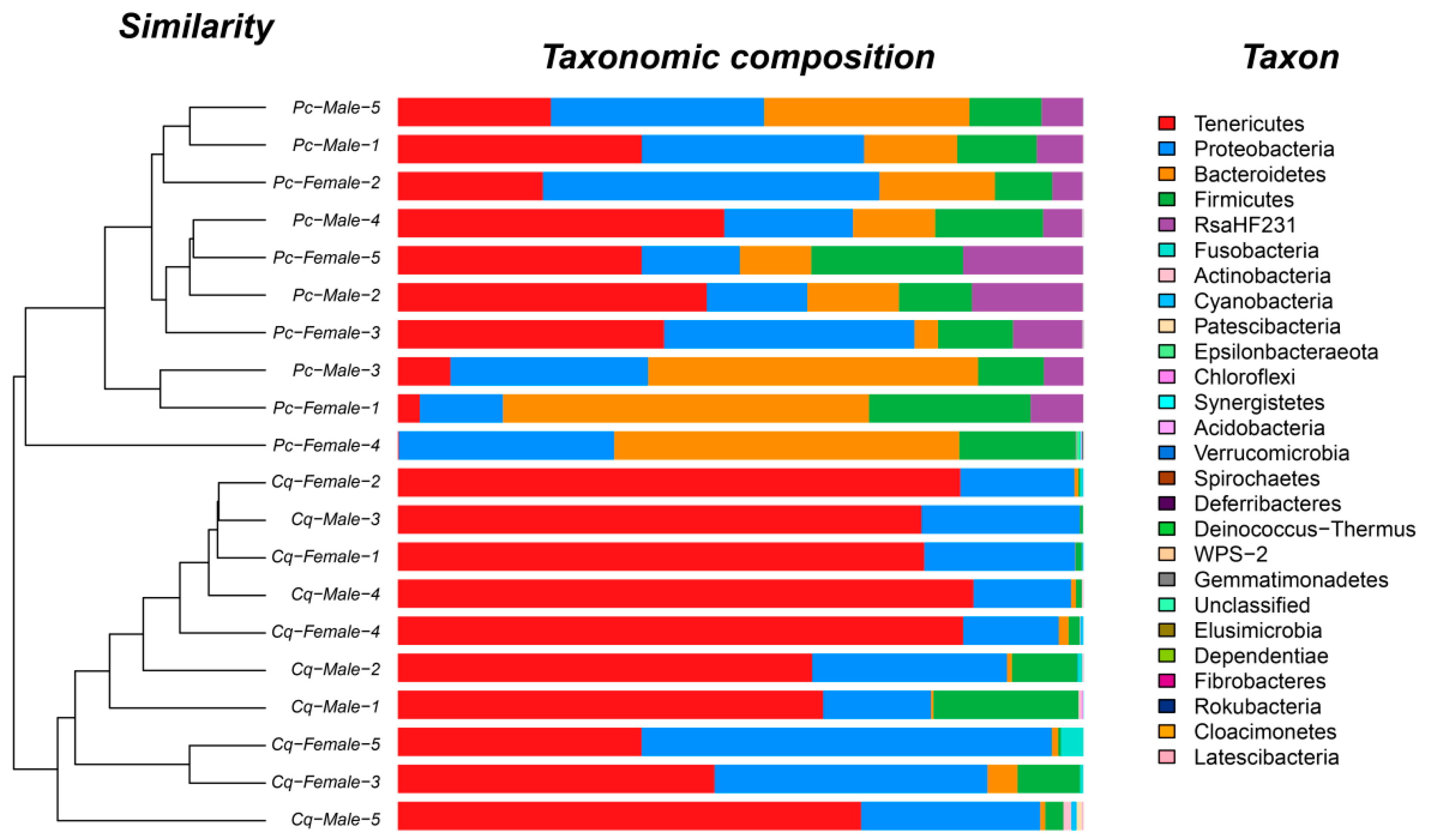
A total of 26 phyla were identified in all samples, and significant differences were observed in the relative abundance of gut microbiota between Pc and Cq groups (p < 0.05). In the intestines of Pc-group individuals, there were five core phyla with mean relative abundances over 1%, namely Proteobacteria (26.92%), Tenericutes (25.73%), Bacteroidetes (25.12%), Firmicutes (14.03%), and RsaHF231 (8.02%), while in the Cq group, only three core phyla were identified, namely Tenericutes (67.35%), Proteobacteria (25.98%), and Firmicutes (4.69%). These core microbiota accounted for 99.81% of the total abundance in the Pc group and 98.02% in the Cq group.
Among the core gut microbiota, Bacteroidetes, RsaHF231, and Tenericutes demonstrated a significant differential abundance between the Pc and Cq groups, especially RsaHF231, which showed a significantly higher abundance of community in the Pc group than in the Cq group. The genera with mean relative abundances over 1% were assigned as core genera, and 13 core genera were detected in the Pc group, namely Candidatus Bacilloplasma (25.63%), Bacteroides (21.00%), Citrobacter (10.30%), RsaHF231_norank (8.02%), [Anaerorhabdus] furcosa group (4.54%), Vibrio (4.41%), Shewanella (4.01%), [Eubacterium] brachy group (3.57%), Brachymonas (2.27%), Dysgonomonas (1.65%), ZOR0006 (1.65%), Aeromonas (1.41%), and Desulfovibrio (1.17%). Eight core genera were detected in the Cq group, namely Candidatus Bacilloplasma (46.10%), Mycoplasmataceae_uncultured (11.31%), Candidatus Hepatoplasma (9.93%), Vibrio (9.59%), Citrobacter (7.29%), Halomonas (3.26%), Lachnospiraceae_uncultured (2.99%), and Enterobacter (1.52%).
In addition, in Pc and Cq groups, the members and percentage of the dominant genera in males and females differed (Figure 3). The Pc female group had four more core genera than the Pc male group, and the Cq female and male groups both had eight core genera, but only four were shared.

3.4. Variance Analysis

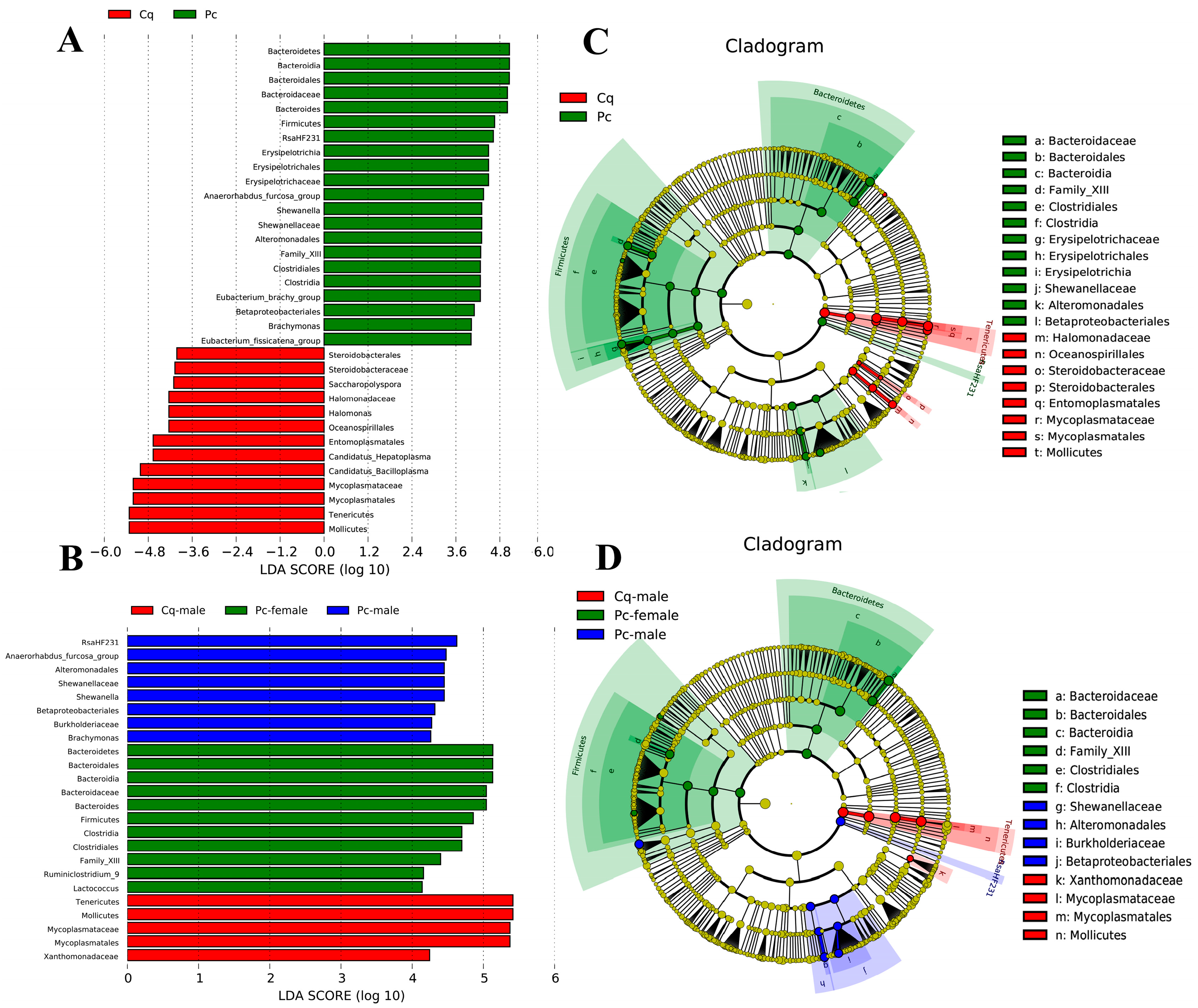
The results based on LEfSe analysis are shown in Figure 4. The cladogram shows microbial communities at different levels of classification, with significant differences between Pc and Cq groups. A total of 34 species were detected with significant differences in abundance between the Pc and Cq groups. After introducing the sexes into the classification, the species number decreased to 24. Between male and female Pc, 19 species with significant differences in abundance were detected, and there were no microbial communities showing significant differences in abundance between the Cq female group and other groups.

4. Discussion

In different developmental stages, different culture environments, different diets, or different health statuses, the top three phyla components of Pc intestinal microbiota (Proteobacteria, Bacteroidetes and Firmicutes) are stable [18,19,20,21]. Regarding the dominant phyla, the Pc were reported with four or five dominant phyla [22,23], while in Cq, the number of dominant phyla was only three [24]. In our study, the intestinal microbiota of Pc were consistent with previous studies. This indicated that diet and digestive capacity of Pc and Cq are very different.
The microbial composition can reflect and affect diet composition and digestion [25]. Our study showed that the community richness and diversity of the intestinal microbiota in the Pc group were higher than those in the Cq group. More diverse gut communities exert greater protective effects on the host [26,27]. Based on the lower-diversity intestinal microbiota community found in the Cq group when compared to the Pc group, the homeostatic balance of Cq intestinal microbiota might be more vulnerable than Pc intestinal microbiota.
Sex-specific differences in immune responses have been reported in a number of invertebrates, such as Ruditapes philippinarum [28]. In mice, gut microbiota differed in males and females. After puberty, the male microbiota composition becomes less diverse than the female microbiota [29]. In our study, there was no significant difference in the microbiota composition diversity between male and female individuals, but the composition richness of male individuals was significantly higher than female individuals, and Vibrio dominated the major differential abundance between male (0.28–1.80%) and female (7.03–18.90%) crayfish in both Pc and Cq groups. Vibrio is one of the main components of Proteobacteria, which is often described as the dominant genus in the shrimp gut [30]. Most vibrios exist harmoniously with the host [4,31], but certain strains of Vibrio can cause diseases with high mortality, which affect a vast number of aquatic crustacean species [32,33,34]. Therefore, the relatively high abundance of Vibrio in female crayfish may relate to the sex-specific differences in immune responses [35]. However, the relationship and mechanism still need to be further verified.
Vibrio coralliilyticus were only detected in the Cq group. V. coralliilyticus is a pathogenic species commonly found in marine and brackish environments [36,37]. V. coralliilyticus is pathogenic to various marine animals such as the Pacific oyster (Crassostrea gigas) [38,39] and brine shrimp (Artemia spp.) [40]. Although there is no evidence of the infection of V. coralliilyticus in freshwater crustaceans, it is pathogenic to rainbow trout (Oncorhynchus mykiss), with moralities of up to 100% [41]. Meanwhile, V. coralliilyticus was isolated from the hepatopancreas of cultured Pacific white shrimp (Litopenaeus vannamei), which did not show symptoms of vibriosis [42]. V. coralliilyticus was isolated from water and sediment in an estuarine environment and was verified as having cephalothin and florfenicol resistant profiles [43]. Although V. coralliilyticus has not caused any outbreaks of disease in Cq until now, it might be a potential threat to freshwater aquatic animals. The Cq crayfish used in this study were cultured in an inland pond. V. coralliilyticus may be introduced by infected brine shrimp as feed. Brine shrimp are a quality starter feed for most aquatic species. In Cq cultivation, brine shrimp are often used as micro-diets at the larval stage. In this study, V. coralliilyticus was detected in the intestines of adult crayfish, which suggests that it has a strong colonization capacity in Cq. This finding suggests that caution must be exercised in pathogen detection and the use of brine shrimp as aquaculture feed in the future.
Bacteroidetes are regarded as specialists for degrading high-molecular-weight organic matter such as proteins and carbohydrates [44]. In humans, the relative abundance of Bacteroidetes is related to obesity and diet composition and influences the health of the host by limiting potential pathogenic bacteria colonization in the gastrointestinal tract [45]. In our study, the Bacteroidetes occupy a quarter of the intestinal microbiota proportion of Pc but are not included in the core phyla of Cq. These results indicate that the dietary patterns of Pc and Cq were different. The Cq may prefer higher protein diets than Pc, and together with the lower diversity of its intestinal microbiota, the ability to maintain gastrointestinal homeostasis of Cq may be weaker than Pc.

5. Conclusions

Balanced gut microbiota is important to the health of the host. In this study, the profiles of microbial communities gathered in the intestines of Pc and Cq were investigated. The results showed that the bacterial compositions of Pc and Cq were significantly different. The community richness and diversity of Pc were higher than Cq, and they shared three core phyla. In addition, Proteobacteria exhibited significant differential abundance between males and females, and V. coralliilyticus, which is commonly found in marine and brackish environments, was newly identified in the intestine of Cq. This study provided information on the bacterial communities of Pc and Cq and analyzed the difference in their core phyla, which could be further applied to microbial supplements and might be instructive for production.

Author Contributions

Conceptualization, H.C.; methodology, H.C. and F.L.; validation, F.L., M.O. and H.Z.; investigation, F.L., M.O. and H.Z.; resources, F.L.; data curation, F.L. and M.O.; writing—original draft preparation, H.C.; writing—review and editing, H.C.; visualization, H.C.; supervision, B.L.; project administration, B.L.; funding acquisition, H.C. All authors have read and agreed to the published version of the manuscript.

Funding

The work was supported by the Zhejiang Science and Technology Major Program (2021C02069-4).

Institutional Review Board Statement

The study was conducted according to the guidelines of the Declaration of Helsinki, and approved by Committee of Laboratory Animal Experimentation at Zhejiang Academy of Agricultural Sciences (No. 2022ZAASLA29).

Acknowledgments

We would like to express gratitude to Haojie Qian at Honghai Culture Co., Ltd., Haining, Zhejiang province, for his kind help in collecting samples.

Conflicts of Interest

The authors declare no competing interest.

References

  1. Crandall, K.A.; Buhay, J.E. Global diversity of crayfish (Astacidae, Cambaridae, and Parastacidae—Decapoda) in freshwater. In Freshwater Animal Diversity Assessment; Springer: Dordrecht, The Netherlands, 2007; pp. 295–301. [Google Scholar]
  2. Mu, F.; Cheng, Y.; Wu, X. Distribution and industrial development of crayfish in the world. J. Shanghai Ocean Univ. 2007, 16, 64–72. (In Chinese) [Google Scholar]
  3. Bao, J.; Xing, Y.; Feng, C.; Kou, S.; Jiang, H.; Li, X. Acute and sub-chronic effects of copper on survival, respiratory metabolism, and metal accumulation in Cambaroides dauricus. Sci. Rep. 2020, 10, 16700. [Google Scholar] [CrossRef]
  4. Holdich, D.M. A review of astaciculture: Freshwater crayfish farming. Aquat. Living Resour. 1993, 6, 307–317. [Google Scholar] [CrossRef]
  5. King, C.R. Growth and survival of redclaw crayfish hatchlings (Cherax quadricarinatus von Martens) in relation to temperature, with comments on the relative suitabitity of Cherax quadricarinatus and Cherax destructor for culture in Queensland. Aquaculture 1994, 122, 75–80. [Google Scholar] [CrossRef]
  6. Holt, C.C.; Bass, D.; Stentiford, G.D.; Giezen, M. Understanding the role of the shrimp gut microbiome in health and disease. J. Invertebr. Pathol. 2021, 186, 107387. [Google Scholar] [CrossRef]
  7. Li, T.; Li, H.; Gatesoupe, F.J.; She, R.; Lin, Q.; Yan, X.; Li, J.; Li, X. Bacterial signatures of “Red-Operculum” disease in the gut of crucian carp (Carassius auratus). Microb. Ecol. 2017, 74, 510–521. [Google Scholar] [CrossRef]
  8. Mora-Sánchez, B.; Balcázar, J.L.; Pérez-Sánchez, T. Effect of a novel postbiotic containing lactic acid bacteria on the intestinal microbiota and disease resistance of rainbow trout (Oncorhynchus mykiss). Biotechnol. Lett. 2020, 42, 1957–1962. [Google Scholar] [CrossRef]
  9. Zhu, C.Z.; Li, D.; Chen, W.J.; Ban, S.N.; Liu, T.; Wen, H.; Jiang, M. Effects of dietary host-associated Lactococcus lactis on growth performance, disease resistance, intestinal morphology and intestinal microbiota of mandarin fish (Siniperca chuatsi). Aquaculture 2021, 540, 736702. [Google Scholar] [CrossRef]
  10. You, C.; Chen, B.; Wang, M.; Wang, S.; Zhang, M.; Sun, Z.; Juventus, A.J.; Ma, H.; Li, Y. Effects of dietary lipid sources on the intestinal microbiome and health of golden pompano (Trachinotus ovatus). Fish Shellfish Immunol. 2019, 89, 187–197. [Google Scholar] [CrossRef]
  11. Foysal, M.J.; Fotedar, R.; Tay, A.C.Y.; Gupta, S.K. Effects of long-term starvation on health indices, gut microbiota and innate immune response of fresh water crayfish, marron (Cherax cainii, Austin 2002). Aquaculture 2020, 514, 734444. [Google Scholar] [CrossRef]
  12. Mu, D.; Meng, J.; Bo, X.; Wu, M.; Xiao, H.; Wang, H. The effect of cadmium exposure on diversity of intestinal microbial community of Rana chensinensis tadpoles. Ecotoxicol. Environ. Saf. 2018, 154, 6–12. [Google Scholar] [CrossRef]
  13. Li, E.; Xu, C.; Wang, X.; Wang, S.; Zhao, Q.; Zhang, M.; Qin, J.G.; Chen, L. Gut microbiota and its modulation for healthy farming of Pacific white shrimp Litopenaeus vannamei. Rev. Fish. Sci. Aquac. 2018, 26, 381–399. [Google Scholar] [CrossRef]
  14. Dragičević, P.; Bielen, A.; Petrić, I.; Vuk, M.; Žučko, J.; Hudina, S. Microbiome of the successful freshwater invader, the signal crayfish, and its changes along the invasion range. Microbiol. Spectr. 2021, 9, e00389-21. [Google Scholar] [CrossRef]
  15. Skelton, J.; Geyer, K.M.; Lennon, J.T.; Creed, R.P.; Brown, B.L. Multi-scale ecological filters shape the crayfish microbiome. Symbiosis 2017, 72, 159–170. [Google Scholar] [CrossRef]
  16. Magoč, T.; Salzberg, S.L. FLASH: Fast length adjustment of short reads to improve genome assemblies. Bioinformatics 2011, 27, 2957–2963. [Google Scholar] [CrossRef]
  17. McDonald, D.; Price, M.N.; Goodrich, J.; Nawrocki, E.P.; DeSantis, T.Z.; Probst, A.; Andersen, G.L.; Knight, R.; Hugenholtz, P. An improved Greengenes taxonomy with explicit ranks for ecological and evolutionary analyses of bacteria and archaea. ISME J. 2012, 6, 610–618. [Google Scholar] [CrossRef]
  18. Liu, Q.; Long, Y.N.; Li, B.; Zhao, L.; Luo, J.; Xu, L.; Luo, W.; Du, Z.; Zhou, J.; Yang, S. Rice-shrimp culture: A better intestinal microbiota, immune enzymatic activities, and muscle relish of crayfish (Procambarus clarkii) in Sichuan Province. Appl. Microbiol. Biotechnol. 2020, 104, 9413–9420. [Google Scholar] [CrossRef]
  19. Guo, K.; Ruan, G.; Fan, W.; Fang, L.; Wang, Q.; Luo, M.; Yi, T. The effect of nitrite and sulfide on the antioxidant capacity and microbial composition of the intestines of red swamp crayfish, Procambarus clarkii. Fish Shellfish Immunol. 2020, 96, 290–296. [Google Scholar] [CrossRef]
  20. Zhang, Z.; Liu, J.; Jin, X.; Liu, C.; Fan, C.; Guo, L.; Liang, Y.; Zheng, J.; Peng, N. Developmental, dietary, and geographical impacts on gut microbiota of red swamp crayfish (Procambarus clarkii). Microorganisms 2020, 8, 1376. [Google Scholar] [CrossRef]
  21. Chen, H.; Wang, Y.; Zhang, J.; Bao, J. Intestinal microbiota in white spot syndrome virus infected red swamp crayfish (Procambarus clarkii) at different health statuses. Aquaculture 2021, 542, 736826. [Google Scholar] [CrossRef]
  22. Chen, X.; Fan, L.M.; Qiu, L.P.; Dong, X.X.; Wang, Q.; Hu, G.D.; Meng, S.L.; Li, D.D.; Chen, J.Z. Metagenomics Analysis Reveals Compositional and Functional Differences in the Gut Microbiota of Red Swamp Crayfish, Procambarus clarkii, Grown on Two Different Culture Environments. Front. Microbiol. 2021, 12, 735190. [Google Scholar] [CrossRef]
  23. Feng, Y.; Li, M.H.; Duan, H.M.; Li, L.Y.; Ouyang, P.; Chen, D.F.; Geng, Y.; Huang, X.L.; Yang, S.Y.; Yin, L.Z.; et al. Microbial analysis reveals the potential colonization of pathogens in the intestine of crayfish (Procambarus clarkii) in traditional aquaculture environments. Ecotoxicol. Environ. Saf. 2021, 224, 112705. [Google Scholar] [CrossRef]
  24. Liu, S.B.; Qi, C.L.; Jia, Y.Y.; Gu, Z.M.; Li, E.C. Growth and intestinal health of the red claw crayfish, Cherax quadricarinatus, reared under different salinities. Aquaculture 2020, 524, 735256. [Google Scholar] [CrossRef]
  25. Pérez-Sánchez, T.; Ruiz-Zarzuela, I.; de Blas, I.; Balcázar, J.L. Probiotics in aquaculture: A current assessment. Rev. Aquac. 2014, 6, 133–146. [Google Scholar] [CrossRef]
  26. De Schryver, P.; Vadstein, O. Ecological theory as a foundation to control pathogenic invasion in aquaculture. ISME J. 2014, 8, 2360–2368. [Google Scholar] [CrossRef]
  27. Zhu, J.; Dai, W.; Qiu, Q.; Dong, C.; Zhang, J.; Xiong, J. Contrasting ecological processes between intestinal bacterial community in healthy and diseased shrimp. Microb. Ecol. 2016, 72, 975–985. [Google Scholar] [CrossRef]
  28. Matozzo, V.; Marin, M.G. First evidence of gender-related differences in immune parameters of the clam Ruditapes philippinarum (Mollusca, Bivalvia). Mar. Biol. 2010, 157, 1181–1189. [Google Scholar] [CrossRef]
  29. Yurkovetskiy, L.; Burrows, M.; Khan, A.A.; Graham, L.; Volchkov, P.; Becker, L.; Antonopoulos, D.; Umesaki, Y.; Chervonsky, A.V. Gender bias in autoimmunity is influenced by microbiota. Immunity 2013, 39, 400–412. [Google Scholar] [CrossRef]
  30. Rungrassamee, W.; Klanchui, A.; Maibunkaew, S.; Chaiyapechara, S.; Jiravanichpaisal, P.; Karoonuthaisiri, N. Characterization of intestinal bacteria in wild and domesticated adult black tiger shrimp (Penaeus monodon). PLoS ONE 2014, 9, e91853. [Google Scholar] [CrossRef]
  31. Sugita, H.; Ito, Y. Identification of intestinal bacteria from Japanese flounder (Paralichthys olivaceus) and their ability to digest chitin. Lett. Appl. Microbiol. 2006, 43, 336–342. [Google Scholar] [CrossRef]
  32. Prachumwat, A.; Wechprasit, P.; Srisala, J.; Kriangsaksri, R.; Flegel, T.W.; Thitamadee, S.; Sritunyalucksana, K. Shewanella khirikhana sp. nov.–a shrimp pathogen isolated from a cultivation pond exhibiting early mortality syndrome. Microb. Biotechnol. 2020, 13, 781–795. [Google Scholar] [CrossRef] [Green Version]
  33. Radhakrishnan, E.V.; Kizhakudan, J.K. Health Management in Lobster Aquaculture. In Lobsters: Biology, Fisheries and Aquaculture; Springer: Singapore, 2019; pp. 571–601. [Google Scholar]
  34. Muthukrishnan, S.; Defoirdt, T.; Ina-Salwany, M.Y.; Yusoff, F.M.; Shariff, M.; Ismail, S.I.; Natrah, I. Vibrio parahaemolyticus and Vibrio harveyi causing Acute Hepatopancreatic Necrosis Disease (AHPND) in Penaeus vannamei (Boone, 1931) isolated from Malaysian shrimp ponds. Aquaculture 2019, 511, 734227. [Google Scholar] [CrossRef]
  35. Kurtz, J.; Wiesner, A.; Götz, P.; Sauer, K.P. Gender differences and individual variation in the immune system of the scorpionfly Panorpa vulgaris (Insecta: Mecoptera). Dev. Comp. Immunol. 2000, 24, 1–12. [Google Scholar] [CrossRef]
  36. Ben-Haim, Y.; Thompson, F.L.; Thompson, C.C.; Cnockaert, M.C.; Hoste, B.; Swings, J.; Rosenberg, E. Vibrio coralliilyticus sp. nov., a temperature-dependent pathogen of the coral Pocillopora damicornis. Int. J. Microbiol. 2003, 53, 309–315. [Google Scholar] [CrossRef]
  37. Kimes, N.E.; Grim, C.J.; Johnson, W.R.; Hasan, N.A.; Tall, B.D.; Kothary, M.H.; Kiss, H.; Munk, A.C.; Tapia, R.; Green, L.; et al. Temperature regulation of virulence factors in the pathogen Vibrio coralliilyticus. ISME J. 2012, 6, 835–846. [Google Scholar] [CrossRef]
  38. Kesarcodi-Watson, A.; Miner, P.; Nicolas, J.L.; Robert, R. Protective effect of four potential probiotics against pathogen-challenge of the larvae of three bivalves: Pacific oyster (Crassostrea gigas), flat oyster (Ostrea edulis) and scallop (Pecten maximus). Aquaculture 2012, 344, 29–34. [Google Scholar] [CrossRef]
  39. Richards, G.P.; Watson, M.A.; Needleman, D.S.; Church, K.M.; Häse, C.C. Mortalities of Eastern and Pacific oyster larvae caused by the pathogens Vibrio coralliilyticus and Vibrio tubiashii. Appl. Environ. Microbiol. 2015, 81, 292–297. [Google Scholar] [CrossRef]
  40. Torres, M.; Reina, J.C.; Fuentes-Monteverde, J.C.; Fernández, G.; Rodríguez, J.; Jiménez, C.; Llamas, I. AHL-lactonase expression in three marine emerging pathogenic Vibrio spp. reduces virulence and mortality in brine shrimp (Artemia salina) and Manila clam (Venerupis philippinarum). PLoS ONE 2018, 13, e0195176. [Google Scholar] [CrossRef]
  41. Austin, B.; Austin, D.; Sutherland, R.; Thompson, F.; Swings, J. Pathogenicity of vibrios to rainbow trout (Oncorhynchus mykiss, Walbaum) and Artemia nauplii. Environ. Microbiol. 2005, 7, 1488–1495. [Google Scholar] [CrossRef]
  42. Albuquerque, C.R.; Cristina, S.G.; Lima, A.R.; Edirsana, M.R.; Regine, H.S. Microbiota of Vibrio sp. in the hepatopancreas of cultured white pacific shrimp (Litopenaeus vannamei). Rev. MVZ Cordoba 2013, 18, 3439–3443. [Google Scholar] [CrossRef]
  43. Dos Santos Rocha, R.; De Sousa, O.V.; Dos Fernandes Vieira, R.H.S. Multidrug-resistant Vibrio associated with an estuary affected by shrimp farming in Northeastern Brazil. Mar. Pollut. Bull. 2016, 105, 337–340. [Google Scholar] [CrossRef] [PubMed]
  44. Thomas, F.; Hehemann, J.H.; Rebuffet, E.; Czjzek, M.; Michel, G. Environmental and gut bacteroidetes: The food connection. Front. Microbiol. 2011, 2, 93. [Google Scholar] [CrossRef] [PubMed]
  45. Mazmanian, S.K.; Round, J.L.; Kasper, D.L. A microbial symbiosis factor prevents intestinal inflammatory disease. Nature 2008, 453, 620–625. [Google Scholar] [CrossRef] [PubMed] [Green Version]
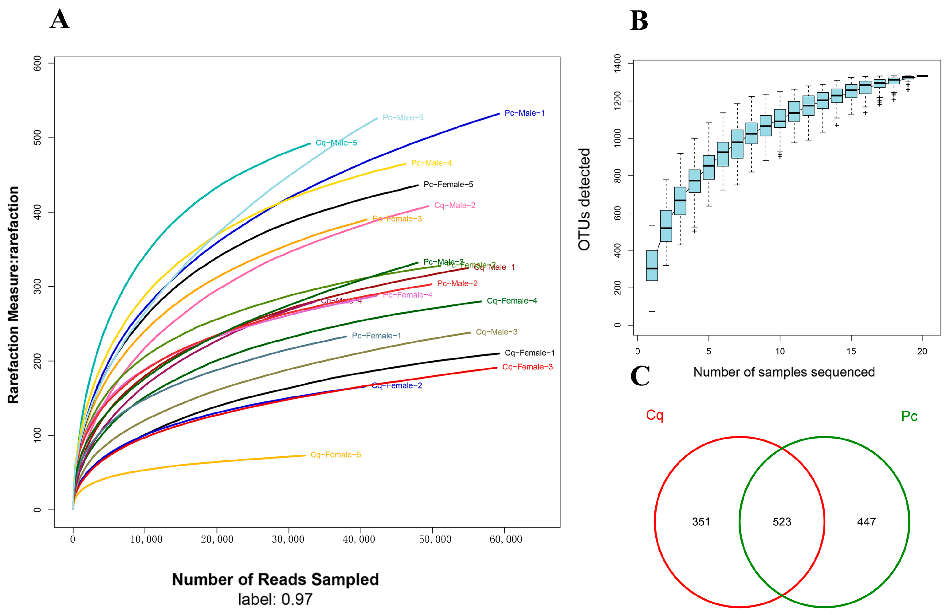
Figure 1. Rarefaction analysis, species accumulation analysis, and operational taxonomic unit (OTU) Venn analysis of Cq and Pc. (A) The rarefaction curves of each sample in the Pc and Cq groups; (B) The species accumulation curves; (C) Venn diagram analysis showing the unique and shared OTUs between Pc and Cq groups.
Fishes 07 00241 g001
Figure 2. The beta diversity analysis of samples. (A) Heatmap analysis for distance matrix of each sample; (B) PCoA analysis based on weighted UniFrac; (C) Similarity tree for multiple samples.
Fishes 07 00241 g002
Figure 3. The microbial community barplot with cluster tree. The left of the figure shows the hierarchical cluster analysis based on community composition among samples, and the right of the figure shows a histogram of community structure at the phylum level of each sample.
Fishes 07 00241 g003
Figure 4. Linear discriminant analysis (LDA) effect size (LEfSe) analysis displaying the differences in intestinal microbiota. (A) Differential abundance of taxa between Cq and Pc groups (LDA > 4); (B) Differential abundance of taxa among male Pc, female Pc, and male Cq groups (LDA > 4); (C) The cladogram showing differentially abundant taxa of the intestinal microbiota between Pc and Cq groups; (D) The cladogram showing differentially abundant taxa of the intestinal microbiota among male Pc, female Pc, and male Cq groups.
Fishes 07 00241 g004
Table 1. Richness and diversity index of bacteria in samples at the 97% similarity level.
PcPc FemalePc MaleCqCq FemaleCq Male
Chao538.24 ± 210.95426.19 ± 73.83650.29 ± 251.57351.28 ± 137.62254.01 ± 93.12448.56 ± 101.42
Shannon4.46 ± 0.304.33 ± 0.284.59 ± 0.282.76 ± 0.492.53 ± 0.292.98 ± 0.58
Simpson0.11 ± 0.020.12 ± 0.020.10 ± 0.030.32 ± 0.100.33 ± 0.070.31 ± 0.13
ACE526.76 ± 192.91421.48 ± 80.33632.04 ± 222.64354.00 ± 139.59249.87 ± 94.37458.13 ± 88.47
Good’s coverage0.9982 ± 0.0010.9980 ± 0.0000.9969 ± 0.0020.9975 ± 0.0010.9989 ± 0.0000.9976 ± 0.001
OTU (97%)346.9 ± 87.2311.40 ± 71.80382.40 ± 93.83236.5 ± 118.5158.60 ± 58.65314.40 ± 113.86
Publisher’s Note: MDPI stays neutral with regard to jurisdictional claims in published maps and institutional affiliations.