Article

Cytoprotective and Antigenotoxic Properties of Organic vs. Conventional Tomato Puree: Evidence in Zebrafish Model

1
Department of Environmental, Biological and Pharmaceutical Sciences and Technologies, University of Campania “Luigi Vanvitelli”, 81100 Caserta, Italy
2
CREATE Fertility, 150 Cheapside, London EC2V 6ET, UK
3
Department of Woman, Child and General and Special Surgery, University of Campania “Luigi Vanvitelli”, 80138 Napoli, Italy
*
Author to whom correspondence should be addressed.
Fishes 2022, 7(3), 103; https://doi.org/10.3390/fishes7030103
Submission received: 17 March 2022 / Revised: 26 April 2022 / Accepted: 27 April 2022 / Published: 29 April 2022
(This article belongs to the Section Physiology and Biochemistry)

Abstract

:
In this in vivo study, we investigated cytoprotective and antigenotoxic effects of commercial tomato puree obtained from conventional vs. organic farming systems (pesticides vs. pesticide-free agriculture, respectively). This is relevant as pesticides are widely used in agriculture to prevent pests, weeds, and the spread of plant pathogens. By exposing zebrafish to tomato puree alone and in combination with H2O2 (a well-known genotoxic agent), we analyzed the percentage of fish survival, cell viability, intracellular concentration of reactive oxygen species (ROS), DNA fragmentation index (DFI%), and genomic template stability (GTS%). Fish exposed to organic puree showed higher fish survival and cellular viability, lower DFI% and ROS, and improved GTS%. Our results suggest a higher cytoprotective and antigenotoxic effect of organic pesticide-free tomatoes, probably because the activity of natural phytochemicals is not affected by the presence of toxic residues, which are otherwise produced by pesticides used in conventional farming systems. Our study points out the importance of considering alternative strategies in agriculture to minimize the genotoxic impact of chemical pesticides.

1. Introduction

In recent years, organic farming has significantly increased as a result of the growing consumer demand for organic products, price premiums, and improving market opportunities [1]. Despite conflicting data regarding the lower yields and nutritional values of organic foods, their choice is primarily driven by the consumers’ concern for risks associated with the presence of pesticide residues in foods and pollution from their run-off in the environment [1,2]. Pesticides are genotoxic compounds, capable of inducing modifications within the nucleotide sequence or the double helix DNA structure, whose effects can occur even long after the end of exposure. These mutagenic substances are able to act both directly, by damaging the genetic material through the formation of DNA adducts, and indirectly, by depleting the intracellular antioxidant defenses, resulting in oxidative stress (OS) [3]. OS is a condition of disequilibrium between the accumulation of reactive oxygen (ROS) or nitrogen (RNS) species, and the body’s ability to counteract their action through the antioxidant defense system [4]. The damage induced by reactive species affects all cellular components (lipids, proteins, and DNA), and has also been confirmed as a contributing factor to the pathophysiology of many chronic conditions. These include neurodegenerative (Parkinson’s, Alzheimer’s, and Huntington’s diseases and amyotrophic lateral sclerosis) and inflammatory diseases, infertility, and ophthalmic disorders [5,6,7,8]. Furthermore, the metabolic activity of pesticides leads to the formation of highly toxic secondary compounds involved in the cancer onset mechanism [3].
The endogenous antioxidant system includes enzymatic (superoxide dismutase—SOD, catalase—CAT, and glutathione peroxidase—GPX), and non-enzymatic systems (i.e., ascorbate, glutathione, tocopherol, proteins, coenzyme Q10, melatonin, and polyamines, amongst others) [9]. Despite its remarkable efficiency, this complex antioxidant apparatus may be not sufficient to counterbalance the free radicals, and exogenous antioxidants are to be introduced through the diet. Tomato (Solanum lycopersicum L.) and its products (i.e., tomato paste, sauce, juice, and puree) are important sources of antioxidants in the Mediterranean diet [10], as they contain high concentrations of several natural antioxidant chemicals, such as carotenoids (β-carotenoids and lycopene), ascorbic acid (vitamin C), tocopherol (vitamin E) and bioactive phenolic compounds (quercetin, kaempferol, naringenin and lutein, as well as caffeic, ferulic and chlorogenic acids) [11]. Due to their antioxidant and antigenotoxic action, these compounds show a protective role against cellular and DNA damage. Epidemiological studies indicated that a diet rich in carotenoids is associated with a lower cancer incidence and cardiovascular disease, better cognitive performance, modulation of immune response, as well as delayed development and progression of osteoporosis, age-related macular degeneration and cataracts [12,13,14,15]. Among carotenoids, lycopene is known to be the most powerful compound capable of scavenging ROS and preventing cell damage [16], inducing the overexpression of antioxidant enzymes and downregulating inflammatory cytokines, such as IL-1β and TNF-α [17]. It is also reportedly associated with reduced caspase-3 and caspase-9 activity, and the expression of the apoptotic Bax [18].
Lycopene constitutes about 85% of all the carotenoid content in tomatoes [14]; however, the industrial transformation of this fruit into tomato products, with the consequent addition of unregulated additives, involves various treatments, such as blanching, cooking, drying, pasteurization, sterilization, and canning, that potentially affect the final profile of antioxidants and other metabolites [19,20,21]. This can directly affect the health benefits of processed products. Moreover, a major factor to take into consideration is that pesticides are used at various stages of conventional tomato crop growth. A negative impact on human health has been widely described in cases of occupational exposure to pesticides [22,23,24,25,26]; however, the general population are mostly exposed to pesticide residues through the diet [27,28,29]. The application of both systemic and contact pesticides can generate residues [30,31,32] that remain in the fruit at levels above those considered safe [33,34]. Rodrigues et al. showed that a percentage ranging between 10–30% of the contact pesticides (azoxystrobin, chlorothalonil and difenoconazole) used in the tomato crop can penetrate the fruit [35]. Multiple residues were also identified in all tomato samples analyzed by a recent study conducted in Nepal [36], with 44% of the samples showing levels of the pesticide chlorpyrifos exceeding the maximum residue levels defined by the European Union [37]. The presence of powerful mutagenic substances such as pesticide residues can result in increased OS and limit the antigenotoxic action of natural antioxidants present in the tomatoes. Therefore, organic tomatoes may preserve the quality of the food and the antigenotoxic potential more than those undergoing traditional farming. However, this has not been investigated yet, and it remains merely a theory so far.
In this in vivo study, we aim to investigate the cytoprotective and genoprotective effects of organic (Pfree group) and conventional (P1 and P2 groups) commercial tomato purees in a zebrafish (Danio rerio) model, as well as co-exposure with hydrogen peroxide (H2O2), by analyzing the percentage of zebrafish survival, cell viability, percentage of intracellular ROS levels, and genome integrity. The co-exposure with H2O2 aimed to establish the antigenotoxic power of organic tomato products compared to conventional ones against a known mutagenic agent.

2. Materials and Methods

2.1. Study Design

Zebrafish were obtained from a local source (CARMAR sas, San Giorgio a Cremano, Italy) and were raised in a large tank containing about 80 L of water (temperature ≥ 25 °C, pH of 7.6). Photoperiod of 10 h (h) of dark and 14 h of light was respected, while fish were fed every two days with commercial food (Tetramin Tropical Flakes, Spectrum Brands, Blacksburg, VA, USA), and raised in a room with no ambient noise.
Experiments were carried out on a total of 240 adult zebrafish of about 3.5 cm in length, without distinction between male and female. Fish were placed in 16 tanks of 10 L of water volume containing 15 fishes each. These replicates allowed limiting the effects of the environment on stress levels in fish kept in captivity. To test the cytoprotective and antigenotoxic effects of two different brands (P1 and P2) of conventional, and one organic pesticide-free (Pfree) tomato products, they were dissolved alone as well as with H2O2, according to the study design in Figure 1. Tomato products, previously dried at 80 °C for 24 h and grounded to a fine powder, were added to each tank at a concentration of 0.5 mg/mL. Commercial tomato products P1 and P2 were purchased at a local market while organic tomatoes were grown as follows. The tomato seedlings were transplanted on 30 April 2020 at the three-true-leaf stage in a local farm. Plants grew under natural conditions and were cultivated in compliance with the organic farming method. Fruits were harvested 70–80 days after the transplant in July 2020 and processed in puree.
A volume of 2.24 mL H2O2 per tank was added (concentration of 200 μM). This was chosen according to the published literature; such concentration of H2O2 did reportedly induce autophagy, apoptosis, and cell death, while it reduced the activity of endogenous antioxidants. Therefore, it is chosen as positive control in toxicology studies to mimic oxidative stress [38,39,40,41]. H2O2 naturally degrades into water and oxygen, and the rate of decomposition can range from a few minutes to more than a week, depending on the different chemical, biological and physical factors. The rapid rates of degradation are mainly the result of microbial action; in fact, the half-life of H2O2 in the aquarium can be several days or more in water free of microorganisms [42]. In order to preserve the quality of the water and the stability of the dissolved substances as well as limit the bacterial growth, a special aerator was inserted into the tanks, water was changed every 7 days with a bottom siphon and the feeding was suspended during the exposure of fish to fresh tomato puree. This is the first study investigating the fish exposure to tomato puree, hence concentration to test (0.5 mg/mL) was chosen after a preliminary study in which lower concentrations did not show any statistically significant dose–response (data unpublished). The density of the water in the tanks was measured with a special densimeter during the experiments. The value was stable for 5, 7 and 14 days at about 1 g/cm3. Fish stocking density during treatments was in the average of the values recommended by “The Zebrafish Book” and by most producers of zebrafish systems ranging from 0.66 fish/L to 5 fish/L [43].
Fish were analyzed at three exposure times: 5, 7 and 14 days. At any investigated time point, 10 fish were anesthetized with Tricaine methylsulfonate (Sigma-Aldrich, St. Louis, MI, USA) and 25 μL of blood was collected by sampling under the gills with 1 mL heparinised syringes, to avoid the formation of blood clots. We followed the recommendations of ARRIVE guidelines and carried out all experiments in accordance with the recommendations reported in the Directive 2010/63/EU of the European Parliament and of the Council of 22 September 2010 on the protection of animals used for scientific purposes.

2.2. Cell Viability Test

Cell viability was assessed by Trypan blue (Thermo Fisher Scientific, Waltham, MA, USA) dye [44]. An aliquot of blood sample was suspended in 1 mL of PBS 1× (Thermo Fisher Scientific, Waltham, MA, USA) and centrifuged for 5 min at 1000 rpm. The pellet was resuspended in 1 mL of PBS 1×. Then, the cell suspension was incubated with 0.4% trypan blue for 5 min at room temperature (1:1 cell dilution). Ten μL of cell suspection was loaded into Burker counting chamber (VWR International Srl, Milano, Italy) and observed under the microscope (OPTIKA IM-5, Ponteranica, Italy) at 20× magnification. A total number of 100 cells was counted in 4 selected squares. The number of cells was calculated according to the specifications of the counting slide: Total cells/mL = (Total cells counted × dilution factor × 10,000 cells/mL)/Number of squares counted. We counted blue-colored cells as dead, while cells showing bright centers and dark edges were considered alive.

2.3. ROS Assay

The blood samples were mixed with PBS 1× and subsequently centrifuged at 2000 rpm for 10 min [45]. The assay based on the 2’-7’dichlorofluorescin diacetate (DCFH2-DA, Sigma-Aldrich, St. Louis, MI, USA) was performed in triplicate to evaluate the intracellular ROS concentration as previously described [46]. Briefly, the blood sample was centrifuged at 2000 rpm for 10 min at room temperature. The supernatant was removed, and the pellet washed with PBS 1× for two times. Pellet was resuspended in 13 μM DCFH2-DA solution and incubated in the dark at 37 °C for 30 min. Then, samples were centrifuged at 2000 rpm for 10 min, and washed with PBS 1× three times. Finally, nuclei were counterstained with 4′,6-diamidino-2′- phenylindole dihydrochloride (DAPI, Sigma-Aldrich). Samples were transferred to glass slides, mounted using the anti-fade reagent 1,4-diazabicyclo[2.2.2]octane (DABCO, Sigma-Aldrich) and glycerol, and observed by using fluorescence microscope (Nikon Eclipse E600) with BP 330–380 nm and LP 420 nm filters. Figures were acquired by using the software GENIKON version 3.7 (Nikon Instruments, Campi Bisenzio, Firenze). Intracellular ROS were calculated as percentage of cells showing as green out of the total.

2.4. DNA Extraction from Muscle Tissue

DNA was extracted from muscle of zebrafish according to the manufacturer’s suggestions of a commercial kit (High Pure PCR Template Preparation Kit, ROCHE Diagnostics, Basel, Switzerland). DNA purity and concentration were evaluated using a Nanodrop 2000 (Thermo Fisher Scientific, Waltham, MA, USA).

2.5. RAPD–PCR Protocol

The random amplified polymorphic DNA–polymerase chain reaction (RAPD–PCR) was performed as previously described [47]. Briefly, the amplification reaction (total volume of 25 µL) included PuREtaq Ready-to go-PCR (Sigma-Aldrich), which contains nucleotides (dNTPs) and Taq DNA recombinant polymerase (2.5 units), DNA (40 ng), the primer 6 (5-d[CCCGTCAGCA]-3) (5 pmol µL−1) and H2O Milli-Q (DNAse and RNAse free). After an initial step (5 min–94 °C), 45 cycles of amplification were performed (1 min–95 °C, 1 min–36 °C and 2 min–72 °C). A total of 15 μL of amplified mixture was then analyzed electrophoretically on 2% agarose gel with 1× ethidium bromide. Results were expressed as genomic template stability percentage (GTS%) for each experimental group as follows:
GTS = (1 − a/n) × 100
where n is the total number of bands in the controls group and a indicates the number of bands which appear and/or disappear in comparison with the control. Control was set equal to 100% and it was used as reference.

2.6. TUNEL Assay

TUNEL assay was performed to detect DNA fragmentation by using the In Situ Cell Death Detection Kit, Fluorescein (ROCHE Diagnostics, Basel, Switzerland). First, the volume of blood sample was washed for two times with an equal volume of PBS 1×, followed by centrifugation (2000 rpm for 10 min). A total of 10 µL of samples was smeared on glass slides and fixed in 4% paraformaldehyde (Sigma-Aldrich) for 1 h at room temperature. After washing with PBS 1×, slides were incubated in a permeabilizing solution (0.1% sodium citrate, (CARLO ERBA Reagents, Cornaredo, Milano), 0.1% Triton X-100 (Sigma-Aldrich) for 30 min. The slides were washed again for two times in PBS 1×, and then incubated with 50 µL of TUNEL reaction mixture. Slides were incubated in a humid chamber for 1 h at 37 °C in the dark, and then washed for three times with PBS 1×, before staining with DAPI for 5 min in the dark. Finally, the slides were observed under fluorescent microscope: about 350 cells per slide in triplicate were analyzed, determining the percentage of nuclei with fragmented DNA (green-colored) out of the total number of cells with intact nuclei.

2.7. Statistical Analysis

ANOVA test was performed using the GraphPad Prism 6 to analyze the differences between the groups, which were considered significant when p-value (p) < 0.05. Data is reported as mean ± standard deviation (SD).

3. Results

3.1. Zebrafish Survival

The survival of zebrafish exposed to H2O2 was significantly reduced after 5 exposure days, further decreasing to 50% after 14 days of treatment (p < 0.05). Conversely, the exposure to conventional (P1 and P2 groups) and organic (Pfree group) tomato purees showed similar percentage of survival to unexposed zebrafish for all exposure times (p > 0.05) (Figure 2). After co-exposure to H2O2 and tomato puree, percentage of survival from the three groups was comparable to the unexposed controls except for the conventional puree, which showed reduced zebrafish survival after H2O2 co-exposure at 14 days (p < 0.05) (Figure 2).

3.2. Cell Viability Test

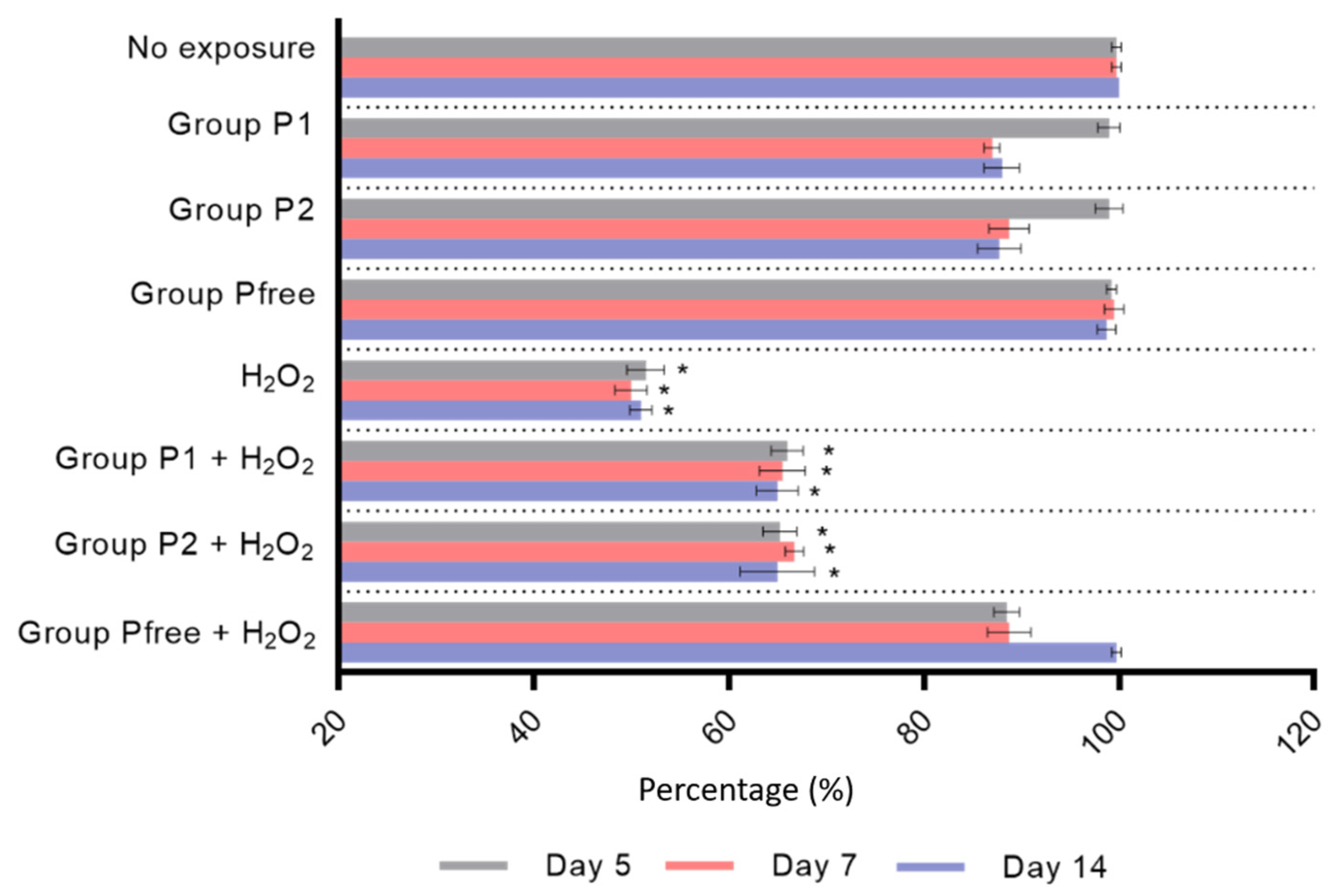
Cell viability was significantly reduced in a time-dependent manner when zebrafish were exposed to H2O2 after 5, 7, and 14 days (p < 0.05). After 5 and 7 days, cell viability values for P1 and P2 groups were comparable with the unexposed samples, while after 14 days, cell viability was significantly reduced after exposure to conventional tomato purees (p < 0.05), individually as well as in combination with H2O2. On the contrary, cell viability was not affected by individual exposure to organic puree (Pfree group) or co-exposure to H2O2 (Figure 3).

3.3. ROS Assay

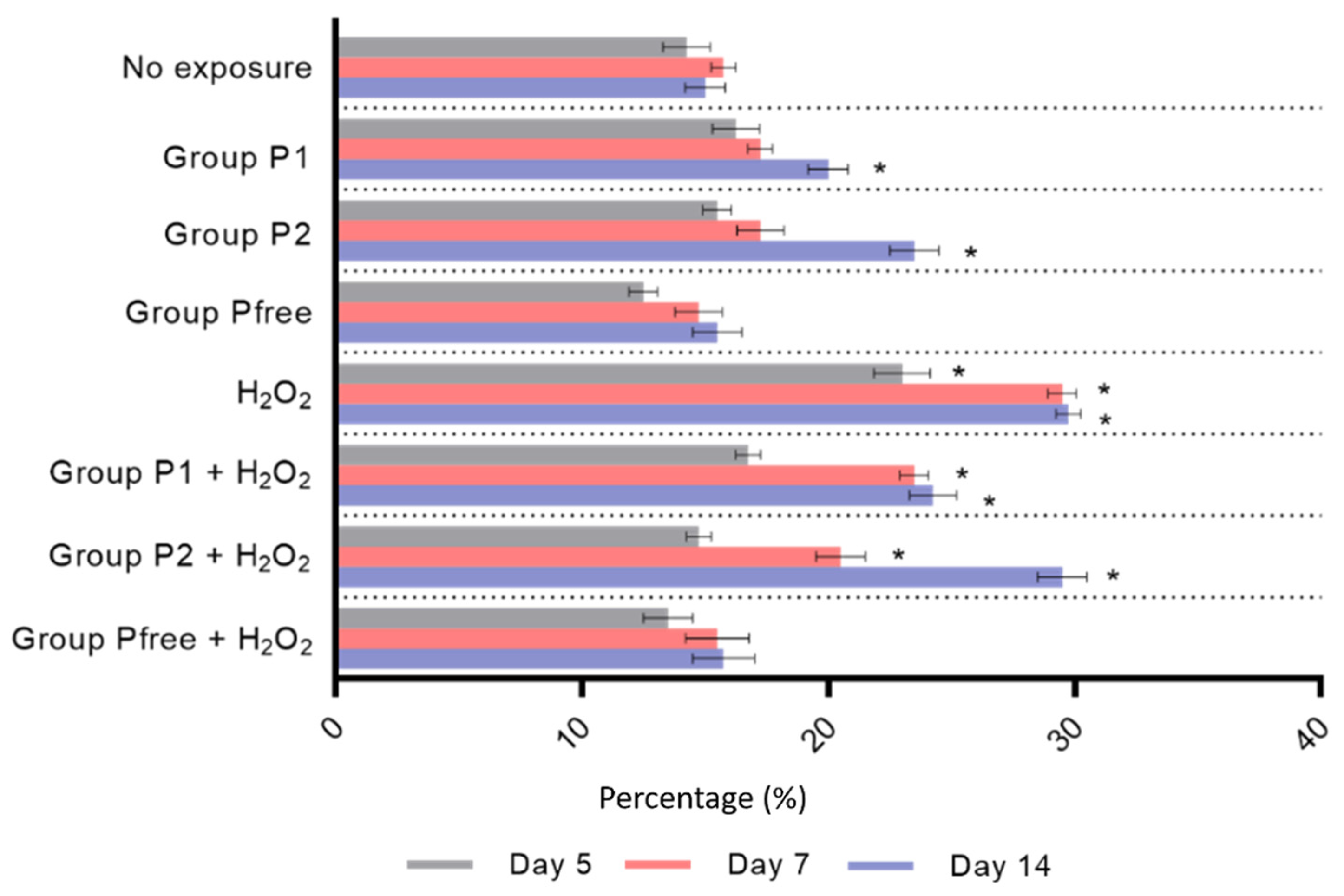
Exposure to H2O2 resulted in a significant increase in the percentage of intracellular ROS at all times of exposure (p < 0.05) (Figure 4). Groups P1 and P2 showed increased levels of ROS along the time, in the case of exposure to individual treatments (14 exposure days: p < 0.05) to conventional purees or after co-exposure to H2O2 (7 and 14 exposure days: p < 0.05). Intracellular ROS levels did not vary after individual exposure to the organic puree (Pfree group) or co-exposure to H2O2.

3.4. RAPD–PCR Assay and GTS%

The amplification products obtained by RAPD–PCR showed a characteristic pattern of polymorphic bands (200, 310, 500, 550, 700, 750, 820, 1500 base pairs—bp) in all unexposed controls, which did not vary over time. Exposure to H2O2 resulted in the appearance and disappearance of different bands (Table 1) after 5, 7, and 14 days, with a GTS% reduction of about 50%.
In P1 and P2 groups, a new band of 600 bp appeared after 7 and 14 days of exposure, while exposure of Pfree group did not show polymorphic variations at any investigated time (Table 1). Co-exposure of P1 and P2 groups to H2O2 resulted in the appearance and disappearance of several bands already after 5 days (Table 1), with a reduction in GTS% of about 35%. A single band appeared when Pfree group was exposed to H2O2 after 7 (650 bp) and 14 (850 bp) days (Table 1), with a slight GTS% reduction of 13% (Figure 5).

3.5. TUNEL Assay

Exposure to H2O2 resulted in a significant increase in DNA Fragmentation Index (DFI%) for all times of exposure (p < 0.05) (Figure 6). Zebrafish exposed to conventional tomato puree (P1, P2 groups) showed no significant differences in DFI% in comparison with unexposed groups for 5 and 7 exposure days. On the contrary, exposure to conventional tomato puree for 14 days resulted in a significant increase in DFI% (p < 0.05). After co-exposure of P1 and P2 groups to H2O2, we observed increased DFI% for all exposure times. Conversely, the Pfree group showed no difference in DFI% when compared with the negative control, after individual and co-exposure to H2O2.

4. Discussion

Our study showed that tomato puree obtained by organic pesticide-free cultivation has a greater cytoprotective and antioxidant potential than tomato puree obtained by conventional farming (in which the use of pesticides is allowed), when used either alone or after co-exposure with H2O2. In fact, fish exposed to organic tomato puree showed greater survival and cell viability, with higher genomic stability, and reduced intracellular ROS generation and DNA damage.
Zebrafish is the most used model for toxicity tests for its similarities in genetics, anatomy, and physiology with humans, making it an ideal model for the study of several human diseases. Additionally, this model represents a valid strategy for the rapid and cost-effective screening of natural products which may be potentially safe or toxic for human health. In this regard, numerous studies have been based on zebrafish to evaluate the ability of substances to induce harmful conditions such as oxidative stress, inflammation, genotoxicity, developmental toxicity, and neurotoxicity [48].
Intake of tomato and tomato-based products has been consistently associated with a beneficial impact on human health, due to the high content of nutrients and natural antioxidants [for a review on the topic, see [49]]. In fact, tomatoes have a high content of minerals, vitamins, proteins, essential amino acids (leucine, threonine, valine, histidine, lysine, and arginine), monounsaturated fatty acids (linoleic and linolenic), and phytosterols (β-sitosterol, campesterol, and stigmasterol). Moreover, tomato fruits are considered one of the main sources of food antioxidants such as carotenoids (α-carotene, β-carotene, lycopene, lutein, and cryptoxanthin) [14]. Among the carotenoids, lycopene is particularly important for human health [50]. In fact, it is widely described as one of the most powerful antioxidants among carotenoids, second only to astaxanthin [51], as its efficacy in singlet oxygen removal is double that of β-carotene and 10-fold higher than α-tocopherol [52]. Besides directly scavenging ROS, it also activates the expression of antioxidant genes, such as NAD(P)H:ubiquinone oxidoreductase, haemoxygenase 1, glutathione reductase, and glutathione S-transferase [53], and modulates the activity of enzymes that contribute to ROS formation, such as NADP(H)oxidase, cyclooxygenase-2, 5-lipoxygenase, and induced nitric oxide synthase [54].
Due to the impact of such components on human health, cultivation practices that better preserve the nutritional, biological, and protective characteristics of the tomatoes and their cytoprotective and antioxidant potential should be preferred.
Our results show a higher cytoprotective and antigenotoxic potential of organic tomatoes in comparison to those obtained by conventional cultivation. This is in agreement with a previous study analyzing the data collected over 10 years, where the average levels of flavonoids (quercetin and kaempferol) in organic tomatoes were, respectively, 79% and 97% higher than those of conventional tomatoes [55]. Higher content of phytochemicals (i.e., flavonoids and carotenoids) was also observed in tomato fruits purchased from the same cultivar for two consecutive years, with the same degree of maturity and size, when the organic approach was followed [56]. Similarly, a meta-analysis published in 2014 analyzing 343 publications observed a significantly higher concentration in antioxidants and nutrients in organic crops, including phenolic acids, flavanones, stilbenes, and flavones, amongst others [57].
Besides the enrichment of phytonutrients content in organic tomatoes, Sharpe et al. also reported a differential expression of genes involved in nitrogen transport and assimilation when the same genotype of tomatoes was grown under organic or conventional conditions [58]. Therefore, the transcriptome analysis suggested that using organic fertilizer may alter the expression of genes, resulting in greater phytonutrients accumulation in tomato fruits.
Our results also showed a slight reduction in genomic stability and integrity after long treatment with the two conventional tomato purees. This highlights a potential, albeit minimal, genotoxic activity induced by the conventional food products even when they are not in co-exposure with a pro-oxidant, unlike the pesticide-free product. Similarly, analysis of DNA fragmentation showed the greatest index after co-exposure of the two conventional tomato products with H2O2, thus suggesting a greater genotoxic effect in the case of simultaneous intake, compared to the treatment with the pesticide-free organic tomato puree alone and in combination with H2O2. The latter is a very well-known genotoxic agent, as this metabolite can diffuse across the cellular membrane and become a source of oxygen-derived free radicals [59]. In our model, H2O2 induced statistically significant damages both in terms of cytotoxicity (reducing cell viability and increasing oxidative stress), and genotoxicity (with increased DNA fragmentation and genomic instability). In this study, the co-exposure of tomato products to H2O2 was functional to establish the antigenotoxic potential of the three tomato purees, and more precisely the ability of their antioxidant molecules to defend cells and genetic material from mutagenic damage. The results showed that the co-exposure of the pesticide-free tomato puree with H2O2 resulted in lower DNA fragmentation and induced mutations. This suggests that the phytochemicals with antioxidant activity present in conventional tomato products may be affected, unlike the pesticide-free products, which retain their antioxidant and antigenotoxic activity. The accumulation of pesticides used in crops may explain the impairment of the natural antioxidants in tomato fruits. Barański et al. reported that the amount of pesticide residues in conventional crops was 4-fold higher than organic ones [57]. The screening of tomatoes revealed higher concentration of pesticide residues in conventional cultivation worldwide, including Chile, China, Turkey, and Kazakhstan, amongst others, compared to organic farming [60,61,62,63].
Although our study seems to point out a higher cytoprotective and antioxidant effect of organic tomatoes on zebrafish health, the limitations in our study are to be highlighted. First of all, this study did not analyze the content of pesticide residues in the tomatoes tested, although in all the commercial products used, the levels of pesticides are below the maximum residue levels authorized by national authorities in EU countries. Hence, any association between the presence of such residues and the reduced cytoprotective potential of commercial tomatoes is speculative, although still plausible and supported by previously cited literature. Similarly, concentration of natural nutrients and antioxidants was not investigated. In addition, we cannot evaluate the impact of a possible contamination of water and soil with pollutants such as heavy metals, which may affect the cytoprotective and antioxidant potential of tomatoes in both types of cultivations [64,65]. Finally, this study investigated the zebrafish exposure to a concentration of tomato puree equal to 0.5 mg/mL. Due to the lack of previous studies as reference, this was selected, after a preliminary study in which lower concentrations did not show any statistically significant dose–response (data unpublished). However, future research should focus on testing higher concentrations of both conventional and organic tomato purees, to verify their cytoprotective potential. Moreover, longer times of exposure should be investigated, due to the long-term impact of pesticides on health [66,67,68], as well as the effect of the combined consumption of tomatoes and other antioxidant molecules, due to the synergistic mode of action of antioxidants [9]. The performance of -OMICS-related experiments (i.e., genomics, transcriptomics, proteomics, and metabolomics) may further clarify the influence of conventional or organic tomato cultivations on human health.

5. Conclusions

In conclusion, our study showed for the first time a higher antioxidant, cytoprotective and genoprotective impact of organic tomato puree compared to ones obtained by conventional farming in an animal model of zebrafish exposed to H2O2 as oxidizing agent. These results highlight the need to implement sustainable agriculture strategies to reduce the use of chemical pesticides and the impact on the health, while keeping intact the nutritional, biological and protective characteristics of food products.

Author Contributions

Conceptualization, F.M.; methodology, M.S.; validation, F.M. and L.R.; data curation, R.F.; writing—original draft preparation, R.F.; writing—review and editing, R.F. and F.M.; supervision, L.R.; project administration and funding acquisition, P.C. All authors have read and agreed to the published version of the manuscript.

Funding

This research was funded by Università degli Studi della Campania Luigi Vanvitelli (grant number VALERE: VAnviteLli pEr la RicErca) and by Regione Campania Lotta alle Patologie Oncologiche progetto iCURE (CUP B21C17000030007–SURF 17061BP000000008).

Institutional Review Board Statement

The study was conducted according to the recommendations reported in the Directive 2010/63/EU of the European Parliament and of the Council of 22 September 2010 on the protection of animals used for scientific purposes.

Data Availability Statement

All data generated or analyzed during this study are included; any additional information is available from the corresponding author on reasonable request.

Acknowledgments

The authors would like to express their gratitude to Maria Immacolata Schiavone and Leyla Corvino for their contribution in maintenance and treatment of zebrafish.

Conflicts of Interest

The authors declare no conflict of interest.

References

  1. Carillo, P.; Cacace, D.; De Pascale, S.; Rapacciuolo, M.; Fuggi, A. Organic vs. traditional potato powder. Food Chem. 2012, 133, 1264–1273. [Google Scholar] [CrossRef]
  2. González, N.; Marquès, M.; Nadal, M.; Domingo, J.L. Occurrence of environmental pollutants in foodstuffs: A review of organic vs. conventional food. Food Chem. Toxicol. 2019, 125, 370–375. [Google Scholar] [CrossRef] [PubMed]
  3. Kapeleka, J.A.; Sauli, E.; Ndakidemi, P.A. Pesticide exposure and genotoxic effects as measured by DNA damage and human monitoring biomarkers. Int. J. Environ. Health Res. 2021, 31, 805–822. [Google Scholar] [CrossRef] [PubMed]
  4. Sies, H.; Berndt, C.; Jones, D. Oxidative stress. Annu. Rev. Biochem. 2017, 86, 715–748. [Google Scholar] [CrossRef]
  5. Uttara, B.; Singh, A.; Zamboni, P.; Mahajan, R. Oxidative stress and neurodegenerative diseases: A review of upstream and downstream antioxidant therapeutic options. Curr. Neuropharmacol. 2009, 7, 65–74. [Google Scholar] [CrossRef] [Green Version]
  6. Leisegang, K.; Roychoudhury, S.; Slama, P.; Finelli, R. The mechanisms and management of age-related oxidative stress in male hypogonadism associated with non-communicable chronic disease. Antioxidants 2021, 10, 1834. [Google Scholar] [CrossRef]
  7. Mittal, M.; Siddiqui, M.R.; Tran, K.; Reddy, S.P.; Malik, A.B. Reactive oxygen species in inflammation and tissue injury. Antioxid. Redox Signal. 2014, 20, 1126–1167. [Google Scholar] [CrossRef] [Green Version]
  8. Ahmad, A.; Ahsan, H. Biomarkers of inflammation and oxidative stress in ophthalmic disorders. J. Immunoass. Immunochem. 2020, 41, 257–271. [Google Scholar] [CrossRef]
  9. Pisoschi, A.M.; Pop, A. The role of antioxidants in the chemistry of oxidative stress: A review. Eur. J. Med. Chem. 2015, 97, 55–74. [Google Scholar] [CrossRef]
  10. Gómez-Romero, M.; Arráez-Román, D.; Segura-Carretero, A.; Fernández-Gutiérrez, A. Analytical determination of antioxidants in tomato: Typical components of the Mediterranean diet. J. Sep. Sci. 2007, 30, 452–461. [Google Scholar] [CrossRef]
  11. Salehi, B.; Sharifi-Rad, R.; Sharopov, F.; Namiesnik, J.; Roointan, A.; Kamle, M.; Kumar, P.; Martins, N.; Sharifi-Rad, J. Beneficial effects and potential risks of tomato consumption for human health: An overview. Nutrition 2019, 62, 201–208. [Google Scholar] [CrossRef] [PubMed]
  12. Chaudhary, P.; Sharma, A.; Singh, B.; Nagpal, A.K. Bioactivities of phytochemicals present in tomato. J. Food Sci. Technol. 2018, 55, 2833–2849. [Google Scholar] [CrossRef] [PubMed]
  13. Eggersdorfer, M.; Wyss, A. Carotenoids in human nutrition and health. Arch. Biochem. Biophys. 2018, 652, 18–26. [Google Scholar] [CrossRef] [PubMed]
  14. Rao, A.V.; Rao, L.G. Carotenoids and human health. Pharmacol. Res. 2007, 55, 207–216. [Google Scholar] [CrossRef]
  15. Rouphael, Y.; Corrado, G.; Colla, G.; De Pascale, S.; Dell’aversana, E.; D’amelia, L.I.; Fusco, G.M.; Carillo, P. Biostimulation as a means for optimizing fruit phytochemical content and functional quality of tomato landraces of the San Marzano area. Foods 2021, 10, 926. [Google Scholar] [CrossRef] [PubMed]
  16. Grabowska, M.; Wawrzyniak, D.; Rolle, K.; Chomczyński, P.; Oziewicz, S.; Jurga, S.; Barciszewski, J. Let food be your medicine: Nutraceutical properties of lycopene. Food Funct. 2019, 10, 3090–3102. [Google Scholar] [CrossRef] [PubMed]
  17. Zhao, B.; Ren, B.; Guo, R.; Zhang, W.; Ma, S.; Yao, Y.; Yuan, T.; Liu, Z.; Liu, X. Supplementation of lycopene attenuates oxidative stress induced neuroinflammation and cognitive impairment via Nrf2/NF-κB transcriptional pathway. Food Chem. Toxicol. 2017, 109, 505–516. [Google Scholar] [CrossRef]
  18. Li, W.; Jiang, B.; Cao, X.; Xie, Y.; Huang, T. Protective effect of lycopene on fluoride-induced ameloblasts apoptosis and dental fluorosis through oxidative stress-mediated Caspase pathways. Chem. Biol. Interact. 2017, 261, 27–34. [Google Scholar] [CrossRef] [PubMed]
  19. Kelebek, H.; Selli, S.; Kadiroğlu, P.; Kola, O.; Kesen, S.; Uçar, B.; Çetiner, B. Bioactive compounds and antioxidant potential in tomato pastes as affected by hot and cold break process. Food Chem. 2017, 220, 31–41. [Google Scholar] [CrossRef]
  20. Knockaert, G.; Pulissery, S.K.; Colle, I.; Van Buggenhout, S.; Hendrickx, M.; Loey, A. Van Lycopene degradation, isomerization and in vitro bioaccessibility in high pressure homogenized tomato puree containing oil: Effect of additional thermal and high pressure processing. Food Chem. 2012, 135, 1290–1297. [Google Scholar] [CrossRef]
  21. Vallverdú-Queralt, A.; Medina-Remón, A.; Andres-Lacueva, C.; Lamuela-Raventos, R.M. Changes in phenolic profile and antioxidant activity during production of diced tomatoes. Food Chem. 2011, 126, 1700–1707. [Google Scholar] [CrossRef] [PubMed]
  22. Van Maele-Fabry, G.; Hoet, P.; Vilain, F.; Lison, D. Occupational exposure to pesticides and Parkinson’s disease: A systematic review and meta-analysis of cohort studies. Environ. Int. 2012, 46, 30–43. [Google Scholar] [CrossRef] [PubMed]
  23. Starling, A.P.; Umbach, D.M.; Kamel, F.; Long, S.; Sandler, D.P.; Hoppin, J.A. Pesticide use and incident diabetes among wives of farmers in the Agricultural Health Study. Occup. Environ. Med. 2014, 71, 629–635. [Google Scholar] [CrossRef] [PubMed]
  24. Dyck, R.; Karunanayake, C.; Pahwa, P.; Hagel, L.; Lawson, J.; Rennie, D.; Dosman, J. Prevalence, risk factors and co-morbidities of diabetes among adults in rural Saskatchewan: The influence of farm residence and agriculture-related exposures. BMC Public Health 2013, 13, 7. [Google Scholar] [CrossRef] [Green Version]
  25. Andersen, H.R.; Debes, F.; Wohlfahrt-Veje, C.; Murata, K.; Grandjean, P. Occupational pesticide exposure in early pregnancy associated with sex-specific neurobehavioral deficits in the children at school age. Neurotoxicol. Teratol. 2015, 47, 1–9. [Google Scholar] [CrossRef] [Green Version]
  26. Nicolopoulou-Stamati, P.; Maipas, S.; Kotampasi, C.; Stamatis, P.; Hens, L. Chemical pesticides and human health: The urgent need for a new concept in agriculture. Front. Public Health 2016, 4, 148. [Google Scholar] [CrossRef] [Green Version]
  27. Lu, C.; Toepel, K.; Irish, R.; Fenske, R.A.; Barr, D.B.; Bravo, R. Organic diets significantly lower children’s dietary exposure to organophosphorus pesticides. Environ. Health Perspect. 2006, 114, 260–263. [Google Scholar] [CrossRef]
  28. Oates, L.; Cohen, M.; Braun, L.; Schembri, A.; Taskova, R. Reduction in urinary organophosphate pesticide metabolites in adults after a week-long organic diet. Environ. Res. 2014, 132, 105–111. [Google Scholar] [CrossRef] [Green Version]
  29. Bradman, A.; Quirós-Alcalá, L.; Castorina, R.; Schall, R.A.; Camacho, J.; Holland, N.T.; Barr, D.B.; Eskenazi, B. Effect of organic diet intervention on pesticide exposures in young children living in low-income urban and agricultural communities. Environ. Health Perspect. 2015, 123, 1086–1093. [Google Scholar] [CrossRef] [Green Version]
  30. Abd-Elhaleem, Z.A. Pesticide residues in tomato and tomato products marketed in Majmaah province, KSA, and their impact on human health. Environ. Sci. Pollut. Res. Int. 2020, 27, 8526–8534. [Google Scholar] [CrossRef]
  31. Williamson, S.; Ball, A.; Pretty, J. Trends in pesticide use and drivers for safer pest management in four African countries. Crop Prot. 2008, 27, 1327–1334. [Google Scholar] [CrossRef]
  32. EL-Saeid, M.H.; AL-Dosari, S.A. Monitoring of pesticide residues in Riyadh dates by SFE, MSE, SFC, and GC techniques. Arab. J. Chem. 2010, 3, 179–186. [Google Scholar] [CrossRef] [Green Version]
  33. European Commission Commission Directive 2002/63/EC Establishing Community Methods of Sampling for the Official Control of Pesticide Residues in and on Products of Plant and Animal Origin and Repealing–Directive 79/700/EEC. Available online: https://www.fao.org/faolex/results/details/en/c/LEX-FAOC034557/ (accessed on 30 November 2021).
  34. Food and Agriculture Organization of the United Nations and World Health Organization Codex Alimentarius–International Food Standards. Available online: https://www.fao.org/fao-who-codexalimentarius/en/?provide=committeeDetail&idList=29 (accessed on 30 November 2021).
  35. Rodrigues, A.A.Z.; De Queiroz, M.E.L.R.; De Oliveira, A.F.; Neves, A.A.; Heleno, F.F.; Zambolim, L.; Freitas, J.F.; Morais, E.H.C. Pesticide residue removal in classic domestic processing of tomato and its effects on product quality. J. Environ. Sci. Health Part B Pestic. Food Contam. Agric. Wastes 2017, 52, 850–857. [Google Scholar] [CrossRef] [PubMed]
  36. Bhandari, G.; Zomer, P.; Atreya, K.; Mol, H.G.J.; Yang, X.; Geissen, V. Pesticide residues in Nepalese vegetables and potential health risks. Environ. Res. 2019, 172, 511–521. [Google Scholar] [CrossRef]
  37. EFSA (European Food Safety Authority); Carrasco Cabrera, L.; Medina Pastor, P. The 2019 European Union report on pesticide residues in food. EFSA J. 2021, 19, e06491. [Google Scholar]
  38. Duan, J.; Duan, J.; Zhang, Z.; Tong, T. Irreversible cellular senescence induced by prolonged exposure to H2O2 involves DNA-damage-and-repair genes and telomere shortening. Int. J. Biochem. Cell Biol. 2005, 37, 1407–1420. [Google Scholar] [CrossRef]
  39. Dulull, N.K.; Dias, D.A.; Thrimawithana, T.R.; Kwa, F.A.A. L-Sulforaphane confers protection against oxidative stress in an in vitro model of age-related macular degeneration. Curr. Mol. Pharmacol. 2018, 11, 237–253. [Google Scholar] [CrossRef]
  40. Castino, R.; Fiorentino, I.; Cagnin, M.; Giovia, A.; Isidoro, C. Chelation of lysosomal iron protects dopaminergic SH-SY5Y neuroblastoma cells from hydrogen peroxide toxicity by precluding autophagy and Akt dephosphorylation. Toxicol. Sci. 2011, 123, 523–541. [Google Scholar] [CrossRef] [Green Version]
  41. Shi, D.H.; Wu, J.H.; Ge, H.M.; Tan, R.X. Protective effect of hopeahainol A, a novel acetylcholinesterase inhibitor, on hydrogen peroxide-induced injury in PC12 cells. Environ. Toxicol. Pharmacol. 2009, 28, 30–36. [Google Scholar] [CrossRef]
  42. Schmidt, L.J.; Gaikowski, M.P.; Gingerich, W.H. Evironmental assessment for the use of hydrogen peroxide in aquaculture for treating external fungal and bacterial diseases of cultured fish and fish eggs. USGS 2006, 54603, 180. [Google Scholar]
  43. Castranova, D.; Lawton, A.; Lawrence, C.; Baumann, D.; Best, J.; Coscolla, J.; Doherty, A.; Ramos, J.; Hakkesteeg, J.; Wang, C.; et al. The effect of stocking densities on reproductive performance in laboratory zebrafish (Danio rerio). Zebrafish 2011, 8, 141–146. [Google Scholar] [CrossRef] [PubMed] [Green Version]
  44. Strober, W. Trypan blue exclusion test of cell viability. Curr. Protoc. Immunol. 2001, 18432654. [Google Scholar] [CrossRef]
  45. Mottola, F.; Finelli, R.; Iovine, C.; Carannante, M.; Santonastaso, M.; Rocco, L. Anti-genotoxicity evaluation of ellagic acid and curcumin—An in vitro study on zebrafish blood cells. Appl. Sci. 2021, 11, 8142. [Google Scholar] [CrossRef]
  46. Santonastaso, M.; Mottola, F.; Colacurci, N.; Iovine, C.; Pacifico, S.; Cammarota, M.; Cesaroni, F.; Rocco, L. In vitro genotoxic effects of titanium dioxide nanoparticles (n-TiO2) in human sperm cells. Mol. Reprod. Dev. 2019, 86, 1369–1377. [Google Scholar] [CrossRef] [PubMed]
  47. Mottola, F.; Santonastaso, M.; Iovine, C.; Feola, V.; Pacifico, S.; Rocco, L. Adsorption of Cd to TiO2-NPs forms low genotoxic aggregates in Zebrafish cells. Cells 2021, 10, 310. [Google Scholar] [CrossRef] [PubMed]
  48. Lin, F.J.; Li, H.; Wu, D.T.; Zhuang, Q.G.; Li, H.B.; Geng, F.; Gan, R.Y. Recent development in zebrafish model for bioactivity and safety evaluation of natural products. Crit. Rev. Food Sci. Nutr. 2021, 1–29. [Google Scholar] [CrossRef]
  49. Ali, M.Y.; Sina, A.A.I.; Khandker, S.S.; Neesa, L.; Tanvir, E.M.; Kabir, A.; Khalil, M.I.; Gan, S.H. Nutritional composition and bioactive compounds in tomatoes and their impact on human health and disease: A review. Foods 2021, 10, 45. [Google Scholar] [CrossRef]
  50. Costa-Rodrigues, J.; Pinho, O.; Monteiro, P.R.R. Can lycopene be considered an effective protection against cardiovascular disease? Food Chem. 2018, 245, 1148–1153. [Google Scholar] [CrossRef]
  51. Cho, K.S.; Shin, M.; Kim, S.; Lee, S.B. Recent advances in studies on the therapeutic potential of dietary carotenoids in neurodegenerative diseases. Oxid. Med. Cell. Longev. 2018, 2018, 4120458. [Google Scholar] [CrossRef]
  52. Krinsky, N.I.; Johnson, E.J. Carotenoid actions and their relation to health and disease. Mol. Aspects Med. 2005, 26, 459–516. [Google Scholar] [CrossRef]
  53. Breinholt, V.; Lauridsen, S.T.; Daneshvar, B.; Jakobsen, J. Dose-response effects of lycopene on selected drug-metabolizing and antioxidant enzymes in the rat. Cancer Lett. 2000, 154, 201–210. [Google Scholar] [CrossRef]
  54. Palozza, P.; Colangelo, M.; Simone, R.; Catalano, A.; Boninsegna, A.; Lanza, P.; Monego, G.; Ranelletti, F.O. Lycopene induces cell growth inhibition by altering mevalonate pathway and Ras signaling in cancer cell lines. Carcinogenesis 2010, 31, 1813–1821. [Google Scholar] [CrossRef] [PubMed] [Green Version]
  55. Mitchell, A.E.; Hong, Y.J.; Koh, E.; Barrett, D.M.; Bryant, D.E.; Denison, R.F.; Kaffka, S. Ten-year comparison of the influence of organic and conventional crop management practices on the content of flavonoids in tomatoes. J. Agric. Food Chem. 2007, 55, 6154–6159. [Google Scholar] [CrossRef] [PubMed]
  56. Vallverdú-Queralt, A.; Jáuregui, O.; Medina-Remón, A.; Lamuela-Raventós, R.M. Evaluation of a method to characterize the phenolic profile of organic and conventional tomatoes. J. Agric. Food Chem. 2012, 60, 3373–3380. [Google Scholar] [CrossRef] [PubMed]
  57. Barański, M.; Średnicka-Tober, D.; Volakakis, N.; Seal, C.; Sanderson, R.; Stewart, G.B.; Benbrook, C.; Biavati, B.; Markellou, E.; Giotis, C.; et al. Higher antioxidant and lower cadmium concentrations and lower incidence of pesticide residues in organically grown crops: A systematic literature review and meta-analyses. Br. J. Nutr. 2014, 112, 794–811. [Google Scholar] [CrossRef] [Green Version]
  58. Sharpe, R.M.; Gustafson, L.; Hewitt, S.; Kilian, B.; Crabb, J.; Hendrickson, C.; Jiwan, D.; Andrews, P.; Dhingra, A. Concomitant phytonutrient and transcriptome analysis of mature fruit and leaf tissues of tomato (Solanum lycopersicum L. Cv. Oregon Spring) grown using organic and conventional fertilizer. PLoS ONE 2020, 15, e0227429. [Google Scholar] [CrossRef] [Green Version]
  59. Rhee, S.G. H2O2, a necessary evil for cell signaling. Science 2006, 312, 1882–1883. [Google Scholar] [CrossRef]
  60. Elgueta, S.; Valenzuela, M.; Fuentes, M.; Meza, P.; Manzur, J.; Liu, S.; Zhao, G.; Correa, A. Pesticide residues and halth risk assessment in tomatoes and lettuces from farms of metropolitan region Chile. Molecules 2020, 25, 355. [Google Scholar] [CrossRef] [Green Version]
  61. Chen, C.; Qian, Y.; Chen, Q.; Tao, C.; Li, C.; Li, Y. Evaluation of pesticide residues in fruits and vegetables from Xiamen, China. Food Control 2011, 22, 1114–1120. [Google Scholar] [CrossRef]
  62. Bakirci, G.T.; Yaman Acay, D.B.; Bakirci, F.; Ötleş, S. Pesticide residues in fruits and vegetables from the Aegean region, Turkey. Food Chem. 2014, 160, 379–392. [Google Scholar] [CrossRef]
  63. Lozowicka, B.; Abzeitova, E.; Sagitov, A.; Kaczynski, P.; Toleubayev, K.; Li, A. Studies of pesticide residues in tomatoes and cucumbers from Kazakhstan and the associated health risks. Environ. Monit. Assess. 2015, 187, 1–19. [Google Scholar] [CrossRef] [PubMed] [Green Version]
  64. Kasem Mahmoud, E.; Mohamed Ghoneim, A. Effect of polluted water on soil and plant contamination by heavy metals in El-Mahla El-Kobra, Egypt. Solid Earth 2016, 7, 703–711. [Google Scholar] [CrossRef] [Green Version]
  65. Rashtian, J.; Chavkin, D.E.; Merhi, Z. Water and soil pollution as determinant of water and food quality/contamination and its impact on female fertility. Reprod. Biol. Endocrinol. 2019, 17, 5. [Google Scholar] [CrossRef] [PubMed] [Green Version]
  66. Hu, R.; Huang, X.; Huang, J.; Li, Y.; Zhang, C.; Yin, Y.; Chen, Z.; Jin, Y.; Cai, J.; Cui, F. Long- and short-term health effects of pesticide exposure: A cohort study from China. PLoS ONE 2015, 10, e0128766. [Google Scholar] [CrossRef]
  67. Naughton, S.X.; Terry, A.V. Neurotoxicity in acute and repeated organophosphate exposure. Toxicology 2018, 408, 101–112. [Google Scholar] [CrossRef] [PubMed]
  68. Mineau, P. A review and analysis of study endpoints relevant to the assessment of “long term” pesticide toxicity in avian and mammalian wildlife. Ecotoxicology 2005, 14, 775–799. [Google Scholar] [CrossRef]
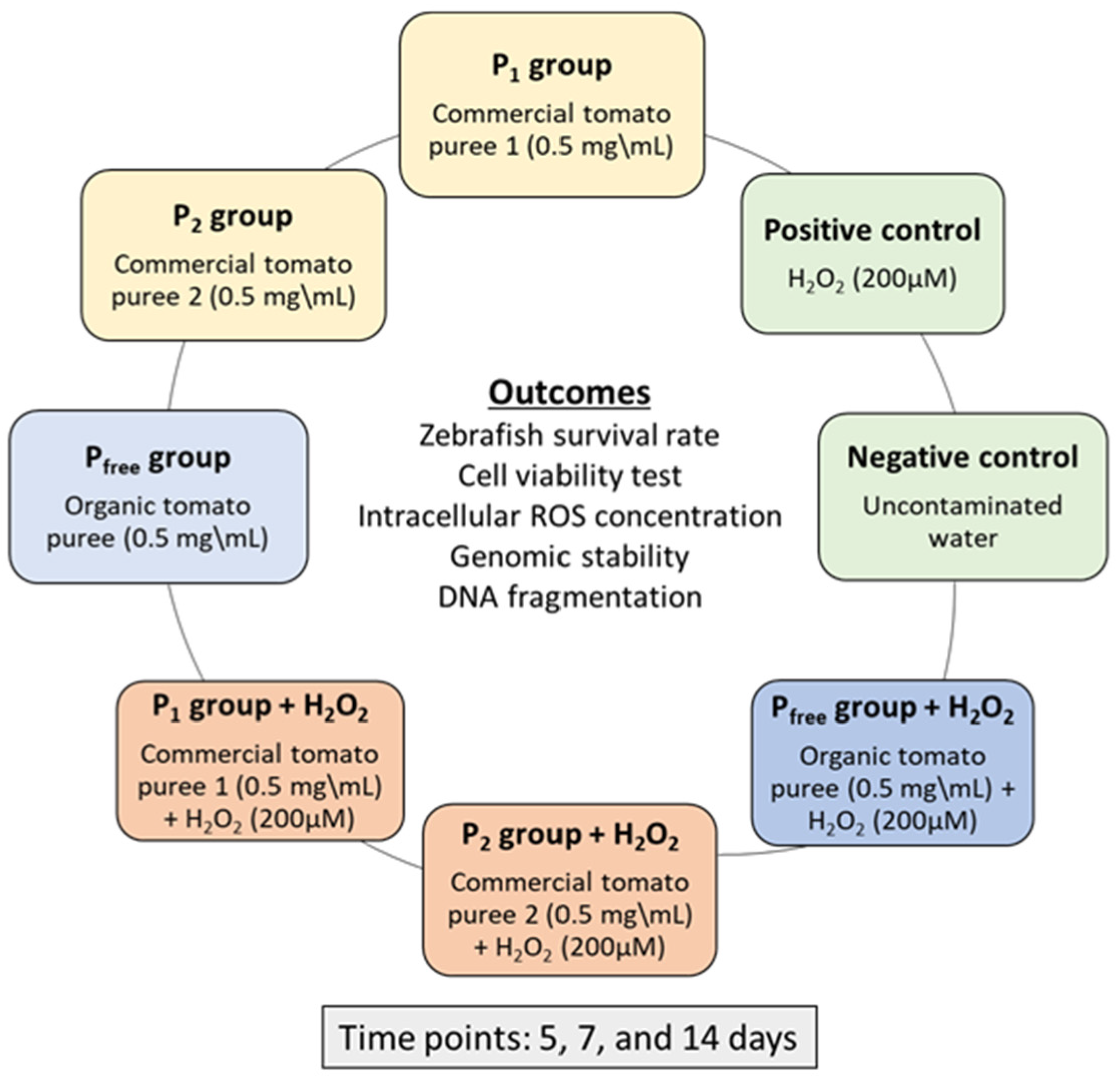
Figure 1. Study design showing the 8 groups exposed to tomato puree and the oxidant agent H2O2, the outcomes investigated, and the time points of zebrafish sampling.
Fishes 07 00103 g001
Figure 2. Percentage of surviving zebrafish exposed to conventional (P1 and P2 groups) and organic (Pfree group) tomato purees (0.5 mg/mL) individually or after co-exposure to H2O2 (200 µM) for 5, 7, and 14 days. * p < 0.05 in comparison with unexposed controls.
Fishes 07 00103 g002
Figure 3. Percentage of viable zebrafish cells exposed to conventional (groups P1 and P2) and organic (group Pfree) tomato purees (0.5 mg/mL) individually or after co-exposure to H2O2 (200 µM) for 5, 7, and 14 days. Data is expressed as percentage. * p < 0.05 in comparison with unexposed controls.
Fishes 07 00103 g003
Figure 4. Percentage of intracellular ROS levels in zebrafish cells exposed to conventional (P1 and P2 groups) and organic (Pfree group) tomato purees (0.5 mg/mL) individually or after co-exposure to H2O2 (200 µM) for 5, 7, and 14 days. * p < 0.05 in comparison with unexposed controls.
Fishes 07 00103 g004
Figure 5. Percentage of genomic template stability in zebrafish DNA exposed for 5, 7, 14 days to 0.5 mg/mL of conventional (P1 and P2 groups) or organic tomato purees (Pfree group), individually or after co-exposure to H2O2 (200 µM). * p < 0.05 in comparison with unexposed controls.
Fishes 07 00103 g005
Figure 6. Percentage of DNA fragmentation index (DFI%) in zebrafish blood cells. Zebrafish were exposed to conventional (P1 and P2 groups) and organic (group Pfree) tomato purees (0.5 mg/mL) individually or after co-exposure to H2O2 (200 µM) for 5, 7, and 14 days. * p < 0.05 in comparison with unexposed controls.
Fishes 07 00103 g006
Table 1. RAPD–PCR of DNA isolated from zebrafish erythrocytes exposed for 5, 7, 14 days to 0.5 mg/mL of conventional (P1 and P2 groups) or organic tomato purees (Pfree group), individually or after co-exposure to H2O2 (200 µM). * +: appearance of bands; −: disappearance of bands; /: no bands variation; control bands: 200 bp, 310 bp, 500 bp, 550 bp, 700 bp, 750 bp, 820 bp, 1500 bp.
ExposureDay 5 *Day 7 *Day 14 *
Group P1/+600+600
Group P2/+600+600
Group Pfree///
Group P1 + H2O2+600, 620
−200
+850
−200, 310
+350, 650, 850
Group P2 + H2O2+600, 620
−200
+850
−200, 310
+350, 650, 850
Group Pfree + H2O2/+650+850
H2O2+250, 350, 600
−310
+250, 350, 600
−310
+350, 650
−250, 600
Publisher’s Note: MDPI stays neutral with regard to jurisdictional claims in published maps and institutional affiliations.