Article

Cloning and Expression Profiling of the Gene vasa during First Annual Gonadal Development of Cobia (Rachycentron canadum)

1
College of Fisheries, Guangdong Ocean University, Zhanjiang 524025, China
2
Southern Marine Science and Engineering Guangdong Laboratory (Zhanjiang), Zhanjiang 524025, China
*
Author to whom correspondence should be addressed.
Fishes 2022, 7(2), 60; https://doi.org/10.3390/fishes7020060
Submission received: 27 January 2022 / Revised: 3 March 2022 / Accepted: 6 March 2022 / Published: 10 March 2022

Abstract

:
The vasa gene is essential for germ cell development and gametogenesis both in vertebrates and in invertebrates. In the present study, vasa (Rcvasa) cDNA was cloned from cobia (Rachycentron canadum) using the RACE amplification method. We found that the full-length cDNA sequence of Rcvasa comprises 2571 bp, containing a 5′-UTR of 145 bp, a 3′-UTR of 341 bp, and an open reading frame (ORF) of 2085 bp, encoding a protein of 694 aa. The deduced amino acid sequence contains 8 conserved motifs of the DEAD-box protein family, 7 RGG repeats, and 10 RG repeats in the N-terminal region. Comparisons of the deduced amino acid sequence with those of other teleosts revealed the highest percentage identity (86.0%) with Seriola quinqueradiata. By using semiquantitative RT-PCR, Rcvasa appeared to be specifically expressed in the testis and ovary, among 13 tissues analyzed. In addition, annual changes in Rcvasa expression levels were examined in the gonads by quantitative real-time PCR (qRT-PCR). The expression of Rcvasa in the testis first increased significantly at 120 dph (stage II–III), then stabilized as the testis developed from 185 dph (stage III) to 360 dph (stage V). During the development of the ovary (stage I to II), the expression of Rcvasa first increased and reached the highest level at 210 dph (stage II), then decreased. Furthermore, the results of chromogenic in situ hybridization (CISH) revealed that Rcvasa mRNA was mainly expressed in germ cells and barely detected in somatic cells. In the testis, Rcvasa mRNA signal was concentrated in the periphery of spermatogonia, primary spermatocytes, and secondary spermatocytes and was significantly weaker in spermatids and spermatozoa. In the ovary, Rcvasa mRNA signal was uniformly distributed in the perinuclear cytoplasm and was intense in early primary oocytes (stage I and II). These findings could provide a reference for understanding the regulatory mechanisms of vasa expression during the development of germ cells in cobia.

1. Introduction

Germ cell development is the basis of vertebrate reproduction and plays a vital role in transmitting species-specific genomic information between generations. In teleosts, germ cells originate from primordial germ cells (PGCs) which were segregated from somatic cells in the early stage of embryogenesis; then, PGCs migrate into the primary gonad and become gonadal germ stem cells (oogonia in the ovary and spermatogonia in the testis) [1]. During gonad maturation, oogonia and spermatogonia undergo meiosis and then transform into ovum and sperm, respectively. The process from formation of PGCs to maturity is regulated by many factors, including genes, hormones, and environment [2]. To date, the mechanism through which germ cells produce either ovum or sperm has been unclear. Recently, germ cell markers including vasa have been widely used in gonadal development studies [3,4,5,6,7], as they could be effectively used to trace the development of germ cells. These studies would facilitate the identification of the mechanisms underlying the specification and development of germ cells.
The vasa gene, also called Ddx4 (DEAD box polypeptide 4), is an ATP-dependent RNA helicase belonging to the DEAD (Asp–Glu–Ala–Asp)-box protein family [8,9]. It also plays an indirect role in the metabolism of RNA [10] and in the regulation of the expression of transcription factors [11]. This gene was initially identified as a maternal-effect gene in Drosophila, and mutations in it could hamper the development of germ cells [12]. In the past few decades, several germ cell-specific molecular markers have been discovered and used for identifying the germline in fish [13]. Among these genes, vasa is one of the most documented germ cell markers in teleosts [14,15,16,17,18,19]. Extensive progress has been made in understanding the function of vasa in PGC determination; however, little information is available about the function of vasa in gonadal tissues. As reported, the vasa gene is also expressed in mature gonads, and significant interspecies differences in the expression patterns of vasa during gametogenesis were found [20].
Cobia (Rachycentron canadum; Rachycentridae, Perciformes) is a migratory pelagic fish widely distributed across tropical and subtropical waters, except in the eastern Pacific [21]. Interest in this species for cage and other intensive aquaculture systems has raised because of its rapid growth, strong disease resistance, and excellent meat quality [22]. To date, the production of cobia in southern China (amounting up to an average of about 40,000 tons per year, according to the China Fishery Statistical Yearbook) has kept increasing and is ranked high among all the fish species maricultured in China. The recent growth of cobia culture in southeast Asia [23] has raised needs for breeding technology development. In order to facilitate the large-scale breeding of cobia and improve the source of germplasm, further research on its gonadal development and gametogenesis is necessary.
In this study, the full-length vasa cDNA of cobia (Rcvasa) was cloned and identified. We further investigated Rcvasa expression patterns by qRT-PCR and detected the localization of Rcvasa mRNA by CISH during the first annual gonadal development. These results can help understand the role of vasa in spermatogenesis and oogenesis and will ultimately provide a basis for clarifying the mechanisms underlying the migration and differentiation of PGCs in cobia.

2. Materials and Methods

2.1. Biological Samples

From June 2019 to April 2020, cobia individuals at 90 dph (days post hatching), 120 dph, 150 dph, 185 dph, 210 dph, and 360 dph (more than three female and three male fish in each period) were obtained from an indoor aquaculture farm in the town of Mata (Maoming, Guangdong, China). A total of 18 male (weight 230.0–4225.0 g, length 29.2–64.5 cm) and 23 female (weight 215.0–5050.0 g, length 29.8–68.5 cm) cobia were collected. All the fish were reared in metal circular tanks with a diameter of 9 m in 2.5 m-deep water. During the experimental period, water temperature, salinity, and pH were 25.0–30.0 °C, 27.0–30.5, and 7.6–8.0, respectively.
The two-lobed gonads of the same fish were stored separately: one lobe, used for chromogenic in situ hybridization, was fixed in 4% paraformaldehyde (PFA) for 24 h, then stored in 70% ethanol with diethyl pyrocarbonate (DEPC) until histological processing; the other was first rinsed with 0.1% DEPC-treated water, then fixed in RNAlater overnight at 4 °C and stored at −80 °C. As tissue analysis of 150 dph juvenile cobia, 13 tissues (ovary, testis, heart, brain, muscle, liver, gill, intestine, stomach, spleen, kidney, skin, and eye) were extracted and fixed immediately in RNAlater overnight at 4 °C, then stored at −80 °C until used.

2.2. Total RNA Extraction, Cloning, and Phylogenetic Analysis of the vasa Gene

Total RNA from the gonads at different stages during development (90–360 dph) and various tissues of 150 dph juvenile cobia were isolated with the Trizol Reagent (Invitrogen). The concentration and quality of total RNA was determined by a SimpliNano microvolume UV–Vis Spectrophotometer (Biochrom), and its integrity was measured by electrophoresis on a 1.5% agarose gel. First-strand cDNA and 5′/3′ RACE-ready cDNA were synthesized using the EasyScript One-step cDNA Synthesis Kit (TransGen) and SMARTer® RACE 5′/3′ Kit (Clontech), respectively, according to the manufacturer’s instructions.
The CDS sequence of the vasa gene was extracted from the genome-wide database of cobia, which was obtained previously by our research team (it has not been uploaded to the NCBI database). For amplification of vasa cDNA fragments, two pairs of specific primers (vasaF/R, Table 1) were designed from the 5′- and 3′- ends of the CDS sequence. The PCR cycling conditions were as follows: predenaturation at 95 °C for 5 min, 35 cycles of denaturation at 95 °C for 30 s, annealing at 64 °C for 30 s, and extension at 72 °C for 3 min; a final extension was performed at 72 °C for 10 min. The PCR products were separated on a 1.5% agarose gel, purified using the Gel Extraction Kit (TransGen, Beijing, China), cloned into the pMD-18T vector (Takara), and propagated in E. coli DH-5α (Takara, Dalian, China). The positive clones were sequenced by the sequencing service of Sangon Biotech Co., Ltd. (Shanghai, China). To obtain the upstream and downstream sequences, two forward primers (Rcvasa3′-F1/F2) and two reverse primers (Rcvasa5′-R1/R2) were designed according to fragments of Rcvasa (Table 1). Subsequently, nested PCR was performed using the SMARTer® RACE 5′/3′ Kit (Clontech, Mountain View, CA, USA). The products from the first PCR (1 μL each) were used as templates in the following PCR reactions. The PCR conditions for 5′/3′ RACE were as follows: predenaturation at 95 °C for 5 min, 35 cycles of denaturation at 95 °C for 30 s, annealing at 61 °C for 30 s (Rcvasa3′-F1/F2, Rcvasa5′-R1) or at 63 °C for 30 s (Rcvasa5′-R2), and extension at 72 °C for 1 min 30 s; final extension at 72 °C for 10 min.
Homology comparison and identity analysis of the deduced amino acid sequence of the obtained cDNA were carried out using the NCBI BLAST server. Alignment of the deduced amino acid sequences was performed using the clustalX1.83, and the output of the alignment results through GenDoc. Phylogenetic trees were constructed using the Neighbor-Joining (NJ) method with MEGA 5.0. Bootstrap values were calculated with 1000 replications to estimate the robustness of internal branches.

2.3. Semiquantitative RT-PCR

Total RNA extraction and cDNA synthesis were performed using 13 tissues (ovary, testis, heart, brain, muscle, liver, gill, intestine, stomach, spleen, kidney, skin, and eye). The tissue distribution pattern of Rcvasa was examined by semiquantitative RT-PCR. Rcvasa transcripts were amplified by using a pair of specific primers (Rcvasa-F1/R1, Table 1) spanning a 210 bp cDNA fragment. As an internal control, the β-actin cDNA was amplified using the primers β-actin-F/R (Table 1). The PCR cycling conditions were as follows: predenaturation at 95 °C for 5 min, 35 cycles of denaturation at 95 °C for 30 s, annealing at 59 °C for 30 s, and extension at 72 °C for 20 s; final extension at 72 °C for 10 min. The PCR products were analyzed by 1.5% agarose gel electrophoresis, and the gel was visualized on a Tanon 4100 GEL imaging system.

2.4. Quantitative Real-Time PCR (qRT-PCR)

The expression patterns of Rcvasa transcripts during gonadal development (90–360 dph) were determined by qRT-PCR. The total RNA of a gonad (2 µg) was reverse-transcribed into cDNA using the EasyScript One-step cDNA Synthesis Kit (TransGen). The sequences of the specific primers for Rcvasa (Rcvasa-F1/R1) and the internal control β-actin (β-actin-F/R) are listed in Table 1. The qRT-PCR assays were performed with a SYBR® Premix Ex TaqTM kit (Takara) on an ABI 7500 Real-Time PCR Detection System (ThermoFisher Scientific Inc., Waltham, MA, USA), according to the manufacturer’s protocol. The vasa expression level was normalized against β-actin expression level to generate a ΔCt value, and the relative quantification was performed using the 2ΔΔCt method described previously [24]. For each data point, triplicate reactions were carried, out and the experiment was repeated three times.
To identify statistically significant differences in vasa relative expression by qRT-PCR, one-way ANOVA was employed, followed by a Duncan’s multiple range test, using SSPS 19.0 software (IBM). In each case, differences were accepted as statistically significant at p < 0.05.

2.5. Chromogenic In Situ Hybridization (CISH)

In situ hybridization by chemical staining with BCIP/NBT substrates on histological sections of gonads from cobia at 120 dph, 210 dph, and 360 dph was performed in accordance with some previously reported procedures [3,17]. The classification of gonad differentiation was based on histological observations of cobia gonadal development [25], and the staging criteria were in accordance with Liu (1993) [26]. A Rcvasa oligonucleotide probe (Table 1) labeled with digoxigenin (DIG) was synthesized by Servicebio Biological Technology Co., Ltd. (Wuhan, Hubei, China). Briefly, the testes and ovaries were dissected, fixed in 4% PFA, and then embedded into paraffin, and 6 μm sections were cut. After deparaffinization, hydration, and digestion with proteinase K (20 μg/mL), the samples were hybridized with the probe at 37 °C overnight. The signals were detected by phosphatase-conjugated anti-DIG-AP and NBT/BICP as the chromogenic substrates.

3. Results

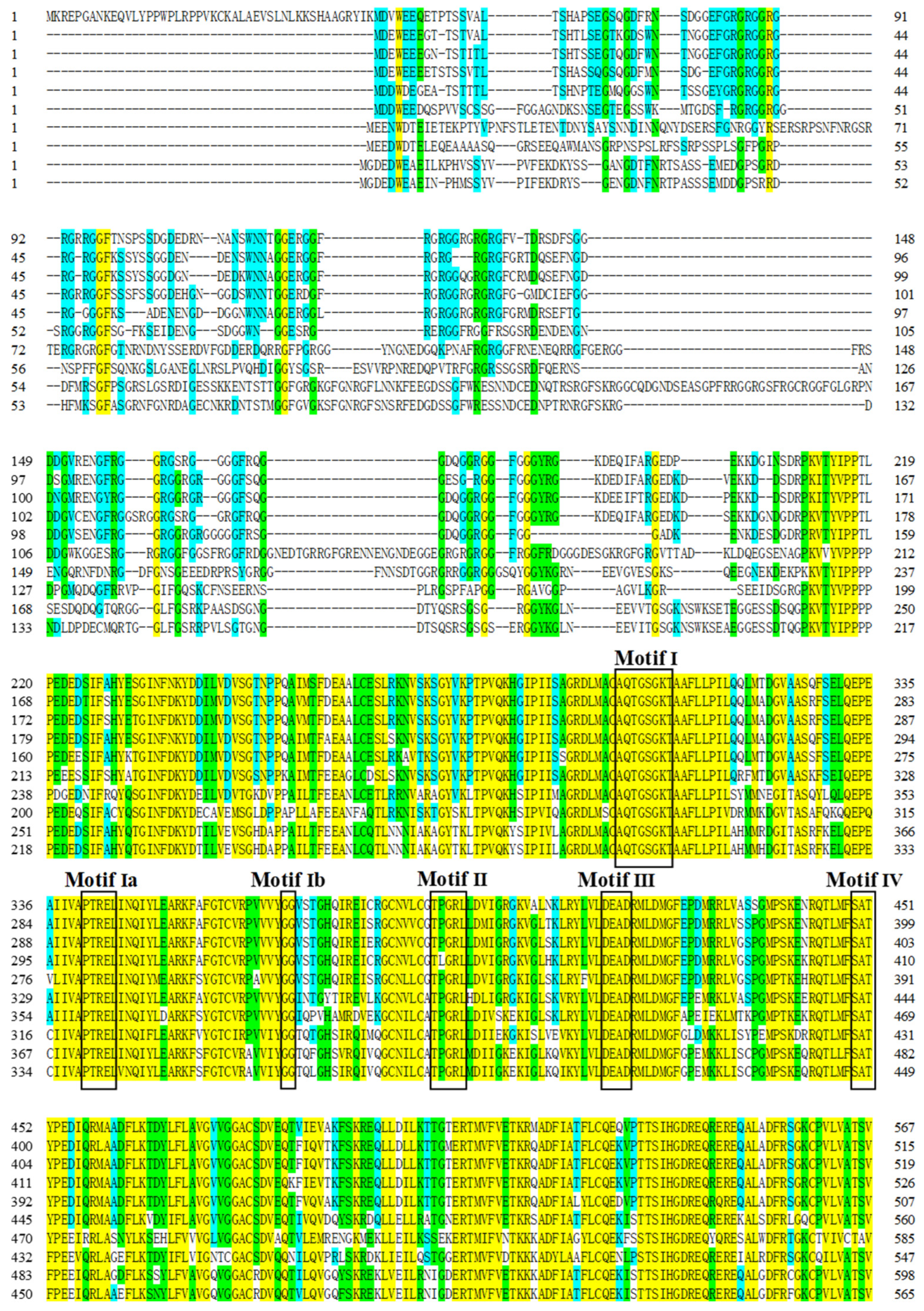
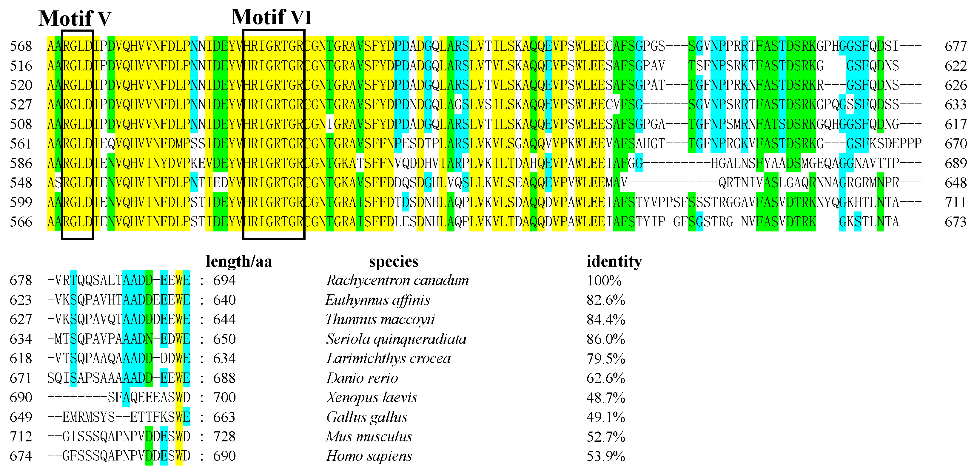
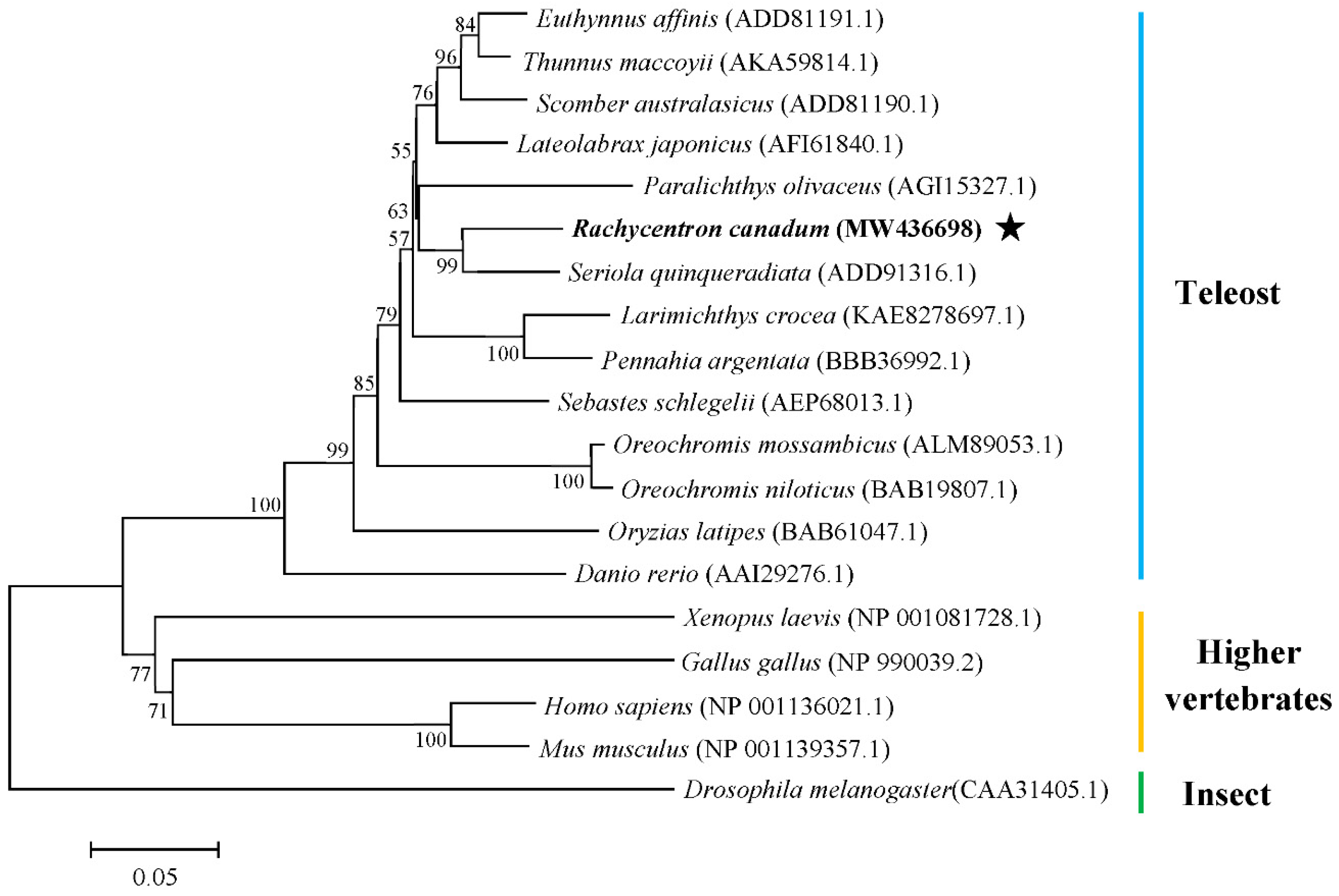
3.1. Cloning of Rcvasa cDNA and Phylogenetic Analysis

The full-length cDNA sequence of cobia vasa (Rcvasa) was cloned by RACE amplification (GenBank accession No. MW436698). The Rcvasa cDNA appeared to consist of 2571 bp, comprising an ORF (open reading frame) of 2085 bp, a 5′-UTR (untranslated region) of 145 bp, and a 3′-UTR of 341 bp, and to encode a 694-amino acid protein.
The predicted RcVasa protein contains 8 conserved motifs of the DEAD-box protein family, including motif I (AQTGSGKT), motif Ia (PTRELI), motif Ib (GG), motif II (TPGRL), motif III (DEAD), motif IV (SAT), motif V (RGLD), and motif VI (HRIGRTGR) (Figure 1), and the N-terminal region contains 10 arginine–glycine (RG) repeats and 7 arginine–glycine–glycine (RGG) repeats. The RcVasa protein also has two highly conserved domains, the DEXDc domain (275–486 aa) and the HELICc domain (522–603 aa). Multiple sequence alignment revealed that the deduced amino acid sequence of Rcvasa has 62.6–86.0% identity with those of Rcvasa proteins in other teleosts. The highest identity was found between cobia Rcvasa amino acid sequence and that of the corresponding protein in Seriola quinqueradiata (86.0%), followed by Thunnus thynnus (84.4%), Euthynnus affinis (82.6%), and Larimichthys crocea (79.5%) proteins, while the lowest identity was found with Rcvasa amino acid sequence in Danio rerio (62.6%) as shown in Figure 1.
Based on the genetic distances calculated with the Poisson correction model, a phylogenetic tree was constructed by the Neighbor-Joining method to investigate the phylogenetic relationship among different species (Figure 2). The results showed that all the teleost species fall into a lineage, the higher vertebrates, including amphibians, birds, and mammals, fall into another lineage, and Drosophila melanogaster forms a single cluster. The teleost cluster comprises 14 species, 7 of which are from the Perciformes order, 3 from the Scombriformes order, and 1 each from the Pleuronectiformes, Scorpaeniformes, Cyprinodontiformes, and Cypriformes orders. Moreover, RcVasa clusters together with Seriola quinqueradiata Vasa.

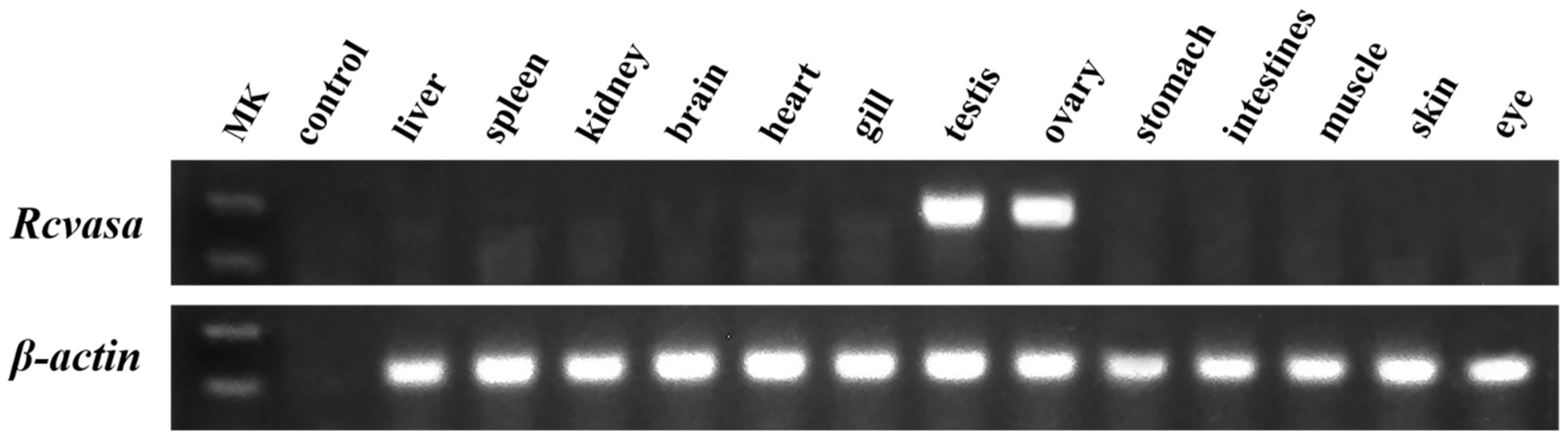
3.2. Tissue Distribution Patterns of Rcvasa mRNA

The tissue distribution patterns of Rcvasa mRNA in 13 tissues was analyzed by semiquantitative RT-PCR. The results showed that Rcvasa was exclusively expressed in the ovary and testis, whereas almost no expression was detected in other somatic tissues (Figure 3).

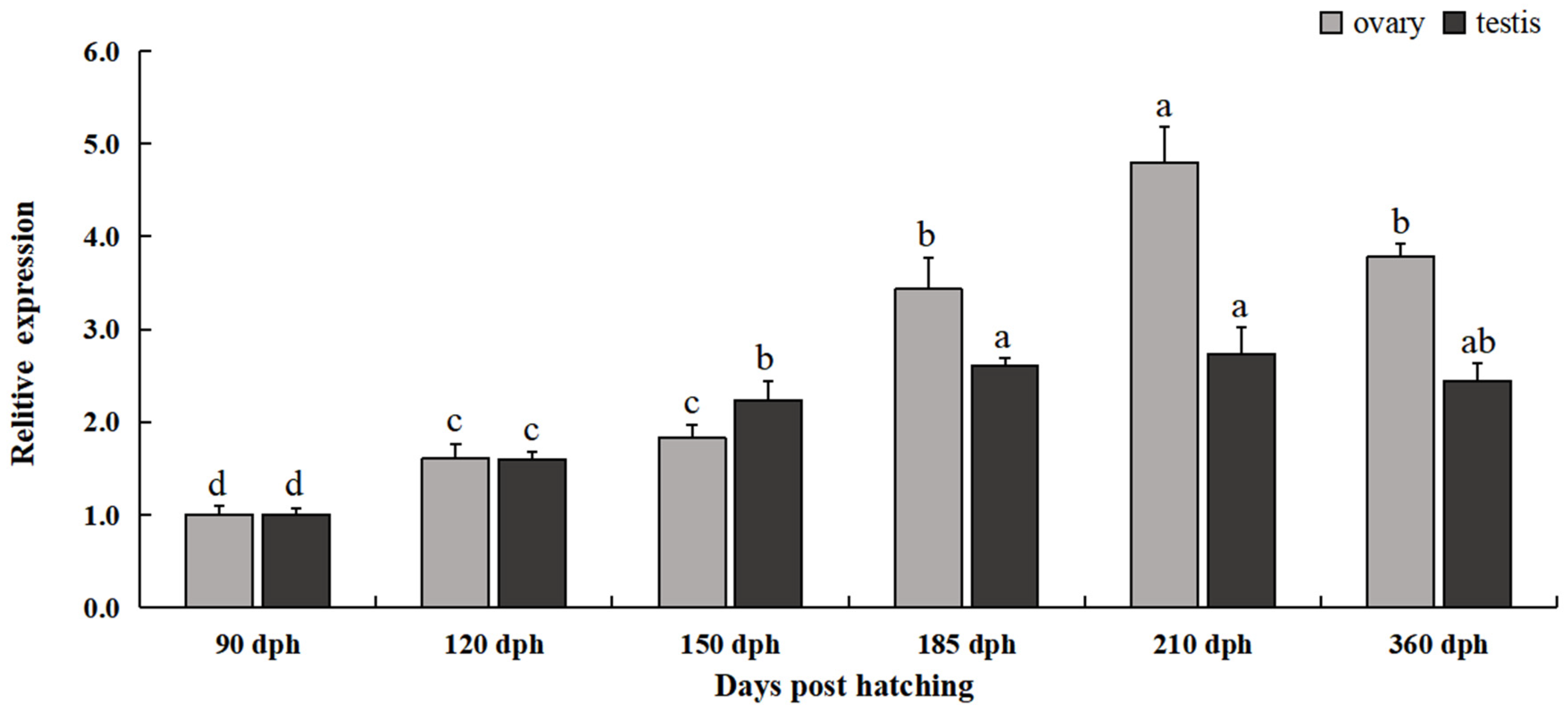
3.3. Expression Patterns of Rcvasa mRNA at Different Gonadal Development Stages

Based on histological observation of gonadal differentiation, the testes collected from 90-to-360 dph cobia could be divided into four stages (stage II, III, IV, V). The testes were in the spermatocyte growth stage (stage II) at 90 dph and in stage II–III at 120 dph. Testes at 150 dph and 185 dph were in the spermatocyte mature stage (stage III), while testes at 210 dph and 360 dph were in the spermatid metamorphosis stage (stage IV) and sperm mature stage (stage V), respectively. The first annual ovarian development of cobia was divided into three stages (Stage I, II, III). The ovaries were in the oogonium proliferation-stage (stage I) at 90 dph and developed to stage I–II at 120 dph. Ovaries at 150 dph, 185 dph and 210 dph were in the oocyte primary growth stage (stage II) and entered the oocyte cortical alveolus stage (stage III) at 360 dph.
The expression of Rcvasa mRNA was detected in the gonads of both male and female cobia throughout the first annual development. In the testis, the expression level of Rcvasa increased significantly after 120 dph; the expression levels at 185 dph (stage III), 210 dph (Stage IV), and 360 dph (Stage V) showed no significant differences. The expression level at 90 dph (Stage II) was the lowest; it then increased significantly, reaching the maximum at 210 dph (Stage IV), which was 2.78 times that at 90 dph (Figure 4).
During the first annual ovarian development, the expression levels of Rcvasa first increased and then decreased. The lowest expression level was detected at 90 dph (stage I). The relative expression reached the maximum at 210 dph (stage II), and its value was about 4.97 times that at 90 dph, while the relative expression at 360 dph (stage III) was significantly lower (Figure 4).

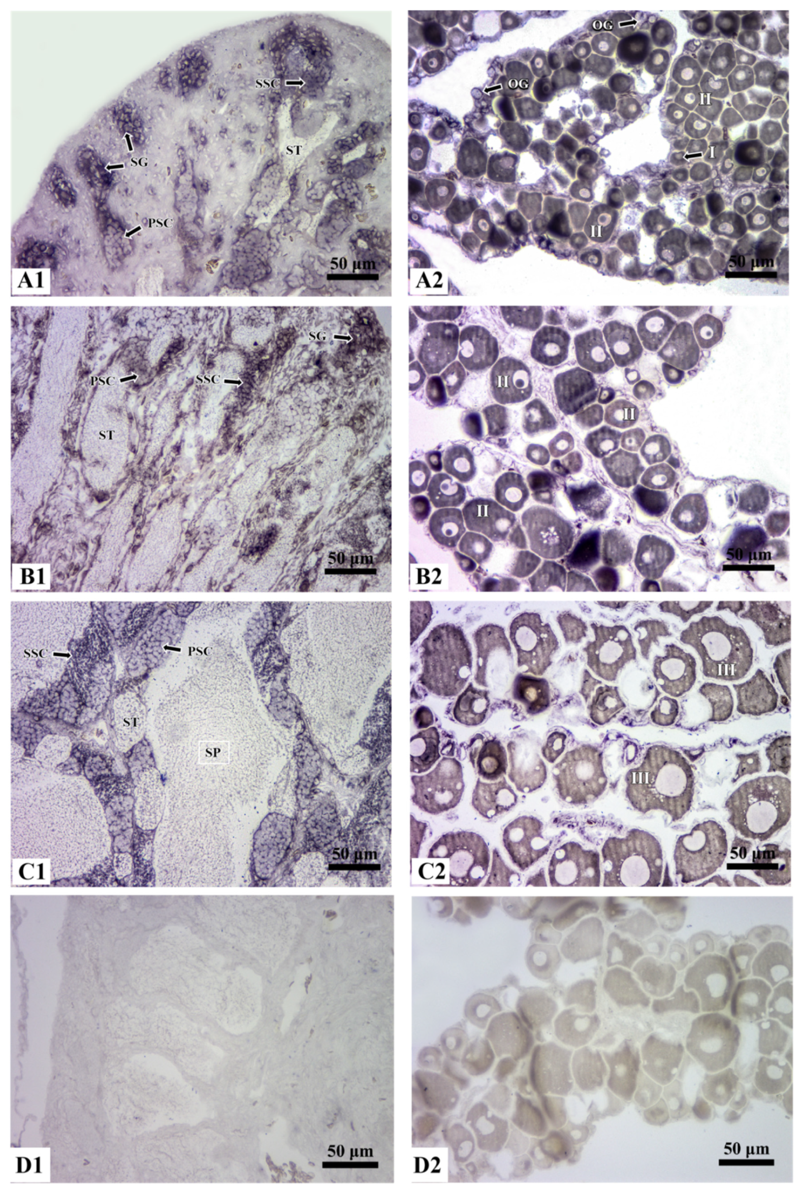
3.4. Localization of Rcvasa mRNA in Germ Cells during Gametogenesis

The localization of Rcvasa mRNA in the gonads at different developmental stages was investigated by CISH. Rcvasa mRNA signals were restricted to germ cells and were not detected in somatic cells. In the testis of 120 dph cobia, Rcvasa mRNA was predominantly detected in the periphery of spermatogonia, primary spermatocytes, and secondary spermatocytes. The expression level of Rcvasa was strong in spermatogonia and scarcely detected in spermatids (Figure 5A1). Similar expression patterns were observed in the testis at 210 dph, when Rcvasa mRNA signals were also mainly distributed in the periphery of spermatogonia and in spermatocytes (Figure 5B1). In the testis at 360 dph, Rcvasa mRNA signal was detected in all germ cells throughout spermatogenesis and were concentrated in spermatocytes; it appeared significantly weaker in spermatids and spermatozoa (Figure 5C1).
During ovarian development, Rcvasa mRNA was specifically expressed in germ cells and evenly distributed in the cytoplasm and nucleoli. In the ovary of 120 dph cobia, Rcvasa mRNA was localized in oogonium (chromatin nucleolar stage), stage I and II oocytes (perinucleolar and previtellogenic stage) (Figure 5A2). In the ovary at 210 dph, Rcvasa mRNA signal was concentrated in the cytoplasm and nucleoli of stage II oocytes (previtellogenic stage); with the accumulation of oocyte protoplasm, the signals gradually became stronger (Figure 5B2). In the ovary at 360 dph, Rcvasa mRNA signal detected in the cytoplasm of stage III oocytes (vitellogenic stage) was significantly weakened (Figure 5C2).

4. Discussion

The present study reported the isolation and characterization of cobia vasa homologue. The deduced amino acid sequence of the protein encoded by Rcvasa contains eight conserved motifs of the DEAD protein family [27]. RcVasa showed an appreciable identity to the Vasa protein from other marine teleosts, as well as to the Vasa homologue of Seriola quinqueradiata (86.0%). In addition, a glycine-rich region in the N-terminal region of RcVasa containing 10 arginine–glycine (RG) repeats and 7 arginine–glycine–glycine (RGG) repeats was observed. This glycine-rich region is regarded as a characteristic of single-stranded nucleic acid-binding proteins, such as RNA helicase, which may regulate the activity of different vasa transcripts [28]. Phylogenetic analysis revealed that RcVasa closely aligned to its teleost counterparts. These findings illustrate that the vasa gene has been highly conserved in the evolution of teleost, and the conserved motifs of the Vasa protein potentially play an important role in sustaining protein structure and function [29].
The tissue distribution patterns of vasa mRNA in 150 dph juvenile cobia showed that Rcvasa was specifically and abundantly expressed in testis and ovary. Previous reports also showed that Rcvasa is a gonad-specific gene and may be involved in the regulation of gonadal development [19,20,30,31].
The expression of Rcvasa increased significantly when the testis developed from stage I to III, but no significant difference was detected after the late stage III. It has been reported that in the early stage of the testicular development of fish, spermatogonia undergo mitosis and gradually transforms into primary spermatocytes; then, primary spermatocytes divide into secondary spermatocytes [32]. As a result, the expression pattern of Rcvasa is positively correlated with the number of spermatocytes, suggesting that Rcvasa might play an important role in the regulation of spermatogenesis. Previous studies have reported that the expression levels of catfish (Clarias gariepinus) vasa gradually increased during testis development from stage I to VI and decreased significantly after stage V [33]. Mu et al. found that the expression of Korean rockfish vasa maintained a high level in testicular stages I to III, but vasa expression decreased significantly when spermatids started to mature [31]. These results indicate that there are significant interspecies differences in the expression patterns of vasa during the testicular development of teleost. The expression levels of Rcvasa in the early stage of ovarian development tended to increase gradually. As the ovaries developed to stage II, the oocytes became larger by accumulating protoplasm consisting of carbohydrates, proteins, and nucleic acids [32], and the expression of Rcvasa significantly increased. The oocytes began to produce egg yolk in ovarian stage III, and the expression of Rcvasa decreased significantly. These results suggest that Rcvasa might participate in the accumulation of oocyte protoplasm and in the formation of ovum yolk.
Furthermore, Rcvasa mRNA was exclusively expressed in germ cells and not in somatic cells. During testicular development, Rcvasa mRNA signal was mainly concentrated in the periphery of spermatogonia, primary spermatocytes, and secondary spermatocytes, and it was found to be weak in spermatids and spermatozoa. Similar results were reported in other fish [34,35]. The localization of Rcvasa mRNA during spermatogenesis suggested that this protein might play a major role in the transformation of spermatogonia into primary spermatocytes, but not in the development and maturation of spermatids. As reported, bluefin tuna vasa mRNA was only expressed in spermatogonia and not in other testicular cells [19]. This indicated that vasa might have a species-specific role in regulating the transformation of spermatogenic cells. The expression pattern of Rcvasa during oogenesis was similar to those observed in several teleost species [3,18,36] and revealed that Rcvasa mRNA was abundant in early primary oocytes (stage I and II, perinucleolar and previtellogenic stage) and deficient in yolk vesicle (cortical alveolus) formation-stage oocytes (stage III, vitellogenic stage). These findings indicate that vasa might regulate oocyte development during the early stage of oogenesis. As for gilthead sea bream (Sparus aurata), the vasa mRNA level was low in early primary oocytes and high when the oocytes developed to the mature gametes [37]. According to a report in tilapia, vitellogenesis was usually accompanied by the accumulation of vasa maternal factors, showing that vasa might be involved in regulating the maturation of oocytes [15]. However, a weak Rcvasa mRNA signal was detected in stage III oocytes. The reason for this might be related to the fact that the total protein content of these maturating oocytes increased, and as a result, the relative concentration of Rcvasa mRNA was diluted. Similar to the expression patterns of vasa during testicular development, a certain interspecies difference was also found in the regulation of vasa expression during oogenesis.

5. Conclusions

The vasa gene from cobia was cloned and characterized, and further investigation of the expression level and localization of Rcvasa mRNA during the first annual gonadal development was performed. The high and exclusive expression of Rcvasa mRNA in germ cells indicates the important role of vasa in spermatogenesis and oogenesis in this important aquaculture species. The gradually increasing mRNA level along with the growth and development of cobia also provides a basis for clarifying the mechanisms underlying the migration and differentiation of PGCs in teleosts.

Author Contributions

Conceptualization, Q.M., J.K. and G.C.; methodology, Q.M. and J.K.; software, J.K.; validation, Q.M., J.K. and J.Z.; analysis, J.H., F.M. and Q.Z.; writing—original draft preparation, Q.M. and J.K.; writing—review and editing, G.C., J.Z., F.M. and Q.Z.; All authors have read and agreed to the published version of the manuscript.

Funding

This research was funded by the Modern Agricultural Industrial Technology System Special Funding, grant number CARS-47; the Program for Scientific Research Start-up Funds of Guangdong Ocean University, grant number R19022; the Fund of Southern Marine Science and Engineering Guangdong Laboratory (Zhanjiang), grant number ZJW-2019-06.

Institutional Review Board Statement

The animal study protocol was approved by the Animal Ethics Committee of Guangdong Ocean University (protocol code 0301-2019, 1 March 2019).

Informed Consent Statement

Not applicable.

Data Availability Statement

The data presented in this study are available on request from the corresponding author.

Conflicts of Interest

The authors declare no conflict of interest.

References

  1. Wylie, C. Germ cells. Cell 1999, 96, 165–174. [Google Scholar] [CrossRef]
  2. Barske, L.A.; Capel, B. Blurring the edges in vertebrate sex determination. Curr. Opin. Genet. Dev. 2008, 18, 499–505. [Google Scholar] [CrossRef] [PubMed] [Green Version]
  3. Xu, H.Y.; Gui, J.F.; Hong, Y.H. Differential expression of vasa RNA and protein during spermatogenesis and oogenesis in the gibel carp (Carassius auratus gibelio), a bisexually and gynogenetically reproducing vertebrate. Dev. Dyn. 2005, 23, 872–882. [Google Scholar] [CrossRef]
  4. Liu, L.X.; Hong, N.; Xu, H.Y.; Li, M.Y.; Yan, Y.; Purwanti, Y.; Yi, M.S.; Li, Z.D.; Wang, L.; Hong, Y.H. Medaka dead end encodes a cytoplasmic protein and identifies embryonic and adult germ cells. Gene. Expr. Patterns 2009, 9, 541–548. [Google Scholar] [CrossRef]
  5. Bhat, N.; Hong, Y.H. Cloning and expression of boule and dazl in the Nile tilapia (Oreochromis niloticus). Gene 2014, 540, 140–145. [Google Scholar] [CrossRef]
  6. Sun, Z.H.; Wang, Y.; Lu, W.J.; Li, Z.; Liu, X.C.; Li, S.; Zhou, L.; Gui, J.F.; Lin, L. Divergent expression patterns and function implications of four nanos genes in a hermaphroditic fish, Epinephelus coioides. Int. J. Mol. Sci. 2017, 18, 685. [Google Scholar] [CrossRef]
  7. Duangkaew, R.; Jangprai, A.; Ichida, K.; Yoshizaki, G.; Boonanuntanasarn, S. Characterization and expression of a vasa homolog in the gonads and primordial germ cells of the striped catfish (Pangasianodon hypophthalmus). Theriogenology 2019, 131, 61–71. [Google Scholar] [CrossRef]
  8. Linder, P.; Lasko, P.F.; Ashburner, M.; Leroy, P.; Nielsen, P.J.; Nishi, K.; Schnier, J.; Slonimski, P.P. Birth of the D-E-A-D box. Nature 1989, 337, 121–122. [Google Scholar] [CrossRef]
  9. Liang, L.; Diehl-Jones, W.; Lasko, P. Localization of vasa protein to the Drosophila pole plasm is independent of its RNA-binding and helicase activities. Development 1994, 120, 1201–1211. [Google Scholar] [CrossRef]
  10. Deshpande, G.; Calhoun, G.; Yanowitz, J.L.; Schedl, P.D. Novel functions of nanos in downregulating mitosis and transcription during the development of the Drosophila Germline. Cell 1999, 99, 271–281. [Google Scholar] [CrossRef] [Green Version]
  11. Lasko, P. The DEAD-box helicase Vasa: Evidence for a multiplicity of functions in RNA processes and developmental biology. Biochim. Biophys. Acta. 2013, 1829, 810–816. [Google Scholar] [CrossRef]
  12. Schüpbach, T.; Wieschaus, E. Maternal-effect mutations altering the anterior-posterior pattern of the Drosophila embryo. Roux’s Arch. Dev. Biol. 1986, 195, 302–317. [Google Scholar] [CrossRef] [PubMed]
  13. Xu, H.Y.; Li, M.Y.; Gui, J.F.; Hong, Y.H. Fish germ cells. Sci. China Life Sci. 2010, 53, 435–446. [Google Scholar] [CrossRef] [PubMed]
  14. Braat, A.K.; Zandbergen, M.A.; Water, S.V.D.; Goos, H.J.; Zivkovic, D. Characterization of zebrafish primordial germ cells: Morphology and early distribution of vasa RNA. Dev. Dyn. 1999, 216, 153–167. [Google Scholar] [CrossRef]
  15. Kobayashi, T.; Kajiura-Kobayashi, H.; Nagahama, Y. Differential expression of vasa homologue gene inthe germ cells during oogenesis and spermatogenesisin a teleost fish, tilapia, Oreochromis niloticus. Mech. Dev. 2000, 99, 139–142. [Google Scholar] [CrossRef]
  16. Wang, Z.K.; Gao, J.N.; Song, H.Y.; Wu, X.M.; Sun, Y.; Qi, J.; Yu, H.Y.; Wang, Z.G.; Zhang, Q.Q. Sexually dimorphic expression of vasa isoforms in the tongue sole (Cynoglossus semilaevis). PLoS ONE 2014, 9, e93380. [Google Scholar] [CrossRef] [Green Version]
  17. Xu, H.Y.; Lim, M.; Dwarakanath, M.; Hong, Y.H. Vasa identifies germ cells and critical stages of oogenesis in the Asian seabass. Int. J. Biol. Sci. 2014, 10, 225–235. [Google Scholar] [CrossRef] [Green Version]
  18. Yoshizaki, G.; Sakatani, S.; Tominaga, H.; Takeuchi, T. Cloning and characterization of a vasa-like gene in rainbow trout and its expression in the germ cell lineage. Mol. Reprod. Dev. 2015, 55, 364–371. [Google Scholar] [CrossRef]
  19. Nagasawa, K.; Takeuchi, Y.; Miwa, M.; Higuchi, K.; Morita, T.; Mitsuboshi, T.; Miyaki, K.; Kadomura, K.; Yoshizaki, G. cDNA cloning and expression analysis of a vasa-like gene in Pacific bluefin tuna Thunnus orientalis. Fish. Sci. 2009, 75, 71–79. [Google Scholar] [CrossRef]
  20. Kobayashi, T.; Kajiura-Kobayashi, H.; Nagahama, Y. Two isoforms of vasa homologs in a teleost fish: Their differential expression during germ cell differentiation. Mech. Dev. 2002, 111, 167–171. [Google Scholar] [CrossRef]
  21. Castellanos-Galindo, G.; Baos, R.; Zapata, L. Mariculture-induced introduction of cobia Rachycentron canadum (Linnaeus, 1766), a large predatory fish, in the Tropical Eastern Pacific. BioInvasions Rec. 2016, 5, 55–58. [Google Scholar] [CrossRef]
  22. Velde, T.; Griffiths, S.P.; Fry, G.C. Reproductive biology of the commercially and recreationally important cobia Rachycentron canadum in northeastern Australia. Fish. Sci. 2010, 76, 33–43. [Google Scholar] [CrossRef]
  23. Hamilton, S.; Severi, W.; Cavalli, R.O. Biology and aquaculture of cobia: A review. Bol. Inst. Pesca 2013, 39, 461–477. [Google Scholar]
  24. Schmittgen, T.D.; Livak, K.J. Analyzing realtime PCR data by the comparative C(T) method. Nat. Protoc. 2008, 3, 1101–1108. [Google Scholar] [CrossRef] [PubMed]
  25. Kuang, J.; Chen, G.; Ma, Q.; Mao, F.; Zhou, Q.; Huang, J.; Shi, G.; Zhang, J. Histological observation on gonadal differentiation and first annual gonadal development of cobia (Rachycentron canadum). Haiyang Xuebao 2021, 43, 1–11. [Google Scholar]
  26. Liu, Y. Propagation Physiology of Main Cultivated Fish in China; Agricultural Publishing Press: Beijing, China, 1993; pp. 20–42. [Google Scholar]
  27. Rocak, S.; Linder, P. DEAD-box proteins: The driving forces behind RNA metabolism. Nat. Rev. Mol. Cell Biol. 2004, 5, 232–241. [Google Scholar] [CrossRef] [PubMed]
  28. Bartfai, R.; Orban, L. The vasa locus in zebrafish: Multiple RGG boxes from duplications. DNA Cell Biol. 2003, 22, 47–54. [Google Scholar] [CrossRef]
  29. Tanner, N.K. The newly identified Q motif of DEAD-box helicases is involved in adenine recognition. Cell Cycle 2003, 2, 18–19. [Google Scholar] [CrossRef] [Green Version]
  30. Lin, F.; Xu, S.H.; Ma, D.Y.; Xiao, Z.Z.; Zhao, C.Y.; Xiao, Y.S.; Chi, L.; Liu, Q.H.; Li, J. Germ line specific expression of a vasa homologue gene in turbot (Scophthalmus maximus): Evidence for vasa localization at cleavage furrows in euteleostei. Mol. Reprod. Dev. 2012, 79, 803–813. [Google Scholar] [CrossRef]
  31. Mu, W.J.; Wen, H.H.; He, F.; Li, J.F.; Liu, M.; Ma, R.Q.; Zhang, Y.Q.; Hu, J.; Qi, B.X. Cloning and expression analysis of vasa during the reproductive cycle of Korean rockfish, Sebastes schlegeli. J. Ocean Univ. China 2013, 12, 115–124. [Google Scholar] [CrossRef]
  32. Nishimura, T.; Tanaka, M. Gonadal development in fish. Sex. Dev. 2014, 8, 252–261. [Google Scholar] [CrossRef] [PubMed]
  33. Raghuveer, K.; Senthilkumaran, B. Cloning and differential expression pattern of vasa in the developing and recrudescing gonads of catfish, Clarias gariepinus. Comp. Biochem. Physiol. Part A Mol. Integr. Physiol. 2010, 157, 79–85. [Google Scholar] [CrossRef] [PubMed]
  34. Xiao, J.; Luo, Y.J.; Chen, L.B.; Yang, L.; Huang, Y.L.; Guo, Z.B.; Guo, E.Y.; Tang, Z.Y.; Zhang, M.; Gan, X. Molecular cloning of vasa gene and the effects of LHRH-A on its expression in blue tilapia Oreochromis aureus. Fish Physiol. Biochem. 2013, 39, 931–940. [Google Scholar] [CrossRef]
  35. Zhu, W.X.; Wang, T.; Zhao, C.; Wang, D.; Zhang, X.Y.; Zhang, H.Y.; Chi, M.L.; Yin, S.W.; Jia, Y.Y. Evolutionary conservation and divergence of Vasa, Dazl and Nanos1 during embryogenesis and gametogenesis in dark sleeper (Odontobutis potamophila). Gene 2018, 672, 21–33. [Google Scholar] [CrossRef]
  36. Qu, L.; Wu, X.; Liu, M.F.; Zhong, C.Y.; Xu, H.Y.; Li, S.S.; Lin, H.R.; Liu, X.C. Identification and characterization of germ cell genes vasa and dazl in a protogynous hermaphrodite fish, orange-spotted grouper (Epinephelus coioides). Gene Expr. Patterns 2020, 35, 11905. [Google Scholar] [CrossRef]
  37. Cardinali, M.; Gioacchini, G.; Candiani, S.; Pestarino, M.; Yoshizaki, G.; Carnevali, O. Hormonal regulation of vasa-like messenger RNA expression in the ovary of the marine teleost Sparus aurata. Biol. Reprod. 2004, 70, 737–743. [Google Scholar] [CrossRef]
Figure 1. Multiple alignment of Rcvasa deduced amino acid sequences. The framed regions indicate the eight conserved functional motifs.
Fishes 07 00060 g001aFishes 07 00060 g001b
Figure 2. Phylogenetic tree of vasa amino acid sequences based on the Neighbor-Joining (NJ) method. The tree is based on a 1000 bootstrap procedure; the scale bar (0.05 in terms of genetic distance) is indicated below the tree; the asterisk indicates the target species in this study.
Fishes 07 00060 g002
Figure 3. Rcvasa mRNA expression in various tissues of cobia. cDNA from various tissues (liver, spleen, kidney, brain, heart, gill, testis, ovary, stomach, intestines, muscle, skin, and eye) was used. β-actin was used as an internal control. MK: DNA marker; control: negative control without cDNA template.
Fishes 07 00060 g003
Figure 4. Rcvasa mRNA expression patterns during the first annual gonadal development of cobia. The results are presented as the mean ± SD (n = 3), and the values with different letters (a–d) appeared significantly different in pairwise comparisons (p < 0.05).
Fishes 07 00060 g004
Figure 5. Distribution of Rcvasa mRNA at different developmental stages in cobia gonads, analyzed by CISH. (A1,B1,C1), sections of testes at 120 dph, 210 dph, and 360 dph were hybridized with an Rcvasa oligonucleotides probe; (A2,B2,C2), sections of ovaries at 120 dph, 210 dph, and 360 dph were hybridized with an Rcvasa oligonucleotides probe. (D1,D2), negative control, sections of testes and ovaries at 210 dph, hybridized with a sense probe. SG: spermatogonia; PSC: primary spermatocyte; SSC: secondary spermatocyte; ST: spermatid; SP: spermatozoa; OG: oogonium (chromatin nucleolar oocyte); I: oocyte at stage I (perinucleolar oocyte); II: oocyte at stage II (previtellogenic oocyte); III: oocyte at stage III (vitellogenic oocyte).
Fishes 07 00060 g005
Table 1. Primer sequences used in this study.
PrimerSequence (5′–3′)Usage
Rcvasa-FATGAAAAGAGAACCAGGCGCGACDS sequence cloning
Rcvasa-RCTACTCCCATTCTTCATCATCAGCTG
Rcvasa3′-F1TGACCTCCCCAACAACATAGACG3′ RACE
Rcvasa3′-F2TGGGAGGGCGGTGTCTTTC
Rcvasa5′-R1GCCAGCAGAAATGATGGGGAT5′ RACE
Rcvasa5′-R2CTCCCTGATCTCCACCTTGTCTG
Long-UPMCTAATACGACTCACTATAGGGCAAGCAGTGGTATCAACGCAGAGTRACE universal primer
Short-UPMCTAATACGACTCACTATAGGGC
Rcvasa-F1GGTTGGCAGGAGCAGTAACTPCR amplification
Rcvasa-R1TGTGGTTGTGATCTCCGGTG
β-actin-FAGGGAAATTGTGCGTGACinternal control gene
β-actin-RAGGCAGCTCGTAGCTCTT
Rcvasa-ProCAGGACGTTACACCCCCGGCATATTTCTCprobe of CISH
Publisher’s Note: MDPI stays neutral with regard to jurisdictional claims in published maps and institutional affiliations.

