Technical Note

Isolation, Culture, and Differentiation of Blastema Cells from the Regenerating Caudal Fin of Zebrafish

by
Parameswaran Vijayakumar
1,2,*,
M. Leonor Cancela
2,3 and
Vincent Laizé
2
1
Centre for Ocean Research, Sathyabama Institute of Science and Technology, Jeppiaar Nagar, Rajiv Gandhi Salai, Chennai, Tamil Nadu 600119, India
2
Centre of Marine Sciences (CCMAR), University of Algarve, Campus de Gambelas, 8005-139 Faro, Portugal
3
Department of Biomedical Sciences and Medicine (DCBM) and Algarve Biomedical Center (ABC), University of Algarve, Campus de Gambelas, 8005-139 Faro, Portugal
*
Author to whom correspondence should be addressed.
Submission received: 3 July 2019 / Revised: 6 January 2020 / Accepted: 19 January 2020 / Published: 30 January 2020

Abstract

:
The caudal fin of teleost fish has become an excellent system for investigating the mechanisms of epimorphic regeneration. Upon amputation of the caudal fin, a mass of undifferentiated cells, called blastema, proliferate beneath the wound-epidermis and differentiate into various cell types to faithfully restore the missing fin structures. Here we describe a protocol that can be used to isolate and culture blastema cells from zebrafish. Primary cultures were initiated from 36 h post-amputation (hpa) blastema and optimal cell growth was achieved using L-15 medium supplemented with 5% fetal bovine serum in plates either coated with fibronectin or uncoated. After seeding, zebrafish blastema cells formed a uniform culture and exhibited polygonal shapes with prominent nucleus, while various cell types were also observed after few days in culture indicating cell differentiation. Upon treatment with all-trans retinoic acid, zebrafish blastema cells differentiated into neuron-like and oligodendritic-like cells. Immunocytochemistry data also revealed the presence of mesenchymal and neuronal cells. The availability of blastema cell cultures could contribute to a better understanding of epimorphic regeneration by providing a mean to investigate the mechanisms underlying blastema cell differentiation. Furthermore, this protocol is simple, rapid, and cost-efficient, and can be virtually applied to the development of any fish blastema cell culture.

1. Introduction

Due to its remarkable simplicity and accessibility, the caudal fin of teleost fish has become an excellent system for investigating the cellular and molecular mechanisms of epimorphic regeneration, i.e., the complete reformation of missing tissues (reviewed by [1,2,3,4,5]). Upon amputation of caudal fin tissues (i.e., bony rays covered with a thin epidermis, nerves, blood vessels, and connective tissue), a thick wound epidermis forms rapidly at the amputation plane, while a mass of undifferentiated cells, called blastema, proliferate beneath the wound epidermis [6]. It has been proposed that blastema is formed by cells that de-differentiate but remain lineage committed [7,8,9]. Blastema cells will contribute to various cell types—e.g., osteoblasts, vasculature, melanocytes, and epidermal cells—to faithfully restore missing fin structures [10,11,12,13] through molecular and genetic processes that have been approached in several studies [14,15,16,17]. Recently, Tu and Johnson demonstrated that blastema cells do not differentiate into cell lineages that were not already present in the caudal fin tissues [9].
Full regeneration is usually achieved after 10–15 days depending on the species, the level of amputation, and culture conditions. Protocols to prepare zebrafish primary cell cultures have been established recently to study the structural and functional properties of cardiomyocytes [18], neurogenesis [19], and mechanisms underlying inhibition of neural regeneration in vitro [20], and while several cell lines have been developed from the caudal fin in zebrafish [21,22] and other fish species [23,24,25,26], none have been reported for the blastema yet. In this study, a protocol was established to isolate blastema cells from the zebrafish Danio rerio (Hamilton 1822) and to successfully initiate primary cell cultures. It is expected that the availability of such in vitro cell systems will contribute to a better understanding of epimorphic regeneration by providing a means to investigate mechanisms underlying blastema cell differentiation, while also complementing the work performed in vivo.

2. Results and Discussion

The development of primary cultures of blastema cells (see Figure 1A–C and detailed protocol in the Materials and Methods section) opens the possibility to investigate in vitro the cellular and molecular mechanisms underlying epimorphic regeneration of the caudal fin of fish. Zebrafish cells obtained after enzymatic dissociation of regenerating fin tissues adhered rapidly on the substratum at 28 °C (Figure 1D), proliferated continuously for about 48 h, and could be maintained for 1 or 2 passages. Longer survival times were reported for salamander limb blastema cells (up to 30 days in culture; [27]). The possibility of the zebrafish cells being in a committed stage and less prone to active proliferation after 3 days of culture conditions could explain a shorter survival time, although the lack of specific growth factors are also conceivable. In this regard, culture medium could be supplemented with (1) conditioned medium obtained from zebrafish ES cells, (2) zebrafish embryo extract, (3) fish serum (instead of bovine serum) prepared from farmed fishes, e.g., Atlantic salmon, trout, tilapia, and gilthead seabream [28,29], or (4) growth factors such as Wnt, TGF-β, IGF-1, bFGF, or LIF [30,31,32,33,34]. To extend their lifetime, blastema cells could also be cultured in a collagenous gel [35] or as organotypic slice culture [36]. The use of a feeder layer to grow zebrafish blastema cells could be tested as it was shown to be critical to the growth and maintenance of undifferentiated state of mouse embryonic stem (ES) cells [37].
Approximately 3–3.5 × 10⁴ cells were recovered per blastema with a viability higher than 90%. Blastema cells were routinely cultured in L-15 medium supplemented with 5% FBS after a reduced proliferation rate was observed in cultures supplemented with 10%, 15%, and 20% of serum (Figure 1E), an observation in agreement with the data published by Kumar and Godwin [27] showing the inability of salamander limb blastema cells to survive in culture media containing high levels of bovine serum. Thus, salamander blastema cells were cultured in medium containing <2% of FBS (e.g., in serum-free medium or medium with 0.5–1% serum) to reduce cell death. Whether zebrafish blastema cells can be maintained in medium containing less than 5% of FBS should be further tested. The morphology of zebrafish blastema cells cultured in 5% FBS was polygonal with a prominent nucleus and maintained over time. (Figure S1). Coating the surface of cell culture dishes with extracellular matrix molecules, e.g., fibronectin, laminin, poly-L-lysine, and gelatin, can promote cell attachment, growth, spreading, and differentiation [38]. While fibronectin slightly stimulated the viability of zebrafish blastemal cells, laminin, poly-L-lysine, and gelatin triggered an anti-proliferative effect. It appeared that fin blastema cell attachment or growth was best in the absence of any coating (Figure 1F). No deposition of mineral was observed in blastema cell cultures treated with mineralogenic cocktail (Figure S2), although the absence of ECM mineralization may be related to the short treatment.
Although Tu and Johnson reported that blastema cells do not differentiate into cell lineages that are not already present in the caudal fin tissues [19], new cell types, i.e., spindle-shaped, oligodendritic-like, neuron-like cells, were observed in blastema cell cultures after 3 days in culture (Figure 2A–G). The presence of these new types of cells may depend on culture conditions e.g., the absence of a particular cell substrate or growth factor, the presence of fetal bovine serum, or the temperature and the osmolarity of the culture medium. As for murine embryonic stem (ES) cells that require a feeder layer or leukemia inhibitory factor to maintain an undifferentiated state [34,39], blastema cells may need particular culture conditions to maintain their lineage commitment. In this regard, organotypic slice culture (OSC) of blastema explants may be tried in the future to maintain the original tissue architecture. The treatment of zebrafish blastema cells with all-trans retinoic acid (RA) also triggered the appearance of neuron-like and oligodendritic-like cells (Figure 2H,I), in agreement with a recent study reporting the differentiation of rabbit pinna-derived blastema cells into neuronal cells upon RA treatment [40]. Similar changes in cell phenotype were also found in human, Japanese medaka, and gilthead seabream cells [41,42,43]. Vitamin A (including compounds such as retinol and RA) is a signaling molecule that plays key roles in vertebrate pattern formation both in developing and regenerating tissues and to the differentiation of many types of cells [44,45,46,47,48,49]. The production of endogenous RA from retinol is essential for the regeneration of zebrafish fin [50] and RA signaling is enhanced in the stump mesenchyme within the first few hours after fin amputation [51]. The effect of exogenous administration of RA is however controversial. It has been shown to either impair fin regeneration by blocking blastema formation [44,52,53] or to enhance RA signaling and positively influence fin regeneration [51]. RA is not only a potent and effective inducer of embryonic stem cell differentiation [54] but also of zebrafish blastema cell differentiation (this study).
Differentiation of blastema cells was evaluated days 1 and 5 by immunocytochemistry using lineage specific markers (Figure 3). The number of cells positive for Oc1 (osteoblast marker), Zns5 (scleroblast/osteoblast marker), and Zn12 (a neuronal cell surface marker) markedly increased from day 1 to day 5 (Figure 3A–O). There was also a strong decrease in the number of cells positive for Ssea1 (a marker for undifferentiated cells in rodents). It is worth mentioning that Ssea1 monoclonal antibodies efficiently functioned when used in zebrafish blastema cells, as already seen in zebrafish blastula cells [55] and zebrafish transient embryonic stem cells [56]. The number of cells positive for PHH3 (a marker for proliferation) was also reduced at the same time points (Figure 3P–Y), indicating the potential of blastema cells (at least a large part of the cell population) to differentiate into several cell types, in particular osteoblasts and neurons, and therefore their suitability to study molecular and cellular mechanisms involved in fin regeneration, in particular de novo bone and nerve formation. Our results also demonstrated the suitability of this protocol to develop blastema cell cultures. This protocol has been successfully validated and also applied to develop blastema primary cell cultures from other teleost like gilthead seabream and mosquitofish (data not shown). The advantage of this protocol is that it is simple, rapid, and cost efficient, and can be virtually applied in the development of any fin blastema cell cultures. Our future work will aim to evaluate means to extend the lifetime of blastema cell primary cultures, e.g., the use of a feeder layer or some specialized medium to sustain cell growth, or to transform primary cell cultures into continuous cell lines.

3. Materials and Methods

3.1. Fish Maintenance

Young adult zebrafish (AB line) of about 3–4 months were maintained in 10-L plastic tanks with recirculating fresh water at 28 °C (ZebTEC housing system from Tecniplast, Buguggiate, Italy) and fed twice a day with dry food (Tetramin flake C) and live Artemia nauplii (INVE Aquaculture, Dendermonde, Belgium).

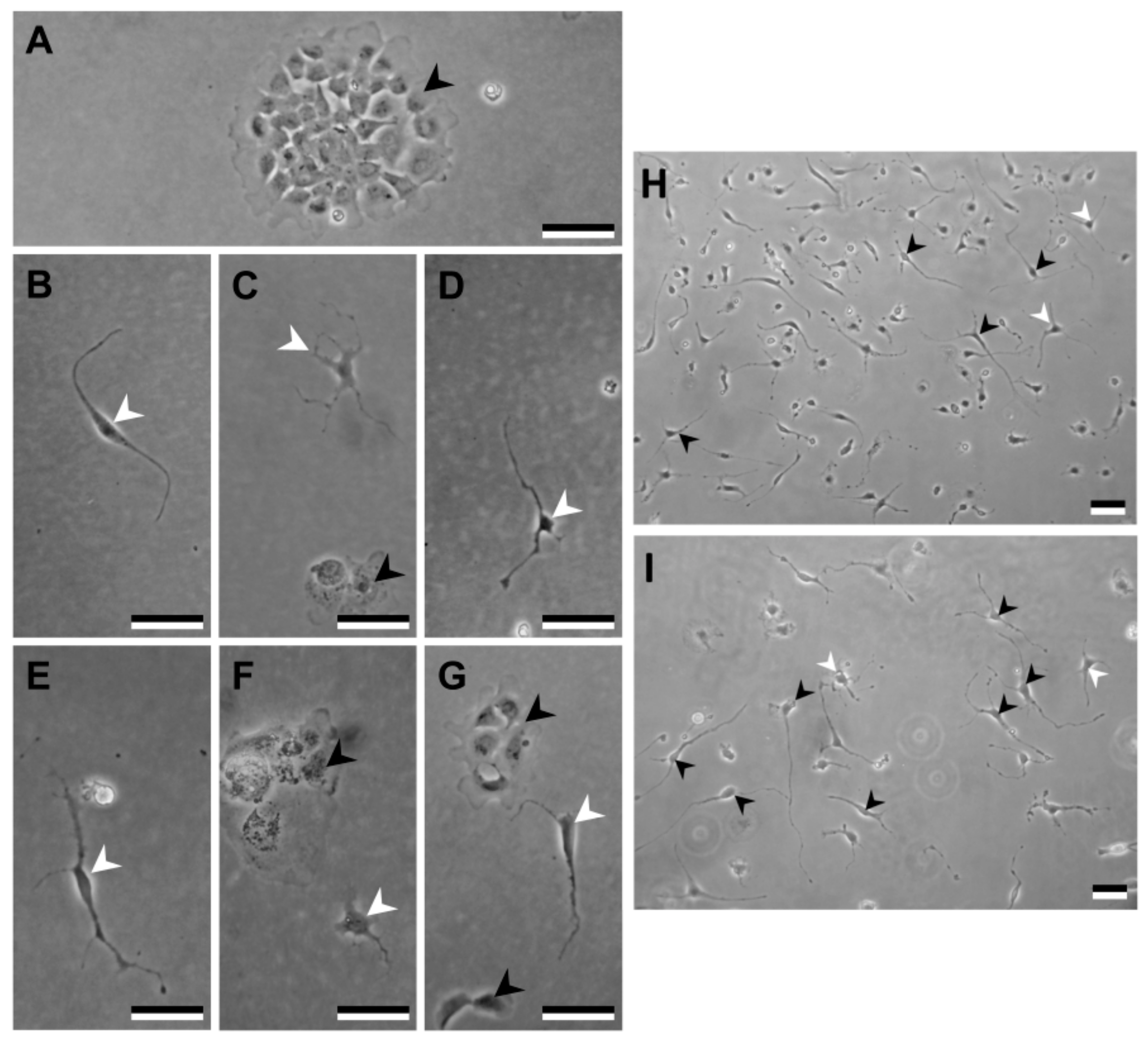
3.2. Primary Blastema Cells Isolation and Culture

The detailed protocol to isolate and obtain primary blastema cells is as follows:

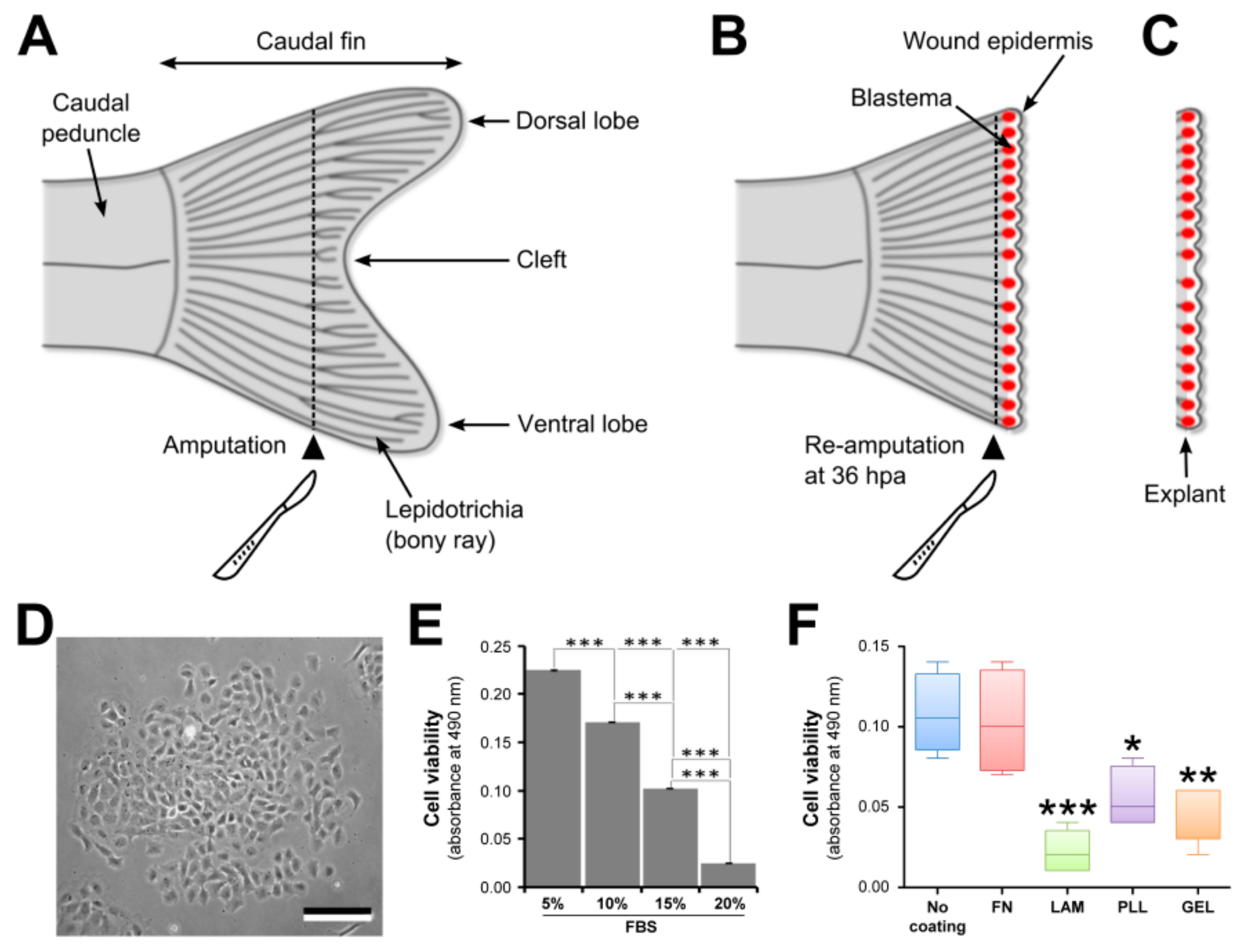
3.2.1. Amputation of the Caudal Fin

Clean the working area with 70% ethanol and autoclave all the dissection instruments.
  • Anesthetize the animals by exposing them to tricaine methanesulfonate (MS-222; 0.01% (w/v) in fish water; Sigma-Aldrich, St. Louis, MO, USA) for 4–6 min. Animals are anesthetized in batches of five fishes.
  • Using blunt-end forceps, transfer anesthetized fish onto an inverted Petri dish (sterile, bacterial) placed on the stage of a stereomicroscope (e.g., MZ6 from Leica Microsystems, Wetzlar, Germany) and carefully deploy and flatten the caudal fin.
  • Using a sterile scalpel (scalpel No. 4 and blade No. 24), amputate the caudal fin in two segments ahead of the cleft (see Figure 1A). Clean cut is achieved by positioning the blade tip on the Petri dish and by sectioning the fin in a single downward movement.
  • Transfer fish with amputated fins back to their aquarium or tank.
  • Repeat Steps 2–4 for the remaining animals.
  • Maintain the animals in normal culture conditions until re-amputation.

3.2.2. Blastema Collection

Clean the working area with 70% ethanol and autoclave all the dissection instruments; coat cell culture dishes with appropriate molecules according to manufacturer’s instructions.
7.
At 36 h post-amputation (hpa), repeat Steps 1 and 2 for all animals.
8.
Using a sterile scalpel, re-amputate the caudal fin one segment ahead of the first amputation plane (see Figure 1B). See Step 3 for making clean cuts.
9.
Using sharpened forceps, collect the fin fragment containing the whole blastema (see Figure 1C) and transfer it into a Petri dish containing 3 mL of Leibovitz’s L-15 culture medium (Invitrogen, Carlsbad, CA, USA) supplemented with 5× antibiotics (100× solution containing 10,000 units of penicillin, and 10,000 µg of streptomycin per mL; Invitrogen) and 1× fungizone (100× solution containing 250 µg of amphotericin B per mL; Invitrogen) and allow it to bath for 15 min to avoid bacterial contamination.
10.
Repeat Steps 8 and 9 for the remaining animals.
11.
Transfer the blastemas into a sterile 1.5-mL microcentrifuge tube containing 1 mL of collagenase solution (0.125% (w/v) in PBS; Invitrogen).
12.
Gently dissociate the blastemas by pipetting up and down (approximately 15 times using a 1000 µL micropipette) and place the tube in a rotary shaker for 10 min at room temperature (approximately 24–25 °C).
13.
Centrifuge the tube for 3 min at 972 × g at 25 °C and gently discard the supernatant.
14.
Add 0.5 mL of trypsin-EDTA solution (0.25% trypsin (Invitrogen) and 1.1 mM EDTA in phosphate-buffered saline (PBS, 1×, sterile); pH 7.4) and gently pipette up and down (using a 1000 µL micropipette) to resuspend the cells and then place the tube in a rotary shaker for 3–5 min at 25 °C.
Almost all the blastemas are dissociated during collagenase and trypsin treatment.
15.
Repeat Step 13 and gently resuspend the cells in 1 mL of L-15 medium.
16.
Repeat Step 13 and gently resuspend the cells in 1 mL of L-15 medium supplemented with 5% fetal bovine serum (FBS; Sigma-Aldrich), 1× antibiotics, and 0.2× fungizone.
17.
Count cells using a Neubauer counting chamber (BLAUBRAND) and seed 1.5 × 10³ cells per well in a 96-well dish (Nunc, Roskilde, Denmark) coated with laminin, gelatin, fibronectin, or poly-L-lysine and 1 × 10⁴ cells per well in a 24-well dish (Nunc) for cell culture.
18.
Incubate enzymatically dissociated cell cultures in a 28 °C cell incubator (e.g., Galaxy 170 S from New Brunswick Scientific, Enfield, CT, USA).

3.2.3. Maintenance of Primary Cell Cultures

19.
Observe daily the growth of primary cell cultures under an inverted microscope (e.g., Axiovert 25 from Zeiss, Oberkochen, Germany).
20.
Renew culture medium every 3 days.
21.
At confluence, cells are detached/dissociated with trypsin-EDTA and then split 1:2.

3.3. ECM Coating of Tissue Culture Dishes

Stock solution of poly-L-lysine at 0.1 mg/mL was prepared using sterile water. Then, 100–200 µL of this solution was aseptically added in a well of 24-well plate, rocked gently for 5 min to ensure a homogenous coating of the well. The solution was removed and wells were rinsed thoroughly with sterile water and allowed to dry for 2 h in the biological safety cabinet. The same procedure was followed for laminin (0.1 mg/mL) and gelatin (1 mg/mL) coated plates although incubation was carried out at 37 °C and washes done with PBS. For fibronectin (0.05 mg/mL), plates were air dried for 1 h at 25 °C, then excess of fibronectin was removed and the culture dish rinsed with PBS.

3.4. Cell Viability

Viability of blastema cells was subsequently determined in media supplemented with different FBS concentrations and in dishes coated with different extracellular matrix (ECM) molecules i.e., fibronectin, laminin, poly-L-lysine, and gelatin (all from Sigma-Aldrich) using XTT Cell Proliferation kit (AppliChem, A8088) according to manufacturer’s instructions. Briefly, the cells were seeded at 1.5 × 10³ cells/well in 96-well plates. At appropriate times, cells were incubated for 3 h with 50 µL of reagent mixture in 100 µL medium then cell viability was determined from absorbance at 490 nm.

3.5. Extracellular Matrix Mineralization and Acridine Orange Staining

Blastema cells were seeded at 1.0 × 10⁵ cells/well in six-well plates. ECM mineralization was induced by supplementing the culture medium with a mineralogenic cocktail composed of 10 mM β-glycerophosphate, 4 mM calcium chloride, and 50 µg/mL of L-ascorbic acid. After 3 and 5 days of treatment, ECM mineralization was assessed through alizarin red S staining (AR-S, 40 mM at pH 4.2; Sigma-Aldrich) and nucleic acids were revealed through acridine orange staining (100 µg/mL in PBS). Stained cells were visualized under an inverted fluorescence light microscope.

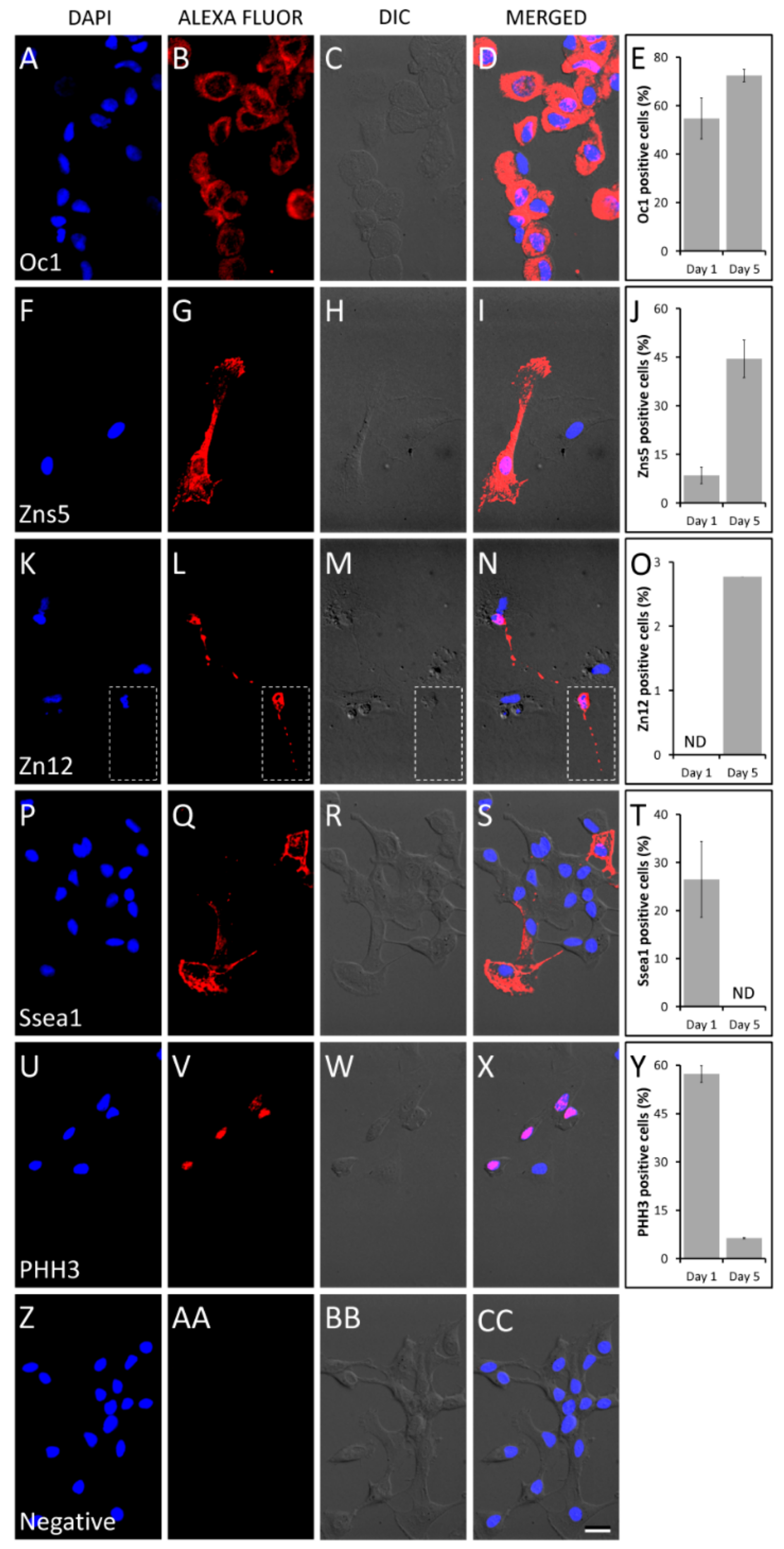
3.6. Cell Differentiation

Blastema cells were seeded at 1.5 × 10³ cells/well in 96-well plates. After 24 h, medium was renewed and supplemented with 0.5 µM all-trans retinoic acid (RA; Sigma-Aldrich, R2625) or with 0.1% dimethyl sulfoxide (DMSO; Sigma-Aldrich, D2650), the vehicle control. Morphological changes in cells were examined daily under an inverted microscope equipped with phase contrast.

3.7. Immunofluorescence Staining

For immunophenotyping, blastema cells grown on 12-mm glass coverslips (VWR, West Chester, PA, USA) for 1 day, and 5 days after seeding were briefly fixed for 10 min with 100% ice-cold methanol (Merck, Darmstadt, Germany), washed with PBS, permeabilized with 0.1% Triton X-100 (Sigma-Aldrich) for 5 min, and blocked in PBS containing 1% bovine serum albumin (BSA; VWR). Purified mouse monoclonal antibodies against Zns5 (ZIRC, 090511), Ssea1, and Zn-12 (Developmental Studies Hybridoma Bank, MC-480 and zn-12), and purified rabbit polyclonal antibodies against Oc1 [57] and PHH3 (Cell Signaling Technology, 9701), were diluted 1:200 in PBS with 1% BSA and directly added to the fixed cells at 25 °C for 2 h. Cells were washed with 1:10 dilution of blocking buffer in PBS then incubated with secondary antibodies Alexa Fluor 594 (goat anti-mouse IgG A-11005; Invitrogen) or Alexa Fluor 488 (goat anti-rabbit IgG A-11008; Invitrogen) diluted 1:1000 in the same solution as the primary antibody for 45 min at room temperature in the dark. Cells were washed thrice with PBS and then incubated with DAPI (Merck) for 5 min. The cells were again washed several times with PBS and mounted in microscope slides (VWR) using Mowiol (Sigma-Aldrich), then observed using an Axioimager Z2 fluorescence microscope equipped with an Axiocam camera (Zeiss).

4. Conclusions

In this article we described the protocol for the successful culture of blastema cells collected from regenerating caudal fin of zebrafish. Cultured blastema cells spontaneously differentiated into different cell types, in particular those related to bone formation. The availability of such in vitro cell system represents a promising tool for investigating mechanisms of blastema cell differentiation during fin regeneration.

Supplementary Materials

The following are available online at https://www.mdpi.com/2410-3888/5/1/6/s1. Figure S1. Phase-contrast (A,C,E) and fluorescence (B,D,F) micrographs of acridine orange stained blastema cells cultured for 24 h (A,B), 72 h (C,D) and 120 h (E,F) in medium supplemented with 5% FBS. Bar is 200 μm, Figure S2. Phase-contrast micrographs of alizarin red S-stained blastema cells treated for 3 days (A) and 5 days (B) with the mineralogenic cocktail to induce extracellular matrix mineralization.

Author Contributions

P.V., V.L. and M.L.C. designed the experiments. P.V. and V.L. carried out the experiments. P.V., V.L. and M.L.C. interpreted the data. All authors wrote the manuscript. All authors have read and agreed to the published version of the manuscript.

Funding

This work was funded by the Portuguese Science and Technology Foundation (FCT) through FISHCELL (PTDC/MAR/105313/2008) and AQUATOX (PTDC/MAR/112992/2009) research projects and by the European Community (EC) through ASSEMBLE (FP7/227799) research project. It was also funded by FCT through UID/Multi/04326/2019 project. P.V. was supported by a post-doctoral grant (SFRH/BPD/39189/2007) from the FCT.

Acknowledgments

We would like to thank the Department of Biomedical Sciences and Medicine at the University of Algarve for using the microscopy facility to acquire fluorescence images. We also thank Paulo J. Gavaia for his assistance in fish care.

Conflicts of Interest

The authors declare no competing or financial interests.

Compliance with Ethical Standards

Animal handling and experimentation was performed by qualified operators legally accredited by the Portuguese Direção Geral de Alimentação e Veterinária (DGAV) and following the EU and Portuguese legislation for animal experimentation and welfare (Directive 86/609/CEE and 2010/63/EU, Portaria 1005/92, 466/95 and 1131/97). Experimental procedures were performed under authorization (0421/000/000) from the DGAV, complying with the decreto de lei 113/2013 de 7 de Agosto, from the Portuguese legislation.

References

  1. Akimenko, M.-A.; Mari-Beffa, M.; Becerra, J.; Géraudie, J. Old questions, new tools, and some answers to the mystery of fin regeneration. Dev. Dyn. 2003, 226, 190–201. [Google Scholar] [CrossRef] [PubMed]
  2. Poss, K.D.; Keating, M.T.; Nechiporuk, A. Tales of regeneration in zebrafish. Dev. Dyn. 2003, 226, 202–210. [Google Scholar] [CrossRef] [PubMed]
  3. Nakatani, Y.; Kawakami, A.; Kudo, A. Cellular and molecular processes of regeneration, with special emphasis on fish fins. Dev. Growth Differ. 2007, 49, 145–154. [Google Scholar] [CrossRef] [PubMed]
  4. Yoshinari, N.; Kawakami, A. Mature and juvenile tissue models of regeneration in small fish species. Biol. Bull. 2011, 221, 62–78. [Google Scholar] [CrossRef] [PubMed] [Green Version]
  5. Gemberling, M.; Bailey, T.J.; Hyde, D.R.; Poss, K.D. The zebrafish as a model for complex tissue regeneration. Trends Genet. 2013, 29, 611–620. [Google Scholar] [CrossRef] [PubMed] [Green Version]
  6. Akimenko, M.-A.; Smith, A. Paired fin repair and regeneration. In Fins into Limbs: Evolution, Development, and Transformation; Hall, B.K., Ed.; University of Chicago Press: Chicago, IL, USA, 2007; pp. 152–162. [Google Scholar]
  7. Knof, F.; Hammond, C.; Chekuru, A.; Kurth, T.; Hans, S.; Weber, C.W.; Mahatama, G.; Fisher, S.; Brand, M.; Schulte-Merker, S.; et al. Bone regenerates via dedifferentiation of osteoblasts in the zebrafish fin. Dev. Cell 2011, 20, 713–724. [Google Scholar] [CrossRef] [Green Version]
  8. Sousa, S.; Afonso, N.; Bensimon-Brito, A.; Fonseca, M.; Simões, M.; Leon, J.; Roehl, H.; Cancela, M.L.; Jacinto, A. Differentiated skeletal cells contribute to blastema formation during zebrafish fin regeneration. Development 2011, 138, 3897–3905. [Google Scholar] [CrossRef] [Green Version]
  9. Tu, S.; Johnson, S.L. Fate restriction in the growing and regenerating zebrafish fin. Dev. Cell 2011, 20, 725–732. [Google Scholar] [CrossRef]
  10. Stoick-Cooper, C.L.; Moon, R.T.; Weidinger, G. Advances in signaling in vertebrate regeneration as a prelude to regenerative medicine. Genes Dev. 2007, 21, 1292–1315. [Google Scholar] [CrossRef] [Green Version]
  11. Wehner, D.; Weidinger, G. Signaling networks organizing regenerative growth of the zebrafish fin. Trends Genet. 2015, 31, 336–343. [Google Scholar] [CrossRef]
  12. Pfefferli, C.; Jaźwińska, A. The art of fin regeneration in zebrafish. Regeneration 2015, 19, 72–83. [Google Scholar] [CrossRef]
  13. Bergen, D.J.M.; Kague, E.; Hammond, C.L. Zebrafish as an emerging model for osteoporosis: A primary testing platform for screening new osteo-active compounds. Front. Endocrinol. 2019, 10, 6. [Google Scholar] [CrossRef] [Green Version]
  14. Whitehead, G.G.; Makino, S.; Lien, C.L.; Keating, M.T. fgf20 is essential for initiating zebrafish fin regeneration. Science 2005, 310, 1957–1960. [Google Scholar] [CrossRef] [PubMed] [Green Version]
  15. Lee, Y.; Hami, D.; De Val, S.; Kagermeier-Schenk, B.; Wills, A.A.; Black, B.L.; Weidinger, G.; Poss, K.D. Maintenance of blastemal proliferation by functionally diverse epidermis in regenerating zebrafish fins. Dev. Biol. 2009, 331, 270–280. [Google Scholar] [CrossRef] [PubMed] [Green Version]
  16. Yoshinari, N.; Ishida, T.; Kudo, A.; Kawakami, A. Gene expression and functional analysis of zebrafish larval fin fold regeneration. Dev. Biol. 2009, 325, 71–81. [Google Scholar] [CrossRef] [PubMed]
  17. Singh, S.P.; Holdway, J.E.; Poss, K.D. Regeneration of amputated zebrafish fin rays from de novo osteoblasts. Dev. Cell 2012, 22, 879–886. [Google Scholar] [CrossRef] [Green Version]
  18. Sander, V.; Suñe, G.; Jopling, C.; Morera, C.; Izpisua Belmonte, J.C. Isolation and in vitro culture of primary cardiomyocytes from adult zebrafish hearts. Nat. Protoc. 2013, 8, 800–809. [Google Scholar] [CrossRef]
  19. Tapanes-Castillo, A.; Shabazz, F.S.; Mboge, M.Y.; Vajn, K.; Oudega, M.; Plunkett, J.A. Characterization of a novel primary culture system of adult zebrafish brain stem cells. J. Neurosci. Methods 2014, 223, 11–19. [Google Scholar] [CrossRef]
  20. Chen, Z.; Lee, H.; Henle, S.J.; Cheever, T.R.; Ekker, S.C.; Henley, J.R. Primary neuron culture for nerve growth and axon guidance studies in zebrafish (Danio rerio). PLoS ONE 2013, 8, e57539. [Google Scholar] [CrossRef] [Green Version]
  21. Yan, C.H.; Chan, K.M. Characterization of zebrafish metallothionein gene promoter in a zebrafish caudal fin cell-line, SJD. 1. Mar. Environ. Res. 2002, 54, 335–339. [Google Scholar] [CrossRef] [PubMed]
  22. Kalaiselvi Sivalingam, N.N.; Seepoo, A.M.; Gani, T.; Selvam, S.; AzeezSait, S.H. Zebrafish fin-derived fibroblast cell line: A model for in vitro wound healing. J. Fish Dis. 2019, 42, 573–584. [Google Scholar] [CrossRef] [PubMed]
  23. Yadav, K.; Lakra, W.S.; Sharma, J.; Goswami, M.; Singh, A. Development and characterization of a cell line TTCF from endangered mahseer Tor tor (Ham.). Fish Physiol. Biochem. 2012, 38, 1035–1045. [Google Scholar] [CrossRef] [PubMed]
  24. Lakra, W.S.; Goswami, M. Development and characterization of a continuous cell line PSCF from Puntius sophore. J. Fish Biol. 2011, 78, 987–1001. [Google Scholar] [CrossRef]
  25. Kumar, R.; Ravi, C.; Das, S.; Dharmaratnam, A.; Basheer, V.S.; Swaminathan, T.R. Establishment and characterization of a caudal fin-derived cell line, AOF, from the Oscar, Astronotus ocellatus. Fish Physiol. Biochem. 2019, 45, 123–131. [Google Scholar] [CrossRef]
  26. Lakra, W.S.; Goswami, M.; Yadav, K.; Gopalakrishnan, A.; Patiyal, R.S.; Singh, M. Development and characterization of two cell lines PDF and PDH from Puntius denisonii (Day 1865). In Vitro Cell. Dev. Biol. Anim. 2011, 47, 89–94. [Google Scholar] [CrossRef] [PubMed] [Green Version]
  27. Kumar, A.; Godwin, J.W. Preparation and culture of limb blastema stem cells from regenerating larval and adult salamanders. Cold Spring Harb. Protoc. 2010, 2010, pdb–prot5367. [Google Scholar] [CrossRef]
  28. Rosa, J.; Tiago, D.M.; Dias, J.; Cancela, M.L.; Laizé, V. Serum-specific stimulation of proliferation and mineralization of fish bone-derived cells. J. Appl. Ichthyol. 2010, 26, 251–256. [Google Scholar] [CrossRef]
  29. Collodi, P.; Kamei, Y.; Ernst, T.; Miranda, C.; Buhler, D.R.; Barnes, D.W. Culture of cells from zebrafish (Brachydanio rerio) embryo and adult tissues. Cell Biol. Toxicol. 1992, 8, 43–61. [Google Scholar] [CrossRef]
  30. Van Camp, J.K.; Beckers, S.; Zegers, D.; Van Hul, W. Wnt signaling and the control of human stem cell fate. Stem Cell Rev. 2014, 10, 207–229. [Google Scholar] [CrossRef]
  31. Itoh, F.; Watabe, T.; Miyazono, K. Roles of TGF-β family signals in the fate determination of pluripotent stem cells. Semin. Cell Dev. Biol. 2014, 32, 98–106. [Google Scholar] [CrossRef]
  32. Wang, L.; Schulz, T.C.; Sherrer, E.S.; Dauphin, D.S.; Shin, S.; Nelson, A.M.; Ware, C.B.; Zhan, M.; Song, C.Z.; Chen, X.; et al. Self-renewal of human embryonic stem cells requires insulin-like growth factor-1 receptor and ERBB2 receptor signaling. Blood 2007, 110, 4111–4119. [Google Scholar] [CrossRef] [PubMed] [Green Version]
  33. Lotz, S.; Goderie, S.; Tokas, N.; Hirsch, S.E.; Ahmad, F.; Corneo, B.; Le, S.; Banerjee, A.; Kane, R.S.; Stern, J.H.; et al. Sustained levels of FGF2 maintain undifferentiated stem cell cultures with biweekly feeding. PLoS ONE 2013, 8, e56289. [Google Scholar] [CrossRef] [PubMed] [Green Version]
  34. Pease, S.; Braghetta, P.; Gearing, D.; Grail, D.; Williams, R.L. Isolation of embryonic stem (ES) cells in media supplemented with recombinant leukemia inhibitory factor (LIF). Dev. Biol. 1990, 141, 344–352. [Google Scholar] [CrossRef]
  35. Shibata, E.; Yokota, Y.; Horita, N.; Kudo, A.; Abe, G.; Kawakami, K.; Kawakami, A. Fgf signalling controls diverse aspects of fin regeneration. Development 2016, 143, 2920–2929. [Google Scholar] [CrossRef] [PubMed] [Green Version]
  36. Lehrberg, J.; Gardiner, D.M. Regulation of axolotl (Ambystoma mexicanum) limb blastema cell proliferation by nerves and BMP2 in organotypic slice culture. PLoS ONE 2015, 10, e0123186. [Google Scholar] [CrossRef] [Green Version]
  37. Suemori, H.; Nakatsuji, N. Establishment of the embryo-derived stem (ES) cell lines from mouse blastocysts: Effects of the feeder cell layer. Dev. Growth Differ. 1987, 29, 133–139. [Google Scholar] [CrossRef]
  38. Adams, J.C.; Watt, F.M. Regulation of development and differentiation by the extracellular matrix. Development 1993, 117, 1183–1198. [Google Scholar]
  39. Robertson, E.J. Embryo-derived stem cell lines. In Teratocarcinomas and Embryonic Stem Cells: A Practical Approach; Robertson, E.J., Ed.; IRL Press: Oxford, UK, 1987; pp. 71–112. [Google Scholar]
  40. Saeinasab, M.; Matin, M.M.; Rassouli, F.B.; Bahrami, A.R. Blastema cells derived from New Zealand white rabbit’s pinna carry stemness properties as shown by differentiation into insulin producing, neural, and osteogenic lineages representing three embryonic germ layers. Cytotechnology 2016, 68, 497–507. [Google Scholar] [CrossRef] [Green Version]
  41. Andrews, P.W. Retinoic acid induces neuronal differentiation of a cloned human embryonal carcinoma cell line in vitro. Dev. Biol. 1984, 103, 285–293. [Google Scholar] [CrossRef]
  42. Wakamatsu, Y.; Ozato, K.; Sasado, T. Establishment of a pluripotent cell line derived from a medaka (Oryzias latipes) blastula embryo. Mol. Mar. Biol. Biotechnol. 1994, 3, 185–191. [Google Scholar]
  43. Parameswaran, V.; Laizé, V.; Gavaia, P.J.; Cancela, M.L. ESSA1 embryonic stem like cells from gilthead seabream: A new tool to study mesenchymal cell lineage differentiation in fish. Differentiation 2012, 84, 240–251. [Google Scholar] [CrossRef] [PubMed]
  44. White, J.A.; Boffa, M.B.; Jones, B.; Petkovich, M. A zebrafish retinoic acid receptor expressed in the regenerating caudal fin. Development 1994, 120, 1861–1872. [Google Scholar] [PubMed]
  45. Means, A.L.; Gudas, L.J. The roles of retinoids in vertebrate development. Annu. Rev. Biochem. 1995, 64, 201–233. [Google Scholar] [CrossRef] [PubMed]
  46. Mongan, N.P.; Gudas, L.J. Diverse actions of retinoid receptors in cancer prevention and treatment. Differentiation 2007, 75, 853–870. [Google Scholar] [CrossRef] [PubMed]
  47. Soprano, D.R.; Teets, B.W.; Soprano, K.J. Role of retinoic acid in the differentiation of embryonal carcinoma and embryonic stem cells. Vitam. Horm. 2007, 75, 69–95. [Google Scholar] [CrossRef] [PubMed]
  48. Gudas, L.J.; Wagner, J.A. Retinoids regulate stem cell differentiation. J. Cell. Physiol. 2011, 226, 322–330. [Google Scholar] [CrossRef] [Green Version]
  49. Gudas, L.J. Emerging roles for retinoids in regeneration and differentiation in normal and disease states. Biochim. Biophys. Acta 2012, 1821, 213–221. [Google Scholar] [CrossRef] [Green Version]
  50. Mathew, L.K.; Sengupta, S.; Franzosa, J.A.; Perry, J.; La Du, J.; Andreasen, E.A.; Tanguay, R.L. Comparative expression profiling reveals an essential role for Raldh2 in epimorphic regeneration. J. Biol. Chem. 2009, 284, 33642–33653. [Google Scholar] [CrossRef] [Green Version]
  51. Blum, N.; Begemann, G. Retinoic acid signaling controls the formation, proliferation and survival of the blastema during adult zebrafish fin regeneration. Development 2012, 139, 107–116. [Google Scholar] [CrossRef] [Green Version]
  52. Géraudie, J.; Monnot, M.J.; Brulfert, A.; Ferretti, P. Caudal fin regeneration in wild type and long-fin mutant zebrafish is affected by retinoic acid. Int. J. Dev. Biol. 1995, 39, 373–381. [Google Scholar]
  53. Cardeira, J.; Gavaia, P.J.; Fernández, I.; Cengiz, I.F.; Moreira-Silva, J.; Oliveira, J.M.; Reis, R.L.; Leonor Cancela, M.; Laizé, V. Quantitative assessment of the regenerative and mineralogenic performances of the zebrafish caudal fin. Sci. Rep. 2016, 6, 39191. [Google Scholar] [CrossRef] [PubMed]
  54. Tighe, A.P.; Gudas, L.J. Retinoic acid inhibits leukemia inhibitory factor signaling pathways in mouse embryonic stem cells. J. Cell. Physiol. 2004, 198, 223–229. [Google Scholar] [CrossRef] [PubMed]
  55. Xing, J.G.; Lee, L.E.; Fan, L.; Collodi, P.; Holt, S.E.; Bols, N.C. Initiation of a zebrafish blastula cell line on rainbow trout stromal cells and subsequent development under feeder-free conditions into a cell line, ZEB2J. Zebrafish 2008, 5, 49–63. [Google Scholar] [CrossRef] [PubMed]
  56. Robles, V.; Marti, M.; Izpisua Belmonte, J.C. Study of pluripotency markers in zebrafish embryos and transient embryonic stem cell cultures. Zebrafish 2011, 8, 57–63. [Google Scholar] [CrossRef] [Green Version]
  57. Simes, D.C.; Williamson, M.K.; Schaff, B.J.; Gavaia, P.J.; Ingleton, P.M.; Price, P.A.; Cancela, M.L. Characterization of osteocalcin (BGP) and matrix gla protein (MGP) fish specific antibodies: Validation for immunodetection studies in lower vertebrates. Calcif. Tissue Int. 2004, 74, 170–180. [Google Scholar] [CrossRef]
Figure 1. Schematic diagram for caudal fin amputation, blastema collection, and blastema cell culture. (A) Amputation of the caudal fin; (B,C) re-amputation procedures towards blastema collection. Dotted lines indicate amputation and re-amputation planes. Blastema is indicated in red; (D) phase-contrast micrographs of zebrafish blastema cell cultures at 72 h post-seeding; (E) viability of blastema cells cultured in medium supplemented with different concentrations of FBS; (F) viability of blastema cells cultured on surfaces coated with different molecules. In (E,F), cell viability was measured 72 h post-seeding and asterisks indicate values significantly different through one-way ANOVA followed by Tukey’s (E) or Dunnett’s (F) multiple comparison test (*** p < 0.001, ** p < 0.01, and * p < 0.05; n = 6). FN, fibronectin; LAM, laminin; PLL, poly-L-lysine; GEL, gelatin. Scale bar in (D) is 100 µm.
Fishes 05 00006 g001
Figure 2. Spontaneous differentiation of zebrafish blastema cells after 5 days in culture. (A) Homogenous progenitor blastema cells showing polygonal cells with prominent nuclei and sparse cytoplasm. (BG) Differentiated cells exhibiting a spindle-like (B), an oligodendritic-like (C), and a neuron-like (DG) phenotype. Black and white arrowheads show progenitor blastema cells and differentiated cells, respectively. (H,I) Phase-contrast micrographs of zebrafish blastema cells exposed to 0.5 µm all-trans retinoic acid at day 7. Neuron-like (black arrowheads) and oligodendritic-like (white arrowheads) cells at 7 days of treatment. Scale bar is 50 µm in (AG) and (I), and 100 µm in (H).
Fishes 05 00006 g002
Figure 3. Differentiation of zebrafish blastema cells confirmed by immunofluorescence labeling at day 5 (except for Ssea1 at day 1). Zebrafish blastema cells immunolabeled with the osteoblast marker Osteocalcin 1/Oc1 (AD), the scleroblast marker Zns5 (FI), the neuronal cell surface marker Zn12 (KN; inserts illustrate a neuron-like cell), the stage-specific embryonic antigen Ssea1 (PS), and the proliferation marker PHH3 (UX). For each marker, picture sequence is as follows: DAPI labeling, ALEXA Fluor 594 labeling, differential interference contrast (DIC) image and merge image; percentages of cells positive for each marker at days 1 and 5 of cell culture were determined from manual counting and are presented as mean ± standard deviation calculated from three different coverslips (E,J,O,T,Y). Negative control (ZCC). ND, not detected. Scale bar is 10 µm.
Fishes 05 00006 g003

