Article

The Efficacy of Nile Tilapia (Oreochromis niloticus) Broodstock and Larval Immunization against Streptococcus agalactiae and Aeromonas hydrophila

1
Graduate School of Aquaculture Science, Bogor Agricultural University, Dramaga Campus, Bogor 16680, West Java, Indonesia
2
Department of Aquaculture, Faculty of Fisheries and Marine Science, Bogor Agricultural University, Dramaga Campus, Bogor 16680, West Java, Indonesia
*
Author to whom correspondence should be addressed.
Fishes 2018, 3(1), 16; https://doi.org/10.3390/fishes3010016
Submission received: 25 December 2017 / Revised: 19 February 2018 / Accepted: 19 February 2018 / Published: 7 March 2018

Abstract

:
Streptococcus agalactiae and Aeromonas hydrophila have been recognized as the causative agents of mortality in tilapia larvae with single infection and coinfection. The objective of this study was to evaluate the efficacy of maternal transfer and offspring protection from the immunization of monovalent and bivalent vaccines on Nile tilapia (Oreochromis niloticus) broodstock and larval immunization. Four groups of broodstock were intraperitoneally injected with formalin killed whole-cells of S. agalactiae (Sa group), A. hydrophila (Ah group), the bivalent mixed vaccine of them (Biv group), and phosphate-buffered saline as a control (Pbs group). Immunization of the larvae produced from immunized broodstock with a bivalent vaccine (Biv1 group) and Pbs (Pbs1 group) was performed by immersion at 20 days after hatch. Larvae produced from the Pbs group were unvaccinated as the control (Pbs2 group). Changes in the specific antibody and relative percent survival were measured. The Sa and Ah groups that could increase specific antibodies and protection against pathogenic bacteria were challenged with the homologous bacteria. The Biv group stimulated and protected against both S. agalactiae and A. hydrophila. The specific antibody of the Biv1 group was higher than the Pbs1 and Pbs2 groups. The last observation in this study showed that the relative percent survival of the Biv group after challenged S. agalactiae, A. hydrophila, and coinfection were 74.74 ± 3.18%, 73.81 ± 8.58%, and 71.48 ± 5.70%, respectively. The use of bivalent vaccines on the broodstock and larvae may be a strategy to reduce mortality in Nile tilapia larvae caused by single pathogen infection of S. agalactiae and A. hydrophila, or coinfection with both S. agalactiae and A. hydrophila.

1. Introduction

Nile tilapia (Oreochromis niloticus) is one of the important fish aquacultures worldwide, with a production above 3.9 million tons in 2015 [1]. The development of intensive aquaculture has led to the emergence of various bacterial diseases [2]. Streptococcus agalactiae is a gram-positive bacteria, the causative agent of streptococcosis in Nile tilapia [3,4]. Outbreaks of S. agalactiae infection in tilapia have been reported in several countries around the world [5,6,7,8,9,10,11]. Another bacteria known to have a devastating effect on the survival rate in tilapia farms is Aeromonas hydrophila [12,13]. A. hydrophila is a gram-negative, free-living bacteria, opportunistic pathogen and the causative agent of motile aeromonad septicemia [14].
Fish mortality in aquaculture is likely not only caused by a single pathogen, but probably due to multiple pathogen infections [15]. Morbidity and mortality in fish due to bacterial coinfection have been reported to occur in tilapia, such as coinfection of Aeromonas veronii and Flavobacterium columnare [15], Streptococcus agalactiae and Francisella noatunensis [16], and Francisella noatunensis subsp. orientalis and Shewanella putrefaciens [17]. Recently, outbreaks in Indonesia have been reported, caused by coinfection of S. agalactiae and A. hydrophila [18,19].
The high mortality during early stages of development often occurs in fish due to early stages of fish that have a limited immune ability [20,21,22,23]. The tymus is an important organ for producing lymphocyte-T after fertilization in tilapia, a primordial thymus is formed at two days post fertilization (2 dpf), and then recombination activating gene 1 (Rag-1) is exhibited at 4 dpf, and differentiation of the medulla and cortex thymus is observed at 6–20 days post hatch (dph) [24] . Takamura et al. [25] showed that it is possible that new Immunoglobulin-M (IgM) is formed at 14 days of larvae. However, the literature does not provide the exact immunocompetent age of tilapia, generally stating the immunocompetent age of tilapia at 21 days [26]. This pause of about 20 days larvae is causing the limitations of larval immunity.
The health of offspring from the broodstock depends upon the health, as well as the immune status, of the broodstock [22,27]. Transfer of maternal immunity is one of the alternatives to increase immunity in offspring and enhance the survival rate of offspring [22,27]. Currently, the immunization of broodstock with monovalent vaccines has reported transfer immunity to the offspring such as in turbot [23], sea bream [28,29], zebrafish [30,31], catfish [32], and tilapia [33,34,35]. Immunization of the broodstock with an S. agalactiae monovalent vaccine followed by S. agalactiae challenge, and immunization with A. hydrophila vaccine followed by A. hydrophila challenge, could enhance immunity and protection to the offspring produced from immunized broodstock [31,32]. Several studies have also reported that immune factors can be transferred from broodstock to offspring through antibodies, lysozymes, protease inhibitors, and complement factors [27].
Maternal transfer of immunity by broodstock immunization studies has mostly targeted single pathogen protection. To date, no study has reported on broodstock immunization to improve maternal immunity and offspring protection with a simultaneous effect on coinfection caused by S. agalactiae and A. hydrophila. This study consists of two stages of immunization. The first stage of immunization consisted of four groups of immunized broodstock. Immunized broodstock were intraperitoneally injected with formalin killed whole-cells of S. agalactiae (Sa group), A. hydrophila (Ah group), the bivalent mixed vaccine of them (Biv group), and phosphate-buffered saline as a control (Pbs group). In the second stage, the larval were immunized with the bivalent vaccine produced from the broodstock immunized with the bivalent vaccine (Biv1), the larval immunization with the bivalent vaccine produced from the broodstock immunized with PBS (Pbs1), and the larval unimmunized production from the broodstock immunized with PBS (Pbs 2). This study aimed to examine how the efficacy of maternal transfer and the capability of monovalent killed whole-cells of S. agalactiae, A. hydrophila, and bivalent vaccine were challenged by a single pathogen of S. agalactiae and A. hydrophila, and coinfection of S. agalactiae and A. hydrophila. Then, the efficacy of the bivalent vaccine on larva from immunized broodstock was measured.

2. Results

2.1. The First-Stage

2.1.1. Efficacy of Broodstock Immunization

Immunization efficacy was evaluated by using the immersion challenge model aimed at evaluating the relative percent survival (RPS) of larvae produced from immunized broodstock. The cumulative mortality obtained for offspring produced from the four groups of broodstock immunization were challenged with S. agalactiae, A. hydrophila, and coinfection with both S. agalactiae and A. hydrophila at 5, 10, 15, and 20 days after hatching (the cumulative mortalities are detailed in Table S1A–D in the Supplementary Materials). The RPS of the offspring produced from broodstock immunized with the S. agalactiae vaccine and broodstock immunized with the bivalent vaccine showed no significant difference following the S. agalactiae challenge (p > 0.05). The offspring produced from broodstock immunized with the A. hydrophila vaccine and broodstock immunized with the bivalent vaccine was not significant when challenging A. hydrophila at the same stage (p > 0.05). The RPS in larvae produced from broodstock immunized with S. agalactiae vaccine became infected with A. hydrophila and larvae produced from broodstock immunized with the A. hydrophila vaccine following S. agalactiae challenge had failing protection. The monovalent vaccination only protected homologous bacteria; however, the immunization of broodstock with the bivalent vaccine induced stronger protection against coinfection (p < 0.05) when compared to immunizing the broodstock with the monovalent S. agalactiae and A. hydrophila vaccine. The relative percent survival of broodstock immunized with the S. agalactiae, A. hydrophila, and bivalent vaccines are shown in Table 1.

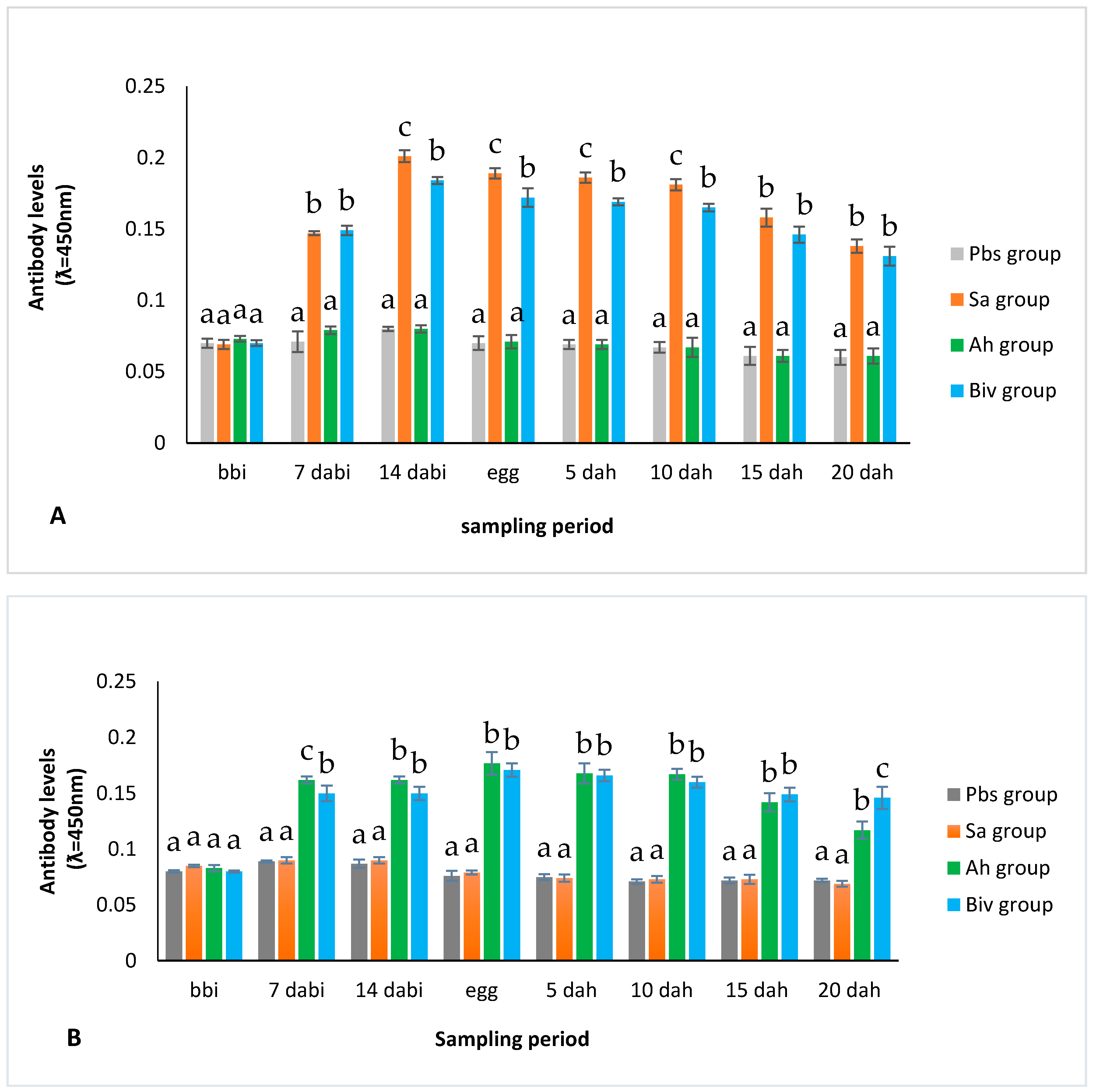
2.1.2. Antibody Titer of Immunized Broodstock

The antibody titer of broodstock serum, egg, and larva homogenates were assessed using the indirect enzyme-linked immunosorbent assay (ELISA) on each specific S. agalactiae and A. hydrophila antigen. After the broodstock was immunized, the antibody level from broodstock immunized with the S. agalactiae vaccine showed significantly different higher values than the control. Furthermore, the broodstock with a bivalent vaccine (which was significantly different from control) was tested with the S. agalactiae antigen (p < 0.05), as shown in Figure 1A. The same result was also seen from the immunized broodstock with A. hydrophila and the bivalent vaccine, which showed significantly different higher values when compared to the control against the A. hydrophila antigen (p < 0.05), as shown in Figure 1B. The detectable antibodies in the treatment group of the offspring from the immunized broodstocks were similar to the controls when tested for different antigens (e.g., the Sa group against the A. hydrophila antigens was not significantly different from the Pbs group). However, immunization with the bivalent vaccine produced antibodies when tested on the S. agalactiae and A. hydrophila antigens, respectively (Figure 1A,B).

2.2. Second Stage

2.2.1. Efficacy of Immunization of Larvae Production from Immunization Broodstock

The results from this study showed that the RPS of the larval immunized from the broodstock immunized with the bivalent vaccine was higher (p < 0.05) when compared to the larval immunized with the bivalent vaccine from the broodstock immunized with Pbs against S. agalactiae, A. hydrophila, and coinfection. The final RPS of the observation larval immunized with the bivalent vaccine produced from the broodstock immunized that was challenged with S. agalactiae, A. hydrophilia, and coinfection was 74.74 ± 3.18%, 73.81 ± 8.58%, and 71.48 ± 5.70%, respectively. The RPS of tilapia larvae is presented in Table 2. (The cumulative mortalities are detailed in Table S2A,B in the Supplementary Materials).

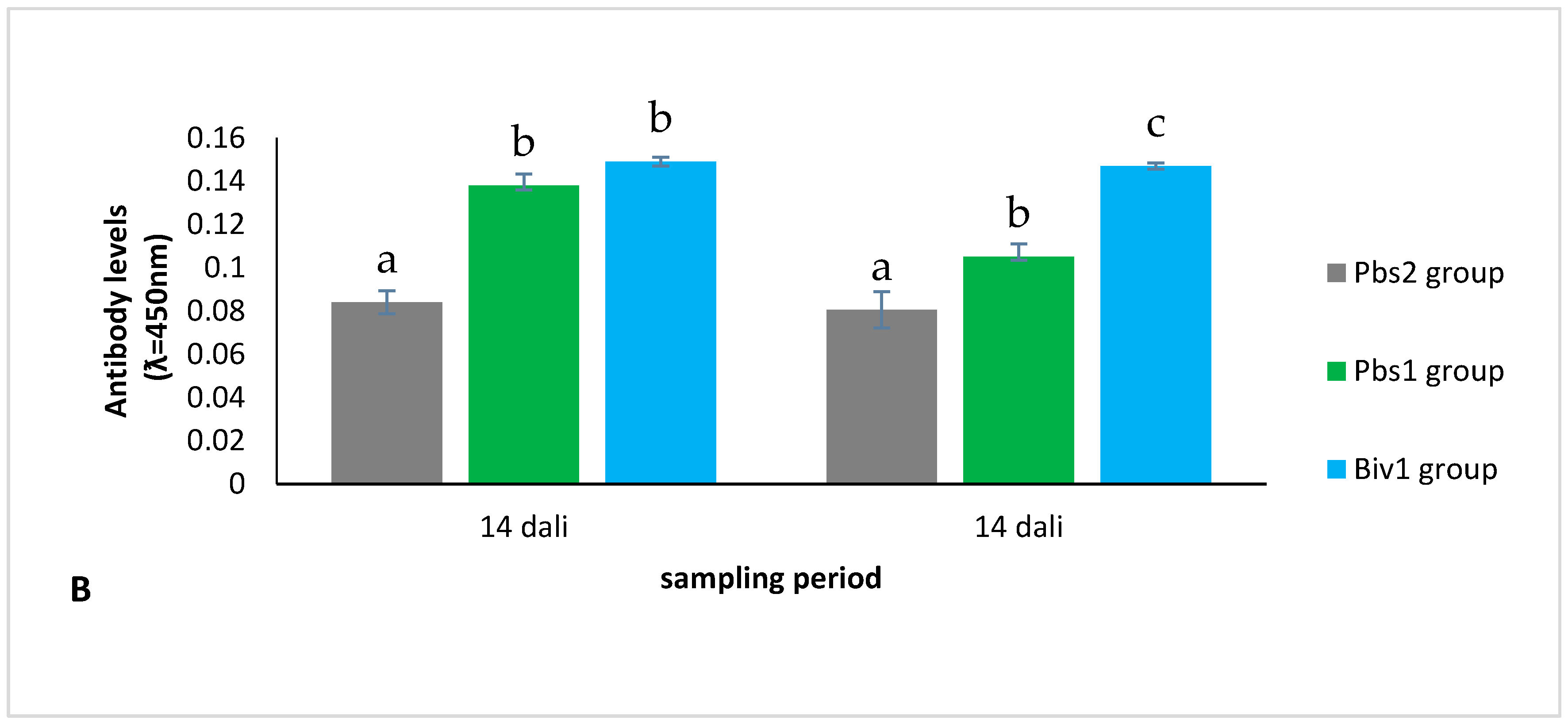
2.2.2. Antibody Level of Immunization of Larvae Production from Immunization Broodstock

At 14 and 21 days after the larvae were immunized, the antibody level of the immunized broodstock and immunized larvae were significantly different (p < 0.05) than the broodstock without immunization and the control tested with the S. agalactiae and A. hydrophila antigen coating. Levels of antibody for larvae within 14 and 21 days after the immunization of larvae production from broodstock immunization are shown in Figure 2A,B.

3. Discussion

An outbreak of S. agalactiae infection in tilapia was reported at the hatchery in Malaysia [5] and Cirata Lake in Indonesia [8]. Another bacterial pathogen that often infects tilapia is A. hydrophila [12,13]. Currently, coinfection with both S. agalactiae and A. hydrophila has been reported in Nile tilapia in Indonesia [18,19,36]. Fish mortality often occurs at the larval stage as the ability of fish immunity in early life is still limited [20,21,22,23]. The development of the tilapia immune system begins at two days of age, and the medulla and cortex thymus organs are differentiated by 20 days of age [24] and are believed to be immunocompetent at 21 days of age [26]. The early stage is still dependent on maternal immunity and one way to increase the protection of larvae against the disease is by immunizing the broodstock with the purpose of transferring maternal immunity to the offspring [22,23,24,27,28,29,30,31,32,33,34,35].
Immunizations to the broodstock using a monovalent whole cell vaccine of S. agalactiae, A. hydrophila, and the bivalent mixture challenged with S. agalactiae, A. hydrophila, and the combination of them (coinfection) were performed in this study. Efficacy vaccination was evaluated by using a challenge model aimed at evaluating the RPS [37]. The results showed that the larvae RPS from the immunization of the monovalent vaccine to the broodstock did not provide cross-protection (e.g., larvae produced from broodstock immunized with S. agalactiae vaccine appeared to fail to protect larvae against A. hydrophila and coinfection. In addition, immunization broodstock with the A. hydrophila vaccine had lower protection against S. agalactiae and coinfection) (Table 1). The results of this study demonstrated that the immunization of broodstock with the S. agalactiae or A. hydrophila monovalent vaccine was not sufficient for the protection of different bacteria or coinfection with both S. agalactiae and A. hydrophila. According to Sugiani et al. [18] and Sumiati et al. [19], broodstock immunized with S. agalactiae and A. hydrophila monovalent vaccines did not conduct effective protection when challenged with different bacteria or coinfection. However, the broodstock immunized with the bivalent vaccine produced protection from both pathogens, similar to vaccine preparation. High larval protection resulting in a transfer of maternal immunity with the immunization of the monovalent vaccine to the broodstock has been widely reported, such as the immunization of fish broodstock with a Streptococcus iniae vaccine on Nile tilapia [33], the A. hydrophila vaccine [35], and the S. agalactiae vaccine [34,38]. The results of the bivalent immunization treatment on the broodstock protected the larvae when challenged to a single infection of S. agalactiae, A. hydrophila, and coinfection with both S. agalactiae and A. hydrophila. This result was one of the advantages of using the multivalent vaccine. Another advantage of the multivalent vaccine was the use of one dose containing many antigens to make the injection immunization more cost-effective and reduce fish stress [39]. The RPS of the maternal immunization treatment decreased until the larvae age of 20 days. The same result from the study by Nisaa et al. [34] showed that RPS larvae from broodstock immunization began to decline with the development of the larvae. Maternal immunity lasted up to the juvenile stage [22]. The study on bivalent immunization, which was used as maternal immunity, was the first study, so there is no comparative literature related to maternal immunity with the bivalent vaccine.
The RPS of bivalent broodstock immunized and immunized larvae with the bivalent vaccine were higher than the immunization treatment only at larval stage (Table 2). Sukenda et al. [40] reported that larvae immunization by immersion with a S. agalactiae vaccine was less fortunate in protecting the larvae against S. agalactiae. Unsuccessful larval immunization may be due to the administration of a vaccination when the immune system is not fully formed. This result is inversely related to the study when immunized larvae were produced from immunized broodstock. Broodstock immunization can probably improve the quality of offspring it produces. Cao et al [24] reported the larvae at 20 days of age already exist differentiation in the thymus organ. Nisaa [41] and Firdausi et al. [42] showed that the re-immunization of tilapia at 20 days after hatching increased larvae protection. The re-immunization treatment in fish has also been shown to improve fish survival [43,44]. Re-immunization treatment may also be used to restore protection when fish susceptibility to disease or re-immunization may take place several months after the first immunization [45].
The antibody titer measurement on the serum broodstock increased when compared to the broodstock before immunization. The antibody titer in the serum, egg homogenates, and larva homogenates after the immunization treatment of the monovalent and bivalent vaccine increased when compared to the control at the same time (Figure 1A,B). In line with this, Nisaa et al. [34,36] and Sukenda et al. [35] found that vaccinated broodstock treated with the S. agalactiae or A. hydrophila vaccine had increased antibodies to homologous antigen coatings. The antibody titer of the bivalent vaccine increased significantly when compared to the control when tested for both bacterial antigens (Figure 1A,B). Sugiani et al. [18] and Sumiati et al. [19] also reported that Nile tilapia immunized with the bivalent S. agalactiae vaccine and A. hydrophila vaccine could increase antibodies against both antigen preparations. The antibodies of egg and larvae homogenates from the broodstock immunization treatment with the monovalent vaccine, when tested with the S. agalactiae coating antigen, were higher than bivalent, but still significantly different from the treatment of the control (Figure 1A). As previously reported by Sugiani et al. [18] and Sumiati et al. [19], higher specific antibodies were obtained from homologous antigens than heterologous antigens. Immunized broodstock produced a serum antibody titer and the results on egg and larva homogenates were also directly proportional. This result may be because broodstock immunization with the bivalent vaccine produced higher antibodies which were then transferred to their offspring via eggs. A study on several species of fish has also shown the transfer of specific antibodies from the broodstock to offspring [20,21,22,23,30,31,32,33,34,35]. The mechanism of the transfer of specific antibody immunity, when immunized with the bivalent vaccine, is unknown. However, some research has suggested that the maternal immunity transfer occurs at the time of the vitellogenesis process. Vitellogenin is carried by the blood and absorbed by the oocyte, which later enlarges to a maximum size. This pathway of vitellogenesis was used for the transfer of specific and non-specific immunities into the yolk [22,24]. Increased antibodies are also often associated with an increase in relative-percent survival [38]. A study by Mingming et al. [23] also showed that the antibody level value would decrease after the egg stage. The antibody originating from the broodstock was usually gradually weakened during the yolk absorption process [27,28].
The antibody results from the broodstock and the vaccinated larvae with the bivalent vaccine were significantly higher than the control treatment and the immunization larvae with the bivalent vaccine without immunized broodstock. Sukenda et al. [40] reported that the immunization of larvae with immersion had not shown optimal protection against S. agalactiae infection. Swain et al. [46] indicated that early immunization in fish took about four weeks to improve the level of antibodies of Indian major carps. In tilapia, IgM formation has occurred at 14 days of age [25] and some believe the age of immunocompetent tilapia to be 21 days [26]. However, the results of this study showed that antibodies remained elevated after larval immunization by immersion from broodstock immunized with the bivalent vaccine (Figure 2). Until now, there has been no mechanism to increase antibody titer on larval immunization treatment from immunized broodstock. This result may be due to the improved larval quality production from the broodstock. Moreover, some studies on fish conducted by repeated exposure to antigens may increase antibodies [44,47]. Nisaa [41] and Firdausi et al. [42] demonstrated that larval immunization at 20 days after hatch with S. agalactiae vaccine production from broodstock S. agalactiae immunization could increase the protection of larvae against S. agalactiae. The results of this study demonstrated that antibody production had an aligned correlation with protection efficacy.

4. Materials and Methods

4.1. Broodstock

Nile tilapia (Oreochromis niloticus) used in this study were Nirvana tilapia obtained from the Seed Development Center Freshwater Fish, Wanayasa, West Java, Indonesia. Fish weighed 251.61 ± 19.18 g and were separated into males and females. The ratio of male and female fish was 1:3 and they were maintained in a 2.5 × 2.5 × 1 m3 with a continuous flow of water and air. Fish were fed with commercial feed PF1000 (Prima Feed, Surabaya, Indonesia) twice daily. The broodstock were kept at 28.1–29 °C, pH 6.85–7.86, dissolved oxygen 5.1–7.7 parts per million, and ammonia 0.04–0.10 mg/L.
The experimental procedures and treatment of fish in this study have been approved by the Indonesia local authorities (Animal Care and Use Committee, Bogor Agricultural University, Indonesia. No: 21/2014IPB).

4.2. Vaccine Preparation

Preparation of a whole-cell S. agalactiae vaccine has previously been described by [48]. S. agalactiae N3M isolate was obtained from the Center for Research and Development of Freshwater Aquaculture, Bogor, West Java, Indonesia. S. agalactiae was cultured in Brain Heart Infusion Broth (BHIB, HiMedia, Mumbai, India) with an incubation period of 72 h at 28–30 °C. Concentration was calculated using the total plate count method and showed 109 Colony Forming Unit (CFU)/mL. Formalin 0.5% was added to the medium and incubated for 24 h. The suspension was then centrifuged at 7000 rpm for 30 min at 4 °C. The pellets were washed twice with phosphate buffered saline (PBS) and centrifuged to remove the formalin residue. Finally, PBS, as much as the initial volume of bacterial culture, was added. Vaccine suspension was cultured on brain heart infusion (BHI) agar and incubated for 72 h to ensure that S. agalactiae was killed and there was no contamination.
A. hydrophila was obtained from the Fish Health Laboratory, Bogor Agricultural University, West Java, Indonesia. The whole-cell A. hydrophila vaccine was made using modified Sukenda et al. [35]. A. hydrophila was cultured in Tryptic Soy Broth (TSB, Merck, Germany), then incubated with a shaker for 24 h at 29–30 °C. Bacterial culture was inactivated by adding 0.5% formalin and then incubated for 24 h at 29–30 °C. The final density of bacteria obtained before inactivation with formalin was 109 CFU/ mL. The inactive bacteria were obtained by centrifugation at 5000× g for 30 min. The pellets were washed twice with PBS and the bacteria sediment was re-suspended as much as the initial volume of bacterial culture. The vaccine viability was tested on tryptic soy agar and if bacteria did not grow within 24 h, the vaccine could be used. The bivalent vaccine was prepared with a mixture of both vaccine preparations with a vaccine formulation of whole-cell S. agalactiae and A. hydrophila.

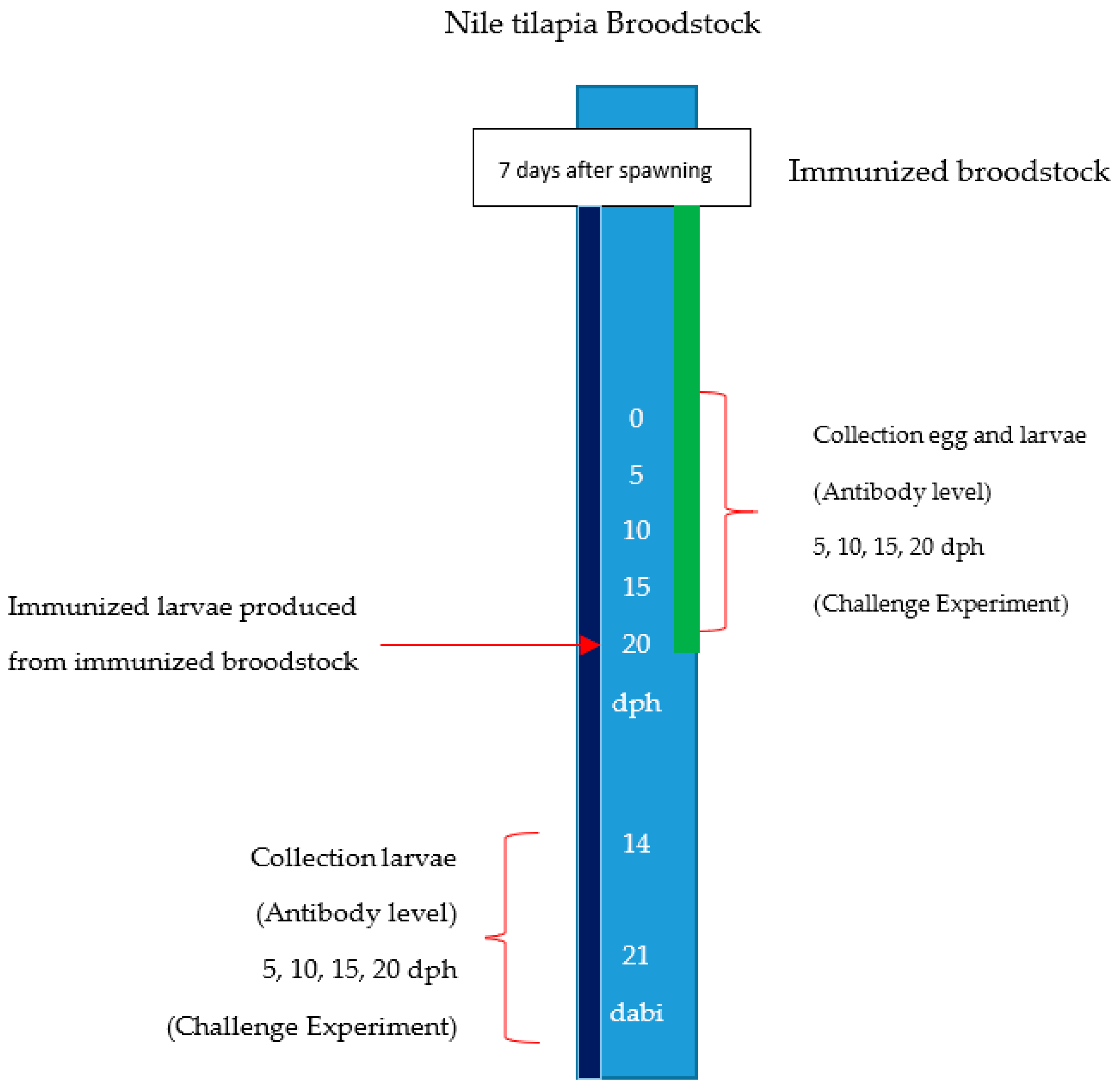
4.3. First Stage. Immunization of Broodstock

Forty-eight fish broodstock specimens were divided into four groups, with nine females and three males in each group. The broodstocks were anesthetized and the Sa group was immunized with the S. agalactiae vaccine; the broodstock Ah group was immunized with the A. hydrophila vaccine; and the broodstock Biv group was immunized with the bivalent vaccine (prepared with a mixture of both S. agalactiae of 75% and A. hydrophila of 25% (v/v)) [49]. The Pbs group was a control group where the broodstock was injected with PBS. Immunization of the broodstock was performed seven days after spawning [34,36]. Immunization was given by intraperitoneal injection with a dose of 0.4 mL/kg fish. The plan of immunization, a collection of eggs, larvae, and the challenge experiment are presented in Figure 3.

4.4. Challenge Experiment

The efficacy of the vaccine was obtained by conducting the test of immersion challenge to larvae produced from broodstock immunization. Triplicate groups of larvae were immersion infected with dilutions for single S. agalactiae (107 CFU/mL), single A. hydrophila (107 CFU/mL), and coinfection (106 CFU/mL of each bacteria (obtained from unpublished preliminary test results)). The challenge test was performed on the larvae at 5, 10, 15, and 20 days after hatch. Dead fish were removed from the container and bacteria were aseptically cultured on BHI agar and Rimler-Shotts (RS) medium from dead fish to confirm the S. agalactiae and A. hydrophila. The cumulative mortality from immunized and unimmunized fish for each experiment was determined over a 12-day period. The efficacy of the vaccine was calculated as a RPS [50]. Moribund/dead fish were removed from the container and bacteria were aseptically cultured on BHI agar and RS medium from dead fish to confirm the S. agalactiae and A. hydrophila. The cultured bacteria were identified by biochemical assays according to Bergey’s Manual of Determinative Bacteriology (Holt et al. 1994), then the Analytical Profile Index (API) 2’Strep system (bioMérieux, Craponne, France) and the API 2’E (bioMérieux, France) were used for more precise bacterial characterization. Isolated bacterium from BHI agar was identified as Streptococcus agalactiae and isolate bacteria from RS medium was identified as Aeromonas hydrophila.

4.5. Second Stage. Immunization of Larvae from Immunized Broodstock.

The larvae produced from the Biv group were immunized with the bivalent vaccine (Biv1 group), larvae produced from the Pbs group were unvaccinated as the control (Pbs2 group), and larvae produced from the other Pbs group were also immunized with the bivalent vaccine (Pbs 1 group). Immunization was performed at 20 days after hatching with immersion in 107 CFU/mL vaccine. Immunization was performed by immersing for 60 min in a vaccine solution with constant aeration and then the larvae were kept in clean water (no vaccine content) for another 10 min and then transferred to the maintenance aquarium. The Biv1, Pbs1, and Pbs2 groups were challenged against the pathogenic S. agalactiae, A. hydrophila, and coinfection at 14 days and 21 days after larvae immunization. The bacterial preparation was similar to that described in the challenge test treatment of larvae from the immunized broodstock (Section 4.4. Challenge Experiment). Larvae after infection were kept in aquaria and fed twice a day. Death was recorded for 12 days and the efficacy of the vaccine was calculated as a RPS [50]. Moribund/dead fish were removed from the container and bacteria were aseptically cultured on BHI agar and RS medium from dead fish to confirm the S. agalactiae and A. hydrophila. Isolates from BHI agar and RS medium were identified by biochemical assays according to Bergey’s Manual of Determinative Bacteriology (Holt et al. 1994), and then the API 2’Strep system (bioMérieux, France) and the API 2’E (bioMérieux, France) were used for more precise bacterial characterization. Isolates were identified as Streptococcus agalactiae and Aeromonas hydrophila.

4.6. Serum Preparation

Serum sampling was performed according to [28] with slight modification. Sampling was carried out at zero (before immunization broodstock), seven, and 14 days after broodstock immunization. The fish were anesthetized and we collected non-anticoagulated blood from three broodstocks for each treatment. Blood was allowed to clot at room temperature for 3 h and then settled at 4 °C (overnight). Serum was collected after centrifugation at 5000 rpm for 10 min, divided into aliquots. and stored at −20 °C.

4.7. Preparation of Eggs and Larvae Homogenates

The sampling of eggs and larvae homogenates was carried out according to [28]. The egg samples were collected from three broodstocks of the broodstock immunization treatment groups. The larvae were collected at 5, 10, 15, and 20 days after hatching, and 14 and 21 days after larval immunization. Each egg and larvae sample was homogenized in PBS-T (PBS + 0.05% Tween-20) solution at a ratio of 1:4. Furthermore, it was centrifuged at 5000 rpm for 10 min. An aliquot of all samples was stored at –20 °C for further use to determine the antibody level.

4.8. Antibody Level

The antibody titer was measured using an indirect ELISA method described by [51] and [19] with a slight modification. The antigens used were S. agalactiae and A. hydrophila, which were prepared by sonification. The total proteins of sonicated S. agalactiae and A. hydrophila antigens were determined by the Bradford [52] and adjusted to a concentration of 10 μg/mL protein using the carbonate-bicarbonate buffer (pH 9.6). A 100 μL antigen suspension of S. agalactiae and A. hydrophila was then added to a different microtiter plate with 96 wells of 100 μL (TPP Techno Plastic Products AG 9209, Trasadingen, Switzerland) and incubated at 4 °C overnight. After incubation, the microtiter plates were then washed three times using PBS-T (PBS pH 7.4 + 0.05% Tween-20 Sigma, Darmstadt, Germany), and the wells were blocked with 3% bovine serum albumin (BSA, Sigma). The microplate was incubated at 25 °C for 1 h, and then the plates were washed three times with PBS-T. Serum samples from broodstock Nile tilapia were diluted at a ratio of 1:50 on PBS-T, while the eggs and larvae were diluted at a ratio of 1:16 on PBS-T, and the yield suspension was then added to three microtiter plate replication wells of 100 μL, followed by incubation at 25 °C for 1 h. The plates were washed with PBS-T. The long-chain anti-Nile Ig (obtained from Nisaa et al. [34]) for polyclonal specific antibodies was diluted at a ratio of 1:200 (v/v) in PBS-T, and 100 μL from the resulting solution was added to each microtiter plate well and incubated at 25 °C for 1 h, before being washed using PBS-T. Anti-Rabbit IgG (whole molecule)-Peroxidase antibody produced in goat (Sigma) was diluted at a ratio of 1:15,000 on PBS-T and a solution of 100 μL was added to each microtiter plate well. The microtiter plate was incubated at 25 °C for 1 h, then washed. One-Step Ultra TMB-ELISA (100 μL) (Sigma) was added to the microtiter well plate. After 20 min, the ELISA reaction was then discontinued by adding 50 μL H2SO4 3 M into each well of the microplates. Optical density readings were then performed using a Microplate Reader (Rayto RT-2100C, Shenzhen, China) at 450 nm.

4.9. Statistical Analysis

All the data were presented as means ± standard error (M ± SE) from three replicates. The statistical analysis of RPS was performed by comparing the immunization group treatment with the challenge test on the same pathogenic bacteria and the same stage. RPS, antibody levels from the first stage, and antibody levels from the second stage were tested using one-way analysis of variance with a Duncan’s test. RPS from the second stage were tested using an independent sample T-test. Significant differences were determined at p < 0.05 ± standard error. The statistical analysis used SPSS Software (version 24.0 for Windows, SPSS Inc., Chicago, IL, USA).

5. Conclusions

This study concluded that immunization broodstock specimens using a monovalent vaccine of S. agalactiae and A. hydrophila were able to increase the serum antibody of the broodstocks, egg homogenates, and larvae homogenates when tested with homologous bacteria. Immunization of broodstock using the bivalent vaccine was able to increase specific antibody titers when tested with the S. agalactiae and A. hydrophila antigens. The larvae production from immunization broodstock using monovalent vaccines was only able to protect a single infection of pathogenic bacteria similar to vaccine preparations. The larvae production from immunization broodstock using bivalent vaccines was capable of protecting the larvae against single infections, as well as coinfection with S. agalactiae and A. hydrophila. Immunization with the bivalent vaccine to larvae produced from broodstock immunized with a bivalent vaccine could increase the antibody titer and protect against S. agalactiae and A. hydrophila, as well as coinfection with both S. agalactiae and A. hydrophila.

Supplementary Materials

The supplementary table can be found at https://www.mdpi.com/2410-3888/3/1/16/s1. Table S1A. The cumulative mortalities from Sa, Ah and Biv group after challenged with S. agalactiae, A. hydrophila and coinfection at larvae aged 5 days after hatch. Table S1B. The cumulative mortalities from Sa, Ah and Biv group after challenged with S. agalactiae, A. hydrophila and coinfection at larvae aged 10 days after hatch. Table S1C. The cumulative mortalities from Sa, Ah and Biv group after challenged with S. agalactiae, A. hydrophila and coinfection at larvae aged 15 days after hatch. Table S1C. The cumulative mortalities from Sa, Ah and Biv group after challenged with S. agalactiae, A. hydrophila and coinfection at larvae aged 15 days after hatch. Table S2A. The dead fish and cumulative mortalities of tilapia larvae Biv1 and Pbs1 were challenged S. agalactiae, A. hydrophila and coinfection at larvae 14 days after larvae immunization. Table S2B. The dead fish and cumulative mortalities of tilapia larvae Biv1 and Pbs1 were challenged S. agalactiae, A. hydrophila and coinfection at larvae 21 days after larvae immunization.

Acknowledgments

The authors are grateful for the technical support from Dendi Hidayatullah. We thank the Fish Health Laboratory, Faculty of Fisheries and Marine Sciences, Bogor Agricultural University very much.

Author Contributions

Wesly Pasaribu, Sukenda Sukenda, and Sri Nuryati compiled and designed the experiment. Wesly Pasaribu experimented and analyzed the data. Sukenda Sukenda and Sri Nuryati supervised the study. All authors contributed to the interpretation of results and made a significant contribution to the preparation of the manuscript. All authors have read, revised, and approved the final version of the manuscript.

Conflicts of Interest

The authors declare no conflict of interest.

References

  1. Food and Agriculture Organization of the United Nations (FAO). Fisheries and Aquaculture Statistics 2015; FAO: Rome, Italy, 2015. [Google Scholar]
  2. Pridgeon, J.W.; Klesius, K. Major bacterial diseases in aquaculture and their vaccine development. CAB Rev. 2012, 7, 1–16. [Google Scholar] [CrossRef]
  3. Hardi, E.H.; Sukenda, S.; Harris, E.; Lusiastuti, A.M. Characterization and pathogenicity of β and non haemolytic Streptococcus agalactiae in cultured Nila tilapia. J. Vet. 2011, 12, 152–164. [Google Scholar]
  4. Buller, N.B. Bacteria and Fungi from Fish and Other Aquatic Animals: A Practical Identification Manual, 2nd ed.; CAB Internasional: Oxford, UK, 2014; p. 920. [Google Scholar]
  5. Amal, M.N.A.; Zamri-Saad, M.; Siti-Zahrah, A.; Zulkafli, A.R. Transmission of Streptococcus agalactiae from a hatchery into a newly established red hybrid tilapia, Oreochromis niloticus (L.) × Oreochromis mossambicus (Peters), farm. J. Fish Dis. 2013, 36, 735–739. [Google Scholar] [CrossRef] [PubMed]
  6. Zhang, D.; Li, A.; Guo, Y.; Zhang, Q.; Chen, X.; Gong, X. Molecular characterization of Streptococcus agalactiae in diseased farmed tilapia in China. Aquaculture 2013, 412–413, 64–69. [Google Scholar] [CrossRef]
  7. Jantrakajorn, S.; Maisak, H.; Wongtavatchai, J. Comprehensive investigation of streptococcosis outbreaks in cultured Nile tilapia, Oreochromis niloticus, and Red Tilapia, Oreochromis sp., of Thailand. J. World Aquac. Soc. 2014, 45, 392–402. [Google Scholar] [CrossRef]
  8. Lusiastuti, A.M.; Textor, M.; Seeger, H.; Akineden, Ö.; Zschöck, M. The occurrence of Streptococcus agalactiae sequence type 261 from fish disease outbreaks of tilapia Oreochromis niloticus in Indonesia. Aquac. Res. 2014, 45, 260–1263. [Google Scholar] [CrossRef]
  9. Barato, P.; Martins, E.R.; Melo-Christino, J.; Iregui, C.A.; Ramirez, M. Persistence of a single clone of Streptococcus agalactiae causing disease in tilapia (Oreochromis sp.) cultured in Colombia over 8 years. J. Fish Dis. 2015, 38, 1083–1087. [Google Scholar] [CrossRef] [PubMed]
  10. Al-Harbi, A.H. Phenotypic and genotypic characterization of Streptococcus agalactiae isolated from hybrid tilapia (Oreochromis niloticus × O. aureus). Aquaculture 2016, 464, 515–520. [Google Scholar] [CrossRef]
  11. Asencios, Y.O.; Sánchez, F.B.; Mendizábal, H.B.; Pusari, K.H.; Alfonso, H.O.; Sayán, A.M. First report of Streptococcus agalactiae isolated from Oreochromis niloticus in Piura, Peru: Molecular identification and histopathological lesions. Aquac. Rep. 2016, 4, 74–79. [Google Scholar] [CrossRef]
  12. Mangunwardoyo, W.; Ismayasari, R.; Riani, E. Pathogenicity and virulency of Aeromonas hydrophila stainer on nila fish (Oreochromis niloticus L.) using Koch postulate. J. Riset Akuakultur 2010, 5, 145–225. [Google Scholar] [CrossRef]
  13. Aboyadak, I.M.; Ali, N.G.M.; Goda, A.M.A.S.; Aboelgalagel, W.H.; Salam, A.M.E. Molecular detection of Aeromonas hydrophila as the main cause of outbreak in tilapia farms in Egypt. J. Aquac. Mar. Biol. 2015, 2, 5. [Google Scholar] [CrossRef]
  14. Harikrishnan, R.; Balasundaram, C. Modern trends in Aeromonas hydrophila disease, management with fish. Rev. Fish Sci. 2005, 13, 281–320. [Google Scholar] [CrossRef]
  15. Dong, H.T.; Nguyen, V.V.; Le, H.D.; Sangsuriya, P.; Jitrakorn, S.; Saksmerprome, V. Naturally concurrent infections of bacterial and viral pathogens in disease outbreaks in cultured Nile tilapia (Oreochromis niloticus) farms. Aquaculture 2015, 448, 427–435. [Google Scholar] [CrossRef]
  16. Assis, G.B.N.; Tavares, G.C.; Pereira, F.L.; Figueiredo, H.C.P.; Leal, C.A.G. Natural coinfection by Streptococcus agalactiae and Francisella noatunensis subsp. orientalis in farmed Nile tilapia (Oreochromis niloticus L.). J. Fish Dis. 2017, 40, 51–63. [Google Scholar] [CrossRef] [PubMed]
  17. Pradeep, P.J.; Suebsing, R.; Sirithammajak, S.; Kampeera, J.; Turner, W.; Jeffs, A.; Withyachumanarnkul, B. Vertical transmission and concurrent infection of multiple bacterial pathogens in naturally infected red tilapia (Oreochromis spp.). Aquac. Res. 2016, 48, 2706–2717. [Google Scholar] [CrossRef]
  18. Sugiani, D.; Sukenda, S.; Harris, E.; Lusiastuti, A.M. Haemato responses and histopathology of tilapia (Oreochromis niloticus) to co-infection Streptococcus agalactiae and Aeromonas hydrophila. J. Riset Akuakultur 2012, 7, 85–91. [Google Scholar] [CrossRef]
  19. Sumiati, T.; Sukenda, S.; Nuryati, S.; Lusiastuti, A.M. Development of ELISA method to detect specific immune response in Nile tilapia (O. niloticus) vaccinated against A. hydrophila and S. agalactiae. J. Riset Akuakultur 2015, 10, 243–250. [Google Scholar] [CrossRef]
  20. Swain, P.; Dash, S.; Bal, J.; Routray, P.; Sahoo, P.K.; Sahoo, S.K.; Meher, P.K. Passive transfer of maternal antibodies and their existence in eggs, larvae and fry of Indian major carp, Labeo rohita (Ham.). Fish Shellfish Immunol. 2006, 20, 519–527. [Google Scholar] [CrossRef] [PubMed]
  21. Seppola, M.; Johnsen, H.; Mennen, S.; Myrnes, B.; Tveiten, H. Maternal transfer and transcriptional onset of immune genes during ontogenesis in Atlantic cod. Dev. Comp. Immunol. 2009, 33, 1205–1211. [Google Scholar] [CrossRef] [PubMed]
  22. Zhang, S.; Wang, Z.; Wang, H. Maternal immunity in fish: A review. Dev. Comp. Immunol. 2013, 39, 72–78. [Google Scholar] [CrossRef] [PubMed]
  23. Ming, H.; Fu, D.; Zhen, M.; Ji, L. The effect of vaccinating turbot broodstocks on the maternal immunity transfer to offspring immunity. Fish Shellfish Immunol. 2014, 39, 118–124. [Google Scholar] [CrossRef] [PubMed]
  24. Cao, J.; Chen, Q.; Lu, M.; Hu, X.; Wang, M. Histology and ultrastructure of the tymus during development in tilapia Oreochromis niloticus. J. Anat. 2017, 230, 720–733. [Google Scholar] [CrossRef] [PubMed]
  25. Takemura, A. Changes in an immunoglobulin M (IgM)-like protein during larval stage in tilapia, Oreochromis mosambicus. Aquculture 1993, 115, 233–241. [Google Scholar] [CrossRef]
  26. Evans, J.J.; Klesius, P.H.; Shoemaker, C.A. Efficacy of Streptococcus agalactiae (group B) vaccine in tilapia (Oreocromis niloticus) by intraperitoneal and bath immersion administration. Vaccine 2004, 22, 3769–3773. [Google Scholar] [CrossRef] [PubMed]
  27. Swain, P.; Nayak, S.K. Role of maternally derived immunity in fish: A review. Fish Shellfish Immunol. 2009, 27, 89–99. [Google Scholar] [CrossRef] [PubMed]
  28. Hanif, A.; Bakopoulos, V.; Dimitriadis, G.J. Maternal transfer of humoral specific and non-specific immune parameters to sea bream (Sparus aurata) larvae. Fish Shellfish Immunol. 2004, 17, 411–435. [Google Scholar] [CrossRef] [PubMed]
  29. Hanif, A.; Bakopoulos, V.; Leonardos, I.; Dimitriadis, G.J. The effect of sea bream (Sparus aurata) broodstock and larval vaccination on the susceptibility by Photobacterium damsela subsp. piscicida and on the humoral immune parameters. Fish Shellfish Immunol. 2005, 19, 345–361. [Google Scholar] [CrossRef] [PubMed]
  30. Wang, H.; Ji, D.; Shao, J.; Zhang, S. Maternal transfer and protective role of antibodies in zebrafish Danio rerio. Mol. Immunol. 2012, 51, 332–336. [Google Scholar] [CrossRef] [PubMed]
  31. Ye, N.; Wu, H.; Zhang, Y. Maternal transfer and protection role in zebrafish (Danio rerio) offspring following vaccination of the brood stock with a live attenuated Vibrio anguillarum vaccine. Aquac. Res. 2016, 47, 3667–3678. [Google Scholar] [CrossRef]
  32. Sukenda, S.; Pratiwi, K.A.; Rahman, R.; Hidayatullah, D. Efficacy of whole cell vaccine Aeromonas hydrophila on catfish broodstock and its offspring resistance againt motile aeromonad septicemia (MAS). J. Akuakultur Indones. 2017, 16, 92–100. [Google Scholar] [CrossRef]
  33. Nur, I.; Sukenda, S.; Dana, D. Resistance of fry from vaccinated mother of Gift tilapia (Oreochromis niloticus Linn.) to artificial infection of Streptococcus iniae. J. Akuakultur Indones. 2004, 3, 37–43. [Google Scholar] [CrossRef]
  34. Nisaa, K.; Sukenda, S.; Junior, M.Z.; Lusiastuti, A.M.; Nuryati, S. Resistance of tilapia (Oreochromis niloticus) fry vaccinated at different gonadal developmental stages toward Streptococcus agalactiae infection. J. Vet. 2016, 17, 355–364. [Google Scholar]
  35. Sukenda, S.; Carman, O.; Rahman, R.; Hidayatullah, D.; Yumaidawati, N.S. Vaccination in tilapia with whole cell vaccine and disease resistance in its fry against Aeromona hyrophila. J. Akuakultur Indones. 2017, 16, 268–276. [Google Scholar] [CrossRef]
  36. Sukenda, S.; Sumiati, T.; Nuryati, S.; Lusiastuti, A.M.; Hidayatullah, D. Specific immune response kinetics and mortality patterns of tilapia Oreochromis niloticus on post-cocktail vaccination period against the infection of Aeromonas hydrophila and Streptococcus agalactiae. OmniAkuatika 2017, 13, 7–15. [Google Scholar] [CrossRef]
  37. Munang’andu, H.M.; Paul, J.; Evensen, Ø. An overview of vaccination strategies and antigen delivery systems for Streptococcus agalactiae vaccines in Nile tilapia (Oreochromis niloticus). Vaccines 2016, 4, 48. [Google Scholar] [CrossRef] [PubMed]
  38. Nisaa, K.; Sukenda, S.; Junior, M.Z.; Lusiastuti, A.M.; Nuryati, S. Fry tilapia (Oreochromis niloticus) antibody improvement against Streptococcus agalactiae through broodstock vaccination. Pak. J. Biotechnol. 2017, 14, 9–16. [Google Scholar]
  39. Shoemaker, C.A.; LaFrentz, B.R.; Klesius, P.H. Bivalent vaccination of sex reversed hybrid tilapia against Streptococcus iniae and Vibrio vulnificus. Aquaculture 2012, 354–355, 45–49. [Google Scholar] [CrossRef]
  40. Sukenda, S.; Febriansyah, T.R.; Nuryati, S. Whole-cell vaccine of Streptococcus agalactiae in Oreochromis sp. with immersion method. J. Akuakultur Indones. 2014, 13, 83–93. [Google Scholar] [CrossRef]
  41. Nisaa, K. Broodstock Vaccination of Tilapia (Oreochromis niloticus) and Resistance of Their Seeds against Streptococcus agalactiae Infection; Bogor Agricultural University: Bogor, Indonesia, 2016; Available online: http://repository.ipb.ac.id/handle/123456789/81514 (accessed on 24 July 2017).
  42. Firdausi, A.P.; Sukenda, S.; Nuryati, S. The efficacy of vaccination on tilapia seeds of (Oreochromis niloticus) using hyperosmotic infiltration method to prevent Streptococcus agalactiae infection. J. Vet. 2017, 18, 634–641. [Google Scholar]
  43. Pretto-Giordano, L.G.; Müller, E.E.; Klesius, P.; Gomes da Silva, P. Efficacy of an experimentally inactivated Streptococcus agalactiae vaccine in Nile tilapia (Oreochromis niloticus) reared in Brazil. Aquac. Res. 2010, 41, 1539–1544. [Google Scholar]
  44. Chettri, J.K.; Jaafar, R.M.; Skov, J.; Kania, P.W.; Dalsgaard, I.; Buchmann, K. Booster immersion vaccination using diluted Yersinia ruckeri bacterin confers protection against ERM in rainbow trout. Aquaculture 2015, 440, 1–5. [Google Scholar] [CrossRef]
  45. Lillehaug, A. Vaccination strategies and procedures. In Fish Vaccination; Gudding, R., Lillehaug, A., Evensen, Ø., Eds.; John Wiley & Sons: Oxford, UK, 2014; pp. 140–152. [Google Scholar]
  46. Swain, P.; Nayak, S.K.; Sahu, A.; Mohapatra, B.C.; Meher, P.K. Bath immunisation of spawn, fry and fingerlings of Indian major carps using a particulate bacterial antigen. Fish Shellfish Immunol. 2002, 13, 133–140. [Google Scholar] [CrossRef] [PubMed]
  47. Ismail, M.S.; Syafiq, M.R.; Siti-Zahrah, A.; Fahmi, S.; Shahidan, H.; Hanan, Y.; Saad, M.Z. The effect of feed-based vaccination on tilapia farm endemic for streptococcosis. Fish Shellfish Immunol. 2017, 60, 21–24. [Google Scholar] [CrossRef] [PubMed]
  48. Hardi, E.H.; Sukenda, S.; Enang, H.; Lusiastuti, A.M. Potential vaccine candidate of Streptococcus agalactiae for prevent strepcococosis on Nila tilapia (Oreochromis niloticus). J. Vet. 2013, 14, 408–416. [Google Scholar]
  49. Sumiati, T.; Lusiastuti, A.M.; Taukhid, T. Pengembangan Vaksin Koktail (A. hydrophila-S. agalactiae) Melalui Rendaman dan pakan untuk Pencegahan Penyakit Potensial pada Budidaya ikan nila; Balai Penelitian dan Pengebangan Budidaya Air Tawar (BPPBAT): Bogor, Indonesia, 2012.
  50. Amend, D.E. Potency testing of fish vaccines. Dev. Biol. Stand. 1981, 49, 447–454. [Google Scholar]
  51. Shelby, R.A.; Shoemaker, C.A.; Evans, J.J.; Klesius, P.H. Development of an Indirect ELISA to detect humoral response to Streptococcus iniae infection of Nile tilapia, Oreochromis niloticus. J. Appl. Aquac. 2001, 11, 35–44. [Google Scholar] [CrossRef]
  52. Bradford, M.M. A rapid and sensitive method for the quantitation of microgram quantities of protein utilizing the principle of protein-dye binding. Anal. Biochem. 1976, 72, 248–254. [Google Scholar] [CrossRef]
Figure 1. Antibody from tilapia serum zero (before immunization), seven, and 14 days after immunization, egg homogenates and larva homogenates aged 5, 10, 15, and 20 days after hatching against S. agalactiae antigens coating (A) and A. hydrophila antigens coating (B). Different superscript letters in the same sampling period showed significantly different immunization effects (Duncan’s test: p < 0.05), bbi: before broodstock immunization, dah: days after hatch, Pbs: phosphate buffered saline, λ: optical density, dabi: days after broodstock immunization.
Fishes 03 00016 g001
Figure 2. Antibody levels from the 14 and 21 days larvae homogenates after larval immunization against S. agalactiae antigens coating (A) and A. hydrophila antigens coating (B). Different superscript letters in the same sampling period showed significantly different immunization effects (Duncan’s test: p < 0.05). dali: days after larvae immunization.
Fishes 03 00016 g002aFishes 03 00016 g002b
Figure 3. Diagram for the study concerning transfer maternal immunity from Nile tilapia broodstock to offspring following broodstock immunized and immunized larvae produced from immunized broodstock. Dph: days post hatch, dabi: days after broodstock immunization.
Fishes 03 00016 g003
Table 1. Relative percent survival of offspring produced from the Sa, Ah, and Biv group after being challenged with Streptococcus agalactiae, Aeromonas hydrophila, and coinfection at larvae aged 5, 10, 15, and 20 days after hatch.
TreatmentsDays After Hatch
Group VaccineChallenging Bacteria5101520
SaS. agalactiae87.22 ± 6.31 a85.35 ± 7.17 a77.03 ± 7.27 a68.52 ± 7.40 a
AhS. agalactiae43.89 ± 5.36 b31.50 ± 10.2 b24.54 ± 10.90 b22.96 ± 3.39 b
BivS. agalactiae84.44 ± 5.09 a82.97 ± 3.85 a68.67 ± 1.96 a62.59 ± 3.57 a
SaA. hydrophila53.00 ± 7.89 b35.84 ± 2.57 b31.94 ± 6.36 b26.76 ± 7.80 b
AhA. hydrophila89.93 ± 1.01 a87.15 ± 8.88 a72.36 ± 3.24 a58.48 ± 4.96 a
BivA. hydrophila89.93 ± 1.01 a84.55 ± 1.19 a65.97 ± 3.18 a58.36 ± 1.92 a
SaCo-infection45.94 ± 4.17 b38.10 ± 10.9 b23.40 ± 10.00 b16.40 ± 8.60 b
AhCo-infection45.94 ± 4.17 b33.93 ± 4.72 b21.55 ± 7.42 b16.60 ± 5.05 b
BivCo-infection81.78 ± 5.89 a79.46 ± 7.31 a63.42 ± 9.51 a57.11 ± 8.76 a
The relative percent survival values compared in the statistical analysis were the immunization treatment challenged by the same pathogenic bacteria and stadium (e.g., the Sa, Ah, and Biv groups were compared when larvae were challenged with S. agalactiae pathogenic bacterium at the larvae aged five days). Different superscript letters in the same sampling period showed significantly different immunization effects (Duncan’s test: p < 0.05). Sa: S. agalactiae group, Ah: A. hydrophila, Biv: bivalent mixed vaccine group.
Table 2. Relative percent survival of tilapia larvae Biv1 and Pbs1 were challenged with S. agalactiae, A. hydrophila, and coinfection at 14 and 21 days after larval immunization.
Broodstock ImmunizationLarvae ImmunizedGroupChallenging BacteriaRPS (%)
Days after Larvae Immunization
1421
Pbs groupBivPbs1S. agalactiae45.77 ± 3.75 a45.77 ± 3.75 a
Biv groupBivBiv1S. agalactiae71.03 ± 4.18 b74.74 ± 3.18 b
Pbs groupBivPbs1A. hydrophila51.79 ± 9.94 a47.62 ± 4.12 a
Biv groupBivBiv1A. hydrophila76.59 ± 6.11 b73.81 ± 8.58 b
Pbs groupBivPbs1Co-infection37.67 ± 8.19 a39.26 ± 5.59 a
Biv groupBivBiv1Co-infection69.37 ± 2.44 b71.48 ± 5.70 b
The RPS compared in the statistical analysis was the immunization treatment challenged with the same pathogenic bacteria and stadium (e.g., the Pbs1 and Biv1 group were compared when larvae were challenged with the S. agalactiae pathogenic bacterium at the larvae aged five days). Different superscript letters in the same sampling period showed significantly different immunization effects (Duncan’s test: p < 0.05).

