Article

Compound Inhibitors Mitigate Skin Ulceration Induced by UVA and Vibrio splendidus in the Sea Cucumber Apostichopus japonicus

1
Liaoning Provincial Key Laboratory of Northern Aquatic Germplasm Resources and Genetics and Breeding, Dalian Ocean University, Dalian 116023, China
2
Key Laboratory of Mariculture & Stock Enhancement in North China’s Sea Ministry of Agriculture and Rural Affairs, Dalian Ocean University, Dalian 116023, China
*
Author to whom correspondence should be addressed.
Fishes 2025, 10(9), 470; https://doi.org/10.3390/fishes10090470
Submission received: 20 June 2025 / Revised: 22 August 2025 / Accepted: 27 August 2025 / Published: 22 September 2025
(This article belongs to the Special Issue Prevention and Control of Aquatic Animal Diseases)

Abstract

The sea cucumber (Apostichopus japonicus) is highly susceptible to environmental stress during aquaculture, storage, and transportation, often resulting in autolysis and considerable economic losses. UVA irradiation and Vibrio splendidus infection were used to induce skin ulceration in A. japonicus. In this study, UVA irradiation and V. splendidus infection were used to induce skin ulceration, and the effectiveness of a compound inhibitor in delaying its onset was evaluated. The degree of skin ulceration in A. japonicus was evaluated. Body wall tissues were collected to measure the activities of self-digesting enzymes, AchE, cathepsin L, SOD, and CAT. Caspase-3 expression was also analyzed to assess apoptosis and tissue damage. The results indicated that soaking A. japonicus in the inhibitor composition significantly delayed the onset of skin ulceration. After 72 h of UVA irradiation, the skin ulceration in group Eg was 0.55%, which was significantly lower than that in groups Cg and Wg. In the V. splendidus infection model, group Eg showed a 4-day delay in the onset of skin ulceration, compared to group Cg. Enzyme activity and gene expression analysis revealed that the inhibitor composition significantly reduced self-digesting enzyme expression in the A. japonicus body wall, increased SOD and CAT activities, and inhibited Caspase-3 expression. This study provides valuable theoretical insights into controlling skin ulceration in A. japonicus during aquaculture, preservation, and transportation.
Key Contribution: This study shows that the inhibitor composition reduced skin ulceration in sea cucumbers, thereby improving survival. It inhibited autolytic enzyme activity, reduced cellular damage, and enhanced antioxidative capacity by promoting antioxidant enzyme generation, which alleviated oxidative stress and maintained cellular homeostasis. Furthermore, Caspase-3 expression was suppressed, blocking the apoptotic pathway and delaying the ulceration process.

1. Introduction

The sea cucumber (Apostichopus japonicus) is a high-value marine species, renowned for its rich nutritional profile, particularly its content of polyunsaturated fatty acids such as eicosapentaenoic acid (EPA) and docosahexaenoic acid (DHA) [1,2]. Owing to its recognized health benefits and nutritional value, it is widely consumed across various regions [3]. With the depletion of wild populations, artificial cultivation has become the predominant approach to meeting market demand. However, in intensive aquaculture systems, sea cucumbers are frequently subjected to multiple stressors, including microbial infections [4], high temperatures [5], and farming operations, which negatively affect their survival and product quality. One such operation, seedling transplantation, often exposes A. japonicus to intense ultraviolet (UV) radiation, which triggers pronounced stress responses. Previous studies have shown that UV radiation [6], microbial infections (Vibrio splendidus) [7], high temperatures [8], and oil pollution [9] induce skin ulceration in A. japonicus. This condition is marked by self-digestion of the body wall, increased surface stickiness, and visible deterioration, resulting in a rapid decline in the commercial value of the body wall—the main edible portion. Skin ulceration severely affects the quality and market price of A. japonicus. It also causes significant losses during aquaculture, transportation, temporary storage, and processing, posing a major challenge to industry development. Therefore, effectively suppressing skin ulceration and extending the survival and preservation of A. japonicus are critical challenges that must be addressed.
Ultraviolet (UV) light, a key component of solar radiation, is classified by wavelength into UVA (320–400 nm), UVB (280–320 nm), and UVC (200–280 nm) [10]. UVA, which accounts for 90–99% of total UV radiation on Earth’s surface, has the strongest penetration ability. UVA penetrates the epidermis and dermis, potentially damaging tissues [11]. Prolonged UVA exposure can lead to photoaging and even skin ulceration [12]. Given its deep tissue penetration and widespread presence in the marine environment, UVA has been increasingly associated with adverse effects on aquatic organisms, especially those in aquaculture settings. UVA exposure accelerates the onset and progression of skin ulceration in A. japonicus [13]. The mechanism involves excessive reactive oxygen species (ROS) generation, disruption of the antioxidant system, and oxidative stress, leading to cell membrane damage, dysfunction, and tissue disintegration, thus accelerating skin ulceration [14,15]. Pathogen infection, in addition to environmental radiation, also contributes to skin ulceration in A. japonicus. The pathogens responsible for skin ulceration in A. japonicus mainly include Vibrio splendidus and other Vibrio species [16]. Vibrio splendidus adheres to the surface of A. japonicus, invades tissues, and releases proteases, hemolysins, and other virulence factors. These factors damage cell membranes, induce oxidative stress, and suppress immunity, impairing tissue repair ability [17]. Hemolysin enhances cell membrane permeability, causing cell lysis [18]. Proteases activate the autolysis system in A. japonicus, particularly cathepsins (e.g., cathepsin L) in lysosomes [19]. This accelerates protein hydrolysis in the body wall, leading to liquefaction and typical skin ulceration [20]. The pathogenic effects of V. splendidus are dose-dependent. Low-dose infections clear damaged cells through Caspase-3-mediated apoptosis, leading to mild inflammation [21]. High-dose infections cause cell necrosis, trigger inflammation, and accelerate skin ulceration [16].
Our team previously developed a chemical intervention composition consisting of three inhibitors: Nafamostat mesylate (FUT-175, a serine protease inhibitor), Ilomastat (GM6001, a matrix metalloproteinase inhibitor), and Emricasan (IDN-6556, a Caspase-3 inhibitor). This composition was able to inhibit UVA-induced autolysis in A. japonicus and prevent the onset of skin ulceration within 96 h. These results suggest its potential as a practical solution for the serious problem in sea cucumber aquaculture. Although preliminary results indicated that the composition was effective, its mechanism of action remains unclear. Specifically, it remains unclear whether the composition can regulate key autolytic enzymes (e.g., cathepsins) and apoptosis-related genes (e.g., Caspase-3). Its effectiveness under UVA-induced skin ulceration or V. splendidus infection conditions remains unknown. Further experimental validation is required.

2. Materials and Methods

2.1. Ethics Statement

The A. japonicus used in this study were bred at a hatchery in Dalian, China. All experimental procedures followed the relevant guidelines of national regulations and Dalian Ocean University.

2.2. Experimental Design

The A. japonicus used in this experiment were sourced from Dalian, China, with an average weight of 20 ± 5 g at the time of the study. Prior to the experiment, they were acclimated for two weeks under the following conditions: water temperature 16 ± 1 °C, salinity 31 ± 1 ‰, and pH 8.0 ± 0.3. They were fed once daily at 14:00 with 3% of their body weight. Daily bottom cleaning was performed to maintain water quality, and uneaten food and feces were removed.

2.2.1. UVA Induction

At the start of the experiment, A. japonicus were randomly assigned to three groups: Eg (experimental group), Cg (control group), and Wg (blank group). We set three replicates for each group, with five A. japonicus in each replicate (n = 3). Each treatment was performed in triplicate, and the simulation experiment was subsequently conducted following the method described by Ding Jun et al. [22].
Group Eg (experimental group): The A. japonicus were immersed in the inhibitor composition (Nafamostat mesylate, Ilomastat, and Emricasan) for 45 min, then placed in trays with physiological saline. They were exposed to UVA irradiation (600FLR40T12E, Nanjing Huaqiang Electronics Co., Ltd., Nanjing, China) at 15 W/m2, 365 nm, for 45 min and observed for 96 h post-irradiation.
Group Cg (control group): The A. japonicus were not treated with the inhibitor composition and were placed directly in physiological saline and exposed to the same UVA irradiation conditions, observed for 96 h post-irradiation.
Group Wg (blank group): The A. japonicus were not subjected to UVA irradiation or inhibitor treatment and were observed for 96 h.
All groups were observed for 96 h, and body wall tissue samples were collected every 24 h for biochemical and molecular analyses.

2.2.2. Vibrio Splendidus Infection

The V. splendidus strain 2CLX001 used in this study was isolated from A. japonicus exhibiting skin ulceration syndrome at the Dalian Ocean University Aquatic Animal Hospital. The strain was cultured on TCBS agar medium at 28 °C for 16 h before transferring a single colony to 500 mL of 2216E liquid medium. Bacterial cultures were incubated in a shaking incubator (ZHWY-2102C, Shanghai Zhicheng Analytical Instrument Manufacturing Co., Ltd., Shanghai, China) at 28 °C and 180 rpm for 20 h. The bacterial suspension was adjusted to 1 × 10⁷ CFU/mL. The A. japonicus were randomly divided into three groups: group Eg (experimental), group Cg (control), and group Wg (blank). We set three replicates for each group, with fifteen A. japonicus in each replicate, placed in separate 10 L tanks (n = 3).
Group Eg (experimental group): The A. japonicus were immersed in the inhibitor composition for 45 min, then transferred to water with V. splendidus suspension (1 × 10⁷ CFU/mL).
Group Cg (control group): The A. japonicus were not treated with the inhibitor composition and were placed directly into water with the same concentration of V. splendidus.
Group Wg (blank group): The A. japonicus were placed in natural seawater, to which an equal volume of sterile 2216E liquid medium was added to ensure consistency in experimental conditions.
Daily management included feeding at 14:00, bottom cleaning, and removal of uneaten food and feces. To maintain stable V. splendidus concentration, bacterial suspension was added after each water change to keep the concentration at 1 × 10⁷ CFU/mL. The health status of the A. japonicus was monitored daily, and skin ulceration was recorded. Tissue samples from the body wall were taken at early, middle, and late stages of skin ulceration for analysis. All infection experiments were repeated three times to ensure reliability.

2.3. Evaluation of Skin Ulceration in A. japonicus

Due to the differing mechanisms and pathological features of skin ulceration induced by UVA irradiation and V. splendidus infection, separate assessment methods were applied for each condition. To ensure clarity and accuracy, the evaluation procedures are described in detail in the following two subsections.

2.3.1. Analysis of UVA-Induced Skin Ulceration Area

High-resolution digital images of the dorsal and ventral sides of the A. japonicus were captured using a Canon EOS R5 camera, with body posture standardized to ensure consistency across samples. Contrast enhancement was performed in Adobe Photoshop 2021 to distinguish degenerated from healthy tissues. Quantitative analysis was carried out using ImageJ (v1.8.0), where each image was converted to 8-bit grayscale and processed using the “Threshold” function to highlight skin ulceration regions. These areas were then segmented, and the software automatically calculated the area of the segmented regions, which was recorded. The body surface area of the A. japonicus was estimated based on the image, and the percentage of the ulceration area relative to the estimated body surface area was then calculated. This percentage represents the extent of skin ulceration induced by UVA exposure [23,24]. We used “−” to indicate less than 1% skin ulceration, “+” for 1–10%, “++” for 10–30%, and “+++” for 30–50% of the skin ulceration.

2.3.2. Vibrio splendidus Infection-Induced Skin Ulceration Grading Criteria

Skin ulceration induced by V. splendidus infection was graded qualitatively into three stages, based on the method of Li Chenghua et al. [17]:
Stage I (mild): Single white spots with ulcers smaller than 0.2 cm in diameter appear on the surface. The A. japonicus retains strong adhesion without exhibiting “evisceration.”
Stage II (moderate): 2–3 larger white spots with diameters greater than 0.2 cm appear. The sea cucumber retains adhesion without showing “evisceration.”
Stage III (severe): Multiple large, deep ulcers are present on the surface. The A. japonicus loses adhesion and exhibits noticeable “evisceration.”
High-resolution digital images of the dorsal and ventral sides of the A. japonicus were captured using the same procedure described above. The extent of skin ulceration induced by V. splendidus infection was quantified using ImageJ (v1.8.0; National Institutes of Health, Bethesda, MD, USA). Specifically, the degenerated regions on each image were manually outlined, and their ulcerated area was calculated. Based on these segmented regions, the ulcer diameters were determined. Visual examination was also performed to assess whether the A. japonicus retained adhesion to the tank surface and whether “evisceration” occurred. Daily observations of skin ulceration were recorded for each group to monitor the progression and degree of degeneration induced by V. splendidus infection.

2.4. Measurement of Autolysis-Related Enzyme and Antioxidant Enzyme Activities

Autolytic enzyme extraction was conducted following the method described by Han Bing et al. [25]. Body wall tissues were homogenized in phosphate-buffered saline (0.1 mol/L, pH 6.3), followed by ammonium sulphate precipitation (20–70% saturation) and dialysis. The resulting enzyme solution was then purified using Sephadex G-100 column chromatography. Enzyme activity was determined via a casein hydrolysis assay. The activities of acetylcholinesterase (AChE), cathepsin L (CL), superoxide dismutase (SOD), and catalase (CAT) were quantified using commercial assay kits (Nanjing Jiancheng Bioengineering Institute, China): AChE (A024-1-1), CL (H635-1-2), CAT (A007-1-1), and SOD (A001-3-2). All experimental procedures were performed strictly in accordance with the manufacturers’ instructions.

2.5. Real-Time Quantitative PCR

The Caspase-3 sequence in the sea cucumber was retrieved from the NCBI database. Caspase-3 primers were designed using NCBI Primer BLAST (National Center for Biotechnology Information, National Library of Medicine, National Institutes of Health, Bethesda, MD, USA): F: AATCACCACGTCCTCGAAAG, R: TACATTCAGACCGAGGACCC. NADH was used as the internal reference gene with the following primers: F: GTCCTACGACCCAATCTGGA, R: ATGAGCCTTGGTTACGTTGG. Real-time PCR was conducted using the FastKing One Step RT-PCR Kit (TianGen) with SYBR Green I dye in a 20 µL volume. The PCR conditions included an initial denaturation at 95 °C for 120 s, followed by 45 cycles: denaturation at 95 °C for 5 s, annealing and extension at 95 °C for 10 s, and a final melt curve analysis. The relative expressions of Caspase-3 in groups Eg, Cg, and Wg were compared using the 2−ΔΔCt method [26]. For the UVA treatment experiments, the 24 h group Cg (control group) was used as the reference group. For the V. splendidus infection experiments, the Stage I group Cg (control group) was used as the reference.

2.6. Data Analysis

Data are expressed as mean ± standard deviation (SD) based on three independent replicates. Statistical analyses and plotting were conducted using Origin 8.5 (OriginLab Corporation, Northampton, MA, USA), GraphPad Prism 8.0 (GraphPad Software, San Diego, CA, USA), SPSS 27.0 (IBM Corp., Armonk, NY, USA), and R (version 4.3.1; R Core Team, Vienna, Austria). To select appropriate statistical methods, we first assessed the normality of the data and tested for homogeneity of variance. The results showed that all data met the assumption of homogeneity of variance, and most of the data followed a normal distribution, while a small portion did not. When both normality and homogeneity of variance assumptions were satisfied, two-way ANOVA was performed, and post hoc multiple comparisons were conducted using Tukey’s HSD test. For data that did not meet the normality assumption, non-parametric methods were applied, including aligned rank transform (ART) ANOVA for factorial analysis and Dunn’s test with Bonferroni correction for post hoc comparisons.

3. Results

3.1. Degree of Skin Ulceration in A. japonicus

3.1.1. UVA-Induced Skin Ulceration Area Ratio

The skin ulceration area ratio in the Eg group was significantly lower than in the Cg and Wg groups at all time points after UVA irradiation (p < 0.001). After 96 h, the skin ulceration area in the Eg group was only 0.55%, compared to 46.02% in the Cg group and 28.36% in the Wg group. No significant differences were found among the experimental groups at various time points, but the skin ulceration area in the Cg and Wg groups was significantly higher than in previous time points (p < 0.001) (Table 1) (Figure 1).

3.1.2. Comparison of Skin Ulceration Induced by Vibrio splendidus

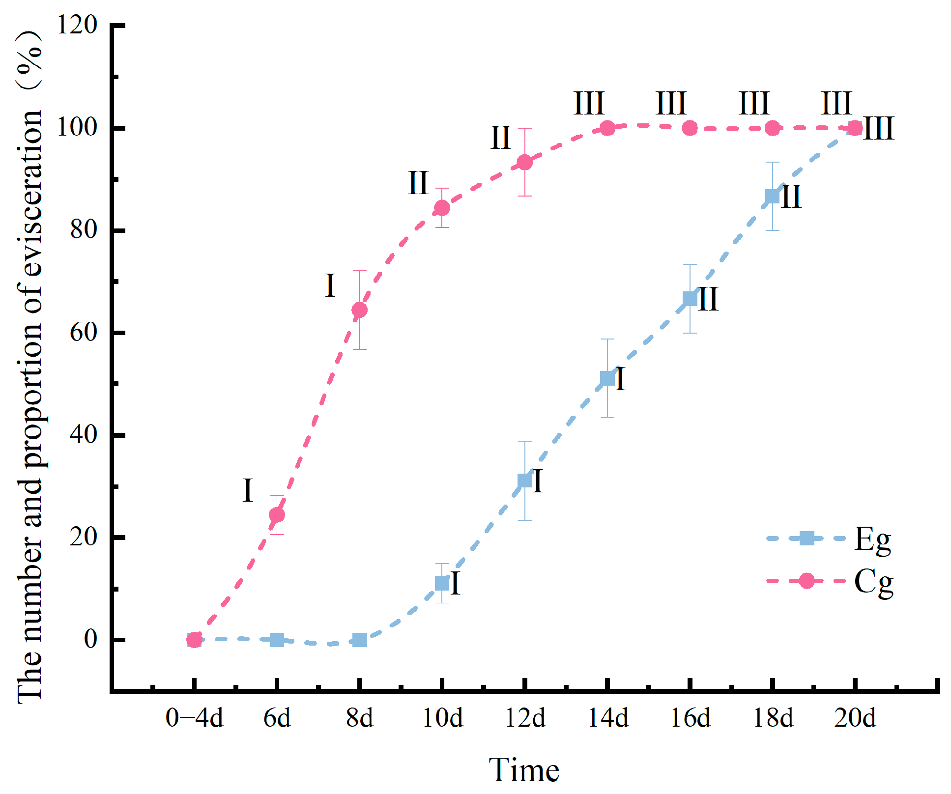
After 6 days of V. splendidus infection, skin ulceration appeared in group Cg, while group Eg did not show degeneration until day 10. In group Cg, skin ulceration progressed from Stage I to Stage II on day 10 and from Stage II to Stage III on day 14. In contrast, group Eg exhibited less severe skin ulceration, with progression from Stage I to Stage II on day 16 and from Stage II to Stage III on day 20 (Figure 2 and Figure 3).

3.2. Enzyme Activity Measurement

3.2.1. Effect of Inhibitor Composition on the Enzyme Activity in the Sea Cucumber Body Wall Induced by UVA Irradiation

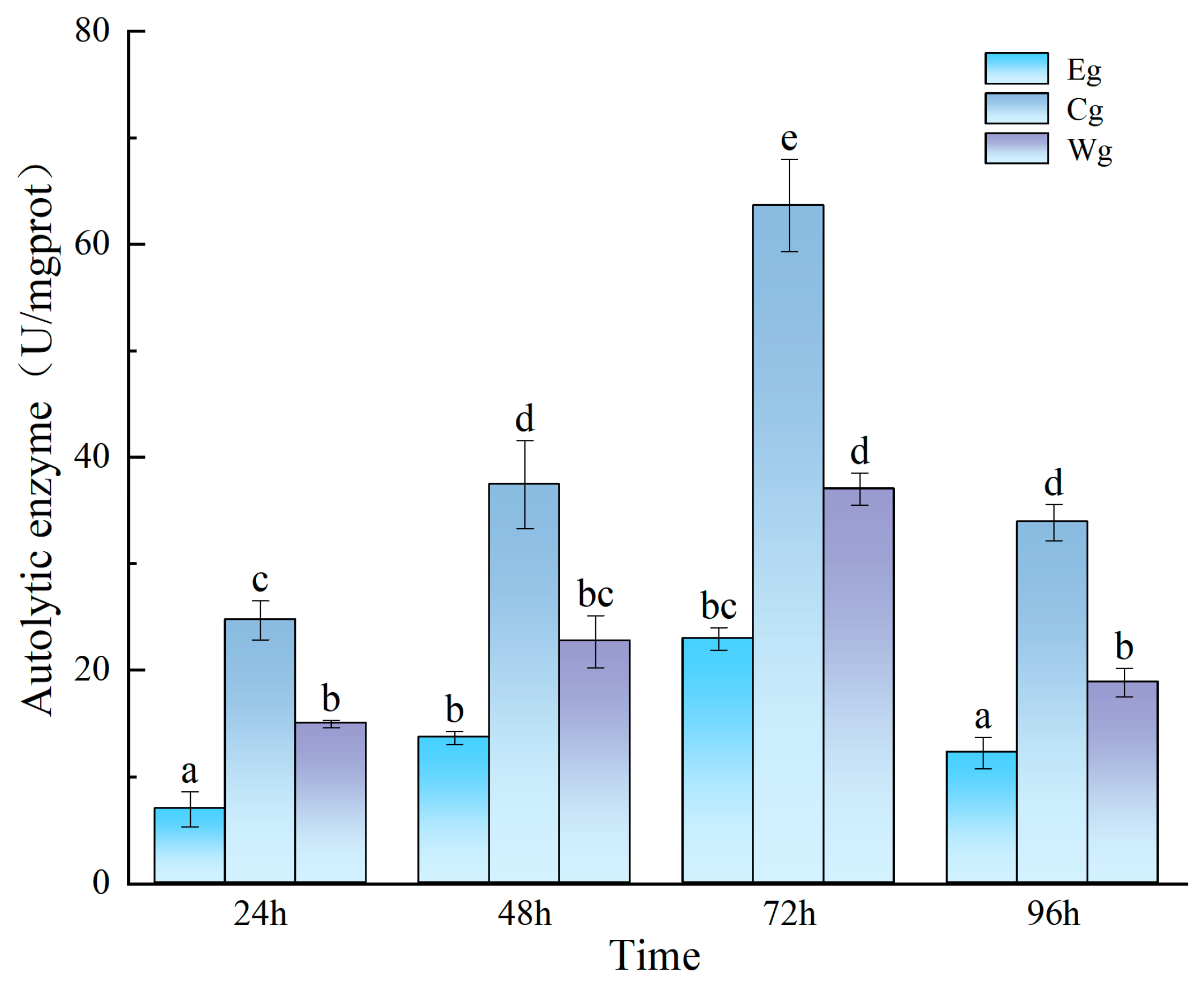
As shown in Figure 4, at 24 h after UVA irradiation, enzyme activity in group Eg was significantly lower than in groups Cg and Wg (p < 0.001). At 48 and 72 h, enzyme activity increased in all groups, but group Eg remained significantly lower than group Cg (p < 0.001). At 96 h, enzyme activity slightly decreased in all groups, but group Eg remained significantly lower than the other two groups (p < 0.001). Although enzyme activity in group Eg increased slowly in the early stages due to the effect of time, it decreased again at 96 h (Figure 4).
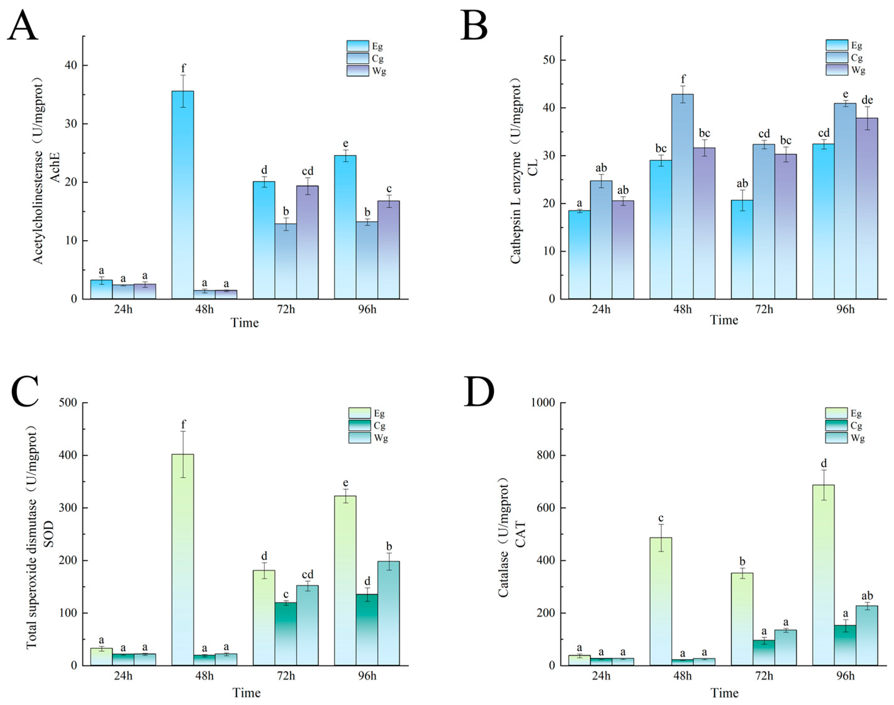
As shown in Figure 5A, at 24 h after UVA irradiation, AchE activity in all groups was low, with no significant differences. However, at 48 h, AchE activity in group Eg was significantly higher than in group Cg and group Wg (p < 0.001). At 72 and 96 h, AchE activity in group Eg showed a decrease but remained significantly higher than in group Cg and group Wg, although it decreased by 56.37% and 32.07%, compared to 48 h.
As shown in Figure 5B, at 24 h after UVA irradiation, although the activity in group Cg was higher, no significant difference was observed, compared with the other two groups. At 48 h, activity increased in all groups, with group Cg showing about 32.32% higher activity than group Eg; group Cg was significantly higher than both group Eg and group Wg (p < 0.001). At 72 h, both group Cg and group Wg showed a decline but remained significantly higher than group Eg (p < 0.001). At 96 h, activity in all groups increased, but a significant difference remained between group Cg and group Eg (p < 0.001).
As shown in Figure 5C, at 24 h after UVA irradiation, SOD activity in all groups was low, with no significant differences. However, at 48 h, group Eg showed higher SOD activity than groups Cg and Wg (p < 0.001). At 72 h, SOD activity in group Eg decreased but remained significantly higher than in group Cg (p < 0.001), 28.40% and 17.90% higher, respectively. At 96 h, SOD activity increased in all groups, with group Eg still showing significant differences, compared to the other two groups (p < 0.001).
As shown in Figure 5D, at 24 h after UVA irradiation, CAT activity in all groups was low, with no significant differences. At 48 h, group Eg showed higher CAT activity than groups Cg and Wg (p < 0.001). At 72 h, group Eg showed a decrease, but its CAT activity remained significantly higher than in group Cg (p < 0.001). At 96 h, CAT activity increased in all groups, with group Eg reaching the highest level at all four time points, significantly higher than the other two groups (p < 0.001), 3.53 times higher than group Cg and 2.03 times higher than group Wg.

3.2.2. Effect of Inhibitor Composition on Enzyme Activity Changes Induced by Vibrio splendidus Infection

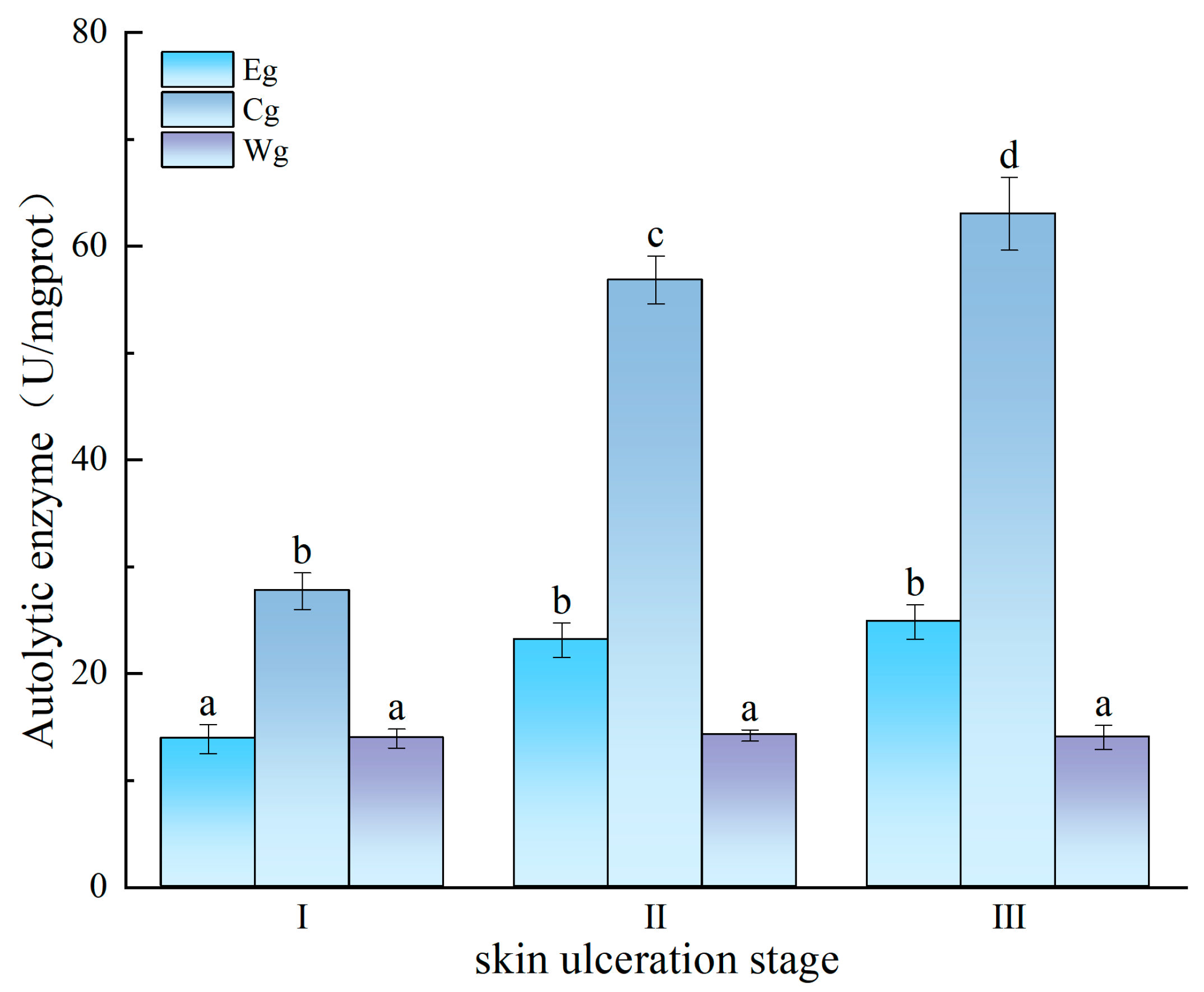
As shown in Figure 6, during the early stage of skin ulceration (Stage I), autolytic enzyme activity was significantly lower in group Eg than in group Cg (p < 0.001), but no difference was observed between Eg and Wg. In Stage II, autolytic enzyme activity in Cg increased significantly (p < 0.001), while Eg activity remained lower than Cg (p < 0.001). In Stage III, group Cg showed a marked increase in autolytic enzyme activity, while group Eg exhibited no significant change from Stage II and remained significantly lower than group Cg (p < 0.001). Compared with Stage I, enzyme activity in group Eg increased significantly in Stage II and Stage III (p < 0.05) but remained significantly lower than that in group Cg at all stages (p < 0.001).
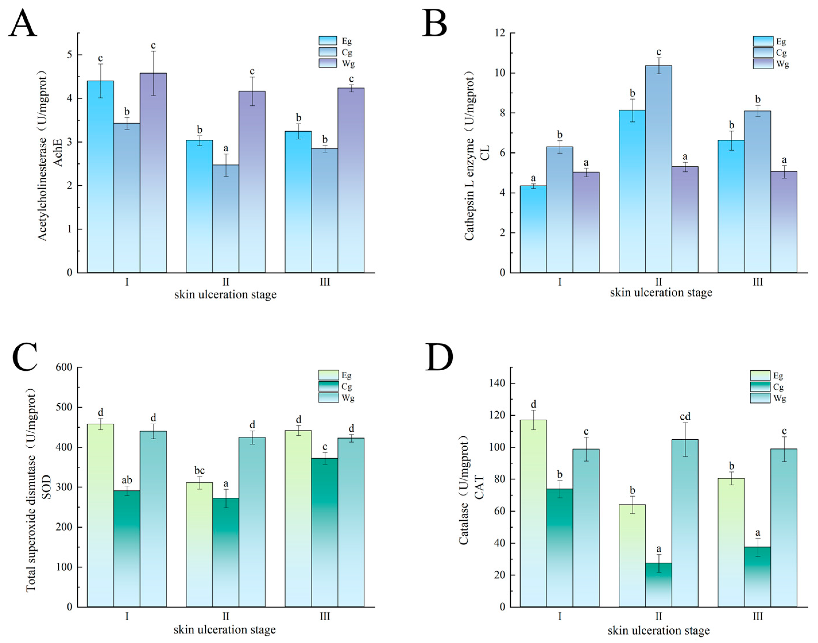
As shown in Figure 7A, during Stage I of skin ulceration, AchE activity in group Eg and group Wg was significantly higher than in group Cg (p < 0.001). In Stage II, AchE activity declined to varying degrees in all groups, but group Eg still exhibited significantly higher activity than group Cg (p < 0.001). In Stage III, due to the influence of time, AchE activity rebounded in both group Eg and group Cg, and no significant difference was observed between them.
As shown in Figure 7B, during Stage I of skin ulceration, group Eg had the lowest CL activity, significantly lower than group Cg (p < 0.001), indicating the composition’s significant inhibition of CL activity. In Stage II, both groups showed a large increase in CL activity, reaching the highest levels. However, CL activity in group Eg remained lower than in group Cg (p < 0.001). In Stage III, CL activity decreased in both groups, with group Eg maintaining significantly lower activity than group Cg (p < 0.001).
As shown in Figure 7C, during Stage I of skin ulceration, SOD activity in group Eg was significantly higher than that in group Cg (p < 0.001), suggesting that the inhibitor composition increased SOD activity. In Stage II, SOD activity decreased in both group Eg and group Cg, indicating a limited and weakening effect over time. In Stage III, SOD activity increased in both groups, with group Eg still showing significantly higher activity than group Cg (p < 0.001).
As shown in Figure 7D, group Eg showed the highest CAT activity during Stage I of skin ulceration. In Stage II, CAT activity decreased in group Eg and group Cg, but group Eg still showed significantly higher activity than group Cg (p < 0.001). In Stage III, CAT activity increased in both groups, with group Eg maintaining significantly higher activity than group Cg (p < 0.001).

3.3. Effect of UVA on the Relative Expression of Caspase-3 in the Sea Cucumber Body Wall

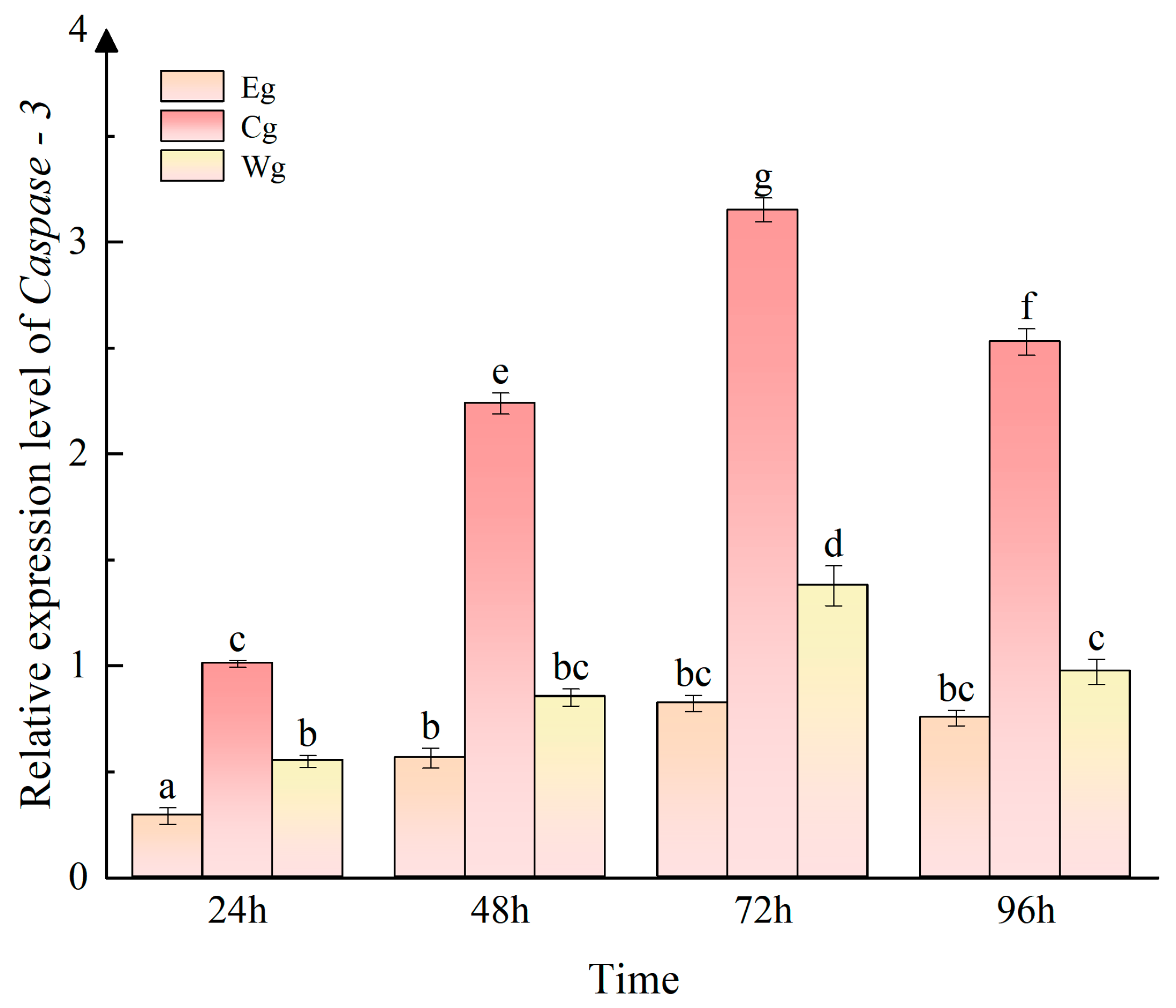
To assess apoptosis in A. japonicus following UVA irradiation, relative expression changes in Caspase-3 in the body wall were measured at 24 h, 48 h, 72 h, and 96 h after UVA exposure. As shown in Figure 8, at 24 h after UVA irradiation, significant differences in Caspase-3 expression were observed between the groups (p < 0.001), with distinct differences indicated by different letters (Eg, Cg, and Wg). At 48 h and 72 h, group Cg exhibited a significant increase in Caspase-3 expression (p < 0.001). After 48 h, the Caspase-3 expression in group Eg showed a slight increase but remained significantly lower than that in group Cg (p < 0.001) and was slightly lower than that in group Wg.

3.4. Effect of Vibrio splendidus on the Relative Expression of Caspase-3 in the Sea Cucumber Body Wall

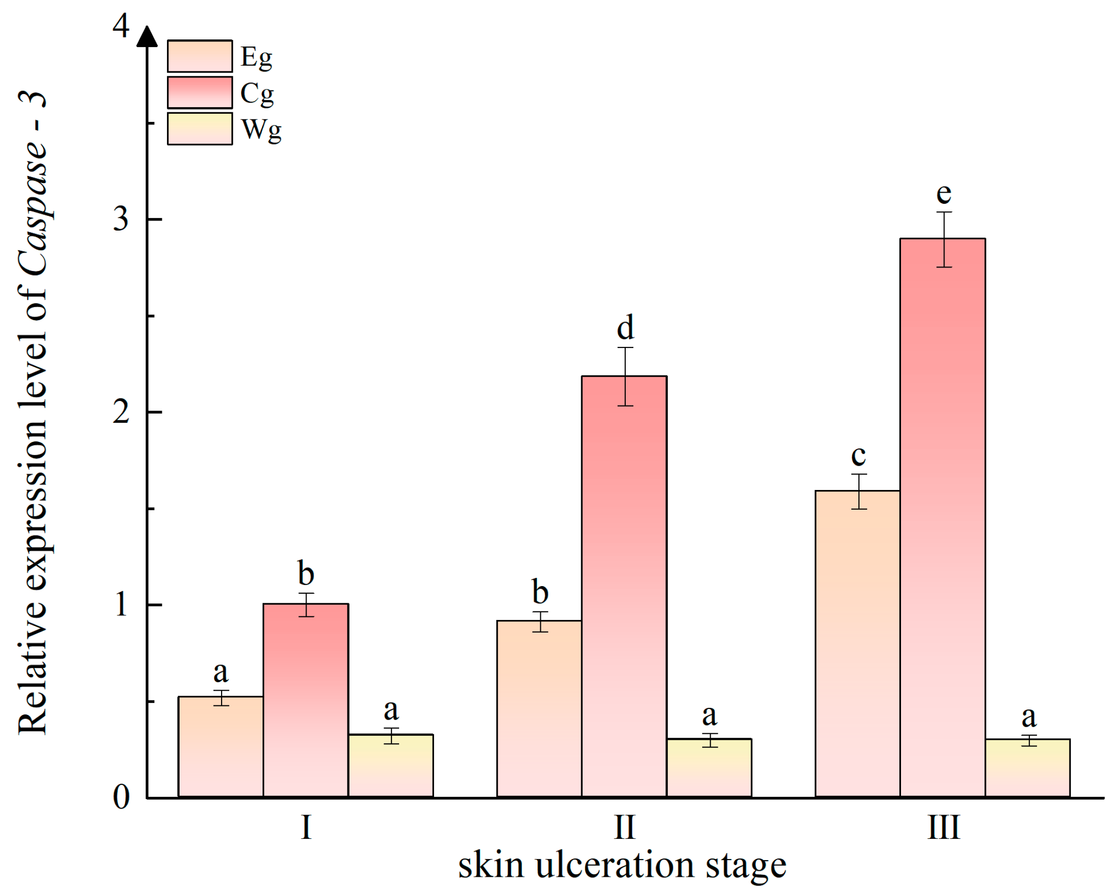
As shown in Figure 9, no significant differences were observed in the Wg group. In Stage I of skin ulceration, the Eg group showed significantly lower Caspase-3 expression compared to the Cg group (p < 0.001), but there was no significant difference between the Eg and Wg groups. In Stage II and Stage III, Caspase-3 expression significantly increased in both the Eg and Cg groups (p < 0.001). During these stages, the Eg group showed significantly lower Caspase-3 expression than the Cg group but significantly higher than the Wg group (p < 0.001).

4. Discussion

This study systematically evaluated the effectiveness of the inhibitor composition in preventing skin ulceration in A. japonicus, investigating its potential mechanisms of action using two typical inducers: UVA irradiation and V. splendidus infection. In the UVA induction experiment, the autolytic enzyme expression in group Eg was significantly lower than in group Cg, especially at 72 h, where the difference was highly significant. This suggests that the inhibitor composition effectively reduces the activity of key enzymes related to autolysis. Autolytic enzymes can destroy the integrity of cell membranes and cause cell lysis [27]. The reduction in their activity helps delay cell death and tissue degradation, thus slowing the skin ulceration process. Additionally, at 48 h after UVA irradiation, acetylcholinesterase (AchE) activity in group Eg was significantly higher than in group Cg and group Wg, suggesting that the inhibitor composition may alleviate UVA-induced interference in neurotransmitter metabolism by stabilizing AchE or promoting its synthesis. This result aligns with previous studies on the short-term effects of UV radiation on AchE, providing new insights into the composition’s protective mechanism. Furthermore, cathepsin L, a key enzyme in the autolysis process in the sea cucumber body wall [28,29], rapidly increased in group Cg after UVA treatment, while group Eg showed a clear inhibitory trend starting from 24 h. The trends in AchE and CL suggest that the composition deays skin ulceration by maintaining neuronal stability and inhibiting protein hydrolysis.
Oxidative stress plays a key role in skin ulceration [6]. UVA irradiation induces rapid accumulation of reactive oxygen species (ROS) in A. japonicus, significantly inhibiting catalase (CAT) activity, which reaches its lowest value within 24 h. However, CAT activity gradually recovered thereafter, indicating that A. japonicus possess antioxidant regulatory capacity [30]. This dynamic change was closely associated with the activation of the Nrf2/ARE signaling pathway. During the early stages of oxidative stress, ROS accumulation was known to disrupt cellular redox balance; however, as the recovery process was initiated, Nrf2 was dissociated from Keap1 and translocated to the nucleus, where it upregulated the expression of phase II detoxifying enzymes, including CAT, to rebuild the antioxidant defense system [31]. Similarly, superoxide dismutase (SOD) activity significantly decreased after UVA irradiation, likely due to UVA-induced enzyme conformation damage or metabolic dysfunction, consistent with the UVB-induced oxidative stress mechanism reported previously [32]. Notably, at 24 h, all three groups showed a dip in SOD and CAT activities. However, at 48 h, the SOD and CAT activities in the Eg group were significantly higher than those in the Cg and Wg groups. This difference was likely attributed to the multi-target regulatory effects of the inhibitor combination: (1) the activity of free radical scavengers that directly eliminate ROS; (2) the enhancement of endogenous antioxidant enzyme expression through the activation of the Nrf2/ARE pathway; and (3) the inhibition of key enzymes involved in ROS generation, such as NADPH oxidase [33]. This may be because the oxidative stress environment activated the autolysis cascade [34]. Specifically, (1) the restoration of CAT and SOD activities was found to effectively reduce the accumulation of lipid peroxidation products (such as MDA), thereby decreasing the extent of cell membrane damage; (2) the reduction in oxidative stress levels was shown to weaken the activation of the Caspase-3 pathway and inhibit the proteolytic activity of cathepsin L (CL); and (3) by maintaining intracellular redox homeostasis, this inhibitor combination may have indirectly protected mitochondrial function and reduced the release of apoptosis-related factors. These mechanisms collectively explained the superior ulcer-suppressive effect observed in the Eg group at 48 h [35,36].
In the V. splendidus infection experiment, the onset of skin ulceration in A. japonicus was significantly delayed, compared to UVA irradiation, possibly due to their stronger innate immune defenses. Apostichopus japonicus resist pathogen invasion through mechanisms like the secretion of antimicrobial peptides [37] and the formation of body wall mucosal barriers [38], delaying early lesion progression. In the experiment, group Eg, treated with the inhibitor composition, showed a 4-day delay in skin ulceration onset, compared to group Cg, and a significant reduction in the occurrence rate and lesion area of Stage III degeneration, demonstrating the composition’s effectiveness in controlling infectious skin ulceration. Throughout the infection period, autolytic enzyme activity in group Eg remained consistently lower than in group Cg. While external factors like fluctuations in water quality may have influenced the results, the overall trend remained stable, indicating that the inhibitor composition had a strong, sustained inhibitory effect on cathepsin activity. Similar to UVA induction, V. splendidus destroys cell membranes through virulence factors like hemolysin [39], triggering oxidative stress [40] and increasing ROS levels, thus burdening the antioxidant system [16,41]. The experimental results showed that SOD and CAT activities in the inhibitor group were significantly higher than in group Cg, suggesting that the composition may reduce ROS accumulation, decrease antioxidant enzyme consumption, and minimize oxidative damage, thereby maintaining cellular homeostasis. Additionally, reduced oxidative stress levels may have indirectly inhibited the activation of autolytic enzymes like CL and CB, further slowing skin ulceration.
Caspase-3, a core effector in the apoptosis pathway, showed highly consistent expression changes in this study [15]. In the UVA irradiation experiment, Caspase-3 expression in group Cg increased significantly, while group Eg maintained low expression, similar to group Wg. This suggests that Emricasan, the Caspase-3 inhibitor, effectively blocked the UVA-induced apoptosis cascade. This inhibitory effect showed minimal fluctuation across time points, likely due to Emricasan being an irreversible inhibitor of Caspase-3 that continuously interferes with apoptosis signaling. This result aligns with previous studies on UV radiation-induced Caspase-3-mediated apoptosis in oysters [42] and A. japonicus [15]. Previous studies found that Caspase-3, as an execution protease, can activate CL release [43] and hydrolyze cellular matrix components, indirectly supporting the potential regulatory relationship between Caspase-3 and autolytic enzymes like CL. This further demonstrates the synergistic effects of the three components of the inhibitor composition, which regulate the skin ulceration process at multiple levels.
A similar trend was observed in the V. splendidus infection experiment. Group Eg showed significantly lower Caspase-3 expression than group Cg in the early stages of infection, suggesting that the inhibitor composition effectively suppressed the pathogen-induced mitochondrial apoptosis pathway. However, as the infection progressed, Caspase-3 expression in group Eg gradually increased but remained lower than in group Cg. This may indicate the activation of additional apoptotic signals due to continuous pathogen stimulation and toxin accumulation [16,44], leading to a gradual reduction in the inhibitor composition’s efficacy or exceeding its regulatory capacity. Nevertheless, Caspase-3 expression in group Eg remained higher than in group Wg, suggesting that the composition did not completely block apoptosis activation and implying that moderate apoptosis under infection conditions is necessary to clear damaged cells and maintain tissue homeostasis [45].
Overall, the composition effectively regulates Caspase-3 expression, reducing apoptosis frequency and intensity and demonstrating strong anti-skin ulceration effects under both UVA irradiation and pathogen infection models.

5. Conclusions

This study systematically explored the effects of an inhibitor composition containing Nafamostat mesylate, Ilomastat, and Emricasan on skin ulceration in A. japonicus under two stress factors: UVA irradiation and V. splendidus infection, along with their potential mechanisms. The results indicated that the inhibitor composition significantly delayed skin ulceration in A. japonicus under various conditions. The composition inhibited the expression of key autolytic enzymes (e.g., cathepsin L) and apoptosis factors (Caspase-3) while maintaining the stability of antioxidant enzyme systems (SOD, CAT), demonstrating strong synergistic effects. In the UVA induction experiment, the inhibitor composition significantly reduced the skin ulceration area and autolytic enzyme activity. It also effectively maintained AchE activity, thereby delaying UVA-induced neuronal damage and protein hydrolysis. The inhibitor group also demonstrated significantly higher SOD and CAT activities compared to group Cg, indicating that the composition alleviates the oxidative stress response induced by UVA. In the V. splendidus infection model, the inhibitor composition also demonstrated consistent anti-skin ulceration effects. It significantly delayed the onset of skin ulceration, reduced the incidence of Stage III degeneration, and inhibited CL enzyme activity and autolysis. The data also showed that the composition effectively suppressed Caspase-3 expression and interfered with the mitochondrial apoptosis pathway triggered by the infection. This demonstrated immune-protective effects.
In summary, the inhibitor composition effectively alleviates skin ulceration under dual stress. Its multi-target synergistic mechanism includes antioxidant regulation, inhibition of autolytic enzymes, and suppression of apoptosis. These findings offer new insights into the mechanisms of skin ulceration in A. japonicus. They also provide theoretical support and experimental evidence for developing efficient, broad-spectrum skin ulceration prevention and control products, with significant industrial applications and broad potential for promotion.

Author Contributions

Conceptualization, J.D.; X.L. was responsible for conceptualization, methodology, formal analysis, investigation, and writing—original draft; Y.T. handled formal analysis, investigation, visualization, and writing—original draft; H.X. contributed to formal analysis, visualization, and investigation; F.T. was responsible for writing—original draft; L.H. was responsible for formal analysis, investigation, and visualization; L.W. was responsible for formal analysis, investigation, and visualization; C.Z. was responsible for formal analysis, investigation, and visualization; J.D. was responsible for conceptualization, methodology, writing—review and editing, supervision, and funding acquisition. All authors have read and agreed to the published version of the manuscript.

Funding

This research was financially supported by the Major Agricultural Project of the Liaoning Provincial Science and Technology Department (Project No. 2023JH1/10200007).

Institutional Review Board Statement

Not applicable. Ethical review and approval were waived for this study due to the categorization of Apostichopus japonicus as invertebrates.

Informed Consent Statement

Not applicable.

Data Availability Statement

All relevant data are presented within the paper.

Conflicts of Interest

The authors declare no conflicts of interest.

References

  1. Xia, S.; Wang, X. Nutritional and medicinal value. In Developments in Aquaculture and Fisheries Science; Elsevier: Amsterdam, The Netherlands, 2015; Volume 39, pp. 353–365. [Google Scholar] [CrossRef]
  2. Vaidya, H.; Cheema, S.K. Sea cucumber and blue mussel: New sources of phospholipid enriched omega-3 fatty acids with a potential role in 3T3-L1 adipocyte metabolism. Food Funct. 2014, 5, 3287–3295. [Google Scholar] [CrossRef]
  3. Ru, X.; Feng, Q.; Zhang, S.; Liu, S.; Zhang, L.; Yang, H. Eco-friendly method for rearing sea cucumber (Apostichopus japonicus) larvae. Aquac. Res. 2022, 53, 3759–3766. [Google Scholar] [CrossRef]
  4. Guo, M.; Gui, M.; Xu, X.; Duan, X.; Zhao, X.; Zhang, W.; Shao, Y.; Wang, B.; Diao, J.; Li, C. BAG2 mediates coelomocyte apoptosis in Vibrio splendidus challenged sea cucumber Apostichopus japonicus. Int. J. Biol. Macromol. 2021, 189, 34–43. [Google Scholar] [CrossRef] [PubMed]
  5. Huo, D.; Sun, L.; Zhang, L.; Ru, X.; Liu, S.; Yang, H. Metabolome responses of the sea cucumber Apostichopus japonicus to multiple environmental stresses: Heat and hypoxia. Mar. Pollut. Bull. 2019, 138, 407–420. [Google Scholar] [CrossRef] [PubMed]
  6. Qi, H.; Fu, H.; Dong, X.; Feng, D.; Li, N.; Wen, C.; Nakamura, Y.; Zhu, B. Apoptosis induction is involved in UVA-induced autolysis in sea cucumber Stichopus japonicus. J. Photochem. Photobiol. B Biol. 2016, 158, 130–135. [Google Scholar] [CrossRef]
  7. Zhang, C.; Liang, W.; Zhang, W.; Li, C. Characterization of a metalloprotease involved in Vibrio splendidus infection in the sea cucumber, Apostichopus japonicus. Microb. Pathog. 2016, 101, 96–103. [Google Scholar] [CrossRef]
  8. Ju, Z.; Liao, G.; Zhang, Y.; Li, N.; Li, X.; Zou, Y.; Yang, W.; Xiong, D. Oxidative stress responses in the respiratory tree and the body wall of sea cucumber Apostichopus japonicus (Selenka) to high temperature. Environ. Sci. Pollut. Res. 2023, 30, 21288–21298. [Google Scholar] [CrossRef]
  9. Ju, Z.; Li, X.; Yang, W.; Xiong, D. Immune responses of sea cucumber (Apostichopus japonicus) to combined environmental stress from high temperature and oil pollution. Mar. Freshw. Res. 2024, 75, MF23161. [Google Scholar] [CrossRef]
  10. Liang, X.; Zhang, C.; Shen, L.; Ding, L.; Guo, H. Role of non-coding RNAs in UV-induced radiation effects. Exp. Ther. Med. 2024, 27, 262. [Google Scholar] [CrossRef] [PubMed]
  11. Yehia, M.A.-H.; Fawzy, M.A.; Elbashar, Y.H.; Abdelmigid, B.A.; ElGhnam, S.M. Histomorphometry, immunohistochemistry, and fine structure of Rats skin exposed to Ultraviolet-A radiation. J. Opt. 2021, 51, 574–584. [Google Scholar] [CrossRef]
  12. Yang, T.T.; Lan, C.C.E. Photocarcinogenesis of the skin: Current status and future trends. Kaohsiung J. Med. Sci. 2025, 41, e12946. [Google Scholar] [CrossRef] [PubMed]
  13. Umam, N.I.; Sulaiman, A.; Wong, Y.F.; Jaya-Ram, A.; Woo, S.P.; Zulkurnain, M. UV-Assisted Autolysis for Nutrient Bioconversion of Sea Cucumber (Stichopus horrens) Body Wall. Food Bioprocess Technol. 2024, 17, 1637–1657. [Google Scholar] [CrossRef]
  14. Dong, X.; Qi, H.; Feng, D.; He, B.; Nakamura, Y.; Yu, C.; Zhu, B. Oxidative stress involved in textural changes of sea cucumber Stichopus japonicus body wall during low-temperature treatment. Int. J. Food Prop. 2018, 21, 2646–2659. [Google Scholar] [CrossRef]
  15. Yang, J.-F.; Gao, R.-C.; Wu, H.-T.; Li, P.-F.; Hu, X.-S.; Zhou, D.-Y.; Zhu, B.-W.; Su, Y.-C. Analysis of apoptosis in ultraviolet-induced sea cucumber (Stichopus japonicus) melting using terminal deoxynucleotidyl-transferase-mediated dUTP nick end-labeling assay and cleaved Caspase-3 immunohistochemistry. J. Agric. Food Chem. 2015, 63, 9601–9608. [Google Scholar] [CrossRef] [PubMed]
  16. Gao, Q.; Liao, M.; Wang, Y.; Li, B.; Zhang, Z.; Rong, X.; Chen, G.; Wang, L. Transcriptome analysis and discovery of genes involved in immune pathways from coelomocytes of sea cucumber (Apostichopus japonicus) after Vibrio splendidus challenge. Int. J. Mol. Sci. 2015, 16, 16347–16377. [Google Scholar] [CrossRef] [PubMed]
  17. Li, C. Research progress on molecular regulation mechanism ofskin ulcer syndrome in sea cucumber Apostichopus japonicus :A review. J. Dalian Ocean Univ. 2021, 36, 355–373. [Google Scholar] [CrossRef]
  18. Zhang, W.; Li, C. Virulence mechanisms of vibrios belonging to the Splendidus clade as aquaculture pathogens, from case studies and genome data. Rev. Aquac. 2021, 13, 2004–2026. [Google Scholar] [CrossRef]
  19. Yang, J.; Liu, H.; Zheng, G.; Xiang, X.; Lv, Z.; Wang, T. Cathepsin L of the sea cucumber Apostichopus japonicus-molecular characterization and transcriptional response to Vibrio splendidus infection. Fish Shellfish Immunol. 2016, 49, 387–395. [Google Scholar] [CrossRef]
  20. Li, Y.; Yang, H.; Zhang, J.; Shi, W.; Li, W.; Zhang, W. VspC from Vibrio splendidus is responsible for collagen degradation in Apostichopus japonicus. Aquaculture 2023, 571, 739489. [Google Scholar] [CrossRef]
  21. Shao, Y.; Li, C.; Zhang, W.; Duan, X.; Li, Y.; Jin, C.; Xiong, J.; Qiu, Q. Molecular cloning and characterization of four Caspase members in Apostichopus japonicus. Fish Shellfish Immunol. 2016, 55, 203–211. [Google Scholar] [CrossRef]
  22. Ding, J.; Chang, Y.; Guo, C.; Wang, L.; Yin, D. Composition and Application for Inhibiting Sea Cucumber Skin Degeneration. CN113826567B, 19 August 2022. [Google Scholar]
  23. Delroisse, J.; Van Wayneberghe, K.; Flammang, P.; Gillan, D.; Gerbaux, P.; Opina, N.; Todinanahary, G.G.B.; Eeckhaut, I. Epidemiology of a SKin Ulceration Disease (SKUD) in the sea cucumber Holothuria scabra with a review on the SKUDs in Holothuroidea (Echinodermata). Sci. Rep. 2020, 10, 22150. [Google Scholar] [CrossRef]
  24. Ferreira, T.; Rasband, W. Section 28: Plugins. In The ImageJ User Guide, 2nd ed.; Ferreira, T., Rasband, W., Eds.; National Institutes of Health: Bethesda, MD, USA, 2011; Volume 1, pp. 155–161. [Google Scholar]
  25. Zhu, B.; Han, B. Characterization and Purification of Holothurians Autoerzyme. Food Ferment. Ind. 2004, 30, 132. (In Chinese) [Google Scholar]
  26. Wang, Z.; Xu, R.; Yang, H.; Li, R.; Ding, J.; Chang, Y.; Zuo, R. Vitamin E Regulates the Collagen Contents in the Body Wall of Sea Cucumber (Apostichopus japonicus) via Its Antioxidant Effects and the TGF-β/Smads Pathway. Antioxidants 2024, 13, 847. [Google Scholar] [CrossRef]
  27. Liu, Z.-q.; Zhou, D.-y.; Liu, Y.-x.; Yu, M.-m.; Liu, B.; Song, L.; Dong, X.-p.; Qi, H.; Shahidi, F. Inhibitory effect of natural metal ion chelators on the autolysis of sea cucumber (Stichopus japonicus) and its mechanism. Food Res. Int. 2020, 133, 109205. [Google Scholar] [CrossRef]
  28. Souza, M.S.; Balseiro, E.; Laspoumaderes, C.; Modenutti, B. Effect of ultraviolet radiation on acetylcholinesterase activity in freshwater copepods. Photochem. Photobiol. 2010, 86, 367–373. [Google Scholar] [CrossRef]
  29. Liu, Y.-X.; Zhou, D.-Y.; Ma, D.-D.; Liu, Y.-F.; Li, D.-M.; Dong, X.-P.; Tan, M.-Q.; Du, M.; Zhu, B.-W. Changes in collagenous tissue microstructures and distributions of cathepsin L in body wall of autolytic sea cucumber (Stichopus japonicus). Food Chem. 2016, 212, 341–348. [Google Scholar] [CrossRef] [PubMed]
  30. Heck, D.E.; Vetrano, A.M.; Mariano, T.M.; Laskin, J.D. UVB light stimulates production of reactive oxygen species: Unexpected role for catalase. J. Biol. Chem. 2003, 278, 22432–22436. [Google Scholar] [CrossRef]
  31. Mo, Q.; You, S.; Fu, H.; Wang, D.; Zhang, J.; Wang, C.; Li, M. Purification and identification of antioxidant peptides from rice fermentation of Lactobacillus plantarum and their protective effects on UVA–Induced oxidative stress in skin. Antioxidants 2022, 11, 2333. [Google Scholar] [CrossRef] [PubMed]
  32. Xue, S.; Zang, Y.; Chen, J.; Shang, S.; Gao, L.; Tang, X. Ultraviolet-B radiation stress triggers reactive oxygen species and regulates the antioxidant defense and photosynthesis systems of intertidal red algae Neoporphyra haitanensis. Front. Mar. Sci. 2022, 9, 1043462. [Google Scholar] [CrossRef]
  33. Wang, L.; Zhang, X.; Xiong, X.; Zhu, H.; Chen, R.; Zhang, S.; Chen, G.; Jian, Z. Nrf2 regulates oxidative stress and its role in cerebral ischemic stroke. Antioxidants 2022, 11, 2377. [Google Scholar] [CrossRef]
  34. Sample, A.; He, Y.Y. Autophagy in UV damage response. Photochem. Photobiol. 2017, 93, 943–955. [Google Scholar] [CrossRef]
  35. He, L.; Sui, C.; Li, J.; Yao, Y.; Li, M.; Wang, R.; Zhu, W. N-Terminal 5-Mer peptide analog P165 of amyloid precursor protein repairs skin photodamage induced by UVB through the Nrf2 signaling pathway. Indian J. Dermatol. 2021, 66, 574. [Google Scholar] [CrossRef]
  36. Zhao, Y.; Zhang, C.-F.; Rossiter, H.; Eckhart, L.; König, U.; Karner, S.; Mildner, M.; Bochkov, V.N.; Tschachler, E.; Gruber, F. Autophagy is induced by UVA and promotes removal of oxidized phospholipids and protein aggregates in epidermal keratinocytes. J. Investig. Dermatol. 2013, 133, 1629–1637. [Google Scholar] [CrossRef] [PubMed]
  37. Tamori, M.; Saha, A.K.; Matsuno, A.; Noskor, S.C.; Koizumi, O.; Kobayakawa, Y.; Nakajima, Y.; Motokawa, T. Stichopin-containing nerves and secretory cells specific to connective tissues of the sea cucumber. Proc. R. Soc. B Biol. Sci. 2007, 274, 2279–2285. [Google Scholar] [CrossRef] [PubMed]
  38. Sperandio, B.; Fischer, N.; Sansonetti, P.J. Mucosal physical and chemical innate barriers: Lessons from microbial evasion strategies. Semin. Immunol. 2015, 27, 111–118. [Google Scholar] [CrossRef] [PubMed]
  39. Zhang, P.; Li, C.; Li, Y.; Zhang, P.; Shao, Y.; Jin, C.; Li, T. Proteomic identification of differentially expressed proteins in sea cucumber Apostichopus japonicus coelomocytes after Vibrio splendidus infection. Dev. Comp. Immunol. 2014, 44, 370–377. [Google Scholar] [CrossRef]
  40. Sun, Y.; Du, X.; Li, S.; Wen, Z.; Li, Y.; Li, X.; Meng, N.; Mi, R.; Ma, S.; Sun, A. Dietary Cordyceps militaris protects against Vibrio splendidus infection in sea cucumber Apostichopus japonicus. Fish Shellfish Immunol. 2015, 45, 964–971. [Google Scholar] [CrossRef]
  41. Zhao, Y.; Ma, H.; Zhang, W.; Ai, Q.; Mai, K.; Xu, W.; Wang, X.; Liufu, Z. Effects of dietary β-glucan on the growth, immune responses and resistance of sea cucumber, Apostichopus japonicus against Vibrio splendidus infection. Aquaculture 2011, 315, 269–274. [Google Scholar] [CrossRef]
  42. Song, H.; Xie, C.; Dong, M.; Zhang, Y.; Huang, H.; Han, Y.; Liu, Y.; Wei, L.; Wang, X. Effects of ambient UVB light on Pacific oyster Crassostrea gigas mantle tissue based on multivariate data. Ecotoxicol. Environ. Saf. 2024, 274, 116236. [Google Scholar] [CrossRef]
  43. Cailhier, J.-F.; Sirois, I.; Laplante, P.; Lepage, S.; Raymond, M.-A.; Brassard, N.; Prat, A.; Iozzo, R.V.; Pshezhetsky, A.V.; Hebert, M.-J. Caspase-3 activation triggers extracellular cathepsin L release and endorepellin proteolysis. J. Biol. Chem. 2008, 283, 27220–27229. [Google Scholar] [CrossRef]
  44. Monian, P.; Jiang, X. The cellular apoptosis susceptibility protein (CAS) promotes tumor necrosis factor-related apoptosis-inducing ligand (TRAIL)-induced apoptosis and cell proliferation. J. Biol. Chem. 2016, 291, 2379–2388. [Google Scholar] [CrossRef] [PubMed]
  45. Shi, Y.; Shao, Y.; Lv, Z.; Li, C. Serpin-type serine protease inhibitor mediates coelomocyte apoptosis in Apostichopus japonicus. Fish Shellfish Immunol. 2020, 104, 410–418. [Google Scholar] [CrossRef] [PubMed]
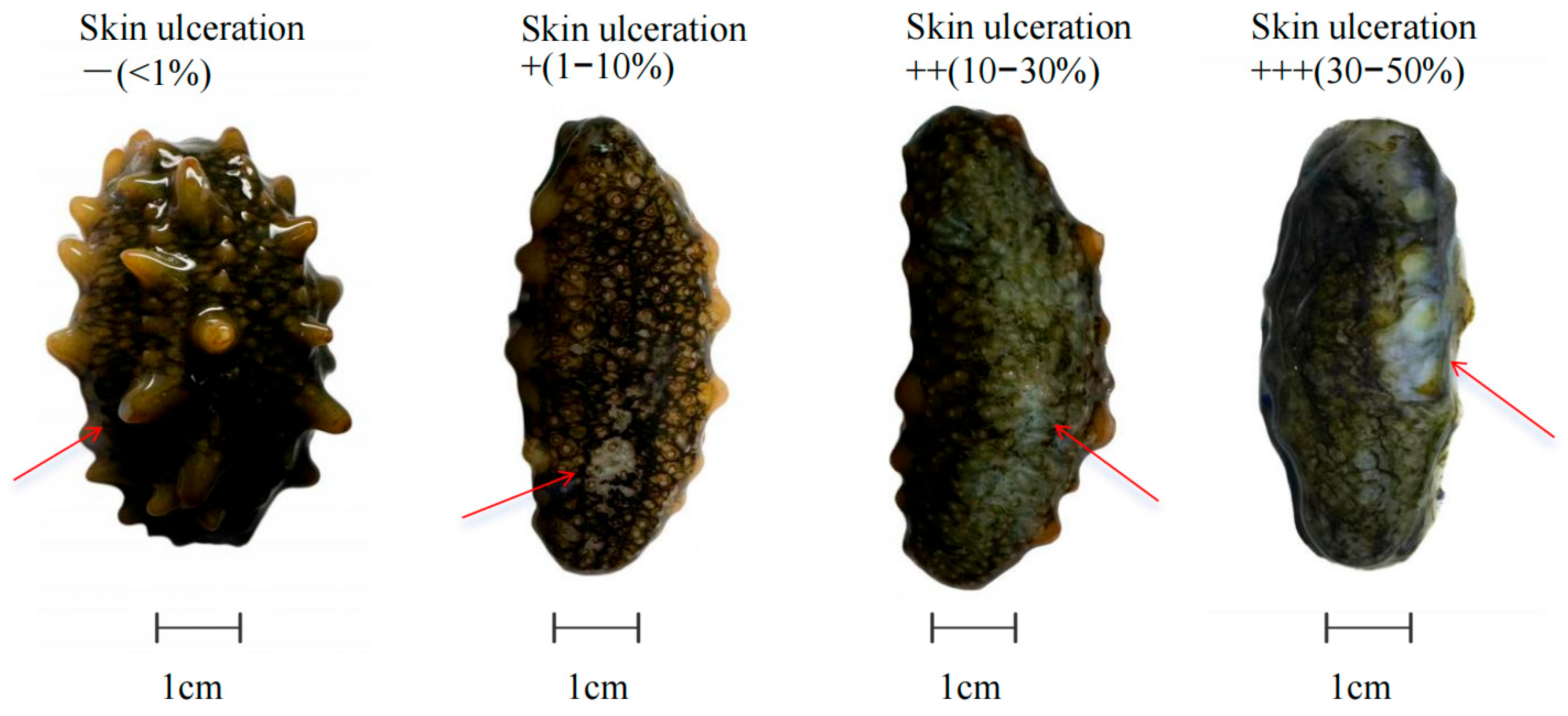
Figure 1. Diagram of skin ulceration at varying degrees in Apostichopus japonicus. Note: Data are presented as mean ± SD (n = 3). The site indicated by the red arrow is the location of skin ulceration.
Fishes 10 00470 g001
Figure 2. Comparison of the proportion and severity of skin ulceration in Apostichopus japonicus induced by Vibrio splendidus, with and without the application of inhibitor compositions. Note: Data are presented as mean ± SD (n = 3). “I” represents Stage I, “II” represents Stage II, and “III” represents Stage III.
Fishes 10 00470 g002
Figure 3. Diagram of developmental characteristics of skin ulceration at different stages in Apostichopus japonicus. Note: Data are presented as mean ± SD (n = 3). The site indicated by the red arrow is the location of skin ulceration.
Fishes 10 00470 g003
Figure 4. Effect of inhibitor compositions on autolysin activity changes in Apostichopus japonicus body wall induced by UVA. Note: Data are presented as mean ± SD (n = 3). Different lowercase letters (a, b, c, d, e) indicate significant differences between groups (p < 0.05).
Fishes 10 00470 g004
Figure 5. Effect of inhibitor compositions on the activity of related enzymes in Apostichopus japonicus body wall induced by UVA. (A) Acetylcholinesterase (AchE); (B) cathepsin L enzyme (CL); (C) total superoxide dismutase (SOD); (D) catalase (CAT). Note: Data are presented as mean ± SD (n = 3). Different lowercase letters (a, b, c, d, e, f) indicate significant differences between groups (p < 0.05).
Fishes 10 00470 g005
Figure 6. Effect of inhibitor compositions on autolysin activity changes in Apostichopus japonicus body wall induced by Vibrio splendidus. Note: Data are presented as mean ± SD (n = 3). Different lowercase letters (a, b, c, d) indicate significant differences between groups (p < 0.05).
Fishes 10 00470 g006
Figure 7. Effect of inhibitor compositions on the activity of related enzymes in Apostichopus japonicus body wall induced by Vibrio splendidus. (A) Acetylcholinesterase (AchE); (B) cathepsin L enzyme (CL); (C) total superoxide dismutase (SOD); (D) catalase (CAT). Note: Data are presented as mean ± SD (n = 3). Different lowercase letters (a, b, c, d) indicate significant differences between groups (p < 0.05).
Fishes 10 00470 g007
Figure 8. Effect of inhibitor compositions on the relative expression of Caspase-3 in Apostichopus japonicus body wall induced by UVA. Note: Data are presented as mean ± SD (n = 3). Different lowercase letters (a, b, c, d, e, f, g) indicate significant differences between groups (p < 0.05).
Fishes 10 00470 g008
Figure 9. Effect of inhibitor compositions on the relative expression of Caspase-3 in Apostichopus japonicus body wall induced by Vibrio splendidus. Note: Data are presented as mean ± SD (n = 3). Different lowercase letters (a, b, c, d, e) indicate significant differences between groups (p < 0.05).
Fishes 10 00470 g009
Table 1. The inhibitor composition inhibits the percentage of the area of sea cucumber skin ulceration induced by UVA irradiation (%).
Groups24 h48 h72 h96 h
Eg0 a0.03 ± 0.006 a0.25 ± 0.04 a0.55 ± 0.02 a
Cg7.99 ± 0.91 b19.58 ± 2.06 c32.80 ± 1.26 d46.02 ± 1.97 e
+++++++++
Wg4.94 ± 0.27 c14.44 ± 0.92 d23.25 ± 1.27 e28.36 ± 1.27 f
+++++++
Note: Data are presented as mean ± SD (n = 3). Less than 1% of the skin is affected: −; 1–10% of the skin is affected: +; 10–30% of the skin is affected: ++; 30–50% of the skin is affected: +++. Different lowercase letters (a, b, c, d, e, f) indicate significant differences between groups (p < 0.05).
Disclaimer/Publisher’s Note: The statements, opinions and data contained in all publications are solely those of the individual author(s) and contributor(s) and not of MDPI and/or the editor(s). MDPI and/or the editor(s) disclaim responsibility for any injury to people or property resulting from any ideas, methods, instructions or products referred to in the content.