Article

Strain-Specific Effects of Bacillus subtilis, Enterococcus faecium, and Pediococcus pentosaceus Supplementation on Growth Performance, Immunity, and Disease Resistance in Olive Flounder (Paralichthys olivaceus)

1
Biotechnology Research Division, National Institute of Fisheries Science, Busan 46083, Republic of Korea
2
Research Planning Division, National Institute of Fisheries Science, Busan 46083, Republic of Korea
*
Author to whom correspondence should be addressed.
Fishes 2025, 10(9), 465; https://doi.org/10.3390/fishes10090465
Submission received: 17 July 2025 / Revised: 13 September 2025 / Accepted: 17 September 2025 / Published: 18 September 2025
(This article belongs to the Section Nutrition and Feeding)

Abstract

Olive flounder (Paralichthys olivaceus), a key aquaculture species in East Asia, is prone to stress and bacterial diseases under intensive farming. Antibiotics are often used to control these problems, but their overuse promotes resistance and threatens sustainability. To provide safer alternatives, this study evaluated the strain-specific effects of dietary probiotics on growth, immunity, and disease resistance in olive flounder. A five-week feeding trial was conducted to evaluate the effects of three isolates—Bacillus subtilis, Enterococcus faecium, and Pediococcus pentosaceus—on growth, blood biochemistry, immune responses, and resistance against Edwardsiella tarda. Each strain was incorporated individually into a basal diet. After the feeding trial, probiotic supplementation improved growth performance. P. pentosaceus significantly increased final body weight and other growth indices (p < 0.05), while E. faecium yielded the lowest feed conversion ratio. Plasma glucose was markedly reduced in the E. faecium and P. pentosaceus groups (p < 0.001), whereas other biochemical indices remained stable. Phagocytic activity was significantly increased in the B. subtilis (p < 0.05) and P. pentosaceus (p < 0.01) groups, while lysozyme activity was significantly elevated in the E. faecium (p < 0.01) and P. pentosaceus (p < 0.05) groups. Following the E. tarda challenge, survival improved in all probiotic-fed groups (22.5–28.9%) compared with the control (11.5%). These findings demonstrate complementary, strain-specific benefits: P. pentosaceus enhanced growth, E. faecium improved feed efficiency and disease resistance, and B. subtilis stimulated immune responses. Validation under farm conditions and exploration of multi-strain formulations are warranted to optimize probiotic use in olive flounder aquaculture.
Key Contribution: This study demonstrates that dietary supplementation with three probiotics—Bacillus subtilis, Enterococcus faecium, and Pediococcus pentosaceus—confers complementary, strain-specific benefits in olive flounder. P. pentosaceus enhanced growth, E. faecium improved feed efficiency and disease resistance, and B. subtilis stimulated immune responses. These findings provide a scientific basis for the targeted application of probiotics as functional feed additives to promote sustainable olive flounder aquaculture.

1. Introduction

Aquaculture plays a pivotal role in ensuring global food security, with olive flounder (Paralichthys olivaceus) recognized as one of the most economically important marine species in East Asian aquaculture, particularly in Korea, Japan, and China [1]. However, the intensive nature of modern aquaculture exposes cultured fish to a range of environmental stressors. Environmental stressors encompass physical factors (e.g., high stocking density and salinity fluctuations), chemical factors (e.g., poor water quality and hypoxia), and thermal factors (e.g., elevated water temperature), all of which can impair the fish’s immune system, reduce growth performance, and increase susceptibility to infectious diseases [2,3,4]. To mitigate these risks, aquaculture practices have traditionally relied on antibiotics and chemotherapeutics. While these agents have been essential tools for controlling disease outbreaks, their extensive use is increasingly questioned, primarily due to critical concerns such as the emergence of antimicrobial resistance, the risk of drug residues in fish products, and the potential for environmental contamination [5,6]. These limitations have therefore prompted increasing interest in safer and more sustainable alternatives to maintain fish health and improve aquaculture productivity.
Probiotics—defined as live microorganisms that confer health benefits to the host when administered in adequate amounts—have emerged as promising environmentally friendly alternatives to antibiotics in aquaculture [7]. Numerous studies have reported that probiotic strains such as Bacillus subtilis, Enterococcus faecium, and Pediococcus pentosaceus can enhance nutrient assimilation, modulate the gut microbiota, stimulate immune responses, and improve resistance to pathogens in various fish species, including carp, tilapia, and rainbow trout [7,8,9,10,11]. The underlying mechanisms include the secretion of digestive enzymes that improve feed utilization, the production of antimicrobial substances (e.g., bacteriocins and organic acids) that inhibit pathogenic bacteria, competition for adhesion sites on the intestinal mucosa, and the activation of innate and adaptive immune pathways such as lysozyme activity, phagocytosis, and cytokine expression [12,13].
Despite this growing body of evidence, research on olive flounder—an economically critical and widely cultured species in East Asian aquaculture—remains relatively limited in scope. While previous studies have provided valuable insights, they have typically focused on discrete aspects of probiotic functionality rather than offering an integrated evaluation. For example, Choi et al. (2022) demonstrated improvements in hematological parameters and immune-related gene expression following probiotic supplementation [14]; Niu et al. (2019) assessed gut-derived Bacillus strains primarily in relation to innate immunity and partial blood indices [15]; and Han et al. (2020) highlighted the antiviral potential of B. subtilis through modulation of host immune genes [16]. However, these studies did not comprehensively address the multifaceted impacts of probiotics on olive flounder health, particularly with respect to the integration of growth performance, blood biochemical indices, non-specific immune responses, and immune-related gene expression. Furthermore, the functional genomic basis underlying these effects—such as the presence of biosynthetic gene clusters associated with secondary metabolite production—remains poorly understood.
To address these gaps, this study investigated the strain-specific effects of dietary supplementation with B. subtilis, E. faecium, and P. pentosaceus on growth performance, blood biochemical parameters, non-specific immune responses, and immune-related gene expression in olive flounder. In parallel, whole-genome sequencing of each probiotic strain was performed to identify biosynthetic gene clusters associated with secondary metabolite production. By integrating genomic characterization with in vivo functional validation, this study establishes a comprehensive framework for elucidating the roles of probiotics in olive flounder health and provides critical insights that can support the industrial application of probiotics as next-generation functional feed additives.

2. Materials and Methods

2.1. Isolation, Identification, and Characterization of Bacterial Strains

Bacterial strains were isolated from the gut contents of olive flounder. The gut contents were serially diluted using phosphate-buffered saline (PBS, pH 7.4, Gibco, Thermo Fisher Scientific, Waltham, MA, USA) and inoculated on tryptic soy agar (TSA, Difco Laboratories, BD, Sparks, MD, USA), yeast peptone dextrose agar (YPD, Difco Laboratories, BD, Sparks, MD, USA), and De Man-Rogosa-Sharpe agar (MRS, Difco Laboratories, BD, Sparks, MD, USA). The agar plates were incubated at 20 °C for 24 h, and colonies were purified by repeated inoculation under the same conditions. Through this procedure, strains designated E-TSA-3, YPD N2, and MRS A4-1 were isolated from TSA, YPD, and MRS agar plates, respectively.
For taxonomic identification, genomic DNA was extracted using the MiniBEST Bacteria Genomic DNA Extraction Kit (Takara Bio Inc., Shiga, Japan). The 16S ribosomal DNA (16S rDNA) was amplified using the primers 27F (5′-AGR GTT YGA TYM TGG CTC AG-3′) and 1492R (5′-RGY TAC CTT GTT ACG ACT T-3′). PCR products were purified (Qiagen, Hilden, Germany) and sequenced using a 3730XL Sanger sequencer (Thermo Fisher Scientific, Waltham, MA, USA). Sequence reads were aligned with BioEdit 7.2.5 (Hall, T.A., Ibis Therapeutics, Carlsbad, CA, USA) and compared against the EZBioCloud database (https://www.ezbiocloud.net/ accessed on 7 October 2024) [17,18]. The isolates were identified as B. subtilis (99.93% similarity, E-TSA-3), E. faecium (99.59%, YPD N2), and P. pentosaceus (99.93%, MRS A4-1). All isolates were preserved in 20% (v/v) glycerol at −80 °C for further use.
The antimicrobial activity of the isolates was evaluated against four representative pathogens—Escherichia coli (Gram-negative), Bacillus cereus (Gram-positive), Candida albicans (fungus), and Edwardsiella tarda (Gram-negative fish pathogen)—using the paper disc diffusion method [19]. Each pathogen was cultured in its optimal broth and adjusted to OD600 = 0.1 prior to plating. A volume of 60 µL of each probiotic culture (48 h, 20 °C) was loaded onto sterile paper discs (8 mm, ADVANTEC, Tokyo, Japan) and placed on inoculated agar plates. Plates were incubated under conditions suitable for each pathogen, and inhibition zones were measured to assess antimicrobial activity.
Enzymatic activities of the isolates were examined using nutrient agar (BD, Sparks, MD, USA) supplemented with different substrates. Protease and gelatinase activities were assessed using 10% (w/v) skim milk and 3% (w/v) gelatin, respectively. Cellulase activity was tested with 1% (w/v) carboxymethyl cellulose (CMC; Sigma-Aldrich, St. Louis, MO, USA), lipase activity with 1% (v/v) glyceryl tributyrate (TBN) and 1% (v/v) glyceryl trioctanoate (TCN), and α-amylase activity with 1% (w/v) soluble starch (Showa Chemicals, Osaka, Japan). Overnight bacterial cultures (1 µL) were spotted onto each medium and incubated at 37 °C for 7 h. Clear zones around colonies were recorded as positive enzymatic activity. For α-amylase activity, 2 mL of iodine solution (Dong Sung Chemical Co., Seoul, Republic of Korea) was added after incubation to visualize starch hydrolysis zones.

2.2. Whole-Genome Sequencing and Secondary Metabolite Gene Cluster Analysis

Genomic DNA from each candidate probiotic strain was extracted using the MiniBEST Bacteria Genomic DNA Extraction Kit (Takara Bio Inc., Shiga, Japan), according to the manufacturer’s instructions. Whole-genome sequencing was conducted using the Nanopore Flongle platform (Oxford Nanopore Technologies, Oxford, UK). Raw sequence reads were assembled using Flye and polished with Medaka to obtain high-quality genome assemblies. The assembled genomes were annotated using Prokka to identify coding sequences and functional genes. To investigate biosynthetic gene clusters (BGCs) potentially involved in secondary metabolite biosynthesis, the genomes were analyzed using antiSMASH (v6.0) [20]. Identified BGCs were further annotated in silico to predict the types of secondary metabolites that may be produced by each strain, based on sequence similarity to known reference clusters.

2.3. Experimental Fish

Olive flounder were obtained from the Genetics and Breeding Research Center of the National Institute of Fisheries Science (Geoje, Republic of Korea) and transported to the laboratory. Fish were acclimated for 2 weeks in indoor tanks to adapt to the experimental conditions, receiving a commercial formulated feed for olive flounder (Daebong LF, Jeju, Republic of Korea). After acclimation, fish with an average initial body weight of 16.98 ± 3.8 g were randomly allocated to 12 acrylic tanks (0.2 m2; 35 fish per tank), with three replicate tanks assigned to each of the four dietary treatments. To ensure randomization and minimize selection bias, fish were distributed sequentially into tanks. Continuous aeration was provided via air stones to maintain adequate dissolved oxygen levels, and the water temperature during the trial averaged 21.4 ± 1.4 °C. Fish were fed twice daily (09:00 and 17:00) at a feeding rate equivalent to 3% of body weight for a total of 5 weeks, a duration determined based on preliminary trials and previous studies on olive flounder growth and immune response [21]. Investigators responsible for husbandry were aware of group allocation, but laboratory analyses were performed under blind conditions. All experimental procedures were approved by the Institutional Animal Care and Use Committee of the National Institute of Fisheries Science (Approval No. 2024-NIFS-IACUC-46).

2.4. Experimental Diet

Four experimental diets were prepared using a commercial formulated feed for olive flounder as the basal diet (Daebong LF, Jeju, Republic of Korea; Table 1). Liquid cultures of B. subtilis, E. faecium, and P. pentosaceus were sprayed onto the basal feed and mixed thoroughly to achieve a final concentration of approximately 6.5 × 10⁷ CFU g−1 of feed. The control diet consisted solely of the basal commercial feed without probiotic supplementation.

2.5. Growth Performance

The initial body weight (IBW) of the experimental fish was recorded after the 2-week acclimation period, and the final body weight (FBW) was measured at the end of the 35-day feeding trial. The total feed intake (FI) was calculated as the cumulative feed provided to each tank throughout the experimental period. Growth performance indices, including body weight gain (BWG), weight gain rate (WGR), specific growth rate (SGR), and feed conversion ratio (FCR), were calculated according to the following equations [22]:
BWG (g) = FBWIBW
WGR (%) = [(FBWIBW)/IBW] × 100
SGR (%/day) = [ln(FBW) − ln(IBW)]/days × 100
FCR = FI/BWG

2.6. Sampling Procedures

At the end of the 5-week feeding trial, five fish from each replicate tank (15 fish per group) were randomly selected for sampling. All fish were anesthetized with 2-phenoxyethanol (100 ppm, Sigma-Aldrich, St. Louis, MO, USA) prior to sampling. Blood was collected from the caudal vein using a heparinized 1 mL syringe, and liver samples were aseptically excised from the same fish. Plasma was obtained by centrifuging the blood samples at 3000 rpm for 10 min (4 °C) and stored at −80 °C until analysis. Liver samples were immediately snap-frozen in liquid nitrogen and stored at −80 °C until RNA extraction.

2.7. Biochemical Analysis

Plasma levels of glucose (GLU), alkaline phosphatase (ALP), glutamic oxaloacetic transaminase (GOT), glutamic pyruvic transaminase (GPT), and total protein (TP) were measured using a dry-type automatic biochemical analyzer (FUJI DRI-CHEM NX500, FUJIFILM, Tokyo, Japan).

2.8. Measurement of Phagocytic Activity

Phagocytic activity was measured using the nitroblue tetrazolium (NBT) reduction assay, as described by Müller et al. (1981) [23]. Briefly, 50 µL of whole blood was mixed with 50 µL of NBT solution and incubated at room temperature for 30 min under dark conditions. Following incubation, the reaction was terminated by adding 100 µL of N,N-dimethylformamide, and the mixture was then centrifuged at 3000 rpm for 10 min (4 °C). Subsequently, 20 µL of the resulting supernatant was transferred into a 96-well microplate and mixed with 180 µL of 1× PBS. Absorbance was measured at 550 nm using a microplate reader (Agilent Technologies, Santa Clara, CA, USA).

2.9. Measurement of Lysozyme Activity

Lysozyme activity in plasma was measured using a turbidimetric method, as described by Hikima et al. (2001) with slight modifications [24]. Freeze-dried Micrococcus lysodeikticus (Sigma-Aldrich, St. Louis, MO, USA) was suspended in 1× PBS to a final concentration of 0.02% (w/v). A total of 190 µL of the prepared suspension was mixed with 10 µL of plasma in a 96-well microplate and incubated at room temperature for 1 min. Absorbance at 530 nm was measured at 1-min intervals for 20 min using a microplate reader (Agilent Technologies, Santa Clara, CA, USA). One unit of lysozyme activity was defined as a reduction in absorbance of 0.001 per min at 530 nm.

2.10. Measurement of Anti-Protease Activity

Anti-protease activity in plasma was measured using an azocasein-based assay, as described by Coêlho et al. (2016), with slight modifications [25]. Briefly, 10 µL of plasma was mixed with 10 µL of trypsin solution (5 mg/mL) and incubated at room temperature for 10 min. Subsequently, 200 µL of a mixture containing 100 µL of 2% (w/v) azocasein and 100 µL of 1× PBS was added to each well, followed by incubation at room temperature for 1 h. The reaction was terminated by adding 500 µL of 10% (w/v) trichloroacetic acid (TCA), and the mixture was centrifuged at 6000 rpm for 5 min. Subsequently, 100 µL of the resulting supernatant was transferred into a 96-well microplate and mixed with 100 µL of 1 N sodium hydroxide (NaOH). Absorbance was measured at 450 nm using a microplate reader (Agilent Technologies, Santa Clara, CA, USA).

2.11. Gene Expression Analysis

Total RNA was extracted from olive flounder liver using RiboEx reagent (GeneAll Biotechnology, Seoul, Korea), and RNA quality was assessed using a NanoDrop spectrophotometer (Thermo Fisher Scientific, Waltham, MA, USA), ensuring an A260/A280 ratio between 1.8 and 2.0. First-strand complementary DNA (cDNA) was synthesized from 1 µg of total RNA using the PrimeScript RT Reagent Kit (Takara Bio Inc., Kusatsu, Japan). The expression levels of c-type lysozyme, CD4, CD8, IL-8, and IL-10 were quantified using SYBR Green-based real-time quantitative PCR (RT-qPCR) on a QuantStudio 3 Real-Time PCR System (Applied Biosystems, Foster City, CA, USA). The thermal cycling protocol consisted of an initial denaturation at 95 °C for 10 min, followed by 40 cycles of 95 °C for 15 s and 60 °C for 1 min. All qPCR reactions were performed in biological triplicates and normalized to the reference gene β-actin. Relative gene expression levels were calculated using the 2−ΔΔCt method [26]. Amplification specificity was confirmed by melt-curve analysis, and primer sequences are listed in Table 2.

2.12. Challenge Test

Following the 5-week feeding trial, disease resistance was evaluated using the same fish from each dietary group (n = 90 per group; 30 fish per tank, three replicate tanks) by challenging them with the Gram-negative fish pathogen E. tarda (KCTC 12267T) through artificial infection. E. tarda was pre-cultured in 1.5% Brain Heart Infusion (BHI; Difco Laboratories, BD, Sparks, MD, USA) broth at 30 °C for 24 h and then resuspended in sterile saline to a final concentration of 2.0 × 10⁶ CFU/mL. A 100 µL volume of the E. tarda suspension was intraperitoneally injected into each fish using a 1 mL syringe, and cumulative mortality was recorded for 21 days post-infection. Continuous aeration was provided via air stones to maintain adequate dissolved oxygen levels. The average water temperature during the trial was 21.4 ± 1.4 °C, and 50% of the water was replaced daily to maintain water quality [27]. Fish were observed at least twice daily for abnormal behavior and feeding activity, and any moribund or dead fish were immediately removed.

2.13. Statistical Analysis

Statistical analyses were performed using one-way analysis of variance (ANOVA), followed by Tukey’s honestly significant difference (HSD) post hoc test in GraphPad Prism version 5.0 (GraphPad Software, La Jolla, CA, USA). Prior to analysis, data were tested for normality using the Shapiro–Wilk test and for homogeneity of variances using Levene’s test. All results are expressed as the mean ± standard error of the mean (SEM) based on three independent replicate tanks per treatment. Differences were considered statistically significant at p < 0.05.

3. Results

3.1. Antimicrobial and Enzymatic Activities of Probiotic Strains

The antimicrobial activities of the three probiotic strains were evaluated using the agar diffusion method (Table 3). P. pentosaceus exhibited the strongest antimicrobial activity, forming inhibition zones of 20.6 mm against E. coli, 24.2 mm against B. cereus, and 9.7 mm against E. tarda. E. faecium exhibited significant activity against B. cereus (11.5 mm), whereas B. subtilis did not exhibit any detectable antimicrobial activity against the tested pathogens.
Enzymatic activities were assessed by measuring clear zone diameters on specific agar plates (Table 3). B. subtilis demonstrated the broadest enzymatic capabilities, showing significant lipase (13.9 mm on TBN agar), cellulase (17.0 mm on CMC agar), protease (22.1 mm on gelatin agar, 19.6 mm on skim milk agar), and amylase (14.7 mm on starch agar) activities. E. faecium also exhibited notable proteolytic activity on skim milk agar (15.3 mm), while P. pentosaceus showed no detectable enzymatic activity under the tested conditions.

3.2. Genome Sequencing and Secondary Metabolite Profiling of Probiotic Strains

To investigate the potential for secondary metabolite production by each probiotic strain, we sequenced their genomes using a Nanopore sequencing platform and assembled the reads with Flye and Medaka. The assembled genome sizes were approximately 4.2 Mb for B. subtilis, 2.8 Mb for E. faecium, and 1.8 Mb for P. pentosaceus (Figure 1). Genome mining with antiSMASH identified multiple BGCs in B. subtilis, including those encoding bacilysin, subtilosin, and fengycin, which are well known for their antimicrobial and antifungal properties. Importantly, no BGCs associated with toxicity or pathogenicity were detected in any of the strains, reinforcing their potential as safe probiotic candidates.

3.3. Growth Performance of Olive Flounder Following Probiotic Supplementation

The growth performance of olive flounder after the 35-day feeding trial is shown in Table 4. IBW did not differ significantly among groups. FBW, BWG, WGR, and SGR differed significantly (p < 0.05). The P. pentosaceus group exhibited significantly higher values than the control, whereas no significant differences were detected for the B. subtilis and E. faecium groups when compared with either the control or P. pentosaceus. FCR did not differ significantly among groups, although all probiotic-supplemented groups exhibited reduced values, with the E. faecium group showing the lowest value.

3.4. Blood Biochemical Parameters of Olive Flounder Fed Probiotic-Supplemented Diets

Plasma GLU levels differed significantly among groups (p < 0.05). The E. faecium and P. pentosaceus groups exhibited significantly lower GLU levels than both the control and B. subtilis groups, whereas no significant difference was detected between the control and B. subtilis. In contrast, ALP, GOT, and GPT activities did not differ significantly among groups, although all probiotic-fed groups showed a downward trend compared with the control. TP concentrations remained stable across all groups, ranging from 3.4 to 3.5 g/dL (Table 5).

3.5. Non-Specific Immune Responses of Olive Flounder Fed Probiotic-Supplemented Diets

Phagocytic activity differed significantly among groups (p < 0.05). Phagocytic activity was significantly higher in the P. pentosaceus group than in the control, while the B. subtilis and E. faecium groups showed no significant differences compared with either the control or P. pentosaceus. Lysozyme activity was also significantly affected (p < 0.05), with both the E. faecium and P. pentosaceus groups displaying higher activities than the control and B. subtilis, whereas no difference was detected between the control and B. subtilis. In contrast, anti-protease activity did not differ significantly among groups, although the P. pentosaceus group showed a slight upward trend compared with the control (Table 6).

3.6. Expression of Immune-Related Genes in the Liver of Olive Flounder

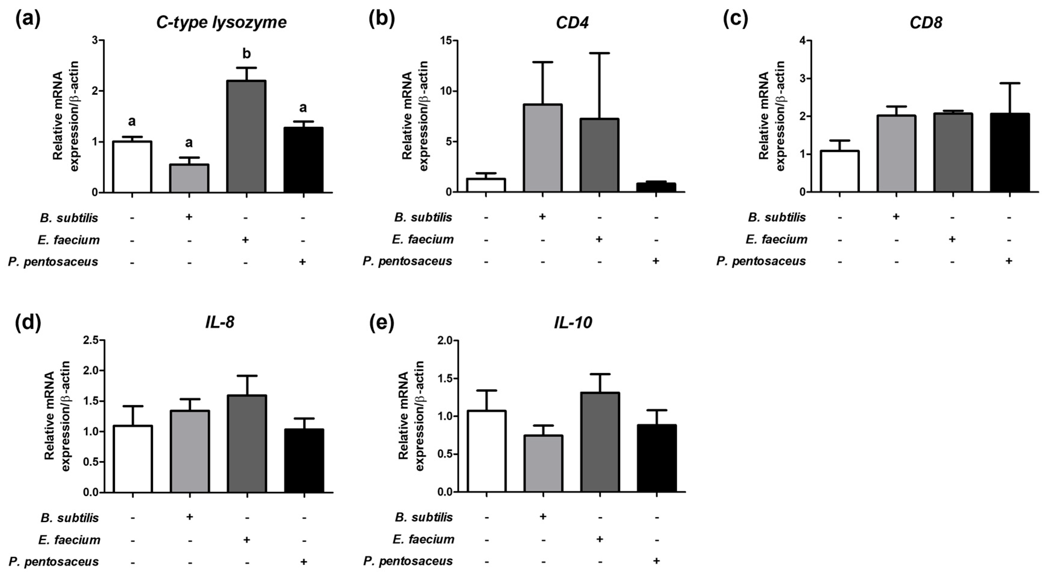
Relative mRNA expression levels of immune-related genes in the liver of olive flounder are shown in Figure 2. The expression of c-type lysozyme differed significantly among groups (p < 0.05). The E. faecium-supplemented group exhibited significantly higher expression than the control, B. subtilis, and P. pentosaceus groups, whereas no significant differences were detected among the control, B. subtilis, and P. pentosaceus. Transcript levels of CD4 and CD8 did not differ significantly among groups, although numerically higher values were observed in the B. subtilis and E. faecium groups. Similarly, IL-8 and IL-10 expressions showed no significant differences; however, numerically higher IL-8 levels were found in the B. subtilis and E. faecium groups, and IL-10 expressions were higher in the E. faecium group.

3.7. Survival Analysis Following E. tarda Challenge

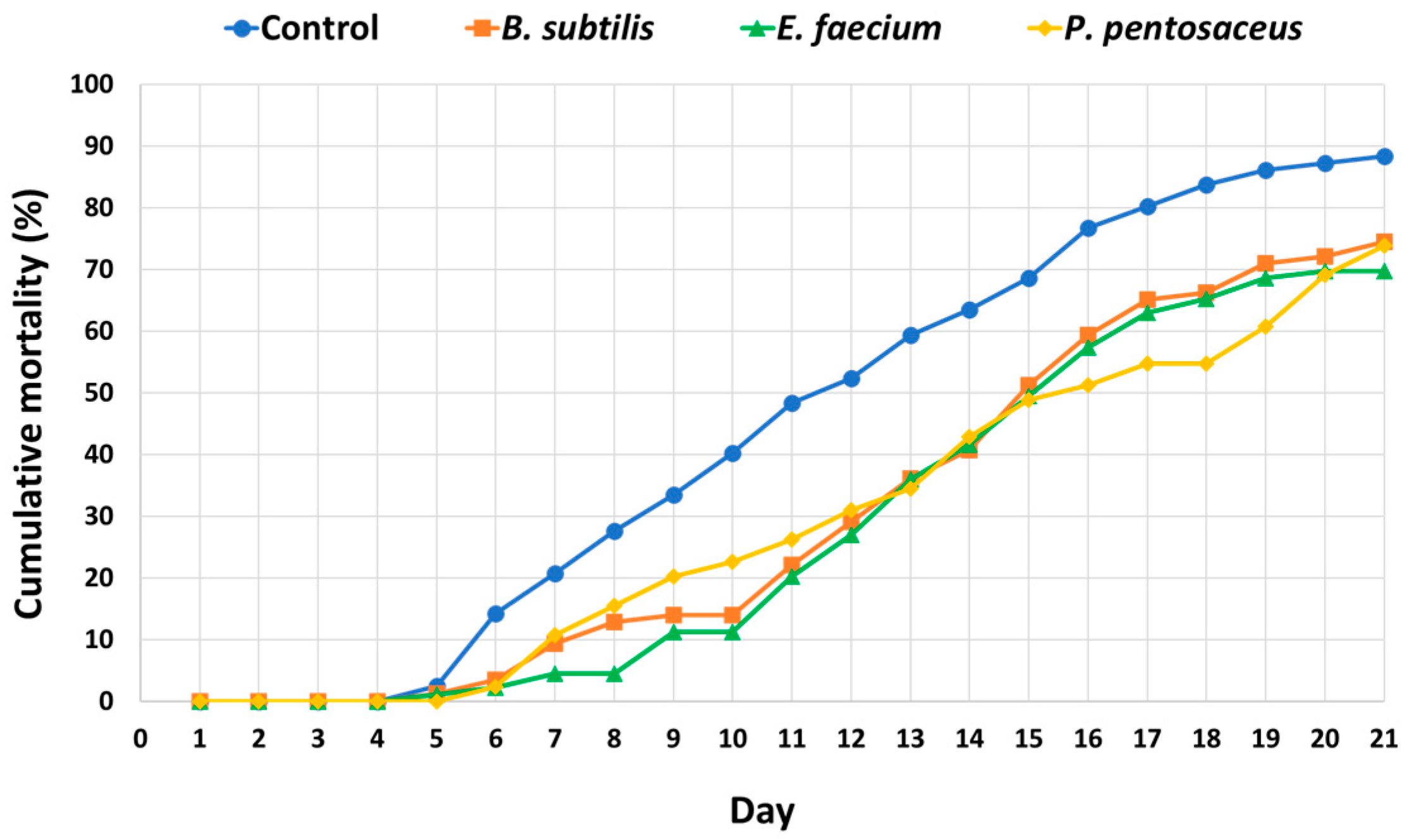
The effects of probiotic supplementation on disease resistance in olive flounder after artificial infection with E. tarda are shown in Figure 3. Mortality in the control group commenced on day 5 post-infection, and cumulative mortality reached approximately 88.5% by day 21, resulting in a survival rate of 11.5%. In contrast, fish fed diets supplemented with B. subtilis, E. faecium, or P. pentosaceus exhibited survival rates of 23.2%, 28.9%, and 22.5%, respectively. Among these, the E. faecium-supplemented group demonstrated the greatest improvement, with a 17.4 percent increase in survival compared to the control.

4. Discussion

In this study, dietary supplementation with three probiotic strains—B. subtilis, E. faecium, and P. pentosaceus—conferred distinct functional benefits in olive flounder. Notably, growth-related indices differed significantly among groups (p < 0.05). The P. pentosaceus group exhibited significantly higher values than the control, whereas the B. subtilis and E. faecium groups did not differ significantly from either the control or P. pentosaceus. This indicates that growth promotion was strain-specific, with P. pentosaceus exerting the predominant effect despite the absence of detectable protease, cellulase, or lipase activity. These findings suggest that its growth-promoting effects are unlikely to result from direct enzymatic degradation but rather from non-enzymatic mechanisms, such as bacteriocin-mediated pathogen suppression and microbiota stabilization, thereby indirectly enhancing nutrient utilization [28]. Similar findings have been reported in other teleosts, where P. pentosaceus improved intestinal barrier function or even conferred growth benefits when inactivated, indicating additional immunomodulatory mechanisms beyond enzymatic activity [29,30]. Future studies should therefore incorporate microbiome profiling to elucidate these mechanisms.
Although FCR did not differ significantly among groups, all probiotic-supplemented groups exhibited reduced values, with the E. faecium group recording the lowest value. This finding suggests that growth improvement (mainly by P. pentosaceus) and feed efficiency (mainly by E. faecium) are distinct probiotic-mediated outcomes, reflecting different metabolic strategies. Such divergence between growth rate and feed efficiency is well documented in aquaculture. For instance, Daboor et al. (2010) reported in Nile tilapia that Lactobacillus plantarum was particularly effective at stimulating weight gain, whereas B. subtilis exerted stronger effects on improving feed conversion efficiency [31]. Furthermore, multi-strain formulations have demonstrated synergistic benefits; in Nile tilapia, a combination of Leuconostoc mesenteroides and Lactococcus lactis significantly enhanced both growth and feed utilization compared with single strains [32]. Collectively, these findings, together with our results, highlight that distinct probiotic strains provide complementary benefits and that rationally designed multi-strain combinations may maximize both growth and feed efficiency in aquaculture.
In addition to these in vivo functional traits, genomic analyses provided further insights into the probiotic potential of the three isolates. The assembled genome sizes of the isolates were consistent with those previously reported for their respective species, supporting their genomic stability [33,34,35]. Genome mining revealed that B. subtilis harbored multiple BGCs, including those encoding bacilysin, subtilosin, and fengycin, which are typically associated with antimicrobial and antifungal activities. Interestingly, however, our assays did not detect strong antimicrobial activity from this strain, suggesting that the genomic potential for secondary metabolite production may not have been fully expressed under the tested conditions. In contrast, P. pentosaceus carried fewer BGCs but exhibited the strongest antimicrobial activity among the three strains, indicating that specific bacteriocins or acidic metabolites were effectively expressed under the experimental conditions. E. faecium showed limited BGC diversity, consistent with previous reports highlighting its role as an immunomodulatory probiotic rather than a strongly antimicrobial one [36,37]. Importantly, no BGCs associated with toxicity or pathogenicity were identified in any of the strains, reinforcing their safety as feed probiotics. These findings highlight the importance of integrating genomic predictions with functional validation and provide a mechanistic basis that complements the phenotypic outcomes observed in this study.
Probiotic supplementation also influenced systemic physiology. Plasma glucose levels differed significantly among groups (p < 0.05). The E. faecium and P. pentosaceus groups exhibited significantly lower glucose concentrations than both the control and B. subtilis, whereas no differences were detected among the control, B. subtilis, and P. pentosaceus. This indicates that glucose-lowering effects were restricted to E. faecium and P. pentosaceus, while B. subtilis had no significant influence. Although these values were lower than some previously reported ranges for healthy olive flounder, they likely reflect improved metabolic homeostasis and reduced stress responses rather than pathological hypoglycemia, consistent with evidence that probiotics can mitigate stress-induced hyperglycemia through modulation of the hypothalamic–pituitary–interrenal (HPI) axis [38,39]. This interpretation is further supported by the absence of adverse effects on growth, survival, TP, or other biochemical indices, indicating that systemic metabolic functions remained stable. Other biochemical indices (TP, ALP, GOT, GPT) also remained within healthy ranges, with downward trends suggesting alleviation of hepatic stress.
The immune-stimulatory effects of probiotics were also strain-dependent. Phagocytic activity differed significantly among groups (p < 0.05), with the P. pentosaceus group showing significantly higher values than the control, whereas the B. subtilis and E. faecium groups did not differ significantly from either the control or P. pentosaceus. Lysozyme activity also differed significantly (p < 0.05), with the E. faecium and P. pentosaceus groups exhibiting higher values than the control and B. subtilis. These results suggest that cellular immunity (phagocytosis) was mainly stimulated by P. pentosaceus, while humoral immunity (lysozyme) was strongly enhanced by E. faecium and P. pentosaceus. Although anti-protease activity did not differ significantly, an upward trend in the P. pentosaceus group may reflect enhanced resistance to extracellular proteases, as previously noted in tilapia [40,41].
These immunological benefits translated into improved disease resistance against E. tarda, with E. faecium showing the greatest post-challenge survival advantage, corroborating earlier findings in olive flounder [42,43]. Interestingly, although B. subtilis and E. faecium did not exhibit direct antimicrobial activity against E. tarda in vitro, both strains reduced mortality in the challenge test. This apparent discrepancy suggests that the protective effects observed in vivo may not be solely attributable to direct pathogen inhibition. Instead, these strains may confer resistance through alternative mechanisms such as enhancement of innate immune responses, modulation of the gut microbiota, or competitive exclusion of pathogens within the intestinal environment. Similar observations have been reported in other teleosts, where probiotics lacking measurable in vitro antimicrobial activity nevertheless improved survival following pathogen challenge by strengthening host immune defenses [44]. These findings highlight the importance of integrating both in vitro and in vivo assessments when evaluating probiotic functionality in aquaculture.
Beyond olive flounder, the beneficial effects of these probiotics have also been demonstrated in other aquaculture species. For instance, Won et al. (2020) reported that dietary supplementation with B. subtilis WB60 and P. pentosaceus at 108 CFU g−1 feed significantly improved growth performance, digestive enzyme activities, immune responses, intestinal histology, and disease resistance in whiteleg shrimp (Litopenaeus vannamei) [45]. Similarly, Tang et al. (2024) demonstrated that double-layer-coated E. faecium enhanced growth, feed utilization, gut morphology, and intestinal microbiota composition in crucian carp (Carassius auratus gibelio) [46]. These cross-species findings reinforce the broad relevance of the three strains for aquafeed applications.
Moreover, multi-strain probiotic formulations are increasingly used in commercial aquaculture and have frequently been reported to outperform single strains by combining complementary mechanisms. For instance, studies on tilapia and shrimp have shown that multi-strain mixtures enhanced both growth and feed utilization more effectively than individual strains [32,47,48]. Our findings provide additional support for this approach, as the distinct yet complementary benefits of B. subtilis, E. faecium, and P. pentosaceus indicate that their rational combination could yield synergistic improvements in growth performance, immunity, and disease resistance in olive flounder.

5. Conclusions

This study demonstrates strain-specific effects of dietary probiotics in olive flounder: P. pentosaceus promoted growth, E. faecium improved feed efficiency and disease resistance, and B. subtilis stimulated immune responses. These complementary outcomes indicate that probiotics can serve as functional feed additives to enhance fish health and performance. As this trial was conducted under controlled laboratory conditions with single strains, future research should validate their efficacy under farm environments and evaluate the potential of multi-strain formulations. Moreover, integrating microbiome analyses with growth and immune outcomes will be essential to optimize probiotic strategies for sustainable aquaculture.

Author Contributions

Conceptualization, M.-K.L., D.-G.K. and Y.S.K.; methodology, M.-K.L., D.-G.K. and Y.S.K.; software, M.-K.L., H.-K.J. and Y.S.K.; validation, M.-K.L.; formal analysis, M.-K.L.; investigation, M.-K.L. and Y.S.K.; data curation, M.-K.L., I.-S.P., Y.L.H., H.-K.J. and Y.S.K.; writing—original draft preparation, M.-K.L.; writing—review and editing, J.K. and Y.S.K.; visualization, M.-K.L. and Y.S.K.; supervision, J.K. and Y.S.K. All authors have read and agreed to the published version of the manuscript.

Funding

This research was supported by the National Institute of Fisheries Science grant (R2025051) funded by the Ministry of Oceans and Fisheries.

Institutional Review Board Statement

The animal study protocol was approved by the Ethics Committee of the National Institute of Fisheries Science (protocol code 2024-NIFS-IACUC-46 and 29 March 2024).

Informed Consent Statement

Not applicable.

Data Availability Statement

Data are contained within the article.

Conflicts of Interest

The authors declare no conflicts of interest.

References

  1. Jang, W.J.; Hasan, M.T.; Choi, W.; Hwang, S.; Lee, Y.; Hur, S.W.; Lee, S.; Lee, B.-J.; Choi, Y.H.; Lee, J.M. Comparison of growth performance, non-specific immunity, and intestinal microbiota of olive flounder (Paralichthys olivaceus) fed with extruded pellet and moist pellet diets under field conditions in South Korea. Front. Microbiol. 2022, 13, 979124. [Google Scholar] [CrossRef]
  2. Oh, S.; Lee, S. Fish Welfare-Related Issues and Their Relevance in Land-Based Olive Flounder (Paralichthys olivaceus) Farms in Korea. Animals 2024, 14, 1693. [Google Scholar] [CrossRef] [PubMed]
  3. Ciji, A.; Akhtar, M.S. Stress management in aquaculture: A review of dietary interventions. Rev. Aquac. 2021, 13, 2190–2247. [Google Scholar] [CrossRef]
  4. Barton, B.A. Stress in fishes: A diversity of responses with particular reference to changes in circulating corticosteroids. Integr. Comp. Biol. 2002, 42, 517–525. [Google Scholar] [CrossRef]
  5. Cabello, F.C.; Godfrey, H.P.; Tomova, A.; Ivanova, L.; Dölz, H.; Millanao, A.; Buschmann, A.H. Antimicrobial use in aquaculture re-examined: Its relevance to antimicrobial resistance and to animal and human health. Environ. Microbiol. 2013, 15, 1917–1942. [Google Scholar] [CrossRef]
  6. De la Casa-Resino, I.; Empl, M.T.; Villa, S.; Kolar, B.; Fabrega, J.; Lillicrap, A.D.; Karamanlis, X.N.; Carapeto-García, R. Environmental risk assessment of veterinary medicinal products intended for use in aquaculture in Europe: The need for developing a harmonised approach. Environ. Sci. Eur. 2021, 33, 84. [Google Scholar] [CrossRef]
  7. Balcázar, J.L.; De Blas, I.; Ruiz-Zarzuela, I.; Cunningham, D.; Vendrell, D.; Múzquiz, J.L. The role of probiotics in aquaculture. Vet. Microbiol. 2006, 114, 173–186. [Google Scholar] [CrossRef]
  8. Panigrahi, A.; Kiron, V.; Kobayashi, T.; Puangkaew, J.; Satoh, S.; Sugita, H. Immune responses in rainbow trout Oncorhynchus mykiss induced by a potential probiotic bacteria Lactobacillus rhamnosus JCM 1136. Vet. Immunol. Immunopathol. 2004, 102, 379–388. [Google Scholar] [CrossRef]
  9. Li, Y.; Yang, Y.; Song, L.; Wang, J.; Hu, Y.; Yang, Q.; Cheng, P.; Li, J. Effects of dietary supplementation of Lactobacillus plantarum and Bacillus subtilis on growth performance, survival, immune response, antioxidant capacity and digestive enzyme activity in olive flounder (Paralichthys olivaceus). Aquac. Fish. 2021, 6, 283–288. [Google Scholar] [CrossRef]
  10. Abbas, M.A.; Kim, H.-J.; Lee, G.-Y.; Cho, H.-Y.; Al Jawad Sayem, S.; Lee, E.-B.; Lee, S.-J.; Park, S.-C. Development and Application of Lactobacillus plantarum PSCPL13 Probiotics in Olive Flounder (Paralichthys olivaceus) Farming. Microorganisms 2025, 13, 61. [Google Scholar] [CrossRef] [PubMed]
  11. Ahmadifar, E.; Sadegh, T.H.; Dawood, M.A.; Dadar, M.; Sheikhzadeh, N. The effects of dietary Pediococcus pentosaceus on growth performance, hemato-immunological parameters and digestive enzyme activities of common carp (Cyprinus carpio). Aquaculture 2020, 516, 734656. [Google Scholar] [CrossRef]
  12. Hoseinifar, S.H.; Sun, Y.-Z.; Wang, A.; Zhou, Z. Probiotics as means of diseases control in aquaculture, a review of current knowledge and future perspectives. Front. Microbiol. 2018, 9, 2429. [Google Scholar] [CrossRef]
  13. Madhulika; Ngasotter, S.; Meitei, M.M.; Kara, T.; Meinam, M.; Sharma, S.; Rathod, S.K.; Singh, S.B.; Singh, S.K.; Bhat, R.A.H. Multifaceted Role of Probiotics in Enhancing Health and Growth of Aquatic Animals: Mechanisms, Benefits, and Applications in Sustainable Aquaculture—A Review and Bibliometric Analysis. Aquac. Nutr. 2025, 2025, 5746972. [Google Scholar]
  14. Choi, W.; Moniruzzaman, M.; Bae, J.; Hamidoghli, A.; Lee, S.; Choi, Y.-H.; Min, T.; Bai, S.C. Evaluation of dietary probiotic bacteria and processed yeast (GroPro-Aqua) as the alternative of antibiotics in juvenile olive flounder Paralichthys olivaceus. Antibiotics 2022, 11, 129. [Google Scholar] [CrossRef] [PubMed]
  15. Niu, K.-M.; Kothari, D.; Lee, W.-D.; Zhang, Z.; Lee, B.-J.; Kim, K.-W.; Wu, X.; Han, H.-S.; Khosravi, S.; Lee, S.-M. Probiotic potential of the farmed olive flounder, Paralichthys olivaceus, autochthonous gut microbiota. Probiotics Antimicrob. Proteins 2021, 13, 1106–1118. [Google Scholar] [CrossRef]
  16. Han, S.-R.; Munang’andu, H.M.; Yeo, I.-K.; Kim, S.-H. Bacillus subtilis inhibits viral hemorrhagic septicemia virus infection in olive flounder (Paralichthys olivaceus) intestinal epithelial cells. Viruses 2020, 13, 28. [Google Scholar] [CrossRef] [PubMed]
  17. Hall, T.A. BioEdit: A user-friendly biological sequence alignment editor and analysis program for Windows 95/98/NT. Nucleic Acids Symp. Ser. 1999, 41, 95–98. [Google Scholar]
  18. Yoon, S.-H.; Ha, S.-M.; Kwon, S.; Lim, J.; Kim, Y.; Seo, H.; Chun, J. Introducing EzBioCloud: A taxonomically united database of 16S rRNA gene sequences and whole-genome assemblies. Int. J. Syst. Evol. Microbiol. 2017, 67, 1613–1617. [Google Scholar] [CrossRef]
  19. Klančnik, A.; Piskernik, S.; Jeršek, B.; Možina, S.S. Evaluation of diffusion and dilution methods to determine the antibacterial activity of plant extracts. J. Microbiol. Methods 2010, 81, 121–126. [Google Scholar] [CrossRef]
  20. Blin, K.; Shaw, S.; Kloosterman, A.M.; Charlop-Powers, Z.; Van Wezel, G.P.; Medema, M.H.; Weber, T. antiSMASH 6.0: Improving cluster detection and comparison capabilities. Nucleic Acids Res. 2021, 49, W29–W35. [Google Scholar] [CrossRef] [PubMed]
  21. Heo, W.-S.; Kim, Y.-R.; Kim, E.-Y.; Bai, S.C.; Kong, I.-S. Effects of dietary probiotic, Lactococcus lactis subsp. lactis I2, supplementation on the growth and immune response of olive flounder (Paralichthys olivaceus). Aquaculture 2013, 376, 20–24. [Google Scholar] [CrossRef]
  22. Wang, J.; Fan, D.; Zhao, R.; Lu, T.; Li, S.; Wang, D. Effects of dietary supplementation with endogenous probiotics Bacillus subtilis on growth performance, immune response and intestinal histomorphology of juvenile rainbow trout (Oncorhynchus mykiss). Fishes 2024, 9, 229. [Google Scholar] [CrossRef]
  23. Müller, J.; Alföldy, P.; Lemmel, E.-M. Nitroblue-tetrazolium test for the functional evaluation of phagocytic cells: A critical analysis of the methodology. Agents Actions 1981, 11, 384–390. [Google Scholar] [CrossRef]
  24. Hikima, J.-i.; Minagawa, S.; Hirono, I.; Aoki, T. Molecular cloning, expression and evolution of the Japanese flounder goose-type lysozyme gene, and the lytic activity of its recombinant protein. Biochim. Et Biophys. Acta (BBA)-Gene Struct. Expr. 2001, 1520, 35–44. [Google Scholar] [CrossRef]
  25. Coêlho, D.F.; Saturnino, T.P.; Fernandes, F.F.; Mazzola, P.G.; Silveira, E.; Tambourgi, E.B. Azocasein substrate for determination of proteolytic activity: Reexamining a traditional method using bromelain samples. BioMed Res. Int. 2016, 2016, 8409183. [Google Scholar] [CrossRef]
  26. Livak, K.J.; Schmittgen, T.D. Analysis of relative gene expression data using real-time quantitative PCR and the 2− ΔΔCT method. Methods 2001, 25, 402–408. [Google Scholar] [CrossRef] [PubMed]
  27. Kim, M.-G.; Gunathilaka, B.E.; Lee, S.; Kim, Y.; Lee, K.-J. Effects of Bacillus SW1-1 coated diets on innate immunity and disease resistance of olive flounder Paralichthys olivaceus against Edwardsiella tarda infection. Fish. Aquat. Sci. 2022, 25, 243–249. [Google Scholar] [CrossRef]
  28. Yi, C.-C.; Liu, C.-H.; Chuang, K.-P.; Chang, Y.-T.; Hu, S.-Y. A potential probiotic Chromobacterium aquaticum with bacteriocin-like activity enhances the expression of indicator genes associated with nutrient metabolism, growth performance and innate immunity against pathogen infections in zebrafish (Danio rerio). Fish Shellfish Immunol. 2019, 93, 124–134. [Google Scholar] [CrossRef]
  29. Ding, F.; Zhou, N.; Luo, Y.; Wang, T.; Li, W.; Qiao, F.; Du, Z.; Zhang, M. Probiotic Pediococcus pentosaceus restored gossypol-induced intestinal barrier injury by increasing propionate content in Nile tilapia. J. Anim. Sci. Biotechnol. 2024, 15, 54. [Google Scholar] [CrossRef]
  30. Dawood, M.; Koshio, S.; Ishikawa, M.; Yokoyama, S. Effects of dietary inactivated Pediococcus pentosaceus on growth performance, feed utilization and blood characteristics of red sea bream, Pagrus major juvenile. Aquac. Nutr. 2016, 22, 923–932. [Google Scholar] [CrossRef]
  31. Daboor, M.; Esmael, N.A.; Lall, S.P. Effect of Different Dietary Probiotics on Growth, Feed Utilization and Digestive Enzymes Activities of Nile Tilapia, Oreochromis niloticus Mohamed A Essa1, Sabry S EL-Serafy2, Magda M El-Ezabi2, Said. Aquaculture 2010, 454, 243–251. [Google Scholar]
  32. Paritova, A.; Nurgaliyev, A.; Nurgaliyeva, G.; Abekeshev, N.; Abuova, A.; Zakirova, F.; Zwierzchowski, G.; Kuanchaleyev, Z.; Issabekova, S.; Kizatova, M. The dietary effects of two strain probiotics (Leuconostoc mesenteroides, Lactococcus lactis) on growth performance, immune response and gut microbiota in Nile tilapia (Oreochromis niloticus). PLoS ONE 2024, 19, e0312580. [Google Scholar] [CrossRef]
  33. Kunst, F.; Ogasawara, N.; Moszer, I.; Albertini, A.; Alloni, G.; Azevedo, V.; Bertero, M.; Bessières, P.; Bolotin, A.; Borchert, S. The complete genome sequence of the gram-positive bacterium Bacillus subtilis. Nature 1997, 390, 249–256. [Google Scholar] [CrossRef] [PubMed]
  34. Kompramool, S.; Singkhamanan, K.; Pomwised, R.; Chaichana, N.; Suwannasin, S.; Wonglapsuwan, M.; Jitpakdee, J.; Kantachote, D.; Yaikhan, T.; Surachat, K. Genomic Insights into Pediococcus pentosaceus ENM104: A Probiotic with Potential Antimicrobial and Cholesterol-Reducing Properties. Antibiotics 2024, 13, 813. [Google Scholar] [CrossRef]
  35. Qin, X.; Galloway-Peña, J.R.; Sillanpaa, J.; Roh, J.H.; Nallapareddy, S.R.; Chowdhury, S.; Bourgogne, A.; Choudhury, T.; Muzny, D.M.; Buhay, C.J. Complete genome sequence of Enterococcus faecium strain TX16 and comparative genomic analysis of Enterococcus faecium genomes. BMC Microbiol. 2012, 12, 135. [Google Scholar] [CrossRef]
  36. Mi, J.; He, T.; Hu, X.; Wang, Z.; Wang, T.; Qi, X.; Li, K.; Gao, L.; Liu, C.; Zhang, Y. Enterococcus faecium C171: Modulating the immune response to acute lethal viral challenge. Int. J. Antimicrob. Agents 2023, 62, 106969. [Google Scholar] [CrossRef]
  37. Mansour, N.M.; Heine, H.; Abdou, S.M.; Shenana, M.E.; Zakaria, M.K.; El-Diwany, A. Isolation of Enterococcus faecium NM113, Enterococcus faecium NM213 and Lactobacillus casei NM512 as novel probiotics with immunomodulatory properties. Microbiol. Immunol. 2014, 58, 559–569. [Google Scholar] [CrossRef]
  38. Aerts, J.; Schaeck, M.; De Swaef, E.; Ampe, B.; Decostere, A. Vibrio lentus as a probiotic candidate lowers glucocorticoid levels in gnotobiotic sea bass larvae. Aquaculture 2018, 492, 40–45. [Google Scholar] [CrossRef]
  39. Wendelaar Bonga, S.E. The stress response in fish. Physiol. Rev. 1997, 77, 591–625. [Google Scholar] [CrossRef] [PubMed]
  40. Chuwen, Q.; Yonghai, S.; Jianming, Z. Probiotics and their impact on mitigating high-temperature stress responses in farmed fish. J. Fish. Res. 2025, 47, 248–260. [Google Scholar]
  41. Fachri, M.; Amoah, K.; Huang, Y.; Cai, J.; Alfatat, A.; Ndandala, C.B.; Shija, V.M.; Jin, X.; Bissih, F.; Chen, H. Probiotics and paraprobiotics in aquaculture: A sustainable strategy for enhancing fish growth, health and disease prevention-a review. Front. Mar. Sci. 2024, 11, 1499228. [Google Scholar] [CrossRef]
  42. Taoka, Y.; Maeda, H.; Jo, J.-Y.; Jeon, M.-J.; Bai, S.C.; Lee, W.-J.; Yuge, K.; Koshio, S. Growth, stress tolerance and non-specific immune response of Japanese flounder Paralichthys olivaceus to probiotics in a closed recirculating system. Fish. Sci. 2006, 72, 310–321. [Google Scholar] [CrossRef]
  43. Nguyen, T.L.; Park, C.-I.; Kim, D.-H. Improved growth rate and disease resistance in olive flounder, Paralichthys olivaceus, by probiotic Lactococcus lactis WFLU12 isolated from wild marine fish. Aquaculture 2017, 471, 113–120. [Google Scholar] [CrossRef]
  44. Aly, S.M.; Mohamed, M.F.; John, G. Effect of probiotics on the survival, growth and challenge infection in Tilapia nilotica (Oreochromis niloticus). Aquac. Res. 2008, 39, 647–656. [Google Scholar] [CrossRef]
  45. Won, S.; Hamidoghli, A.; Choi, W.; Bae, J.; Jang, W.J.; Lee, S.; Bai, S.C. Evaluation of potential probiotics Bacillus subtilis WB60, Pediococcus pentosaceus, and Lactococcus lactis on growth performance, immune response, gut histology and immune-related genes in whiteleg shrimp, Litopenaeus vannamei. Microorganisms 2020, 8, 281. [Google Scholar] [CrossRef] [PubMed]
  46. Tang, H.; Qiao, L.; Li, A.; Zhao, J.; Qian, R.; Luo, Q.; Xu, Q. Effects of coated Enterococcus faecium on the growth performance, immunity, digestive enzyme activity, intestinal morphometry, and microbiota of crucian carp (Carassius auratus gibelio). Aquac. Rep. 2024, 37, 102264. [Google Scholar] [CrossRef]
  47. Hossain, M.K.; Islam, S.M.; Rafiquzzaman, S.; Nuruzzaman, M.; Hossain, M.T.; Shahjahan, M. Multi-species probiotics enhance growth of Nile tilapia (Oreochromis niloticus) through upgrading gut, liver and muscle health. Aquac. Res. 2022, 53, 5710–5719. [Google Scholar] [CrossRef]
  48. Wang, Y.-C.; Hu, S.-Y.; Chiu, C.-S.; Liu, C.-H. Multiple-strain probiotics appear to be more effective in improving the growth performance and health status of white shrimp, Litopenaeus vannamei, than single probiotic strains. Fish Shellfish Immunol. 2019, 84, 1050–1058. [Google Scholar] [CrossRef]
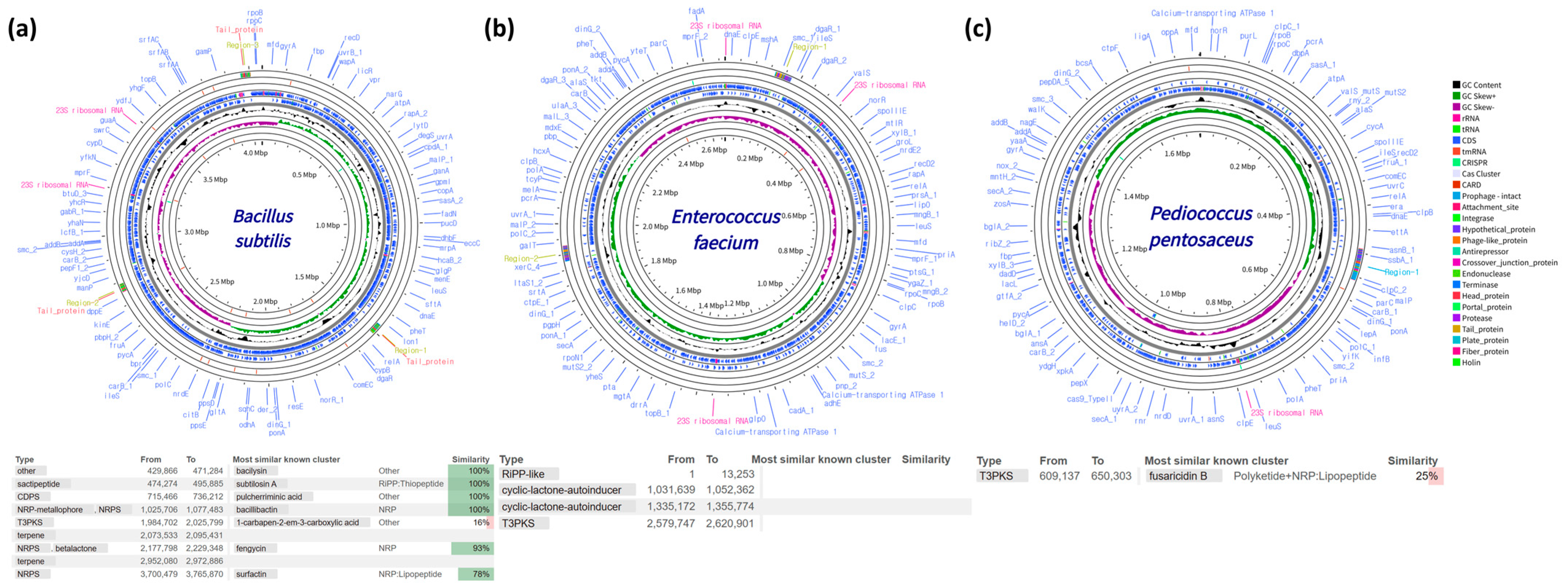
Figure 1. Genomes of candidate probiotic strains for feed additives and their explored secondary metabolite biosynthetic gene clusters. (a) B. subtilis; (b) E. faecium; (c) P. pentosaceus. Colors in the circular genome maps correspond to the functional categories shown in the legend. In the similarity table, green indicates gene clusters with high similarity to known clusters, while red indicates clusters with lower similarity.
Fishes 10 00465 g001
Figure 2. Effects of probiotic-supplemented diets on the expression of immune-related genes in the liver of olive flounder. Relative mRNA expression levels of (a) c-type lysozyme, (b) CD4, (c) CD8, (d) IL-8, and (e) IL-10 were measured after a 35-day feeding trial with probiotic-supplemented diets. Gene expression was normalized to β-actin and calculated using the 2−ΔΔCt method. Data are presented as mean ± SEM (n = 3). Different superscript letters indicate significant differences among groups (Tukey’s HSD, p < 0.05). “+” indicates presence and “−” indicates absence of probiotic supplementation.
Fishes 10 00465 g002
Figure 3. Survival curves of olive flounder following intraperitoneal challenge with E. tarda after a 35-day feeding trial with probiotic-supplemented diets. Fish were fed diets containing B. subtilis, E. faecium, or P. pentosaceus (6.5 × 10⁷ CFU g−1) prior to challenge. Mortality was recorded daily for 21 days post-infection. Data are presented as cumulative survival percentages (n = 3 replicate tanks per group, 30 fish per tank).
Fishes 10 00465 g003
Table 1. Proximate composition of the basal diet used for olive flounder (% dry matter basis).
Proximate CompositionContent (%)
Crude protein≥54.0%
Crude lipid≥8.0%
Crude ash≥18.0%
Crude fiber≤5.0%
Calcium≤1.0%
Phosphorus≤2.7%
Table 2. Primer sequences used for real-time quantitative PCR analysis.
GenePrimer (5′-3′)Accession Number
C-type lysozymeF: CTG TGG GCA GGA AAG ACT TC
R: GGA AGT GTT GGT GGA GAG G
AB050589.1
CD4F: AGG TGC CAG TGA GGT GGT TTA T
R: GCC GTC CTG TTT ACC AAA ACT C
XM_069537653.1
CD8F: CGC TGC GCT GCA ATG ATAB082958.1
R: CAC GCC CCA CCT GTA ACC
IL-8F: CAT CGT TGT TGC TGT GAT GGTAF216646.1
R: AGG CTC ACC GCT TCA CTG AT
IL-10F: AGC GAA CGA TGA CCT AGA CAC GAB685381.1
R: ACC GTG CTC AGG TAG AAG TCC A
β-actinF: CAT CAG GGA GTG ATG GTG GGT A
R: ATA CCG TGC TCG ATG GGG TAC T
HQ386788.1
F: forward. R: reverse. CD4: cluster of differentiation 4. CD8: cluster of differentiation 8. IL-8: interleukin-8. IL-10: interleukin-10.
Table 3. Effects of probiotic strains on antimicrobial and enzymatic activities in vitro. -: no activity.
StrainSpeciesAntimicrobial Activity (mm)Enzyme Activity (mm)
E. coliB. cereusC. albicansE. tardaTCNTBNSkim
Milk
CMCSoluble
Starch
Gelatin
E-TSA-3B. subtilis-----Fishes 10 00465 i001
13.9 ± 0.10 ***
Fishes 10 00465 i002
19.6 ± 0.20 ***
Fishes 10 00465 i003
17.0 ± 0.59 ***
Fishes 10 00465 i004
14.7 ± 1.37 ***
Fishes 10 00465 i005
22.1 ± 0.15 ***
Jeju1
YPD-N2
E. faecium-Fishes 10 00465 i006
11.5 ± 0.25 ***
----Fishes 10 00465 i007
15.3 ± 0.51 ***
---
MRS-A4-1P. pentosaceusFishes 10 00465 i008
20.6 ± 0.60 ***
Fishes 10 00465 i009
24.2 ± 0.75 ***
-Fishes 10 00465 i010
9.7 ± 0.57 ***
------
Values are expressed as mean ± SEM (n = 3). *** p < 0.001 indicates statistically significant differences compared to the control group.
Table 4. Effects of probiotic-supplemented growth performance in olive flounder after a 35-day feeding trial.
IBW (g)FBW (g)BWG (g)WGR (%)SGR (%)FCR
Control16.96 ± 3.046.57 ± 9.8 a29.59 ± 9.8 a174.26 ± 57.9 a2.81 ± 0.6 a1.55 ± 0.8
B. subtilis16.97 ± 3.248.61 ± 10.2 ab32.01 ± 9.6 ab188.51 ± 56.5 ab2.97 ± 0.6 ab1.41 ± 0.9
E. faecium16.98 ± 1.949.51 ± 8.8 ab32.53 ± 8.8 ab191.58 ± 51.6 ab3.01 ± 0.5 ab1.31 ± 0.4
P. pentosaceus16.97 ± 2.750.87 ± 10.0 b33.89 ± 10.0 b199.59 ± 59.0 b3.07 ± 0.6 b1.34 ± 0.9
Values are expressed as mean ± SEM (n = 3, number of replicate tanks per group). The final body weight of 30 fish (10 fish per tank) was measured from each group. Different superscript letters indicate significant differences among groups (Tukey’s HSD, p < 0.05).
Table 5. Effects of probiotic-supplemented diets on blood biochemical parameters in olive flounder after a 35-day feeding trial.
GLU (mg/dL)ALP (U/L)GOT (U/L)GPT (U/L)TP (g/dL)
Control74.50 ± 32.9 a255.0 ± 101.727.3 ± 9.413.3 ± 1.93.4 ± 0.26
B. subtilis54.53 ± 26.3 a218.3 ± 61.621.5 ± 7.312.1 ± 1.53.5 ± 0.27
E. faecium23.43 ± 10.5 b220.5 ± 49.526.3 ± 9.511.1 ± 2.73.5 ± 0.20
P. pentosaceus27.07 ± 12.6 b194.7 ± 40.528.2 ± 18.013.3 ± 4.73.5 ± 0.27
Values are expressed as mean ± SEM (n = 3, number of replicate tanks per group). Blood samples for biochemical analysis were collected from 15 fish per group (5 fish per tank). Different superscript letters indicate significant differences among groups (Tukey’s HSD, p < 0.05). GLU: glucose. ALP: alkaline phosphatase. GOT: glutamyl oxaloacetic transaminase. GPT: glutamic pyruvate. TP: total protein.
Table 6. Effects of probiotic-supplemented non-specific immune responses in olive flounder after a 35-day feeding trial.
Phagocytic Activity
(O.D. 550 nm)
Lysozyme Activity
(U/mL)
Anti-Protease Activity
(% Inhibition)
Control0.054 ± 0.007 a4.55 ± 9.53 a52.00 ± 19.7
B. subtilis0.066 ± 0.009 ab7.48 ± 4.77 a50.22 ± 18.3
E. faecium0.065 ± 0.007 ab21.52 ± 11.98 b42.25 ± 16.8
P. pentosaceus0.070 ± 0.021 b19.13 ± 8.30 b56.69 ± 18.8
Values are expressed as mean ± SEM (n = 3, number of replicate tanks per group). Immune parameters were analyzed using blood samples from 15 fish per group (5 fish per tank). Different superscript letters indicate significant differences among groups (Tukey’s HSD, p < 0.05).
Disclaimer/Publisher’s Note: The statements, opinions and data contained in all publications are solely those of the individual author(s) and contributor(s) and not of MDPI and/or the editor(s). MDPI and/or the editor(s) disclaim responsibility for any injury to people or property resulting from any ideas, methods, instructions or products referred to in the content.